User login
Differentiating serotonin syndrome and neuroleptic malignant syndrome
Serotonin syndrome (SS) and neuroleptic malignant syndrome (NMS) are each rare psychiatric emergencies that can lead to fatal outcomes. Their clinical presentations can overlap, which can make it difficult to differentiate between the 2 syndromes; however, their treatments are distinct, and it is imperative to know how to identify symptoms and accurately diagnose each of them to provide appropriate intervention. This article summarizes the 2 syndromes and their treatments, with a focus on how clinicians can distinguish them, provide prompt intervention, and prevent occurrence.
Serotonin syndrome
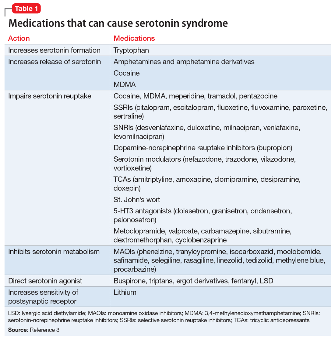
Mechanism. The decarboxylation and hydroxylation of tryptophan forms serotonin, also known as 5-hydroxytryptamine (5-HT), which can then be metabolized by monoamine oxidase-A (MAO-A) into 5-hydroxyindoleacetic acid (5-HIAA).1Medications can disrupt this pathway of serotonin production or its metabolism, and result in excessive levels of serotonin, which subsequently leads to an overactivation of central and peripheral serotonin receptors.1 Increased receptor activation leads to further upregulation, and ultimately more serotonin transmission. This can be caused by monotherapy or use of multiple serotonergic agents, polypharmacy with a combination of medication classes, drug interactions, or overdose. The wide variety of medications often prescribed by different clinicians can make identification of excessive serotonergic activity difficult, especially because mood stabilizers such as lithium,2 and non-psychiatric medications such as
- inhibition of serotonin uptake (seen with selective serotonin reuptake inhibitors [SSRIs], serotonin-norepinephrine reuptake inhibitors [SNRIs], and tricyclic antidepressants [TCAs])
- inhibition of serotonin metabolism (seen with monoamine oxidase inhibitors [MAOIs])
- increased serotonin synthesis (seen with stimulants)
- increased serotonin release (seen with stimulants and opiates)
- activation of serotonin receptors (seen with lithium)
- inhibition of certain cytochrome P450 (CYP450) enzymes (seen with ciprofloxacin, fluconazole, etc.).
It is important to recognize that various serotonergic agents are involved in the CYP450 system. Inhibition of the CYP450 pathway by common antibiotics such as ciprofloxacin, or antifungals such as fluconazole, may result in an accumulation of serotonergic agents and place patients at increased risk for developing SS.
Clinical presentation. The clinical presentation of SS can range from mild to fatal. There is no specific laboratory test for diagnosis, although an elevation of the total creatine kinase (CK) and leukocyte count, as well as increased transaminase levels or lower bicarbonate levels, have been reported in the literature.4
Symptoms of SS generally present within 24 hours of starting/changing therapy and include a triad of mental status changes (altered mental status [AMS]), autonomic instability, and abnormalities of neuromuscular tone. Examples of AMS include agitation, anxiety, disorientation, and restlessness. Symptoms of autonomic instability include hypertension, tachycardia, tachypnea, hyperthermia, diaphoresis, flushed skin, vomiting, diarrhea, and arrhythmias. Symptoms stemming from changes in neuromuscular tone include tremors, clonus, hyperreflexia, and muscle rigidity.1 The multiple possible clinical presentations, as well as symptoms that overlap with those of other syndromes, can make SS difficult to recognize quickly in a clinical setting.
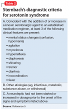
Diagnostic criteria. Sternbach’s diagnostic criteria for SS are defined as the presence of 3 or more of the 10 most common clinical features (Table 25). Due to concerns that Sternbach’s diagnostic criteria overemphasized an abnormal mental state (leading to possible confusion of SS with other AMS syndromes), the Hunter serotonin toxicity criteria6 (Figure6) were developed in 2003, and were found to be more sensitive and specific than Sternbach’s criteria. Both tools are often used in clinical practice.
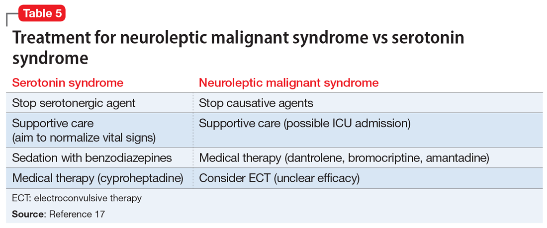
Treatment. Treatment of SS begins with prompt discontinuation of all serotonergic agents. The intensity of treatment depends on the severity of the symptoms. Mild symptoms can be managed with supportive care,3 and in such cases, the syndrome generally resolves within 24 hours.7 Clinicians may use supportive care to normalize vital signs (oxygenation to maintain SpO2 >94%, IV fluids for volume depletion, cooling agents, antihypertensives, benzodiazepines for sedation or control of agitation, etc.). Patients who are more ill may require more aggressive treatment, such as the use of a serotonergic antagonist (ie, cyproheptadine) and those who are severely hyperthermic (temperature >41.1ºC) may require neuromuscular sedation, paralysis, and possibly endotracheal intubation.3
Continue to: Management pitfalls include...
Management pitfalls include misdiagnosis of SS, failure to recognize its rapid rate of progression, and adverse effects of pharmacologic therapy.3 The most effective treatment for SS is prevention. SS can be prevented by astute pharmacologic understanding, avoidance of polypharmacy, and physician education.3
Neuroleptic malignant syndrome
Possible mechanisms. Neuromuscular malignant syndrome is thought to result from dopamine receptor antagonism leading to a hypodopaminergic state in the striatum and hypothalamus.8 The pathophysiology behind NMS has not fully been elucidated; however, several hypotheses attempt to explain this life-threatening reaction. The first focuses on dopamine D2 receptor antagonism, because many of the neuroleptic (antipsychotic) medications that can precipitate NMS are involved in dopamine blockade. In this theory, blocking dopamine D2 receptors in the anterior hypothalamus explains the hyperthermia seen in NMS, while blockade in the corpus striatum is believed to lead to muscle rigidity.9
The second hypothesis suggests that neuroleptics may have a direct toxic effect to muscle cells. Neuroleptics influence calcium transport across the sarcoplasmic reticulum and can lead to increased calcium release, which may contribute to the muscle rigidity and hyperthermia seen in NMS.9
The third hypothesis involves hyperactivity of the sympathetic nervous system; it is thought that psychologic stressors alter frontal lobe function, with neuroleptics disrupting the inhibitory pathways of the sympathetic nervous system. The autonomic nervous system innervates multiple organ systems, so this excessively dysregulated sympathetic nervous system may be responsible for multiple NMS symptoms (hyperthermia, muscle rigidity, hypertension, diaphoresis, tachycardia, elevated CK.10
NMS can be caused by neuroleptic agents (both first- and second-generation antipsychotics) as well as antiemetics (Table 31). The time between use of these medications and onset of symptoms is highly variable. NMS can occur after a single dose, after a dose adjustment, or possibly after years of treatment with the same medication. It is not dose-dependent.11 In certain individuals, NMS may occur at therapeutic doses.
Continue to: Clinical presentation
Clinical presentation. Patients with NMS typically present with a tetrad of symptoms: mental status changes, muscular rigidity, hyperthermia, and autonomic instability.12 Mental status changes can include confusion and agitation, as well as catatonic signs and mutism. The muscular rigidity of NMS is characterized by “lead pipe rigidity” and may be accompanied by tremor, dystonia, or dyskinesias. Laboratory findings include elevated serum CK (from severe rigidity), often >1,000 U/L, although normal levels can be observed if rigidity has not yet developed.13
Treatment. The first step for treatment is to discontinue the causative medication.14 Initiate supportive therapy immediately to restrict the progression of symptoms. Interventions include cooling blankets, fluid resuscitation, and antihypertensives to maintain autonomic stability15 or benzodiazepines to control agitation. In severe cases, muscular rigidity may extend to the airways and intubation may be required. The severity of these symptoms may warrant admission to the ICU for close monitoring. Pharmacologic treatment with
Differentiating between SS and NMS
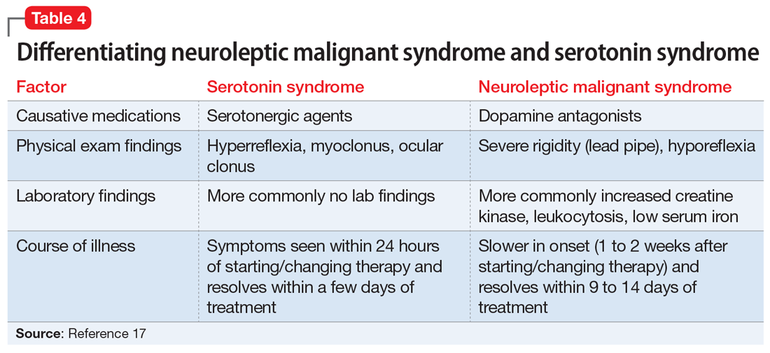
Differentiating between these 2 syndromes (Table 417) is critical to direct appropriate intervention. Table 517 outlines the treatment overview for SS and NMS.
Detailed history. A detailed history is imperative in making accurate diagnoses. Useful components of the history include a patient’s duration of symptoms and medication history (prescription medications as well as over-the-counter medications, supplements, and illicit drugs). Also assess for medical comorbidities, because certain medical diagnoses may alert the clinician that it is likely the patient had been prescribed serotonergic agents or neuroleptics, and renal or liver impairment may alert the clinician of decreased metabolism rates. Medication history is arguably the most useful piece of the interview, because serotonergic agents can cause SS, whereas dopamine blockers cause NMS. It should be noted that excess serotonin acts as a true toxidrome and is concentration-dependent in causing SS, whereas NMS is an idiosyncratic reaction to a drug.
Physical exam. Although there are many overlapping clinical manifestations, SS produces neuromuscular hyperactivity (ie, clonus, hyperreflexia), whereas NMS is characterized by more sluggish responses (ie, rigidity, bradyreflexia).18
Continue to: Laboratory findings
Laboratory findings. Overlap between NMS and SS also occurs with lab findings; both syndromes can result in leukocytosis, elevated CK from muscle damage, and low serum iron levels. However, these findings are more commonly associated with NMS and are seen in 75% of cases.17,19
Course of illness. Duration of symptoms can also help differentiate the 2 syndromes. SS typically develops within 24 hours of starting/changing therapy, whereas NMS symptoms can be present for days to weeks. Resolution of symptoms may also be helpful in differentiation because SS typically resolves within a few days of initiating treatment, whereas NMS resolves within 9 to 14 days of starting treatment.19
Bottom Line
The clinical presentations of serotonin syndrome (SS) and neuroleptic malignant syndrome (NMS) overlap, which can make them difficult to differentiate; however, they each have distinct approaches to treatment. Features in SS that are distinct from NMS include a history of serotonergic agents, rapid onset of symptoms, hyperreflexia, and clonus. NMS is slower in onset and can be found in patients who are prescribed dopamine antagonists, with distinct symptoms of rigidity and hyporeflexia.
Related Resources
- Kimmel R. Serotonin syndrome or NMS? Clues to diagnosis. Current Psychiatry. 2010;9(2):92.
- Strawn JR, Keck Jr PE, Caroff SN. Neuroleptic malignant syndrome: Answers to 6 tough questions. Current Psychiatry. 2008;7(1):95-101.
Drug Brand Names
Amantadine • Symmetrel
Amitriptyline • Elavil, Endep
Aripiprazole • Abilify
Bromocriptine • Cycloset, Parlodel
Bupropion • Wellbutrin, Zyban
Buspirone • BuSpar
Carbamazepine • Carbatrol, Tegretol
Chlorpromazine • Thorazine
Ciprofloxacin • Cipro
Citalopram • Celexa
Clomipramine • Anafranil
Clozapine • Clozaril
Cyclobenzaprine • Amrix, Flexeril
Cyproheptadine • Periactin
Dantrolene • Dantrium
Desipramine • Norpramin
Desvenlafaxine • Pristiq
Dextromethorphan • Benylin, Dexalone
Dolasetron • Anzemet
Doxepin • Silenor
Droperidol • Inapsine
Duloxetine • Cymbalt
Escitalopram • Lexapro
Fentanyl • Actiq, Duragesic
Fluconazole • Diflucan
Fluoxetine • Prozac
Fluphenazine • Prolixin
Fluvoxamine • Luvox
Granisetron • Kytril
Haloperidol • Haldol
Isocarboxazid • Marplan
Levomilnacipran • Fetzima
Linezolid • Zyvox
Lithium • Eskalith, Lithobid
Meperidone • Demerol
Metoclopramide • Reglan
Milnacipran • Savella
Nefazodone • Serzone
Olanzapine • Zyprexa
Ondansetron • Zofran
Paliperidone • Invega
Palonosetron • Aloxi
Paroxetine • Paxil
Pentazocine • Talwin, Talacen
Perphenazine • Trilafon
Phenelzine • Nardil
Procarbazine • Matulane
Prochlorperazine • Compazine
Promethazine • Phenergan
Quetiapine • Seroquel
Rasagiline • Azilect
Risperidone • Risperdal
Safinamide • Xadago
Selegiline • Eldepryl, Zelapar
Sertraline • Zoloft
Sibutramine • Meridia
Tedizolid • Sivextro
Thioridazine • Mellaril
Tranylcypromine • Parnate
Tramadol • Ultram
Trazodone • Desyrel, Oleptro
Venlafaxine • Effexor
Vilazodone • Viibryd
Vortioxetine • Trintellix
Valproate • Depacon
Ziprasidone • Geodon
1. Volpi-Abadie J, Kaye AM, Kaye AD. Serotonin syndrome. Ochsner J. 2013;13(4):533-540.
2. Werneke U, Jamshidi F, Taylor D, et al. Conundrums in neurology: diagnosing serotonin syndrome – a meta-analysis of cases. BMC Neurol. 2016;16:97.
3. Boyer EW, Shannon M. The serotonin syndrome. N Engl J Med. 2005;352(11):1112-1120.
4. Birmes P, Coppin D, Schmitt L, et al. Serotonin syndrome: a brief review. CMAJ. 2003;168(11):1439-1442.
5. Sternbach H. The serotonin syndrome. Am J Psychiatry. 1991;148:705-713.
6. Dunkley EJ, Isbister GK, Sibbritt D, et al. The Hunter serotonin toxicity criteria: simple and accurate diagnostic decision rules for serotonin toxicity. QJM. 2003; 96(9):635-642.
7. Lappin RI, Auchincloss EL. Treatment of the serotonin syndrome with cyproheptadine. N Engl J Med. 1994;331(15):1021-1022.
8. Nisijima K. Serotonin syndrome overlapping with neuroleptic malignant syndrome: A case report and approaches for differentially diagnosing the two syndromes. Asian J Psychiatr. 2015;18:100-101.
9. Adnet P, Lestavel P, Krivosic-Horber R. Neuroleptic malignant syndrome. Br J Anaesth. 2000;85(1):129-135.
10. Gurrera R. Sympathoadrenal hyperactivity and the etiology of neuroleptic malignant syndrome. Am J Psychiatry. 1999;156:169-180.
11. Pope HG Jr, Aizley HG, Keck PE Jr, et al. Neuroleptic malignant syndrome: long-term follow-up of 20 cases. J Clin Psychiatry. 1991;52(5):208-212.
12. Velamoor VR, Norman RM, Caroff SN, et al. Progression of symptoms in neuroleptic malignant syndrome. J Nerv Ment Dis. 1994;182(3):168-173.
13. Caroff SN, Mann SC. Neuroleptic malignant syndrome. Med Clin North Am. 1993;77(1):185-202.
14. Pileggi DJ, Cook AM. Neuroleptic malignant syndrome. Ann Pharmacother. 2016;50(11):973-981.
15. San Gabriel MC, Eddula-Changala B, Tan Y, et al. Electroconvulsive in a schizophrenic patient with neuroleptic malignant syndrome and rhabdomyolysis. J ECT. 2015;31(3):197-200.
16. Buggenhout S, Vandenberghe J, Sienaert P. Electroconvulsion therapy for neuroleptic malignant syndrome. Tijdschr Psychiatr. 2014;56(9):612-615.
17. Perry PJ, Wilborn CA. Serotonin syndrome vs neuroleptic malignant syndrome: a contrast of causes, diagnoses, and management. Ann Clin Psychiatry. 2012;24(2):155-162.
18. Mills KC. Serotonin syndrome. A clinical update. Crit Care Clin. 1997;13(4):763-783.
19. Dosi R, Ambaliya A, Joshi H, et al. Serotonin syndrome versus neuroleptic malignant syndrome: a challenge clinical quandary. BMJ Case Rep. 2014;2014:bcr201404154. doi:10.1136/bcr-2014-204154.
Serotonin syndrome (SS) and neuroleptic malignant syndrome (NMS) are each rare psychiatric emergencies that can lead to fatal outcomes. Their clinical presentations can overlap, which can make it difficult to differentiate between the 2 syndromes; however, their treatments are distinct, and it is imperative to know how to identify symptoms and accurately diagnose each of them to provide appropriate intervention. This article summarizes the 2 syndromes and their treatments, with a focus on how clinicians can distinguish them, provide prompt intervention, and prevent occurrence.
Serotonin syndrome
Mechanism. The decarboxylation and hydroxylation of tryptophan forms serotonin, also known as 5-hydroxytryptamine (5-HT), which can then be metabolized by monoamine oxidase-A (MAO-A) into 5-hydroxyindoleacetic acid (5-HIAA).1Medications can disrupt this pathway of serotonin production or its metabolism, and result in excessive levels of serotonin, which subsequently leads to an overactivation of central and peripheral serotonin receptors.1 Increased receptor activation leads to further upregulation, and ultimately more serotonin transmission. This can be caused by monotherapy or use of multiple serotonergic agents, polypharmacy with a combination of medication classes, drug interactions, or overdose. The wide variety of medications often prescribed by different clinicians can make identification of excessive serotonergic activity difficult, especially because mood stabilizers such as lithium,2 and non-psychiatric medications such as
- inhibition of serotonin uptake (seen with selective serotonin reuptake inhibitors [SSRIs], serotonin-norepinephrine reuptake inhibitors [SNRIs], and tricyclic antidepressants [TCAs])
- inhibition of serotonin metabolism (seen with monoamine oxidase inhibitors [MAOIs])
- increased serotonin synthesis (seen with stimulants)
- increased serotonin release (seen with stimulants and opiates)
- activation of serotonin receptors (seen with lithium)
- inhibition of certain cytochrome P450 (CYP450) enzymes (seen with ciprofloxacin, fluconazole, etc.).
It is important to recognize that various serotonergic agents are involved in the CYP450 system. Inhibition of the CYP450 pathway by common antibiotics such as ciprofloxacin, or antifungals such as fluconazole, may result in an accumulation of serotonergic agents and place patients at increased risk for developing SS.
Clinical presentation. The clinical presentation of SS can range from mild to fatal. There is no specific laboratory test for diagnosis, although an elevation of the total creatine kinase (CK) and leukocyte count, as well as increased transaminase levels or lower bicarbonate levels, have been reported in the literature.4
Symptoms of SS generally present within 24 hours of starting/changing therapy and include a triad of mental status changes (altered mental status [AMS]), autonomic instability, and abnormalities of neuromuscular tone. Examples of AMS include agitation, anxiety, disorientation, and restlessness. Symptoms of autonomic instability include hypertension, tachycardia, tachypnea, hyperthermia, diaphoresis, flushed skin, vomiting, diarrhea, and arrhythmias. Symptoms stemming from changes in neuromuscular tone include tremors, clonus, hyperreflexia, and muscle rigidity.1 The multiple possible clinical presentations, as well as symptoms that overlap with those of other syndromes, can make SS difficult to recognize quickly in a clinical setting.
Diagnostic criteria. Sternbach’s diagnostic criteria for SS are defined as the presence of 3 or more of the 10 most common clinical features (Table 25). Due to concerns that Sternbach’s diagnostic criteria overemphasized an abnormal mental state (leading to possible confusion of SS with other AMS syndromes), the Hunter serotonin toxicity criteria6 (Figure6) were developed in 2003, and were found to be more sensitive and specific than Sternbach’s criteria. Both tools are often used in clinical practice.
Treatment. Treatment of SS begins with prompt discontinuation of all serotonergic agents. The intensity of treatment depends on the severity of the symptoms. Mild symptoms can be managed with supportive care,3 and in such cases, the syndrome generally resolves within 24 hours.7 Clinicians may use supportive care to normalize vital signs (oxygenation to maintain SpO2 >94%, IV fluids for volume depletion, cooling agents, antihypertensives, benzodiazepines for sedation or control of agitation, etc.). Patients who are more ill may require more aggressive treatment, such as the use of a serotonergic antagonist (ie, cyproheptadine) and those who are severely hyperthermic (temperature >41.1ºC) may require neuromuscular sedation, paralysis, and possibly endotracheal intubation.3
Continue to: Management pitfalls include...
Management pitfalls include misdiagnosis of SS, failure to recognize its rapid rate of progression, and adverse effects of pharmacologic therapy.3 The most effective treatment for SS is prevention. SS can be prevented by astute pharmacologic understanding, avoidance of polypharmacy, and physician education.3
Neuroleptic malignant syndrome
Possible mechanisms. Neuromuscular malignant syndrome is thought to result from dopamine receptor antagonism leading to a hypodopaminergic state in the striatum and hypothalamus.8 The pathophysiology behind NMS has not fully been elucidated; however, several hypotheses attempt to explain this life-threatening reaction. The first focuses on dopamine D2 receptor antagonism, because many of the neuroleptic (antipsychotic) medications that can precipitate NMS are involved in dopamine blockade. In this theory, blocking dopamine D2 receptors in the anterior hypothalamus explains the hyperthermia seen in NMS, while blockade in the corpus striatum is believed to lead to muscle rigidity.9
The second hypothesis suggests that neuroleptics may have a direct toxic effect to muscle cells. Neuroleptics influence calcium transport across the sarcoplasmic reticulum and can lead to increased calcium release, which may contribute to the muscle rigidity and hyperthermia seen in NMS.9
The third hypothesis involves hyperactivity of the sympathetic nervous system; it is thought that psychologic stressors alter frontal lobe function, with neuroleptics disrupting the inhibitory pathways of the sympathetic nervous system. The autonomic nervous system innervates multiple organ systems, so this excessively dysregulated sympathetic nervous system may be responsible for multiple NMS symptoms (hyperthermia, muscle rigidity, hypertension, diaphoresis, tachycardia, elevated CK.10
NMS can be caused by neuroleptic agents (both first- and second-generation antipsychotics) as well as antiemetics (Table 31). The time between use of these medications and onset of symptoms is highly variable. NMS can occur after a single dose, after a dose adjustment, or possibly after years of treatment with the same medication. It is not dose-dependent.11 In certain individuals, NMS may occur at therapeutic doses.
Continue to: Clinical presentation
Clinical presentation. Patients with NMS typically present with a tetrad of symptoms: mental status changes, muscular rigidity, hyperthermia, and autonomic instability.12 Mental status changes can include confusion and agitation, as well as catatonic signs and mutism. The muscular rigidity of NMS is characterized by “lead pipe rigidity” and may be accompanied by tremor, dystonia, or dyskinesias. Laboratory findings include elevated serum CK (from severe rigidity), often >1,000 U/L, although normal levels can be observed if rigidity has not yet developed.13
Treatment. The first step for treatment is to discontinue the causative medication.14 Initiate supportive therapy immediately to restrict the progression of symptoms. Interventions include cooling blankets, fluid resuscitation, and antihypertensives to maintain autonomic stability15 or benzodiazepines to control agitation. In severe cases, muscular rigidity may extend to the airways and intubation may be required. The severity of these symptoms may warrant admission to the ICU for close monitoring. Pharmacologic treatment with
Differentiating between SS and NMS
Differentiating between these 2 syndromes (Table 417) is critical to direct appropriate intervention. Table 517 outlines the treatment overview for SS and NMS.
Detailed history. A detailed history is imperative in making accurate diagnoses. Useful components of the history include a patient’s duration of symptoms and medication history (prescription medications as well as over-the-counter medications, supplements, and illicit drugs). Also assess for medical comorbidities, because certain medical diagnoses may alert the clinician that it is likely the patient had been prescribed serotonergic agents or neuroleptics, and renal or liver impairment may alert the clinician of decreased metabolism rates. Medication history is arguably the most useful piece of the interview, because serotonergic agents can cause SS, whereas dopamine blockers cause NMS. It should be noted that excess serotonin acts as a true toxidrome and is concentration-dependent in causing SS, whereas NMS is an idiosyncratic reaction to a drug.
Physical exam. Although there are many overlapping clinical manifestations, SS produces neuromuscular hyperactivity (ie, clonus, hyperreflexia), whereas NMS is characterized by more sluggish responses (ie, rigidity, bradyreflexia).18
Continue to: Laboratory findings
Laboratory findings. Overlap between NMS and SS also occurs with lab findings; both syndromes can result in leukocytosis, elevated CK from muscle damage, and low serum iron levels. However, these findings are more commonly associated with NMS and are seen in 75% of cases.17,19
Course of illness. Duration of symptoms can also help differentiate the 2 syndromes. SS typically develops within 24 hours of starting/changing therapy, whereas NMS symptoms can be present for days to weeks. Resolution of symptoms may also be helpful in differentiation because SS typically resolves within a few days of initiating treatment, whereas NMS resolves within 9 to 14 days of starting treatment.19
Bottom Line
The clinical presentations of serotonin syndrome (SS) and neuroleptic malignant syndrome (NMS) overlap, which can make them difficult to differentiate; however, they each have distinct approaches to treatment. Features in SS that are distinct from NMS include a history of serotonergic agents, rapid onset of symptoms, hyperreflexia, and clonus. NMS is slower in onset and can be found in patients who are prescribed dopamine antagonists, with distinct symptoms of rigidity and hyporeflexia.
Related Resources
- Kimmel R. Serotonin syndrome or NMS? Clues to diagnosis. Current Psychiatry. 2010;9(2):92.
- Strawn JR, Keck Jr PE, Caroff SN. Neuroleptic malignant syndrome: Answers to 6 tough questions. Current Psychiatry. 2008;7(1):95-101.
Drug Brand Names
Amantadine • Symmetrel
Amitriptyline • Elavil, Endep
Aripiprazole • Abilify
Bromocriptine • Cycloset, Parlodel
Bupropion • Wellbutrin, Zyban
Buspirone • BuSpar
Carbamazepine • Carbatrol, Tegretol
Chlorpromazine • Thorazine
Ciprofloxacin • Cipro
Citalopram • Celexa
Clomipramine • Anafranil
Clozapine • Clozaril
Cyclobenzaprine • Amrix, Flexeril
Cyproheptadine • Periactin
Dantrolene • Dantrium
Desipramine • Norpramin
Desvenlafaxine • Pristiq
Dextromethorphan • Benylin, Dexalone
Dolasetron • Anzemet
Doxepin • Silenor
Droperidol • Inapsine
Duloxetine • Cymbalt
Escitalopram • Lexapro
Fentanyl • Actiq, Duragesic
Fluconazole • Diflucan
Fluoxetine • Prozac
Fluphenazine • Prolixin
Fluvoxamine • Luvox
Granisetron • Kytril
Haloperidol • Haldol
Isocarboxazid • Marplan
Levomilnacipran • Fetzima
Linezolid • Zyvox
Lithium • Eskalith, Lithobid
Meperidone • Demerol
Metoclopramide • Reglan
Milnacipran • Savella
Nefazodone • Serzone
Olanzapine • Zyprexa
Ondansetron • Zofran
Paliperidone • Invega
Palonosetron • Aloxi
Paroxetine • Paxil
Pentazocine • Talwin, Talacen
Perphenazine • Trilafon
Phenelzine • Nardil
Procarbazine • Matulane
Prochlorperazine • Compazine
Promethazine • Phenergan
Quetiapine • Seroquel
Rasagiline • Azilect
Risperidone • Risperdal
Safinamide • Xadago
Selegiline • Eldepryl, Zelapar
Sertraline • Zoloft
Sibutramine • Meridia
Tedizolid • Sivextro
Thioridazine • Mellaril
Tranylcypromine • Parnate
Tramadol • Ultram
Trazodone • Desyrel, Oleptro
Venlafaxine • Effexor
Vilazodone • Viibryd
Vortioxetine • Trintellix
Valproate • Depacon
Ziprasidone • Geodon
1. Volpi-Abadie J, Kaye AM, Kaye AD. Serotonin syndrome. Ochsner J. 2013;13(4):533-540.
2. Werneke U, Jamshidi F, Taylor D, et al. Conundrums in neurology: diagnosing serotonin syndrome – a meta-analysis of cases. BMC Neurol. 2016;16:97.
3. Boyer EW, Shannon M. The serotonin syndrome. N Engl J Med. 2005;352(11):1112-1120.
4. Birmes P, Coppin D, Schmitt L, et al. Serotonin syndrome: a brief review. CMAJ. 2003;168(11):1439-1442.
5. Sternbach H. The serotonin syndrome. Am J Psychiatry. 1991;148:705-713.
6. Dunkley EJ, Isbister GK, Sibbritt D, et al. The Hunter serotonin toxicity criteria: simple and accurate diagnostic decision rules for serotonin toxicity. QJM. 2003; 96(9):635-642.
7. Lappin RI, Auchincloss EL. Treatment of the serotonin syndrome with cyproheptadine. N Engl J Med. 1994;331(15):1021-1022.
8. Nisijima K. Serotonin syndrome overlapping with neuroleptic malignant syndrome: A case report and approaches for differentially diagnosing the two syndromes. Asian J Psychiatr. 2015;18:100-101.
9. Adnet P, Lestavel P, Krivosic-Horber R. Neuroleptic malignant syndrome. Br J Anaesth. 2000;85(1):129-135.
10. Gurrera R. Sympathoadrenal hyperactivity and the etiology of neuroleptic malignant syndrome. Am J Psychiatry. 1999;156:169-180.
11. Pope HG Jr, Aizley HG, Keck PE Jr, et al. Neuroleptic malignant syndrome: long-term follow-up of 20 cases. J Clin Psychiatry. 1991;52(5):208-212.
12. Velamoor VR, Norman RM, Caroff SN, et al. Progression of symptoms in neuroleptic malignant syndrome. J Nerv Ment Dis. 1994;182(3):168-173.
13. Caroff SN, Mann SC. Neuroleptic malignant syndrome. Med Clin North Am. 1993;77(1):185-202.
14. Pileggi DJ, Cook AM. Neuroleptic malignant syndrome. Ann Pharmacother. 2016;50(11):973-981.
15. San Gabriel MC, Eddula-Changala B, Tan Y, et al. Electroconvulsive in a schizophrenic patient with neuroleptic malignant syndrome and rhabdomyolysis. J ECT. 2015;31(3):197-200.
16. Buggenhout S, Vandenberghe J, Sienaert P. Electroconvulsion therapy for neuroleptic malignant syndrome. Tijdschr Psychiatr. 2014;56(9):612-615.
17. Perry PJ, Wilborn CA. Serotonin syndrome vs neuroleptic malignant syndrome: a contrast of causes, diagnoses, and management. Ann Clin Psychiatry. 2012;24(2):155-162.
18. Mills KC. Serotonin syndrome. A clinical update. Crit Care Clin. 1997;13(4):763-783.
19. Dosi R, Ambaliya A, Joshi H, et al. Serotonin syndrome versus neuroleptic malignant syndrome: a challenge clinical quandary. BMJ Case Rep. 2014;2014:bcr201404154. doi:10.1136/bcr-2014-204154.
How to diagnose and manage hypertension in a psychiatric patient
Hypertension is a widespread, under-recognized, and undertreated cause of morbidity and mortality in the United States and is associated with several psychiatric illnesses. Left untreated, hypertension can have significant consequences, including increased risk of stroke, coronary heart disease, heart failure, chronic kidney failure, and death. Approximately 70 million adults in the United States have hypertension, but only 60% of them have been diagnosed, and of those only 50% have their blood pressure under control.1 In 2013, 360,000 deaths in the United States were attributed to hypertension.2
Hypertension is associated with major depressive disorder, generalized anxiety disorder, bipolar disorder, and schizophrenia.3-5 Additionally, impulsive eating disorders, substance abuse, anxiety, and depression are associated with a hypertension diagnosis, although patients with panic disorder develop hypertension at a younger age.6 A 2007 study found a 61% prevalence of hypertension in those with bipolar disorder compared with 41% among the general population.7 The strong link between bipolar disorder and hypertension might be because of a common disease mechanism; both are associated with hyperactive cellular calcium signaling and increased platelet intracellular calcium ion concentrations.8
Hypertension not only is common among patients with psychiatric illness, it likely contributes to worse clinical outcomes. Studies across different cultures have found higher mortality rates in individuals with mental illness.9-11 Persons with schizophrenia and other severe mental illnesses may lose ≥25 years of life expectancy, with the primary cause of death being cardiovascular disease, not suicide.12 Patients with depression have a 50% greater risk of cardiovascular disease, which is equivalent to the risk of smoking.13
Schizophrenia is strongly associated with numerous comorbidities and has been linked significantly to an elevated 10-year cardiac risk after controlling for body mass index.5 The high rate of non-treatment of hypertension for patients with schizophrenia (62.4%) is especially concerning.14
Because of the well-documented morbidity and mortality of hypertension and its increased prevalence and undertreatment in the psychiatric population, mental health providers are in an important position to recognize hypertension and evaluate its inherent risks to direct their patients toward proper treatment. This article reviews:
- the signs and symptoms of hypertension
- the mental health provider’s role in the evaluation and diagnosis
- how psychotropic drugs influence blood pressure and drug–drug interactions
- the management of hypertension in psychiatric patients, including strategies for counseling and lifestyle management.
Diagnosing hypertension
Hypertension is defined as a blood pressure >140/90 mm Hg, the average of ≥2 properly measured readings at ≥2 visits in a medical setting.15 The proper equipment, including a well-fitting blood pressure cuff, and technique to measure blood pressure are essential to avoid misdiagnosis. The patient should be at rest for ≥5 minutes, without active pain or emotional distress.
Most cases of hypertension (90% to 95%) are primary, commonly called essential hypertension. However, the differential diagnosis also should consider secondary causes, which may include:
- obesity
- medications
- chronic alcohol use
- methamphetamine or cocaine use
- primary kidney disease
- atherosclerotic renal artery stenosis
- obstructive sleep apnea
- hypothyroidism
- primary hyperaldosteronism
- narrowing of the aorta
- Cushing syndrome
- primary hyperparathyroidism
- polycythemia
- pheochromocytoma.
Medical evaluation. Once the diagnosis of hypertension is made, a medical evaluation is indicated to determine if the patient has end-organ damage from the elevated pressures, such as renal disease or heart disease, to identify other modifiable cardiovascular risk factors, such as hyperlipidemia, and to screen for secondary causes of hypertension. This evaluation includes15:
- a physical exam
- review of medications
- lipid profile
- urinalysis to screen for proteinuria
- serum electrolytes and creatinine
- electrocardiogram to screen for left ventricular hypertrophy or prior infarction
- fasting glucose or hemoglobin A1c to screen for type 2 diabetes mellitus.
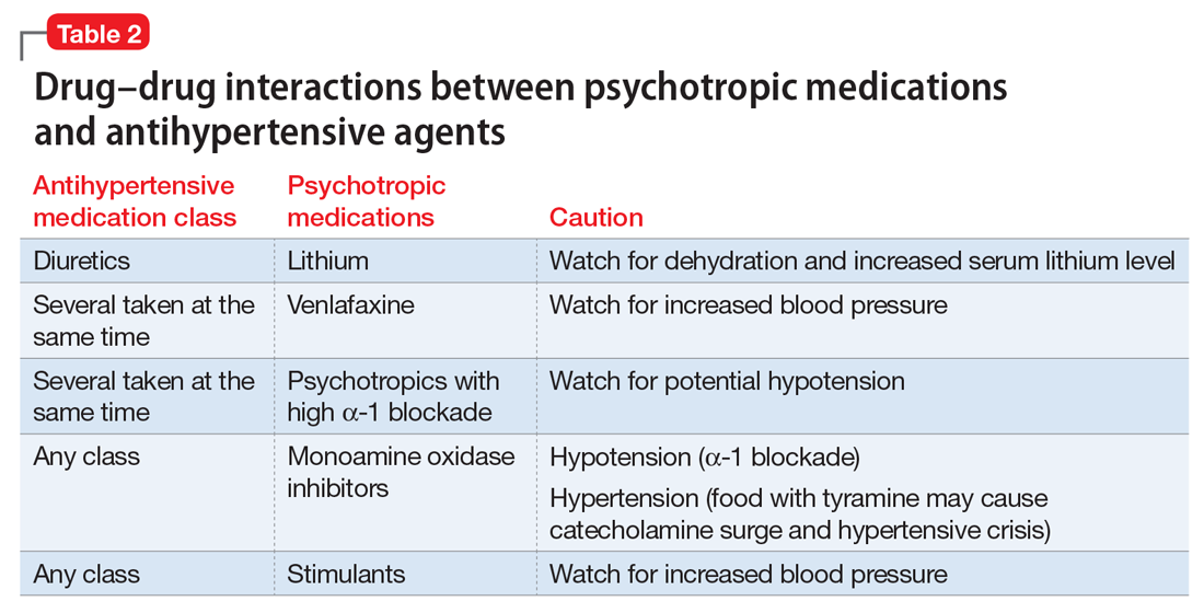
Psychotropic drugs. In psychiatric patients, the evaluation must consider the potential impact psychotropic drug effects and drug–drug interactions can have on blood pressure (Table 2). For example, patients taking both diuretics and lithium are at increased risk for dehydration and increased serum lithium levels, which could cause severe neurologic symptoms and renal insufficiency.16 Several antihypertensives when taken with venlafaxine can increase blood pressure, but antihypertensives with α-1 blocking psychotropics can decrease blood pressure. Monoamine oxidase inhibitors can cause hypotension or hypertension with various classes of antihypertensives. Stimulants, such as methylphenidate, atomoxetine, dextroamphetamine, armodafinil, or modafinil, alone or combined with antihypertensives, can cause hypertension.17
Substance abuse, particularly alcohol, methamphetamine, and cocaine, can cause difficulty controlling blood pressure. Patients with refractory hypertension should have a reassessment of substance abuse as a potential cause.
Screening guidelines for mental health providers
For many patients with severe mental illness, visits to their mental health providers might be their only contact with the medical system. Therefore, screening in the mental health settings could detect cases that otherwise would be missed.
Screening recommendations. The U.S. Preventive Services Task Force recommends screening for hypertension in the general population beginning at age 18.18 Adults age 18 to 39 with normal blood pressure (<130/85 mm Hg) and no other risk factors (eg, overweight, obese, or African American) can be screened every 3 years. Those with risk factors or a blood pressure of 130/85 to 139/89 mm Hg and adults age ≥40 should have annual screenings.
Ideally, psychiatrists and other mental health providers should monitor blood pressure at each visit, especially in patients taking psychotropics because of their higher risk for hypertension.
Optimizing treatment. Once the diagnosis of essential hypertension is established, identifying psychiatric comorbidities and the severity of psychiatric symptoms are important to optimize treatment adherence. Patients with increased depressive symptoms are less likely to comply with antihypertensive medication,19 and patients with confirmed depression are 3 times more likely to not adhere to medical treatment recommendations than non-depressed patients.20
Physicians’ attitudes toward hypertension also can affect patients’ compliance and blood pressure control.21 Psychiatrists should be empathetic and motivational toward patients attempting to control their blood pressure. The Seventh Joint National Committee on the Prevention, Detection, Evaluation, and Treatment of High Blood Pressure states, “Motivation improves when patients have positive experiences with, and trust in, the clinician. Empathy builds trust and is a potent motivator.”22
Treatment and management
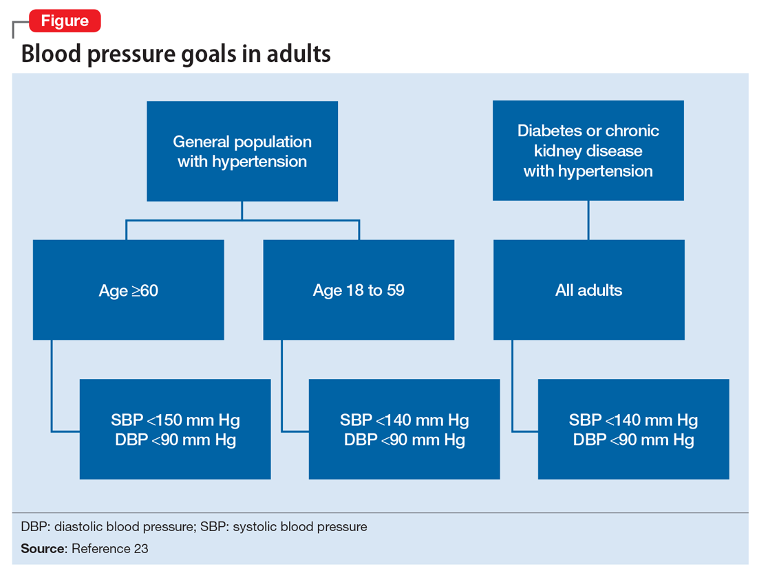
Treatment of hypertension significantly reduces the risk of stroke, myocardial infarction, renal injury, heart failure, and premature death. Studies show that treatment that reduces systolic blood pressure by 12 mm Hg over 10 years will prevent 1 death for every 11 patients with essential hypertension. In those with concomitant cardiovascular disease or target organ damage, such a reduction would prevent death in 1 of every 9 patients treated.15Blood pressure goals. The 2014 Eighth Joint National Committee Guideline for Management of High Blood Pressure in Adults provides guidance on blood pressure goals depending on patients’ underlying medical history (Figure).23 Based on expert opinion and randomized controlled studies, blood pressure goals for patients without diabetes or chronic kidney disease (CKD)—an estimated or measured glomerular filtration rate (GFR) of ≤60 mL/min/1.73 m2—depend on age: <140/90 mm Hg for age 18 to 59 and <150/90 mm Hg for age ≥60. For patients with diabetes or CKD, the blood pressure goal is <140/90 mm Hg, regardless of age.
However, not all experts agree on these specific blood pressure goals. A major trial (SPRINT) published in 2015 found that intensive blood pressure goals do benefit higher-risk, non-diabetic patients.24 Specifically, the study randomized patients age ≥50 with systolic blood pressure of 130 to 180 mm Hg and increased cardiovascular risk to systolic blood pressure targets of <140 mm Hg (standard) or <120 mm Hg (intensive). Characteristics of increased cardiovascular risk were clinical or subclinical cardiovascular disease other than stroke, CKD with GFR of 20 to 60 mL/min/1.73 m2, age ≥75, or Framingham 10-year coronary heart disease risk score ≥15%. Intensive treatment significantly reduced overall mortality and the rate of acute coronary syndrome, myocardial infarction, heart failure, stroke, or cardiovascular death. However, the results of this study have not been assimilated into any recent guidelines. Therefore, consider a goal of <120 mm Hg for non-diabetic patients age ≥50 with any of these factors.
Lifestyle modifications. Psychiatrists are well equipped to motivate and encourage behavioral modification in patients with hypertension. Counseling and structured training courses could help to effectively lower blood pressure.25 Patients should receive education on lifestyle modifications including:
- weight reduction
- physical activity
- moderate alcohol consumption
- decreased sodium consumption
- implementation of the Dietary Approaches to Stop Hypertension (DASH) or Mediterranean diets.15
Maintaining a normal body weight is ideal, but weight reduction of 10 lb can reduce blood pressure in overweight patients. The DASH diet, consisting of fruits, vegetables, low-fat dairy products, high calcium and potassium intake, and reduced saturated and total fat intake can decrease systolic blood pressure from 8 to 14 mm Hg. Reduction of sodium intake to ≤2,400 mg/d can reduce systolic blood pressure from 2 to 8 mm Hg. Regular aerobic exercise of 30 minutes a day most days of the week can reduce systolic blood pressure up to 9 mm Hg. Patients also should be encouraged to quit smoking. Patients who implement ≥2 these modifications get better results.
Antihypertensive medications. Patients who do not reach their goals with lifestyle measures alone should receive antihypertensive medications. Most patients will require ≥2 agents to control their blood pressure. Clinical trials show that some patient subgroups have better outcomes with different first-line agents.
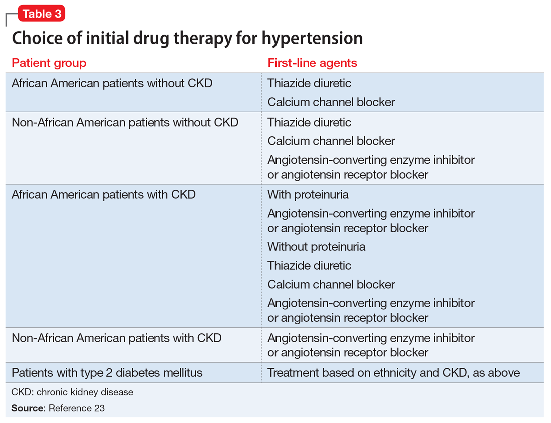
For example, in non-African American patients, thiazide diuretics, calcium channel blockers, angiotensin receptor blockers, and angiotensin-converting enzyme inhibitors are first-line treatments (Table 3). For African American patients without CKD, first-line treatments should be thiazide diuretics and calcium channel blockers, because angiotensin-converting enzyme inhibitors and angiotensin receptor blockers do not reduce cardiovascular events as effectively. African American patients with CKD and proteinuria, however, benefit from angiotensin-converting enzyme inhibitors or angiotensin receptor blockers and are preferred first-line agents. However, blood pressure control is a more important factor in improving outcomes than the choice of medication.
Psychiatrists’ role. Psychiatrists should aim to collaborate with the primary care provider when treating hypertension. However, when integrative care is not possible, they should start a first-line medication with follow-up in 1 month or sooner for patients with severe hypertension (>160/100 mm Hg) or significant comorbidities (eg, CKD, congestive heart failure, coronary disease). Patients with blood pressure >160/100 mm Hg often are started on a thiazide diuretic with one other medication because a single agent usually does not achieve goal blood pressure. Patients with CKD need close monitoring of potassium and creatinine when starting angiotensin-converting enzyme inhibitor or angiotensin receptor blocker therapy, usually within 1 to 2 days of starting or adjusting their medication. Adjust or add medication dosages monthly until blood pressure goals are reached.
A general internist, cardiologist, or nephrologist who has expertise in managing complex cases should oversee care of a psychiatric patient in any of the following scenarios:
- suspected secondary cause of hypertension
- adverse reaction to antihypertensive medications
- complicated comorbid conditions (ie, creatinine >1.8 mg/dL, worsening renal failure, hyperkalemia, heart failure, coronary disease)
- blood pressure >180/120 mm Hg
- requires ≥3 antihypertensive medications.
Summing up
Hypertension is a significant comorbidity in many psychiatric patients, but usually is asymptomatic. Often the psychiatrist or other mental health provider will diagnose hypertension because of their frequent contact with these patients. Once the diagnosis is made, an initial evaluation can direct lifestyle modifications. Patients who continue to have significant elevation of blood pressure should start pharmacotherapy, either by the psychiatrist or by ensuring follow-up with a primary care physician. The psychiatrist may be able to manage cases of essential hypertension, but always must be vigilant for potential drug–disease or drug–drug interactions during treatment. A team-based approach may improve health outcomes in psychiatric patients.
1. Centers for Disease Control and Prevention (CDC). Vital signs: awareness and treatment of uncontrolled hypertension among adults—United States, 2003-2010. MMWR Morb Mortal Wkly Rep. 2012;61:703-709.
2. Mozzafarian D, Benjamin EJ, Go AS, et al; American Heart Association Statistics Committee and Stroke Statistics Subcommittee. Heart Disease and Stroke Statistics—2015 update: a report from the American Heart Association. Circulation. 2015;131(4):e29-e322.
3. Carroll D, Phillips AC, Gale CR, et al. Generalized anxiety and major depressive disorders, their comorbidity and hypertension in middle-aged men. Psychosom Med. 2010;72(1):16-19.
4. Leboyer M, Soreca I, Scott J, et al. Can bipolar disorder be viewed as a multi-system inflammatory disease? J Affect Disord. 2012;141(1):1-10.
5. Goff DC, Sullivan LM, McEvoy JP, et al. A comparison of ten-year cardiac risk estimates in schizophrenia patients from the CATIE study and matched controls. Schizophr Res. 2005;80(1):45-53.
6. Stein DJ, Aguilar-Gaxiola S, Alonso J, et al. Associations between mental disorders and subsequent onset of hypertension. Gen Hosp Psychiatry. 2014;36(2):142-149.
7. Birkenaes AB, Opjordsmoen S, Brunborg C, et al. The level of cardiovascular risk factors in bipolar disorder equals that of schizophrenia: a comparative study. J Clin Psychiatry. 2007;68(6):917-923.
8. Izzo JL, Black HR, Goodfriend TL. Hypertension primer: the essentials of high blood pressure. 4th ed. Philadelphia, PA: Lippincott Williams & Wilkins; 2008.
9. Osby U, Correia N, Brandt L, et al. Mortality and causes of death in schizophrenia in Stockholm County, Sweden. Schizophr Res. 2000;45(1-2):21-28.
10. Brown S, Inskip H, Barraclough B. Causes of the excess mortality of schizophrenia. Br J Psychiatry. 2000;177:212-217.
11. Auquier P, Lançon C, Rouillon F, et al. Mortality in schizophrenia. Pharmacoepidemiol Drug Saf. 2007;16(12):1308-1312.
12. Newcomer JW, Hennekens CH. Severe mental illness and risk of cardiovascular disease. JAMA. 2007;298(15):1794-1796.
13. Bowis J, Parvanova A, McDaid D, et al. Mental and Physical Health Charter: bridging the gap between mental and physical health. https://www.idf.org/sites/default/files/Mental%2520and%2520Physical%2520Health%2520Charter%2520-%2520FINAL.pdf. Published October 7, 2009. Accessed March 6, 2017.
14. Nasrallah HA, Meyer JM, Goff DC, et al. Low rates of treatment for hypertension, dyslipidemia and diabetes in schizophrenia: data from the CATIE schizophrenia trial sample at baseline. Schizophr Res. 2006;86(1-3):15-22.
15. Chobanian AV, Bakris GL, Black HR, et al; National Heart, Lung, and Blood Institute Joint National Committee on Prevention, Detection, Evaluation, and Treatment of High Blood Pressure; National High Blood Pressure Education Program Coordinating Committee. The Seventh Report of the Joint National Committee on Prevention, Detection, Evaluation, and Treatment of High Blood Pressure: the JNC 7 report. JAMA. 2003;289(19):2560-2571.
16. Handler J. Lithium and antihypertensive medication: a potentially dangerous interaction. J Clin Hypertens (Greenwich). 2009;11(12):738-742.
17. National Collaborating Centre for Mental Health (UK). Depression in adults with a chronic physical health problem: treatment and Management. Appendix 16: table of drug interactions. http://www.ncbi.nlm.nih.gov/books/NBK82914. Published 2010. Accessed March 6, 2017.
18. Siu AL; U.S. Preventive Services Task Force. Screening for high blood pressure in adults: U.S. Preventive Services Task Force recommendation statement. Ann Intern Med. 2015:163(10):778-786.
19. Wang PS, Bohn RL, Knight E, et al. Noncompliance with antihypertensive medications: the impact of depressive symptoms and psychosocial factors. J Gen Intern Med. 2002;17(7):504-511.
20. DiMatteo MR, Lepper HS, Croghan TW. Depression is a risk factor for noncompliance with medical treatment: meta-analysis of the effects of anxiety and depression on patient adherence. Arch Intern Med. 2000;160(14):2101-2107.
21. Consoli SM, Lemogne C, Levy A, et al. Physicians’ degree of motivation regarding their perception of hypertension, and blood pressure control. J Hypertens. 2010;28(6):1330-1339.
22. National High Blood Pressure Education Program. The Seventh Report of the Joint National Committee on Prevention, Detection, Evaluation, and Treatment of High Blood Pressure. Improving Hypertension Control. Bethesda, MD: U.S. Department of Health and Human Services; 2004:61-64.
23. James PA, Oparil S, Carter BL, et al. 2014 evidence-based guideline for the management of high blood pressure in adults: report from the panel members appointed to the Eighth Joint National Committee (JNC 8). JAMA. 2014;311(5):507-520.
24. The SPRINT Research Group; Wright JT Jr, Williamson JD, et al. A randomized trial of intensive versus standard blood-pressure control. N Engl J Med. 2015;373(22):2103-2016.
25. Boulware LE, Daumit GL, Frick KD, et al. An evidence-based review of patient-centered behavioral interventions for hypertension. Am J Prev Med. 2001;21(3):221-232.
Hypertension is a widespread, under-recognized, and undertreated cause of morbidity and mortality in the United States and is associated with several psychiatric illnesses. Left untreated, hypertension can have significant consequences, including increased risk of stroke, coronary heart disease, heart failure, chronic kidney failure, and death. Approximately 70 million adults in the United States have hypertension, but only 60% of them have been diagnosed, and of those only 50% have their blood pressure under control.1 In 2013, 360,000 deaths in the United States were attributed to hypertension.2
Hypertension is associated with major depressive disorder, generalized anxiety disorder, bipolar disorder, and schizophrenia.3-5 Additionally, impulsive eating disorders, substance abuse, anxiety, and depression are associated with a hypertension diagnosis, although patients with panic disorder develop hypertension at a younger age.6 A 2007 study found a 61% prevalence of hypertension in those with bipolar disorder compared with 41% among the general population.7 The strong link between bipolar disorder and hypertension might be because of a common disease mechanism; both are associated with hyperactive cellular calcium signaling and increased platelet intracellular calcium ion concentrations.8
Hypertension not only is common among patients with psychiatric illness, it likely contributes to worse clinical outcomes. Studies across different cultures have found higher mortality rates in individuals with mental illness.9-11 Persons with schizophrenia and other severe mental illnesses may lose ≥25 years of life expectancy, with the primary cause of death being cardiovascular disease, not suicide.12 Patients with depression have a 50% greater risk of cardiovascular disease, which is equivalent to the risk of smoking.13
Schizophrenia is strongly associated with numerous comorbidities and has been linked significantly to an elevated 10-year cardiac risk after controlling for body mass index.5 The high rate of non-treatment of hypertension for patients with schizophrenia (62.4%) is especially concerning.14
Because of the well-documented morbidity and mortality of hypertension and its increased prevalence and undertreatment in the psychiatric population, mental health providers are in an important position to recognize hypertension and evaluate its inherent risks to direct their patients toward proper treatment. This article reviews:
- the signs and symptoms of hypertension
- the mental health provider’s role in the evaluation and diagnosis
- how psychotropic drugs influence blood pressure and drug–drug interactions
- the management of hypertension in psychiatric patients, including strategies for counseling and lifestyle management.
Diagnosing hypertension
Hypertension is defined as a blood pressure >140/90 mm Hg, the average of ≥2 properly measured readings at ≥2 visits in a medical setting.15 The proper equipment, including a well-fitting blood pressure cuff, and technique to measure blood pressure are essential to avoid misdiagnosis. The patient should be at rest for ≥5 minutes, without active pain or emotional distress.
Most cases of hypertension (90% to 95%) are primary, commonly called essential hypertension. However, the differential diagnosis also should consider secondary causes, which may include:
- obesity
- medications
- chronic alcohol use
- methamphetamine or cocaine use
- primary kidney disease
- atherosclerotic renal artery stenosis
- obstructive sleep apnea
- hypothyroidism
- primary hyperaldosteronism
- narrowing of the aorta
- Cushing syndrome
- primary hyperparathyroidism
- polycythemia
- pheochromocytoma.
Medical evaluation. Once the diagnosis of hypertension is made, a medical evaluation is indicated to determine if the patient has end-organ damage from the elevated pressures, such as renal disease or heart disease, to identify other modifiable cardiovascular risk factors, such as hyperlipidemia, and to screen for secondary causes of hypertension. This evaluation includes15:
- a physical exam
- review of medications
- lipid profile
- urinalysis to screen for proteinuria
- serum electrolytes and creatinine
- electrocardiogram to screen for left ventricular hypertrophy or prior infarction
- fasting glucose or hemoglobin A1c to screen for type 2 diabetes mellitus.
Psychotropic drugs. In psychiatric patients, the evaluation must consider the potential impact psychotropic drug effects and drug–drug interactions can have on blood pressure (Table 2). For example, patients taking both diuretics and lithium are at increased risk for dehydration and increased serum lithium levels, which could cause severe neurologic symptoms and renal insufficiency.16 Several antihypertensives when taken with venlafaxine can increase blood pressure, but antihypertensives with α-1 blocking psychotropics can decrease blood pressure. Monoamine oxidase inhibitors can cause hypotension or hypertension with various classes of antihypertensives. Stimulants, such as methylphenidate, atomoxetine, dextroamphetamine, armodafinil, or modafinil, alone or combined with antihypertensives, can cause hypertension.17
Substance abuse, particularly alcohol, methamphetamine, and cocaine, can cause difficulty controlling blood pressure. Patients with refractory hypertension should have a reassessment of substance abuse as a potential cause.
Screening guidelines for mental health providers
For many patients with severe mental illness, visits to their mental health providers might be their only contact with the medical system. Therefore, screening in the mental health settings could detect cases that otherwise would be missed.
Screening recommendations. The U.S. Preventive Services Task Force recommends screening for hypertension in the general population beginning at age 18.18 Adults age 18 to 39 with normal blood pressure (<130/85 mm Hg) and no other risk factors (eg, overweight, obese, or African American) can be screened every 3 years. Those with risk factors or a blood pressure of 130/85 to 139/89 mm Hg and adults age ≥40 should have annual screenings.
Ideally, psychiatrists and other mental health providers should monitor blood pressure at each visit, especially in patients taking psychotropics because of their higher risk for hypertension.
Optimizing treatment. Once the diagnosis of essential hypertension is established, identifying psychiatric comorbidities and the severity of psychiatric symptoms are important to optimize treatment adherence. Patients with increased depressive symptoms are less likely to comply with antihypertensive medication,19 and patients with confirmed depression are 3 times more likely to not adhere to medical treatment recommendations than non-depressed patients.20
Physicians’ attitudes toward hypertension also can affect patients’ compliance and blood pressure control.21 Psychiatrists should be empathetic and motivational toward patients attempting to control their blood pressure. The Seventh Joint National Committee on the Prevention, Detection, Evaluation, and Treatment of High Blood Pressure states, “Motivation improves when patients have positive experiences with, and trust in, the clinician. Empathy builds trust and is a potent motivator.”22
Treatment and management
Treatment of hypertension significantly reduces the risk of stroke, myocardial infarction, renal injury, heart failure, and premature death. Studies show that treatment that reduces systolic blood pressure by 12 mm Hg over 10 years will prevent 1 death for every 11 patients with essential hypertension. In those with concomitant cardiovascular disease or target organ damage, such a reduction would prevent death in 1 of every 9 patients treated.15Blood pressure goals. The 2014 Eighth Joint National Committee Guideline for Management of High Blood Pressure in Adults provides guidance on blood pressure goals depending on patients’ underlying medical history (Figure).23 Based on expert opinion and randomized controlled studies, blood pressure goals for patients without diabetes or chronic kidney disease (CKD)—an estimated or measured glomerular filtration rate (GFR) of ≤60 mL/min/1.73 m2—depend on age: <140/90 mm Hg for age 18 to 59 and <150/90 mm Hg for age ≥60. For patients with diabetes or CKD, the blood pressure goal is <140/90 mm Hg, regardless of age.
However, not all experts agree on these specific blood pressure goals. A major trial (SPRINT) published in 2015 found that intensive blood pressure goals do benefit higher-risk, non-diabetic patients.24 Specifically, the study randomized patients age ≥50 with systolic blood pressure of 130 to 180 mm Hg and increased cardiovascular risk to systolic blood pressure targets of <140 mm Hg (standard) or <120 mm Hg (intensive). Characteristics of increased cardiovascular risk were clinical or subclinical cardiovascular disease other than stroke, CKD with GFR of 20 to 60 mL/min/1.73 m2, age ≥75, or Framingham 10-year coronary heart disease risk score ≥15%. Intensive treatment significantly reduced overall mortality and the rate of acute coronary syndrome, myocardial infarction, heart failure, stroke, or cardiovascular death. However, the results of this study have not been assimilated into any recent guidelines. Therefore, consider a goal of <120 mm Hg for non-diabetic patients age ≥50 with any of these factors.
Lifestyle modifications. Psychiatrists are well equipped to motivate and encourage behavioral modification in patients with hypertension. Counseling and structured training courses could help to effectively lower blood pressure.25 Patients should receive education on lifestyle modifications including:
- weight reduction
- physical activity
- moderate alcohol consumption
- decreased sodium consumption
- implementation of the Dietary Approaches to Stop Hypertension (DASH) or Mediterranean diets.15
Maintaining a normal body weight is ideal, but weight reduction of 10 lb can reduce blood pressure in overweight patients. The DASH diet, consisting of fruits, vegetables, low-fat dairy products, high calcium and potassium intake, and reduced saturated and total fat intake can decrease systolic blood pressure from 8 to 14 mm Hg. Reduction of sodium intake to ≤2,400 mg/d can reduce systolic blood pressure from 2 to 8 mm Hg. Regular aerobic exercise of 30 minutes a day most days of the week can reduce systolic blood pressure up to 9 mm Hg. Patients also should be encouraged to quit smoking. Patients who implement ≥2 these modifications get better results.
Antihypertensive medications. Patients who do not reach their goals with lifestyle measures alone should receive antihypertensive medications. Most patients will require ≥2 agents to control their blood pressure. Clinical trials show that some patient subgroups have better outcomes with different first-line agents.
For example, in non-African American patients, thiazide diuretics, calcium channel blockers, angiotensin receptor blockers, and angiotensin-converting enzyme inhibitors are first-line treatments (Table 3). For African American patients without CKD, first-line treatments should be thiazide diuretics and calcium channel blockers, because angiotensin-converting enzyme inhibitors and angiotensin receptor blockers do not reduce cardiovascular events as effectively. African American patients with CKD and proteinuria, however, benefit from angiotensin-converting enzyme inhibitors or angiotensin receptor blockers and are preferred first-line agents. However, blood pressure control is a more important factor in improving outcomes than the choice of medication.
Psychiatrists’ role. Psychiatrists should aim to collaborate with the primary care provider when treating hypertension. However, when integrative care is not possible, they should start a first-line medication with follow-up in 1 month or sooner for patients with severe hypertension (>160/100 mm Hg) or significant comorbidities (eg, CKD, congestive heart failure, coronary disease). Patients with blood pressure >160/100 mm Hg often are started on a thiazide diuretic with one other medication because a single agent usually does not achieve goal blood pressure. Patients with CKD need close monitoring of potassium and creatinine when starting angiotensin-converting enzyme inhibitor or angiotensin receptor blocker therapy, usually within 1 to 2 days of starting or adjusting their medication. Adjust or add medication dosages monthly until blood pressure goals are reached.
A general internist, cardiologist, or nephrologist who has expertise in managing complex cases should oversee care of a psychiatric patient in any of the following scenarios:
- suspected secondary cause of hypertension
- adverse reaction to antihypertensive medications
- complicated comorbid conditions (ie, creatinine >1.8 mg/dL, worsening renal failure, hyperkalemia, heart failure, coronary disease)
- blood pressure >180/120 mm Hg
- requires ≥3 antihypertensive medications.
Summing up
Hypertension is a significant comorbidity in many psychiatric patients, but usually is asymptomatic. Often the psychiatrist or other mental health provider will diagnose hypertension because of their frequent contact with these patients. Once the diagnosis is made, an initial evaluation can direct lifestyle modifications. Patients who continue to have significant elevation of blood pressure should start pharmacotherapy, either by the psychiatrist or by ensuring follow-up with a primary care physician. The psychiatrist may be able to manage cases of essential hypertension, but always must be vigilant for potential drug–disease or drug–drug interactions during treatment. A team-based approach may improve health outcomes in psychiatric patients.
1. Centers for Disease Control and Prevention (CDC). Vital signs: awareness and treatment of uncontrolled hypertension among adults—United States, 2003-2010. MMWR Morb Mortal Wkly Rep. 2012;61:703-709.
2. Mozzafarian D, Benjamin EJ, Go AS, et al; American Heart Association Statistics Committee and Stroke Statistics Subcommittee. Heart Disease and Stroke Statistics—2015 update: a report from the American Heart Association. Circulation. 2015;131(4):e29-e322.
3. Carroll D, Phillips AC, Gale CR, et al. Generalized anxiety and major depressive disorders, their comorbidity and hypertension in middle-aged men. Psychosom Med. 2010;72(1):16-19.
4. Leboyer M, Soreca I, Scott J, et al. Can bipolar disorder be viewed as a multi-system inflammatory disease? J Affect Disord. 2012;141(1):1-10.
5. Goff DC, Sullivan LM, McEvoy JP, et al. A comparison of ten-year cardiac risk estimates in schizophrenia patients from the CATIE study and matched controls. Schizophr Res. 2005;80(1):45-53.
6. Stein DJ, Aguilar-Gaxiola S, Alonso J, et al. Associations between mental disorders and subsequent onset of hypertension. Gen Hosp Psychiatry. 2014;36(2):142-149.
7. Birkenaes AB, Opjordsmoen S, Brunborg C, et al. The level of cardiovascular risk factors in bipolar disorder equals that of schizophrenia: a comparative study. J Clin Psychiatry. 2007;68(6):917-923.
8. Izzo JL, Black HR, Goodfriend TL. Hypertension primer: the essentials of high blood pressure. 4th ed. Philadelphia, PA: Lippincott Williams & Wilkins; 2008.
9. Osby U, Correia N, Brandt L, et al. Mortality and causes of death in schizophrenia in Stockholm County, Sweden. Schizophr Res. 2000;45(1-2):21-28.
10. Brown S, Inskip H, Barraclough B. Causes of the excess mortality of schizophrenia. Br J Psychiatry. 2000;177:212-217.
11. Auquier P, Lançon C, Rouillon F, et al. Mortality in schizophrenia. Pharmacoepidemiol Drug Saf. 2007;16(12):1308-1312.
12. Newcomer JW, Hennekens CH. Severe mental illness and risk of cardiovascular disease. JAMA. 2007;298(15):1794-1796.
13. Bowis J, Parvanova A, McDaid D, et al. Mental and Physical Health Charter: bridging the gap between mental and physical health. https://www.idf.org/sites/default/files/Mental%2520and%2520Physical%2520Health%2520Charter%2520-%2520FINAL.pdf. Published October 7, 2009. Accessed March 6, 2017.
14. Nasrallah HA, Meyer JM, Goff DC, et al. Low rates of treatment for hypertension, dyslipidemia and diabetes in schizophrenia: data from the CATIE schizophrenia trial sample at baseline. Schizophr Res. 2006;86(1-3):15-22.
15. Chobanian AV, Bakris GL, Black HR, et al; National Heart, Lung, and Blood Institute Joint National Committee on Prevention, Detection, Evaluation, and Treatment of High Blood Pressure; National High Blood Pressure Education Program Coordinating Committee. The Seventh Report of the Joint National Committee on Prevention, Detection, Evaluation, and Treatment of High Blood Pressure: the JNC 7 report. JAMA. 2003;289(19):2560-2571.
16. Handler J. Lithium and antihypertensive medication: a potentially dangerous interaction. J Clin Hypertens (Greenwich). 2009;11(12):738-742.
17. National Collaborating Centre for Mental Health (UK). Depression in adults with a chronic physical health problem: treatment and Management. Appendix 16: table of drug interactions. http://www.ncbi.nlm.nih.gov/books/NBK82914. Published 2010. Accessed March 6, 2017.
18. Siu AL; U.S. Preventive Services Task Force. Screening for high blood pressure in adults: U.S. Preventive Services Task Force recommendation statement. Ann Intern Med. 2015:163(10):778-786.
19. Wang PS, Bohn RL, Knight E, et al. Noncompliance with antihypertensive medications: the impact of depressive symptoms and psychosocial factors. J Gen Intern Med. 2002;17(7):504-511.
20. DiMatteo MR, Lepper HS, Croghan TW. Depression is a risk factor for noncompliance with medical treatment: meta-analysis of the effects of anxiety and depression on patient adherence. Arch Intern Med. 2000;160(14):2101-2107.
21. Consoli SM, Lemogne C, Levy A, et al. Physicians’ degree of motivation regarding their perception of hypertension, and blood pressure control. J Hypertens. 2010;28(6):1330-1339.
22. National High Blood Pressure Education Program. The Seventh Report of the Joint National Committee on Prevention, Detection, Evaluation, and Treatment of High Blood Pressure. Improving Hypertension Control. Bethesda, MD: U.S. Department of Health and Human Services; 2004:61-64.
23. James PA, Oparil S, Carter BL, et al. 2014 evidence-based guideline for the management of high blood pressure in adults: report from the panel members appointed to the Eighth Joint National Committee (JNC 8). JAMA. 2014;311(5):507-520.
24. The SPRINT Research Group; Wright JT Jr, Williamson JD, et al. A randomized trial of intensive versus standard blood-pressure control. N Engl J Med. 2015;373(22):2103-2016.
25. Boulware LE, Daumit GL, Frick KD, et al. An evidence-based review of patient-centered behavioral interventions for hypertension. Am J Prev Med. 2001;21(3):221-232.
Head pain and psychiatric illness: Applying the biopsychosocial model to care
More than 45% of people worldwide suffer from headache at some point in their life.1 Head pain can lead to disability and functional decline, yet headache disorders often are underdiagnosed and poorly assessed. For example, 60% of migraine and tension-type headaches go undiagnosed and 50% of persons suffering from migraine have severe functional disability or require bed rest.2-4
Because head pain can be associated with secondary medical and psychiatric conditions, diagnosis can be challenging. This article reviews the medical and psychological aspects of major headaches and assists with clinical assessment. We present clinical interviewing tools and a diagram to enhance focused, efficient assessment and inform treatment plans.
Classification of headache
Headache is a common complaint, yet it is often underdiagnosed and ineffectively treated. The World Health Organization estimates that, globally, 50% of people with headache self-treat their pain.5 The International Headache Society classifies headache as primary or secondary; approximately 90% of complaints are from primary headache.6
Assessment and diagnosis of headache can be complex because of overlapping, subjective symptoms. It is important to have a general understanding of primary and secondary causes of headache so that interrelated symptoms do not obscure the most accurate diagnosis and effective treatment course. Although most headache complaints are benign, ruling out secondary causes helps gauge the likelihood of developing severe sequelae from underlying pathology.
By definition, primary headaches are idiopathic and commonly include migraine, tension-type, cluster, and hemicrania continua headache. Secondary headaches have an underlying pathology, which could improve by targeting the disorder. Common secondary causes of headache include:
• trauma
• vascular abnormalities
• structural abnormalities
• chemical (including medications)
• inflammation or infection
• metabolic conditions
• diseases of the neck and pericranial and intracranial structures
• psychiatric conditions.
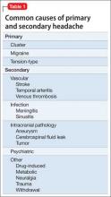
Table 1 illustrates common causes of head pain. More definitive criteria for symptoms and diagnosis can be found in the International Classification of Headache Disorders.7
Primary headache
Tension-type is the most common primary headache, accounting for more than one-half of all headaches.7 Patients usually describe a tight pain in a bilateral band-like distribution, which could be caused by sustained neck muscle contraction. Pain usually builds in intensity and can last 30 minutes to several days. There is a well-established association between emotional stress or depression and the development of tension-type headaches.8
Migraine typically causes pulsating pain in a localized area of the head that lasts as long as 72 hours and can be associated with nausea, vomiting, photophobia, phono-phobia, and aura. Patients report varying precipitating factors but commonly cite certain foods, menstruation, and sleep deprivation. Although rare, migraine with aura has been linked to ischemic stroke; most cases have been reported in female smokers and oral contraceptive users age <45.9
Because migraines can be debilitating, some patients—typically those with ≥4 attacks a month—opt for prophylactic medication. Effective prophylactics include amitriptyline, propranolol, divalproex sodium, and topiramate, which should be monitored closely and given a trial for several months before switching to another drug. Commonly used abortive treatments include triptans and anti-emetics such as metoclopramide.
Meperidine and ketorolac are popular second-line agents for migraine. Botulinum toxin A also has been used in severe cases to reduce the number of headache days in chronic migraine patients.6
Cluster headache is rare, but typically exhibits repeated burning and intense unilateral periorbital or retro-orbital pain that lasts 15 minutes to 3 hours over several weeks. Men are predominantly affected. Cluster headaches typically improve with oxygen treatment.
Biopsychosocial model of head pain
The biomedical model has helped iden tify pathophysiological pain mechanisms and pharmacotherapeutic agents for headache. However, during assessment, limiting one’s attention to the linear relationship between pathology, mechanism of action, and pain oversimplifies common questions clinicians face when assessing chronic head pain.
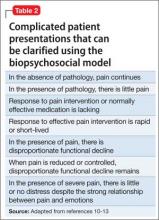
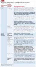
Advancements in the last 3 decades have expanded the conceptualization of head pain to integrate sociocultural, environmental, behavioral, affective, cognitive, and biological variables—otherwise known as the biopsychosocial model.10,11 The biopsychosocial model is a multidimensional theory that helps answer difficult clinical assessment questions and complex patient presentations (Table 2).10-13 Many unusual responses to pain treatment, questionable validity of pain behavior, and disproportionate pain perception and functional decline are explained by non-pathophysiological and non-biomechanical models.
Psychiatric comorbidity and head pain
Psychiatric conditions are highly prevalent among persons with primary headache. Verri et al14 found that 90% of chronic daily headache patients had ≥1 psychiatric condition; depression and anxiety were most common. Of concern, 1 study found that headache is associated with increased frequency of suicidal ideation among patients with chronic pain.15 It is critical for clinicians to screen for psychiatric comorbidities in patients with chronic headache. Conversely, clinicians might want to screen for headache in their patients with psychiatric illness.
Migraine. Mood disorders are common among patients who suffer from migraine. The rate of depression is 2 to 4 times higher in those with migraine compared with healthy controls.16,17 In a large-scale study, patients with migraine had a 1.9-fold higher risk (compared with controls) of having a comorbid depressive episode; a 2-fold higher risk of manic episodes; and a 3-fold higher risk of both mania and depression.18 In a study of 62 inpatients, Fasmer19 reported that 46% of patients with unipolar depression and 44% of patients with bipolar disorder experienced migraine (77% of the bipolar disorder patients with migraine had bipolar II disorder). Patients with migraine are at increased risk of suicide attempts (odds ratio 4.3; 95% CI, 1.2-15.7).20
Tension-type headache. The relationship between psychiatric comorbidity in tension-type headache is well established. In contrast to what is seen with migraines, Puca et al21 found a higher prevalence of anxiety disorders (52.5%) than depressive disorders (36.4%) in patients with tension-type headache. Generalized anxiety disorder was one of the most prevalent anxiety conditions (83.3%), and dysthymia was the most prevalent mood disorder (45.6%). In the same study, 21.7% of patients were found to have a comorbid somatoform disorder.21
Emotional and cognitive factors can co-occur in patients with tension-type headache and a comorbid psychiatric condition. For example, difficulty identifying or recognizing emotions—commonly referred to as alexithymia—has been linked to tension-type headache.22 Additionally, maladaptive cognitive appraisal of stress is more common among patients with tension-type headache when compared with those without headaches.23 Being mindful of and recognizing these co-occurring emotional and cognitive factors will help clinicians construct a more accurate assessment and effective behavioral treatment plan.
Clinical assessment with a useful mnemonic
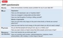
Clinical assessment of psychiatric illness is essential when evaluating chronic pain patients. Using the acronym AMPS (Anxiety, Mood, Psychosis, and Substance use disorders) (Table 3) is an efficient way for the clinician to ask pertinent questions regarding common psychiatric conditions that could have a direct effect on chronic pain.24 Head pain can be more intense when combined with untreated anxiety, depression, psychosis, or a substance use disorder. Untreated anxiety, for example, can amplify sympathetic response to pain and complicate treatment.
Investigating head pain patients for an underlying mood disorder is essential to providing successful treatment. Consider:
• starting psychotherapy modalities that address both pain and psychiatric illness, such as cognitive-behavioral therapy (CBT)
• reframing unhelpful pain beliefs
• managing activity-rest levels
• biofeedback
• supportive group therapy
• reducing family members’ reinforcement of the patient’s pain behavior or sick role.25
Assessing for somatic symptom disorders
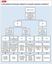
In addition to using the AMPS approach for psychiatric assessment, clinicians should evaluate for somatization, which can present as head pain. Somatic symptom disorders (SSD) are a class of conditions that are impacted by affective, cognitive, and reinforcing factors that might or might not be consciously or intentionally produced. Patients with an SSD have somatic symptoms that are distressing or cause significant disruption of daily life because of excessive thoughts, feelings, or behaviors related to the somatic symptoms, for ≥6 months. The Figure outlines SSD, related conditions, and their respective prominent symptoms to assist in the differential diagnosis.26
Note that some headache conditions present with severe distress because of their abrupt onset and severity of symptoms (eg, cluster headaches). Therefore, the expectation and likelihood of psychological disturbance should be factored into a diagnosis of SSD and related conditions as seen in the Figure.
Secondary factors of unusual pain behavior or treatment response. The role of thoughts, affect, and behaviors is clinically meaningful in understanding SSD and similar conditions. Specific questions about cultural beliefs and rituals as they relate to exacerbations of head pain are of value. Table 413,27 lists behavioral, cognitive, and affective dimensions of head pain using the biopsychosocial model, and further clarifies common questions that arise with unusual pain response and complex patient presentations, which were outlined in the beginning of the article.
Because depression and anxiety can be comorbid with head pain, it is important to recognize psychological factors that contribute to pain perception. Indifference or denial of emotional stress as a result of severe pain and disability can imply a somatization process, which could suggest emotional disconnection or dissociation from somatic functioning.28 This finding can be a component of alexithymia, in which a person is disconnected from emotions and how emotions impact the body. Therefore, recognizing alexithymia assists in identifying psychological factors when patients deny mood symptoms, particularly in tension-type headache.
Functional assessment to rule out the disproportional impact of pain on daily activities is helpful in understanding the somatization process. Neurocognitive functioning should be assessed, particularly because frontal and subcortical dysregulation has been observed in head pain sufferers.29,30 Patients with cognitive changes as a result of a medical illness (eg, stroke, head concussion, brain tumor, or seizures) are especially at risk for neurocognitive dysfunction.
Neuropsychological assessment can be useful, not only to assess neurocognitive functioning (eg, Repeated Battery for the Assessment of Neuropsychological Status) but to identify objective test profiles associated with altered motivation (eg, Rey 15-Item Test, Minnesota Multiphasic Personality Inventory-2-Restructured Form F Scale, Personality Assessment Inventory [PAI] Negative Impression Management) and somatization processes (eg, PAI Somatization Scale). These instruments help to identify the severity of psychiatric and neurocognitive symptoms by comparing scores to normative (eg, healthy control group), clinical (eg, somatization, traumatic brain injury, mild cognitive impairment), and altered motivation (eg, persons instructed to exaggerate symptoms) databases.
If the clinician pursues neurocognitive assessment, direct referral to a neuropsychologist, referral to neurologist, or administration of a cognitive screening tool such as the Montreal Cognitive Assessment, Saint Louis University Mental Status, or Cognitive Log is recommended. If the cognitive screening is positive, next steps include: referring for full neuropsychological assessment, which includes complete cognitive and motor testing, personality testing, and integration of neuroimaging data (eg, MRI, CT scans, and/or EEG).
Assessing the patients’ self-talk or thought patterns as they describe their head pain will help clinicians understand belief systems that may be distorting the reality of the medical condition. For example, a patient might report that “my pain feels like someone is hitting me with an axe”; this is a catastrophic thought that can distort the clarity and perceptibility of pain. Encouraging patients to monitor and analyze their anxiety and associated negative thoughts is an important strategy for improving mood and decreasing somatization. Recording daily thoughts and CBT can help the patient identify and appropriately address his (her) cognitive distortions and futile thinking.
When implementing a treatment plan for somatization disorder, we propose the mnemonic device CARE MD:
• CBT
• Assess (by ruling out a medical cause for somatic complaints)
• Regular visits
• Empathy
• Med-psych interface (help the patient connect physical complaints and emotional stressors)
• Do no harm.
Clinical recommendations
Chronic head pain can be debilitating; psychodiagnostic assessment should therefore be considered an important part of the diagnosis and treatment plan. After ruling out common and emergent primary or secondary causes of head pain, consider psychiatric comorbidities. Depression and anxiety have a strong bidirectional relationship with chronic headache; therefore, we suggest evaluating patients with the intention of alleviating both psychiatric symptoms and head pain.
It is important to diligently assess for common psychiatric comorbidities; using the AMPS and CARE MD mnemonics, along with screening for somatization disorders, is an easy and effective way to evaluate for relevant psychiatric conditions associated with chronic head pain. Because many patients have unusual and complicated responses to head pain that can be explained by non-pathophysiological and non-biomechanical models, using the biopsychosocial model is essential for effective diagnosis, assessment, and treatment. Abortive and prophylactic medical interventions, as well as behavioral, sociocultural, and cognitive assessment, are vital to a comprehensive treatment approach.
Bottom Line
The psychodiagnostic assessment can help the astute clinician identify comorbid psychiatric conditions, psychological factors, and somatic symptoms to develop a comprehensive biopsychosocial treatment plan for patients with chronic head pain. Rule out primary and secondary causes of pain and screen for somatization disorders. Consider medication and psychotherapeutic treatment options.
Related Resources
• Pompili M, Di Cosimo D, Innamorati M, et al. Psychiatric comorbidity in patients with chronic daily headache and migraine: a selective overview including personality traits and suicide risk. J Headache Pain. 2009;10(4):283-290.
• Sinclair AJ, Sturrock A, Davies B, et al. Headache management: pharmacological approaches [published online July 3, 2015]. Pract Neurol. doi: 10.1136/practneurol-2015-001167.
Drug Brand Names
Amitriptyline • Elavil Meperidine • Demerol
Botulinum toxin A • Botox Metoclopramide • Reglan
Divalproex sodium • Depakote Propranolol • Inderide
Ketorolac • Toradol Topiramate • Topamax
Disclosures
The authors report no financial relationships with any company whose products are mentioned in this article or with manufacturers of competing products.
1. Stovner LJ, Hagen K, Jensen R, et al. The global burden of headache: a documentation of headache prevalence and disability worldwide. Cephalalgia. 2007;27(3):193-210.
2. Lipton RB, Stewart WF, Diamond S, et al. Prevalence and burden of migraine in the United States: data from the American Migraine Study II. Headache. 2001;41(7):646-657.
3. The World Health Report 2001: Mental health: new understanding new hope. Geneva, Switzerland: World Health Organization; 2001.
4. World Health Organization. The global burden of disease: 2004 update. http://www.who.int/healthinfo/global_burden_ disease/GBD_report_2004update_full.pdf. Published 2004. Accessed July 31, 2015.
5. World Health Organization. Headache disorders. http:// www.who.int/mediacentre/factsheets/fs277/en/. Published October 2012. Accessed July 31, 2015.
6. Clinch C. Evaluation & management of headache. In: South- Paul JE, Matheny SC, Lewis EL. eds. Current diagnosis & treatment in family medicine, 4th ed. New York, NY: McGraw-Hill; 2015:293-297.
7. Headache Classification Committee of the International Headache Society (IHS). The International Classification of Headache Disorders, 3rd edition (beta version). Cephalalgia. 2013;33(9):629-808.
8. Mawet J, Kurth T, Ayata C. Migraine and stroke: in search of shared mechanisms. Cephalalgia. 2015;35(2):165-181.
9. Janke EA, Holroyd KA, Romanek K. Depression increases onset of tension-type headache following laboratory stress. Pain. 2004;111(3):230-238.
10. Engel GL. The need for a new medical model: a challenge for biomedicine. Science. 1977;196(4286):129-136.
11. Engel GL. The clinical application of the biopsychosocial model. Am J Psychiatry. 1980;137(5):535-544.
12. Turk DC, Flor H. Chronic pain: a biobehavioral perspective. In: Gatchel RJ, Turk DC, eds. Psychosocial factors in pain: critical perspectives. New York, NY: Guilford Press; 1999:18-34.
13. Andrasik F, Flor H, Turk DC. An expanded view of psychological aspects in head pain: the biopsychosocial model. Neurol Sci. 2005;26(suppl 2):s87-s91.
14. Verri AP, Proietti Cecchini A, Galli C, et al. Psychiatric comorbidity in chronic daily headache. Cephalalgia. 1998; 18(suppl 21):45-49.
15. Ilgen MA, Zivin K, McCammon RJ, et al. Pain and suicidal thoughts, plans and attempts in the United States. Gen Hosp Psychiatry. 2008;30(6):521-527.
16. Kowacs F, Socal MP, Ziomkowski SC, et al. Symptoms of depression and anxiety, and screening for mental disorders in migrainous patients. Cephalalgia. 2003;23(2):79-89.
17. Hamelsky SW, Lipton RB. Psychiatric comorbidity of migraine. Headache. 2006;46(9):1327-1333.
18. Nguyen TV, Low NC. Comorbidity of migraine and mood episodes in a nationally representative population-based sample. Headache. 2013;53(3):498-506.
19. Fasmer OB. The prevalence of migraine in patients with bipolar and unipolar affective disorders. Cephalalgia. 2001; 21(9):894-899.
20. Breslau N. Migraine, suicidal ideation, and suicide attempts. Neurology. 1992;42(2):392-395.
21. Puca F, Genco S, Prudenzano MP, et al. Psychiatric comorbidity and psychosocial stress in patients with tension-type headache from headache centers in Italy. Cephalalgia. 1999;19(3):159-164.
22. Yücel B, Kora K, Ozyalçín S, et al. Depression, automatic thoughts, alexithymia, and assertiveness in patients with tension-type headache. Headache. 2002;42(3):194-199.
23. Wittrock DA, Myers TC. The comparison of individuals with recurrent tension-type headache and headache-free controls in physiological response, appraisal, and coping with stressors: a review of the literature. Ann Behav Med. 1998;20(2):118-134.
24. Onate J, Xiong G, McCarron R. The primary care psychiatric interview. In: McCarron R, Xiong G, Bourgeois J. Lippincott’s primary care psychiatry. Philadelphia, PA: Lippincott, Williams and Wilkins; 2009:3-4.
25. Songer D. Psychotherapeutic approaches in the treatment of pain. Psychiatry (Edgmont). 2005;2(5):19-24.
26. Diagnostic and statistical manual of mental disorders, 5th ed. Washington, DC: American Psychiatric Association; 2013.
27. Hashmi JA, Baliki MN, Huang L, et al. Shape shifting pain: chronification of back pain shifts brain representation from nociceptive to emotional circuits. Brain. 2013;136(pt 9):2751-2768.
28. Packard RC. Conversion headache. Headache. 1980;20(5):266-268.
29. Mongini F, Keller R, Deregibus A, et al. Frontal lobe dysfunction in patients with chronic migraine: a clinical-neuropsychological study. Psychiatry Res. 2005;133(1):101-106.
30. Martelli MF, Grayson RL, Zasler ND. Posttraumatic headache: neuropsychological and psychological effects and treatment implications. J Head Trauma Rehabil. 1999;14(1):49-69.
More than 45% of people worldwide suffer from headache at some point in their life.1 Head pain can lead to disability and functional decline, yet headache disorders often are underdiagnosed and poorly assessed. For example, 60% of migraine and tension-type headaches go undiagnosed and 50% of persons suffering from migraine have severe functional disability or require bed rest.2-4
Because head pain can be associated with secondary medical and psychiatric conditions, diagnosis can be challenging. This article reviews the medical and psychological aspects of major headaches and assists with clinical assessment. We present clinical interviewing tools and a diagram to enhance focused, efficient assessment and inform treatment plans.
Classification of headache
Headache is a common complaint, yet it is often underdiagnosed and ineffectively treated. The World Health Organization estimates that, globally, 50% of people with headache self-treat their pain.5 The International Headache Society classifies headache as primary or secondary; approximately 90% of complaints are from primary headache.6
Assessment and diagnosis of headache can be complex because of overlapping, subjective symptoms. It is important to have a general understanding of primary and secondary causes of headache so that interrelated symptoms do not obscure the most accurate diagnosis and effective treatment course. Although most headache complaints are benign, ruling out secondary causes helps gauge the likelihood of developing severe sequelae from underlying pathology.
By definition, primary headaches are idiopathic and commonly include migraine, tension-type, cluster, and hemicrania continua headache. Secondary headaches have an underlying pathology, which could improve by targeting the disorder. Common secondary causes of headache include:
• trauma
• vascular abnormalities
• structural abnormalities
• chemical (including medications)
• inflammation or infection
• metabolic conditions
• diseases of the neck and pericranial and intracranial structures
• psychiatric conditions.
Table 1 illustrates common causes of head pain. More definitive criteria for symptoms and diagnosis can be found in the International Classification of Headache Disorders.7
Primary headache
Tension-type is the most common primary headache, accounting for more than one-half of all headaches.7 Patients usually describe a tight pain in a bilateral band-like distribution, which could be caused by sustained neck muscle contraction. Pain usually builds in intensity and can last 30 minutes to several days. There is a well-established association between emotional stress or depression and the development of tension-type headaches.8
Migraine typically causes pulsating pain in a localized area of the head that lasts as long as 72 hours and can be associated with nausea, vomiting, photophobia, phono-phobia, and aura. Patients report varying precipitating factors but commonly cite certain foods, menstruation, and sleep deprivation. Although rare, migraine with aura has been linked to ischemic stroke; most cases have been reported in female smokers and oral contraceptive users age <45.9
Because migraines can be debilitating, some patients—typically those with ≥4 attacks a month—opt for prophylactic medication. Effective prophylactics include amitriptyline, propranolol, divalproex sodium, and topiramate, which should be monitored closely and given a trial for several months before switching to another drug. Commonly used abortive treatments include triptans and anti-emetics such as metoclopramide.
Meperidine and ketorolac are popular second-line agents for migraine. Botulinum toxin A also has been used in severe cases to reduce the number of headache days in chronic migraine patients.6
Cluster headache is rare, but typically exhibits repeated burning and intense unilateral periorbital or retro-orbital pain that lasts 15 minutes to 3 hours over several weeks. Men are predominantly affected. Cluster headaches typically improve with oxygen treatment.
Biopsychosocial model of head pain
The biomedical model has helped iden tify pathophysiological pain mechanisms and pharmacotherapeutic agents for headache. However, during assessment, limiting one’s attention to the linear relationship between pathology, mechanism of action, and pain oversimplifies common questions clinicians face when assessing chronic head pain.
Advancements in the last 3 decades have expanded the conceptualization of head pain to integrate sociocultural, environmental, behavioral, affective, cognitive, and biological variables—otherwise known as the biopsychosocial model.10,11 The biopsychosocial model is a multidimensional theory that helps answer difficult clinical assessment questions and complex patient presentations (Table 2).10-13 Many unusual responses to pain treatment, questionable validity of pain behavior, and disproportionate pain perception and functional decline are explained by non-pathophysiological and non-biomechanical models.
Psychiatric comorbidity and head pain
Psychiatric conditions are highly prevalent among persons with primary headache. Verri et al14 found that 90% of chronic daily headache patients had ≥1 psychiatric condition; depression and anxiety were most common. Of concern, 1 study found that headache is associated with increased frequency of suicidal ideation among patients with chronic pain.15 It is critical for clinicians to screen for psychiatric comorbidities in patients with chronic headache. Conversely, clinicians might want to screen for headache in their patients with psychiatric illness.
Migraine. Mood disorders are common among patients who suffer from migraine. The rate of depression is 2 to 4 times higher in those with migraine compared with healthy controls.16,17 In a large-scale study, patients with migraine had a 1.9-fold higher risk (compared with controls) of having a comorbid depressive episode; a 2-fold higher risk of manic episodes; and a 3-fold higher risk of both mania and depression.18 In a study of 62 inpatients, Fasmer19 reported that 46% of patients with unipolar depression and 44% of patients with bipolar disorder experienced migraine (77% of the bipolar disorder patients with migraine had bipolar II disorder). Patients with migraine are at increased risk of suicide attempts (odds ratio 4.3; 95% CI, 1.2-15.7).20
Tension-type headache. The relationship between psychiatric comorbidity in tension-type headache is well established. In contrast to what is seen with migraines, Puca et al21 found a higher prevalence of anxiety disorders (52.5%) than depressive disorders (36.4%) in patients with tension-type headache. Generalized anxiety disorder was one of the most prevalent anxiety conditions (83.3%), and dysthymia was the most prevalent mood disorder (45.6%). In the same study, 21.7% of patients were found to have a comorbid somatoform disorder.21
Emotional and cognitive factors can co-occur in patients with tension-type headache and a comorbid psychiatric condition. For example, difficulty identifying or recognizing emotions—commonly referred to as alexithymia—has been linked to tension-type headache.22 Additionally, maladaptive cognitive appraisal of stress is more common among patients with tension-type headache when compared with those without headaches.23 Being mindful of and recognizing these co-occurring emotional and cognitive factors will help clinicians construct a more accurate assessment and effective behavioral treatment plan.
Clinical assessment with a useful mnemonic
Clinical assessment of psychiatric illness is essential when evaluating chronic pain patients. Using the acronym AMPS (Anxiety, Mood, Psychosis, and Substance use disorders) (Table 3) is an efficient way for the clinician to ask pertinent questions regarding common psychiatric conditions that could have a direct effect on chronic pain.24 Head pain can be more intense when combined with untreated anxiety, depression, psychosis, or a substance use disorder. Untreated anxiety, for example, can amplify sympathetic response to pain and complicate treatment.
Investigating head pain patients for an underlying mood disorder is essential to providing successful treatment. Consider:
• starting psychotherapy modalities that address both pain and psychiatric illness, such as cognitive-behavioral therapy (CBT)
• reframing unhelpful pain beliefs
• managing activity-rest levels
• biofeedback
• supportive group therapy
• reducing family members’ reinforcement of the patient’s pain behavior or sick role.25
Assessing for somatic symptom disorders
In addition to using the AMPS approach for psychiatric assessment, clinicians should evaluate for somatization, which can present as head pain. Somatic symptom disorders (SSD) are a class of conditions that are impacted by affective, cognitive, and reinforcing factors that might or might not be consciously or intentionally produced. Patients with an SSD have somatic symptoms that are distressing or cause significant disruption of daily life because of excessive thoughts, feelings, or behaviors related to the somatic symptoms, for ≥6 months. The Figure outlines SSD, related conditions, and their respective prominent symptoms to assist in the differential diagnosis.26
Note that some headache conditions present with severe distress because of their abrupt onset and severity of symptoms (eg, cluster headaches). Therefore, the expectation and likelihood of psychological disturbance should be factored into a diagnosis of SSD and related conditions as seen in the Figure.
Secondary factors of unusual pain behavior or treatment response. The role of thoughts, affect, and behaviors is clinically meaningful in understanding SSD and similar conditions. Specific questions about cultural beliefs and rituals as they relate to exacerbations of head pain are of value. Table 413,27 lists behavioral, cognitive, and affective dimensions of head pain using the biopsychosocial model, and further clarifies common questions that arise with unusual pain response and complex patient presentations, which were outlined in the beginning of the article.
Because depression and anxiety can be comorbid with head pain, it is important to recognize psychological factors that contribute to pain perception. Indifference or denial of emotional stress as a result of severe pain and disability can imply a somatization process, which could suggest emotional disconnection or dissociation from somatic functioning.28 This finding can be a component of alexithymia, in which a person is disconnected from emotions and how emotions impact the body. Therefore, recognizing alexithymia assists in identifying psychological factors when patients deny mood symptoms, particularly in tension-type headache.
Functional assessment to rule out the disproportional impact of pain on daily activities is helpful in understanding the somatization process. Neurocognitive functioning should be assessed, particularly because frontal and subcortical dysregulation has been observed in head pain sufferers.29,30 Patients with cognitive changes as a result of a medical illness (eg, stroke, head concussion, brain tumor, or seizures) are especially at risk for neurocognitive dysfunction.
Neuropsychological assessment can be useful, not only to assess neurocognitive functioning (eg, Repeated Battery for the Assessment of Neuropsychological Status) but to identify objective test profiles associated with altered motivation (eg, Rey 15-Item Test, Minnesota Multiphasic Personality Inventory-2-Restructured Form F Scale, Personality Assessment Inventory [PAI] Negative Impression Management) and somatization processes (eg, PAI Somatization Scale). These instruments help to identify the severity of psychiatric and neurocognitive symptoms by comparing scores to normative (eg, healthy control group), clinical (eg, somatization, traumatic brain injury, mild cognitive impairment), and altered motivation (eg, persons instructed to exaggerate symptoms) databases.
If the clinician pursues neurocognitive assessment, direct referral to a neuropsychologist, referral to neurologist, or administration of a cognitive screening tool such as the Montreal Cognitive Assessment, Saint Louis University Mental Status, or Cognitive Log is recommended. If the cognitive screening is positive, next steps include: referring for full neuropsychological assessment, which includes complete cognitive and motor testing, personality testing, and integration of neuroimaging data (eg, MRI, CT scans, and/or EEG).
Assessing the patients’ self-talk or thought patterns as they describe their head pain will help clinicians understand belief systems that may be distorting the reality of the medical condition. For example, a patient might report that “my pain feels like someone is hitting me with an axe”; this is a catastrophic thought that can distort the clarity and perceptibility of pain. Encouraging patients to monitor and analyze their anxiety and associated negative thoughts is an important strategy for improving mood and decreasing somatization. Recording daily thoughts and CBT can help the patient identify and appropriately address his (her) cognitive distortions and futile thinking.
When implementing a treatment plan for somatization disorder, we propose the mnemonic device CARE MD:
• CBT
• Assess (by ruling out a medical cause for somatic complaints)
• Regular visits
• Empathy
• Med-psych interface (help the patient connect physical complaints and emotional stressors)
• Do no harm.
Clinical recommendations
Chronic head pain can be debilitating; psychodiagnostic assessment should therefore be considered an important part of the diagnosis and treatment plan. After ruling out common and emergent primary or secondary causes of head pain, consider psychiatric comorbidities. Depression and anxiety have a strong bidirectional relationship with chronic headache; therefore, we suggest evaluating patients with the intention of alleviating both psychiatric symptoms and head pain.
It is important to diligently assess for common psychiatric comorbidities; using the AMPS and CARE MD mnemonics, along with screening for somatization disorders, is an easy and effective way to evaluate for relevant psychiatric conditions associated with chronic head pain. Because many patients have unusual and complicated responses to head pain that can be explained by non-pathophysiological and non-biomechanical models, using the biopsychosocial model is essential for effective diagnosis, assessment, and treatment. Abortive and prophylactic medical interventions, as well as behavioral, sociocultural, and cognitive assessment, are vital to a comprehensive treatment approach.
Bottom Line
The psychodiagnostic assessment can help the astute clinician identify comorbid psychiatric conditions, psychological factors, and somatic symptoms to develop a comprehensive biopsychosocial treatment plan for patients with chronic head pain. Rule out primary and secondary causes of pain and screen for somatization disorders. Consider medication and psychotherapeutic treatment options.
Related Resources
• Pompili M, Di Cosimo D, Innamorati M, et al. Psychiatric comorbidity in patients with chronic daily headache and migraine: a selective overview including personality traits and suicide risk. J Headache Pain. 2009;10(4):283-290.
• Sinclair AJ, Sturrock A, Davies B, et al. Headache management: pharmacological approaches [published online July 3, 2015]. Pract Neurol. doi: 10.1136/practneurol-2015-001167.
Drug Brand Names
Amitriptyline • Elavil Meperidine • Demerol
Botulinum toxin A • Botox Metoclopramide • Reglan
Divalproex sodium • Depakote Propranolol • Inderide
Ketorolac • Toradol Topiramate • Topamax
Disclosures
The authors report no financial relationships with any company whose products are mentioned in this article or with manufacturers of competing products.
1. Stovner LJ, Hagen K, Jensen R, et al. The global burden of headache: a documentation of headache prevalence and disability worldwide. Cephalalgia. 2007;27(3):193-210.
2. Lipton RB, Stewart WF, Diamond S, et al. Prevalence and burden of migraine in the United States: data from the American Migraine Study II. Headache. 2001;41(7):646-657.
3. The World Health Report 2001: Mental health: new understanding new hope. Geneva, Switzerland: World Health Organization; 2001.
4. World Health Organization. The global burden of disease: 2004 update. http://www.who.int/healthinfo/global_burden_ disease/GBD_report_2004update_full.pdf. Published 2004. Accessed July 31, 2015.
5. World Health Organization. Headache disorders. http:// www.who.int/mediacentre/factsheets/fs277/en/. Published October 2012. Accessed July 31, 2015.
6. Clinch C. Evaluation & management of headache. In: South- Paul JE, Matheny SC, Lewis EL. eds. Current diagnosis & treatment in family medicine, 4th ed. New York, NY: McGraw-Hill; 2015:293-297.
7. Headache Classification Committee of the International Headache Society (IHS). The International Classification of Headache Disorders, 3rd edition (beta version). Cephalalgia. 2013;33(9):629-808.
8. Mawet J, Kurth T, Ayata C. Migraine and stroke: in search of shared mechanisms. Cephalalgia. 2015;35(2):165-181.
9. Janke EA, Holroyd KA, Romanek K. Depression increases onset of tension-type headache following laboratory stress. Pain. 2004;111(3):230-238.
10. Engel GL. The need for a new medical model: a challenge for biomedicine. Science. 1977;196(4286):129-136.
11. Engel GL. The clinical application of the biopsychosocial model. Am J Psychiatry. 1980;137(5):535-544.
12. Turk DC, Flor H. Chronic pain: a biobehavioral perspective. In: Gatchel RJ, Turk DC, eds. Psychosocial factors in pain: critical perspectives. New York, NY: Guilford Press; 1999:18-34.
13. Andrasik F, Flor H, Turk DC. An expanded view of psychological aspects in head pain: the biopsychosocial model. Neurol Sci. 2005;26(suppl 2):s87-s91.
14. Verri AP, Proietti Cecchini A, Galli C, et al. Psychiatric comorbidity in chronic daily headache. Cephalalgia. 1998; 18(suppl 21):45-49.
15. Ilgen MA, Zivin K, McCammon RJ, et al. Pain and suicidal thoughts, plans and attempts in the United States. Gen Hosp Psychiatry. 2008;30(6):521-527.
16. Kowacs F, Socal MP, Ziomkowski SC, et al. Symptoms of depression and anxiety, and screening for mental disorders in migrainous patients. Cephalalgia. 2003;23(2):79-89.
17. Hamelsky SW, Lipton RB. Psychiatric comorbidity of migraine. Headache. 2006;46(9):1327-1333.
18. Nguyen TV, Low NC. Comorbidity of migraine and mood episodes in a nationally representative population-based sample. Headache. 2013;53(3):498-506.
19. Fasmer OB. The prevalence of migraine in patients with bipolar and unipolar affective disorders. Cephalalgia. 2001; 21(9):894-899.
20. Breslau N. Migraine, suicidal ideation, and suicide attempts. Neurology. 1992;42(2):392-395.
21. Puca F, Genco S, Prudenzano MP, et al. Psychiatric comorbidity and psychosocial stress in patients with tension-type headache from headache centers in Italy. Cephalalgia. 1999;19(3):159-164.
22. Yücel B, Kora K, Ozyalçín S, et al. Depression, automatic thoughts, alexithymia, and assertiveness in patients with tension-type headache. Headache. 2002;42(3):194-199.
23. Wittrock DA, Myers TC. The comparison of individuals with recurrent tension-type headache and headache-free controls in physiological response, appraisal, and coping with stressors: a review of the literature. Ann Behav Med. 1998;20(2):118-134.
24. Onate J, Xiong G, McCarron R. The primary care psychiatric interview. In: McCarron R, Xiong G, Bourgeois J. Lippincott’s primary care psychiatry. Philadelphia, PA: Lippincott, Williams and Wilkins; 2009:3-4.
25. Songer D. Psychotherapeutic approaches in the treatment of pain. Psychiatry (Edgmont). 2005;2(5):19-24.
26. Diagnostic and statistical manual of mental disorders, 5th ed. Washington, DC: American Psychiatric Association; 2013.
27. Hashmi JA, Baliki MN, Huang L, et al. Shape shifting pain: chronification of back pain shifts brain representation from nociceptive to emotional circuits. Brain. 2013;136(pt 9):2751-2768.
28. Packard RC. Conversion headache. Headache. 1980;20(5):266-268.
29. Mongini F, Keller R, Deregibus A, et al. Frontal lobe dysfunction in patients with chronic migraine: a clinical-neuropsychological study. Psychiatry Res. 2005;133(1):101-106.
30. Martelli MF, Grayson RL, Zasler ND. Posttraumatic headache: neuropsychological and psychological effects and treatment implications. J Head Trauma Rehabil. 1999;14(1):49-69.
Rx: Treating chronic medical vulnerability in the mentally ill
With few exceptions, I have found that patients who have chronic moderate or severe mental illness tend to be relatively more vulnerable in terms of (1) receiving suboptimal primary medical care and (2) suffering a resulting increase in morbidity, mortality, and disability.
Across the board, I’ve found, psychiatrists are more likely to treat patients who are chronically vulnerable.
Why are they so vulnerable?
The unique vulnerability of patients with severe mental illness stems from several causative factors:
• the stigma attached to mental illness
• poor implementation of parity in reimbursement for mental health services
• a suboptimal-sized mental health workforce
• related poor patient-centered support
• most important, these patients’ lack of primary and preventive medical care.
Here are a few examples that demonstrate how dire the situation is:
Smoking cigarettes is one of the most dangerous modifiable risk factors for vascular disease and early death. People with mental illness smoke almost half (44%) of the cigarettes sold in the United States and are twice as likely to smoke than those who do not have a mental illness.1,2
HIV infection is at least 2 or 3 times more prevalent among people with severe mental illness as it is in the general population.3
Hepatitis C infection is at least twice as prevalent in people with a diagnosis of schizophrenia as it is in the general population.4
Schizophrenia. As many as 60% of premature deaths among people with schizophrenia are attributable to a medical illness.5 For example, those with schizophrenia have an increased 10-year cardiac mortality; comparatively higher rates of hypertension, diabetes, and smoking; and, on average, a lower level of high-density lipoprotein cholesterol. Nasrallah et al reported that the rate of untreated hypertension among patients with schizophrenia is 62.4%.6
Premature death. People who have a diagnosis of severe mental illness are at risk of dying prematurely by as much as 25 years.5,7-10
Who should take the lead?
How can psychiatrists address this ongoing vulnerability within the mentally ill patient population, and advocate for their patients? A comprehensive answer to this question is beyond the scope of this article, but I can offer this prescription for your consideration.
Be an advocate. You, as a psychiatrist, are well positioned to counter the mental health-related stigma and advocate for implementation of mental health parity nationwide. In addition to participating in community education and outreach, become a member of, and get involved in, established organizations, such as the American Psychiatric Association, that advocates for psychiatric patients at all levels.
Keep patients connected. Make sure your patients are connected with a primary care provider, and use your psychotherapeutic skills to help patients understand the importance of receiving primary and secondary preventive medical care.
Monitor health and disease. As a physician first and a psychiatrist second, closely monitor your patients for general medical conditions that are related to the presence and treatment of psychiatric disorders. Consider routinely reviewing pertinent lab work with patients—even results of tests ordered by a primary care provider (eg, the metabolic panel and a thyroid-stimulating hormone level in patients taking lithium).
Collaborate with your primary care colleagues; they need your help as much as you can use their help! Make sure your patients witness this collaboration, because it mirrors how you would like them to interact with their primary care provider.
Educate yourself. Education in the essentials of psychiatry-based preventive medical care is key, as we work to more effectively address the increased disability, morbidity, mortality, and overall vulnerability in our patients. Stay “current” on general medical topics by reading the “Med/Psych Update” section of Current Psychiatry and relevant articles in other clinical guides to both integrated and preventive medicine.11
1. Lasser K, Boyd JW, Woolhandler S, et al. Smoking and mental illness: a population-based prevalence study. JAMA. 2000;284(20):2606-2610.
2. Grant BF, Hasin DS, Chou SP, et al. Nicotine dependence and psychiatric disorders in the United States: results from the national epidemiologic survey on alcohol and related conditions. Arch Gen Psychiatry. 2004;61(11):1107-1115.
3. Meade CS, Sikkema KJ. HIV risk behavior among adults with severe mental illness: a systematic review. Clin Psychol Rev. 2005;25(4):433-457.
4. Dinwiddie SH, Shicker L, Newman T. Prevalence of hepatitis C among psychiatric patients in the public sector. Am J Psychiatry. 2003;160(1):172-174.
5. Saha S, Chant D, McGrath J. A systematic review of mortality in schizophrenia: is the differential mortality gap worsening over time? Arch Gen Psychiatry. 2007;64(10):1123-1131.
6. Nasrallah HA, Meyer JM, Goff DC, et al. Low rates of treatment for hypertension, dyslipidemia and diabetes in schizophrenia: data from the CATIE schizophrenia trial sample at baseline. Schizophr Res. 2006;86(1-3):15-22.
7. Colton CW, Manderscheid RW. Congruencies in increased mortality rates, years of potential life lost, and causes of death among public mental health clients in eight states. Prev Chronic Dis. 2006;3(2):A42.
8. Druss BG, Bradford WD, Rosenheck RA, et al. Quality of medical care and excess mortality in older patients with mental disorders. Arch Gen Psychiatry. 2001;58(6):565-572.
9. Roshanaei-Moghaddam B, Katon W. Premature mortality from general medical illnesses among persons with bipolar disorder: a review. Psychiatr Serv. 2009;60(2):147-156.
10. Newcomer JW, Hennekens CH. Severe mental illness and risk of cardiovascular disease. JAMA. 2007;298(15):1794-1796.
11. McCarron RM, Xiong G, Keenan CR, et al. Preventive medical care in psychiatry: a practical guide for clinicians. Arlington, VA: American Psychiatric Publishing; 2014.
With few exceptions, I have found that patients who have chronic moderate or severe mental illness tend to be relatively more vulnerable in terms of (1) receiving suboptimal primary medical care and (2) suffering a resulting increase in morbidity, mortality, and disability.
Across the board, I’ve found, psychiatrists are more likely to treat patients who are chronically vulnerable.
Why are they so vulnerable?
The unique vulnerability of patients with severe mental illness stems from several causative factors:
• the stigma attached to mental illness
• poor implementation of parity in reimbursement for mental health services
• a suboptimal-sized mental health workforce
• related poor patient-centered support
• most important, these patients’ lack of primary and preventive medical care.
Here are a few examples that demonstrate how dire the situation is:
Smoking cigarettes is one of the most dangerous modifiable risk factors for vascular disease and early death. People with mental illness smoke almost half (44%) of the cigarettes sold in the United States and are twice as likely to smoke than those who do not have a mental illness.1,2
HIV infection is at least 2 or 3 times more prevalent among people with severe mental illness as it is in the general population.3
Hepatitis C infection is at least twice as prevalent in people with a diagnosis of schizophrenia as it is in the general population.4
Schizophrenia. As many as 60% of premature deaths among people with schizophrenia are attributable to a medical illness.5 For example, those with schizophrenia have an increased 10-year cardiac mortality; comparatively higher rates of hypertension, diabetes, and smoking; and, on average, a lower level of high-density lipoprotein cholesterol. Nasrallah et al reported that the rate of untreated hypertension among patients with schizophrenia is 62.4%.6
Premature death. People who have a diagnosis of severe mental illness are at risk of dying prematurely by as much as 25 years.5,7-10
Who should take the lead?
How can psychiatrists address this ongoing vulnerability within the mentally ill patient population, and advocate for their patients? A comprehensive answer to this question is beyond the scope of this article, but I can offer this prescription for your consideration.
Be an advocate. You, as a psychiatrist, are well positioned to counter the mental health-related stigma and advocate for implementation of mental health parity nationwide. In addition to participating in community education and outreach, become a member of, and get involved in, established organizations, such as the American Psychiatric Association, that advocates for psychiatric patients at all levels.
Keep patients connected. Make sure your patients are connected with a primary care provider, and use your psychotherapeutic skills to help patients understand the importance of receiving primary and secondary preventive medical care.
Monitor health and disease. As a physician first and a psychiatrist second, closely monitor your patients for general medical conditions that are related to the presence and treatment of psychiatric disorders. Consider routinely reviewing pertinent lab work with patients—even results of tests ordered by a primary care provider (eg, the metabolic panel and a thyroid-stimulating hormone level in patients taking lithium).
Collaborate with your primary care colleagues; they need your help as much as you can use their help! Make sure your patients witness this collaboration, because it mirrors how you would like them to interact with their primary care provider.
Educate yourself. Education in the essentials of psychiatry-based preventive medical care is key, as we work to more effectively address the increased disability, morbidity, mortality, and overall vulnerability in our patients. Stay “current” on general medical topics by reading the “Med/Psych Update” section of Current Psychiatry and relevant articles in other clinical guides to both integrated and preventive medicine.11
1. Lasser K, Boyd JW, Woolhandler S, et al. Smoking and mental illness: a population-based prevalence study. JAMA. 2000;284(20):2606-2610.
2. Grant BF, Hasin DS, Chou SP, et al. Nicotine dependence and psychiatric disorders in the United States: results from the national epidemiologic survey on alcohol and related conditions. Arch Gen Psychiatry. 2004;61(11):1107-1115.
3. Meade CS, Sikkema KJ. HIV risk behavior among adults with severe mental illness: a systematic review. Clin Psychol Rev. 2005;25(4):433-457.
4. Dinwiddie SH, Shicker L, Newman T. Prevalence of hepatitis C among psychiatric patients in the public sector. Am J Psychiatry. 2003;160(1):172-174.
5. Saha S, Chant D, McGrath J. A systematic review of mortality in schizophrenia: is the differential mortality gap worsening over time? Arch Gen Psychiatry. 2007;64(10):1123-1131.
6. Nasrallah HA, Meyer JM, Goff DC, et al. Low rates of treatment for hypertension, dyslipidemia and diabetes in schizophrenia: data from the CATIE schizophrenia trial sample at baseline. Schizophr Res. 2006;86(1-3):15-22.
7. Colton CW, Manderscheid RW. Congruencies in increased mortality rates, years of potential life lost, and causes of death among public mental health clients in eight states. Prev Chronic Dis. 2006;3(2):A42.
8. Druss BG, Bradford WD, Rosenheck RA, et al. Quality of medical care and excess mortality in older patients with mental disorders. Arch Gen Psychiatry. 2001;58(6):565-572.
9. Roshanaei-Moghaddam B, Katon W. Premature mortality from general medical illnesses among persons with bipolar disorder: a review. Psychiatr Serv. 2009;60(2):147-156.
10. Newcomer JW, Hennekens CH. Severe mental illness and risk of cardiovascular disease. JAMA. 2007;298(15):1794-1796.
11. McCarron RM, Xiong G, Keenan CR, et al. Preventive medical care in psychiatry: a practical guide for clinicians. Arlington, VA: American Psychiatric Publishing; 2014.
Cannabinoid hyperemesis syndrome: A result of chronic, heavy Cannabis use
Cannabis is the most commonly abused drug in the United States. Since 2008, Cannabis use has significantly increased,1 in part because of legalization for medicinal and recreational use. Cannabinoid hyperemesis syndrome (CHS) is characterized by years of daily Cannabis use, recurrent nausea, vomiting and abdominal pain, compulsive bathing for symptom relief, and symptom resolution with cessation of use.
Prompt recognition of CHS can reduce costs associated with unnecessary workups, emergency department (ED) and urgent care visits, and hospital admissions.2,3 This article provides a review of CHS with discussion of diagnostic and management considerations.
CASE REPORT Nauseated and vomiting—and stoned
Mr. M, age 24, self-presents to the ED complaining of two days of severe nausea, colicky abdominal pain, and nonbloody, nonbilious vomiting, as often as 20 times a day. His symptoms become worse with food, and he has difficulty eating and drinking because of his vomiting. Mr. M reports transient symptom relief when he takes hot showers, and has been taking more than 14 showers a day. He reports similar episodes, occurring every two or three months over the last two years, resulting in several ED visits and three hospital admissions.
Mr. M has smoked two to three joints a day for seven years; he has increased his Cannabis use in an attempt to alleviate his symptoms, but isn’t sure if doing so was helpful. He denies use of tobacco and other illicit drugs, and reports drinking one to three drinks no more than twice a month. He reports dizziness when standing, but no other symptoms. He does not take any medications, and medical and psychiatric histories are unremarkable.
Physical exam reveals a thin, uncomfortable, young man. Vital signs were significant for tachycardia and mild orthostatic hypotension. His abdomen was diffusely tender, soft, and nondistended. Urine toxicology is positive for delta-9-tetrahydrocannabinol (THC) only. Labs, including a complete blood count (CBC), basic metabolic panel, liver function tests, and lipase, are within normal limits. Prior workup included abdominal radiographs, abdominal ultrasonography, abdominal CT, and gastric biopsy; all are normal. He has mild gastritis and esophagitis on esophagogastroduodenoscopy and mildly delayed gastric emptying. HIV and hepatitis screenings are negative. Six months ago he received antibiotic therapy for Helicobacter pylori infection.
Mr. M is admitted to the hospital and seen by the psychiatric consultation service. He is treated with IV ondansetron and prochlorperazine, with little effect. He showers frequently until his symptoms begin to abate within 36 hours of stopping Cannabis use, and is discharged soon after. Psychiatric clinicians provide brief motivational interviewing while Mr. M is in the hospital, and refer him to outpatient psychiatric care and Narcotics Anonymous. Mr. M is then lost to follow up.
In 2011, 18.1 million people reported Cannabis use in the previous month; 39% reported use in 20 of the last 30 days.1 A high rate of use and a relatively low number of cases suggests that CHS is rare. However, it is likely that CHS is under-recognized and under-reported.2,4,5 CHS symptoms may be misattributed to cyclic vomiting syndrome,3 because 50% of patients diagnosed with cyclic vomiting syndrome report daily Cannabis use.6 There is no epidemiological data on the incidence or prevalence of CHS among regular Cannabis users.7
Allen and colleagues first described this syndrome in 2004.4 Since then, CHS has been documented in a growing number of case reports and reviews,2,3,5,7-13 yet it continues to be under-recognized. Many CHS patients experience delays in diagnosis—often years—resulting in prolonged suffering, and costs incurred by frequent ED and urgent care visits, hospital admissions, and unnecessary workups.2,3,7
Clinical characteristics
CHS is characterized by recurrent, hyperemetic episodes in the context of chronic, daily Cannabis use.4 The average age of onset is 25.6 years (range: 16 to 51 years).3 Ninety-five percent of CHS patients used Cannabis daily, for, on average, 9.8 years before symptom onset.3 The amount of Cannabis used, although generally high, is difficult to quantify, and has been described as heavy and hourly in units of blunts, cones, joints, bongs, etc. Patients are most likely to present during acute hyperemetic episodes, which occur in a cyclic pattern, every four to eight weeks,3 interspersed with symptom-free periods. Three phases have been described:
- prodromal or pre-emetic phase
- hyperemetic phase
- recovery phase.4,10
Many patients report a prodromal phase, with one or two weeks of morning nausea, food aversion, preserved eating patterns, possible weight loss, and occasional vomiting. The acute, hyperemetic phase is characterized by severe nausea, frequent vomiting, abdominal pain, and compulsive bathing for temporary symptom relief. In the recovery phase, symptom improvement and resolution occur with cessation of Cannabis use.4,10 Symptom improvement can occur within 12 hours of Cannabis cessation, but can take as long as three weeks.3 Patients remain symptom-free while abstinent, but symptoms rapidly recur when they resume use.3,4
Cannabis is used as an antiemetic and appetite stimulant for chemotherapy-associated nausea and for anorexia in HIV infection. The pathogenesis of paradoxical hyperemetic symptoms of CHS remain unclear, but several mechanisms have been proposed. The principle active cannabinoid in Cannabis is the highly lipophilic compound THC, which binds to cannabinoid type 1 (CB1) and type 2 (CB2) receptors in the CNS and other tissues. It is thought that the antiemetic and appetite-stimulating effects of Cannabis are mediated by CB1 receptor activation in the hypothalamus. Nausea and vomiting are thought to be mediated by CB1 receptor activation in the enteric nervous system, which causes slowed peristalsis, delayed gastric emptying, and splanchnic vasodilation.4,14
In sensitive persons, chronic heavy Cannabis use can cause THC to accumulate to a toxic level in fatty tissues, causing enteric receptor binding effects to override the CNS receptor-binding effects.4 This is supported by case studies describing severe vomiting with IV injection of crude marijuana extract.15 Nearly 100 different THC metabolites have been identified. The Cannabis plant contains more than 400 chemicals, with 60 cannabinoid structures, any of which could cause CHS in toxic concentrations.4,7 Among them, cannabidiol, a 5-HT1A partial agonist, was shown to cause vomiting at higher doses in animal studies.4,7
Mechanisms of action
Cannabis has been used for centuries, so it is unclear why CHS is only recently being recognized. It may be because of higher THC content through selective breeding of plants and a more selective use of female buds that contain more concentrated THC levels than leaves and stems.3 Alternately, CHS may be caused by exogenous substances, such as pesticides, additives, preservatives, or other chemicals used in marijuana preparation, although there is little evidence to support this.3
The mechanism of symptom relief with hot bathing also is unclear. Patients report consistent, global symptom improvement with hot bathing.3 Relief is rapid, transient, and temperature dependent.4 CB1 receptors are located near the thermoregulatory center of the hypothalamus. Increased body temperature with hot bathing may counteract the thermoregulatory dysregulation associated with Cannabis use.4,9 It has been proposed that splanchnic vasodilation might contribute to CHS symptoms. Thus, redistribution of blood from the gut to the skin with warm bathing causes a “cutaneous steal syndrome,” resulting in symptom relief.11
Diagnostic approach
Four key features should be present when making a diagnosis of CHS:
- heavy marijuana use
- recurrent episodes of severe nausea, vomiting, and abdominal cramping
- compulsive bathing for transient symptom relief
- resolution of symptoms with cessation of Cannabis use.2,4,8
Compulsive, hot bathing for symptom relief was described in 98% of all reported cases,3 and should be considered pathognomonic.2 CHS patients can present with other symptoms, including polydipsia, mild fever, weight loss, and orthostasis.3 Although lab studies usually are normal, mild leukocytosis, hypokalemia, hypochloremia, elevated salivary amylase, mild gastritis on esophagogastroduodenoscopy, and delayed gastric emptying have been described during acute episodes (Table 1).2-4,7,8
Diagnosis starts with a history and physical exam, followed by a basic workup geared towards ruling out other causes of acute nausea and vomiting.2,7 Establish temporal relationships between symptoms, Cannabis use (onset, frequency, amount, duration), and bathing behaviors. A positive urine toxicology screen supports a CHS diagnosis and can facilitate discussion of Cannabis use.2 If you suspect CHS, rule out potentially life-threatening causes of acute nausea, vomiting, and abdominal pain, such as intestinal obstruction or perforation, pancreaticobiliary disease, and pregnancy. The initial workup should include a CBC, basic metabolic panel, liver function tests, amylase, lipase, pregnancy test, urinalysis, urine toxicology screen, and abdominal radiographs (Table 2).2,4,7 The differential diagnosis of recurrent vomiting is broad and should be considered (Table 3).2,4,7,16 Further workup can proceed non-emergently, and should be prompted by clinical suspicion.2,7
Supportive treatment, education
Treatment of acute hyperemetic episodes in CHS primarily is supportive; address dehydration with IV fluids and electrolyte replenishment as needed.2,4,7 Standard antiemetics, including 5-HT3 receptor antagonists, D2 receptor antagonists, and H1 receptor antagonists, are largely ineffective.5,9 Although narcotics have been used to treat abdominal pain, use caution when prescribing because they can exacerbate nausea and vomiting.7 Case reports have described symptom relief with inpatient treatment with lorazepam12 and self-medication with alprazolam,4 but more evidence is needed. A recent case report described prompt resolution of symptoms with IV haloperidol.13 Treating gastritis symptoms with acid suppression therapy, such as a proton pump inhibitor, has been suggested.7 Symptoms abate during hospitalization regardless of treatment, marking the progression into the recovery phase with abstinence. There are no proven treatments for CHS, aside from cessation of Cannabis use. Treatment should focus on motivating your patient to stop using Cannabis.
Acute, hyperemetic episodes are ideal teachable moments because of the acuity of symptoms and clear association with Cannabis use. However, some patients may be skeptical about CHS because of the better-known antiemetic effects of Cannabis. For such patients, provide informational materials describing CHS and take time to address their concerns or doubts.
Motivational interviewing can help provoke behavior change by exploring patient ambivalence in a directive, patient-focused manner. Randomized controlled trials have documented significant reductions in Cannabis use with single-session motivational interviewing, with greater effect among heavy users.17 Single-session motivational interviewing showed results comparable to providing drug information and advice, suggesting that education and information are useful interventions.18 Although these single-session studies appear promising, they focus on younger users who have not been using Cannabis as long as typical CHS patients. Multi-session interventions may be needed to address longstanding, heavy Cannabis use in adult CHS patients.
Cognitive-behavioral therapy. In a series of randomized controlled trials,
motivational enhancement training and cognitive-behavioral therapy (CBT) were effective for Cannabis use cessation and maintenance of abstinence.19
Although these interventions take more time—six to 14 sessions for CBT and one to four sessions for motivational enhancement training—they should be considered for CHS patients with persistent use.
Bottom Line
Cannabinoid hyperemesis syndrome (CHS) is characterized by years of daily, heavy Cannabis use, cyclic nausea and vomiting, and compulsive bathing. Symptoms resolve with Cannabis cessation. Workup of suspected CHS should rule out life- threatening causes of nausea and vomiting. Acute hyperemetic episodes should be managed supportively. Motivational enhancement therapy or cognitive-behavioral therapy should be considered for persistent Cannibis use.
Related Resources
- Motivational interviewing for substance use disorders. www.motivationalinterview.org.
- Danovitch I, Gorelick DA. State of the art treatments for cannabis dependence. Psychiatr Clin North Am. 2012;35(2):309-326.
Drug Brand Names
Alprazolam • Xanax Haloperidol • Haldol Lorazepam • Ativan
Ondansetron • Zofran Prochlorperazine • Compazine
Disclosure
The authors report no financial relationships with any company whose products are mentioned in this article or with manufacturers of competing products.
1. U.S. Department of Health and Human Services, Substance Abuse and Mental Health Services Administration, Center for Behavioral Health Statistics and Quality. Results from the 2011 National Survey on Drug Use and Health: Mental health findings. http://www.samhsa.gov/data/NSDUH/2k11MH_FindingsandDetTables/2K11MHFR/NSDUHmhfr2011.htm. Published November 2012. Accessed April 18, 2013.
2. Wallace EA, Andrews SE, Garmany CL, et al. Cannabinoid hyperemesis syndrome: literature review and proposed diagnosis and treatment algorithm. South Med J. 2011; 104(9):659-664.
3. Nicolson SE, Denysenko L, Mulcare JL, et al. Cannabinoid hyperemesis syndrome: a case series and review of previous reports. Psychosomatics. 2012;53(3):212-219.
4. Allen JH, de Moore GM, Heddle R, et al. Cannabinoid hyperemesis: cyclical hyperemesis in association with chronic cannabis abuse. Cut. 2004;53(11):1566-1570.
5. Sontineni SP, Chaudhary S, Sontineni V, et al. Cannabinoid hyperemesis syndrome: clinical diagnosis of an underrecognised manifestation of chronic cannabis abuse. World J Gastroenterol. 2009;15(10):1264-1266.
6. Fajardo NR, Cremonini F, Talley NJ. Cyclic vomiting syndrome and chronic cannabis use. Am J Gastroenterol. 2005;100:S343.
7. Galli JA, Sawaya RA, Friedenberg FK. Cannabinoid hyperemesis syndrome. Curr Drug Abuse Rev. 2011;4(4):241-249.
8. Sullivan S. Cannabinoid hyperemesis. Can J Gastroenterol. 2010;24(5):284-285.
9. Chang YH, Windish DM. Cannabinoid hyperemesis relieved by compulsive bathing. Mayo Clin Proc. 2009;84(1):76-78.
10. Soriano-Co M, Batke M, Cappell MS. The cannabis hyperemesis syndrome characterized by persistent nausea and vomiting, abdominal pain, and compulsive bathing associated with chronic marijuana use: a report of eight cases in the United States. Dig Dis Sci. 2010;55(11):3113-3119.
11. Patterson DA, Smith E, Monahan M, et al. Cannabinoid hyperemesis and compulsive bathing: a case series and paradoxical pathophysiological explanation. J Am Board Fam Med. 2010;23(6):790-793.
12. Cox B, Chhabra A, Adler M, et al. Cannabinoid hyperemesis syndrome: case report of a paradoxical reaction with heavy marijuana use. Case Rep Med. 2012;2012:757696.
13. Hickey JL, Witsil JC, Mycyk MB. Haloperidol for treatment of cannabinoid hyperemesis syndrome [published online April 10, 2013]. Am J Emerg Med. 2013;31(6):1003.e5-6. doi: 10.1016/j.ajem.2013.02.021.
14. McCallum RW, Soykan I, Sridhar KR, et al. Delta-9-tetrahydrocannabinol delays the gastric emptying of solid food in humans: a double-blind, randomized study. Aliment Pharmacol Ther. 1999;13(1):77-80.
15. Vaziri ND, Thomas R, Sterling M, et al. Toxicity with intravenous injection of crude marijuana extract. Clin Toxicol. 1981;18(3):353-366.
16. Abell TL, Adams KA, Boles RG, et al. Cyclic vomiting syndrome in adults. Neurogastroenterol Motil. 2008; 20(4):269-284.
17. McCambridge J, Strang J. The efficacy of single-session motivational interviewing in reducing drug consumption and perceptions of drug-related risk and harm among young people: results from a multi-site cluster randomized trial. Addiction. 2004;99(1):39-52.
18. McCambridge J, Slym RL, Strang J. Randomized controlled trial of motivational interviewing compared with drug information and advice for early intervention among young cannabis users. Addiction. 2008;103(11):1809-1818.
19. Elkashef A, Vocci F, Huestis M, et al. Marijuana neurobiology and treatment. Subst Abus. 2008;29(3):17-29.
Cannabis is the most commonly abused drug in the United States. Since 2008, Cannabis use has significantly increased,1 in part because of legalization for medicinal and recreational use. Cannabinoid hyperemesis syndrome (CHS) is characterized by years of daily Cannabis use, recurrent nausea, vomiting and abdominal pain, compulsive bathing for symptom relief, and symptom resolution with cessation of use.
Prompt recognition of CHS can reduce costs associated with unnecessary workups, emergency department (ED) and urgent care visits, and hospital admissions.2,3 This article provides a review of CHS with discussion of diagnostic and management considerations.
CASE REPORT Nauseated and vomiting—and stoned
Mr. M, age 24, self-presents to the ED complaining of two days of severe nausea, colicky abdominal pain, and nonbloody, nonbilious vomiting, as often as 20 times a day. His symptoms become worse with food, and he has difficulty eating and drinking because of his vomiting. Mr. M reports transient symptom relief when he takes hot showers, and has been taking more than 14 showers a day. He reports similar episodes, occurring every two or three months over the last two years, resulting in several ED visits and three hospital admissions.
Mr. M has smoked two to three joints a day for seven years; he has increased his Cannabis use in an attempt to alleviate his symptoms, but isn’t sure if doing so was helpful. He denies use of tobacco and other illicit drugs, and reports drinking one to three drinks no more than twice a month. He reports dizziness when standing, but no other symptoms. He does not take any medications, and medical and psychiatric histories are unremarkable.
Physical exam reveals a thin, uncomfortable, young man. Vital signs were significant for tachycardia and mild orthostatic hypotension. His abdomen was diffusely tender, soft, and nondistended. Urine toxicology is positive for delta-9-tetrahydrocannabinol (THC) only. Labs, including a complete blood count (CBC), basic metabolic panel, liver function tests, and lipase, are within normal limits. Prior workup included abdominal radiographs, abdominal ultrasonography, abdominal CT, and gastric biopsy; all are normal. He has mild gastritis and esophagitis on esophagogastroduodenoscopy and mildly delayed gastric emptying. HIV and hepatitis screenings are negative. Six months ago he received antibiotic therapy for Helicobacter pylori infection.
Mr. M is admitted to the hospital and seen by the psychiatric consultation service. He is treated with IV ondansetron and prochlorperazine, with little effect. He showers frequently until his symptoms begin to abate within 36 hours of stopping Cannabis use, and is discharged soon after. Psychiatric clinicians provide brief motivational interviewing while Mr. M is in the hospital, and refer him to outpatient psychiatric care and Narcotics Anonymous. Mr. M is then lost to follow up.
In 2011, 18.1 million people reported Cannabis use in the previous month; 39% reported use in 20 of the last 30 days.1 A high rate of use and a relatively low number of cases suggests that CHS is rare. However, it is likely that CHS is under-recognized and under-reported.2,4,5 CHS symptoms may be misattributed to cyclic vomiting syndrome,3 because 50% of patients diagnosed with cyclic vomiting syndrome report daily Cannabis use.6 There is no epidemiological data on the incidence or prevalence of CHS among regular Cannabis users.7
Allen and colleagues first described this syndrome in 2004.4 Since then, CHS has been documented in a growing number of case reports and reviews,2,3,5,7-13 yet it continues to be under-recognized. Many CHS patients experience delays in diagnosis—often years—resulting in prolonged suffering, and costs incurred by frequent ED and urgent care visits, hospital admissions, and unnecessary workups.2,3,7
Clinical characteristics
CHS is characterized by recurrent, hyperemetic episodes in the context of chronic, daily Cannabis use.4 The average age of onset is 25.6 years (range: 16 to 51 years).3 Ninety-five percent of CHS patients used Cannabis daily, for, on average, 9.8 years before symptom onset.3 The amount of Cannabis used, although generally high, is difficult to quantify, and has been described as heavy and hourly in units of blunts, cones, joints, bongs, etc. Patients are most likely to present during acute hyperemetic episodes, which occur in a cyclic pattern, every four to eight weeks,3 interspersed with symptom-free periods. Three phases have been described:
- prodromal or pre-emetic phase
- hyperemetic phase
- recovery phase.4,10
Many patients report a prodromal phase, with one or two weeks of morning nausea, food aversion, preserved eating patterns, possible weight loss, and occasional vomiting. The acute, hyperemetic phase is characterized by severe nausea, frequent vomiting, abdominal pain, and compulsive bathing for temporary symptom relief. In the recovery phase, symptom improvement and resolution occur with cessation of Cannabis use.4,10 Symptom improvement can occur within 12 hours of Cannabis cessation, but can take as long as three weeks.3 Patients remain symptom-free while abstinent, but symptoms rapidly recur when they resume use.3,4
Cannabis is used as an antiemetic and appetite stimulant for chemotherapy-associated nausea and for anorexia in HIV infection. The pathogenesis of paradoxical hyperemetic symptoms of CHS remain unclear, but several mechanisms have been proposed. The principle active cannabinoid in Cannabis is the highly lipophilic compound THC, which binds to cannabinoid type 1 (CB1) and type 2 (CB2) receptors in the CNS and other tissues. It is thought that the antiemetic and appetite-stimulating effects of Cannabis are mediated by CB1 receptor activation in the hypothalamus. Nausea and vomiting are thought to be mediated by CB1 receptor activation in the enteric nervous system, which causes slowed peristalsis, delayed gastric emptying, and splanchnic vasodilation.4,14
In sensitive persons, chronic heavy Cannabis use can cause THC to accumulate to a toxic level in fatty tissues, causing enteric receptor binding effects to override the CNS receptor-binding effects.4 This is supported by case studies describing severe vomiting with IV injection of crude marijuana extract.15 Nearly 100 different THC metabolites have been identified. The Cannabis plant contains more than 400 chemicals, with 60 cannabinoid structures, any of which could cause CHS in toxic concentrations.4,7 Among them, cannabidiol, a 5-HT1A partial agonist, was shown to cause vomiting at higher doses in animal studies.4,7
Mechanisms of action
Cannabis has been used for centuries, so it is unclear why CHS is only recently being recognized. It may be because of higher THC content through selective breeding of plants and a more selective use of female buds that contain more concentrated THC levels than leaves and stems.3 Alternately, CHS may be caused by exogenous substances, such as pesticides, additives, preservatives, or other chemicals used in marijuana preparation, although there is little evidence to support this.3
The mechanism of symptom relief with hot bathing also is unclear. Patients report consistent, global symptom improvement with hot bathing.3 Relief is rapid, transient, and temperature dependent.4 CB1 receptors are located near the thermoregulatory center of the hypothalamus. Increased body temperature with hot bathing may counteract the thermoregulatory dysregulation associated with Cannabis use.4,9 It has been proposed that splanchnic vasodilation might contribute to CHS symptoms. Thus, redistribution of blood from the gut to the skin with warm bathing causes a “cutaneous steal syndrome,” resulting in symptom relief.11
Diagnostic approach
Four key features should be present when making a diagnosis of CHS:
- heavy marijuana use
- recurrent episodes of severe nausea, vomiting, and abdominal cramping
- compulsive bathing for transient symptom relief
- resolution of symptoms with cessation of Cannabis use.2,4,8
Compulsive, hot bathing for symptom relief was described in 98% of all reported cases,3 and should be considered pathognomonic.2 CHS patients can present with other symptoms, including polydipsia, mild fever, weight loss, and orthostasis.3 Although lab studies usually are normal, mild leukocytosis, hypokalemia, hypochloremia, elevated salivary amylase, mild gastritis on esophagogastroduodenoscopy, and delayed gastric emptying have been described during acute episodes (Table 1).2-4,7,8
Diagnosis starts with a history and physical exam, followed by a basic workup geared towards ruling out other causes of acute nausea and vomiting.2,7 Establish temporal relationships between symptoms, Cannabis use (onset, frequency, amount, duration), and bathing behaviors. A positive urine toxicology screen supports a CHS diagnosis and can facilitate discussion of Cannabis use.2 If you suspect CHS, rule out potentially life-threatening causes of acute nausea, vomiting, and abdominal pain, such as intestinal obstruction or perforation, pancreaticobiliary disease, and pregnancy. The initial workup should include a CBC, basic metabolic panel, liver function tests, amylase, lipase, pregnancy test, urinalysis, urine toxicology screen, and abdominal radiographs (Table 2).2,4,7 The differential diagnosis of recurrent vomiting is broad and should be considered (Table 3).2,4,7,16 Further workup can proceed non-emergently, and should be prompted by clinical suspicion.2,7
Supportive treatment, education
Treatment of acute hyperemetic episodes in CHS primarily is supportive; address dehydration with IV fluids and electrolyte replenishment as needed.2,4,7 Standard antiemetics, including 5-HT3 receptor antagonists, D2 receptor antagonists, and H1 receptor antagonists, are largely ineffective.5,9 Although narcotics have been used to treat abdominal pain, use caution when prescribing because they can exacerbate nausea and vomiting.7 Case reports have described symptom relief with inpatient treatment with lorazepam12 and self-medication with alprazolam,4 but more evidence is needed. A recent case report described prompt resolution of symptoms with IV haloperidol.13 Treating gastritis symptoms with acid suppression therapy, such as a proton pump inhibitor, has been suggested.7 Symptoms abate during hospitalization regardless of treatment, marking the progression into the recovery phase with abstinence. There are no proven treatments for CHS, aside from cessation of Cannabis use. Treatment should focus on motivating your patient to stop using Cannabis.
Acute, hyperemetic episodes are ideal teachable moments because of the acuity of symptoms and clear association with Cannabis use. However, some patients may be skeptical about CHS because of the better-known antiemetic effects of Cannabis. For such patients, provide informational materials describing CHS and take time to address their concerns or doubts.
Motivational interviewing can help provoke behavior change by exploring patient ambivalence in a directive, patient-focused manner. Randomized controlled trials have documented significant reductions in Cannabis use with single-session motivational interviewing, with greater effect among heavy users.17 Single-session motivational interviewing showed results comparable to providing drug information and advice, suggesting that education and information are useful interventions.18 Although these single-session studies appear promising, they focus on younger users who have not been using Cannabis as long as typical CHS patients. Multi-session interventions may be needed to address longstanding, heavy Cannabis use in adult CHS patients.
Cognitive-behavioral therapy. In a series of randomized controlled trials,
motivational enhancement training and cognitive-behavioral therapy (CBT) were effective for Cannabis use cessation and maintenance of abstinence.19
Although these interventions take more time—six to 14 sessions for CBT and one to four sessions for motivational enhancement training—they should be considered for CHS patients with persistent use.
Bottom Line
Cannabinoid hyperemesis syndrome (CHS) is characterized by years of daily, heavy Cannabis use, cyclic nausea and vomiting, and compulsive bathing. Symptoms resolve with Cannabis cessation. Workup of suspected CHS should rule out life- threatening causes of nausea and vomiting. Acute hyperemetic episodes should be managed supportively. Motivational enhancement therapy or cognitive-behavioral therapy should be considered for persistent Cannibis use.
Related Resources
- Motivational interviewing for substance use disorders. www.motivationalinterview.org.
- Danovitch I, Gorelick DA. State of the art treatments for cannabis dependence. Psychiatr Clin North Am. 2012;35(2):309-326.
Drug Brand Names
Alprazolam • Xanax Haloperidol • Haldol Lorazepam • Ativan
Ondansetron • Zofran Prochlorperazine • Compazine
Disclosure
The authors report no financial relationships with any company whose products are mentioned in this article or with manufacturers of competing products.
1. U.S. Department of Health and Human Services, Substance Abuse and Mental Health Services Administration, Center for Behavioral Health Statistics and Quality. Results from the 2011 National Survey on Drug Use and Health: Mental health findings. http://www.samhsa.gov/data/NSDUH/2k11MH_FindingsandDetTables/2K11MHFR/NSDUHmhfr2011.htm. Published November 2012. Accessed April 18, 2013.
2. Wallace EA, Andrews SE, Garmany CL, et al. Cannabinoid hyperemesis syndrome: literature review and proposed diagnosis and treatment algorithm. South Med J. 2011; 104(9):659-664.
3. Nicolson SE, Denysenko L, Mulcare JL, et al. Cannabinoid hyperemesis syndrome: a case series and review of previous reports. Psychosomatics. 2012;53(3):212-219.
4. Allen JH, de Moore GM, Heddle R, et al. Cannabinoid hyperemesis: cyclical hyperemesis in association with chronic cannabis abuse. Cut. 2004;53(11):1566-1570.
5. Sontineni SP, Chaudhary S, Sontineni V, et al. Cannabinoid hyperemesis syndrome: clinical diagnosis of an underrecognised manifestation of chronic cannabis abuse. World J Gastroenterol. 2009;15(10):1264-1266.
6. Fajardo NR, Cremonini F, Talley NJ. Cyclic vomiting syndrome and chronic cannabis use. Am J Gastroenterol. 2005;100:S343.
7. Galli JA, Sawaya RA, Friedenberg FK. Cannabinoid hyperemesis syndrome. Curr Drug Abuse Rev. 2011;4(4):241-249.
8. Sullivan S. Cannabinoid hyperemesis. Can J Gastroenterol. 2010;24(5):284-285.
9. Chang YH, Windish DM. Cannabinoid hyperemesis relieved by compulsive bathing. Mayo Clin Proc. 2009;84(1):76-78.
10. Soriano-Co M, Batke M, Cappell MS. The cannabis hyperemesis syndrome characterized by persistent nausea and vomiting, abdominal pain, and compulsive bathing associated with chronic marijuana use: a report of eight cases in the United States. Dig Dis Sci. 2010;55(11):3113-3119.
11. Patterson DA, Smith E, Monahan M, et al. Cannabinoid hyperemesis and compulsive bathing: a case series and paradoxical pathophysiological explanation. J Am Board Fam Med. 2010;23(6):790-793.
12. Cox B, Chhabra A, Adler M, et al. Cannabinoid hyperemesis syndrome: case report of a paradoxical reaction with heavy marijuana use. Case Rep Med. 2012;2012:757696.
13. Hickey JL, Witsil JC, Mycyk MB. Haloperidol for treatment of cannabinoid hyperemesis syndrome [published online April 10, 2013]. Am J Emerg Med. 2013;31(6):1003.e5-6. doi: 10.1016/j.ajem.2013.02.021.
14. McCallum RW, Soykan I, Sridhar KR, et al. Delta-9-tetrahydrocannabinol delays the gastric emptying of solid food in humans: a double-blind, randomized study. Aliment Pharmacol Ther. 1999;13(1):77-80.
15. Vaziri ND, Thomas R, Sterling M, et al. Toxicity with intravenous injection of crude marijuana extract. Clin Toxicol. 1981;18(3):353-366.
16. Abell TL, Adams KA, Boles RG, et al. Cyclic vomiting syndrome in adults. Neurogastroenterol Motil. 2008; 20(4):269-284.
17. McCambridge J, Strang J. The efficacy of single-session motivational interviewing in reducing drug consumption and perceptions of drug-related risk and harm among young people: results from a multi-site cluster randomized trial. Addiction. 2004;99(1):39-52.
18. McCambridge J, Slym RL, Strang J. Randomized controlled trial of motivational interviewing compared with drug information and advice for early intervention among young cannabis users. Addiction. 2008;103(11):1809-1818.
19. Elkashef A, Vocci F, Huestis M, et al. Marijuana neurobiology and treatment. Subst Abus. 2008;29(3):17-29.
How to manage medical complications of the 5 most abused substances
Individuals who abuse substances often have comorbid psychiatric disorders—80% of alcoholics have another axis I disorder1—and the reverse also is true. More than one-half of schizophrenia patients and 30% of anxiety and affective disorder patients abuse substances.1
In addition to worsening psychiatric illnesses and interfering with proper treatment, alcohol and other substances can lead to serious cardiac, neurologic, pulmonary, or gastrointestinal complications that can linger even after your patient stops abusing drugs. This article provides an overview of common medical complications related to using alcohol, marijuana, cocaine, methamphetamines, and opioids.
Alcohol
Because some consequences of alcohol abuse (Table 1) are thought to be dose-dependent, ask about your patient’s alcohol consumption. Moderate drinking is defined as up to 2 drinks/day for men and 1 drink/day for women.2 Heavy drinking is ≥5 drinks/day (or ≥15 drinks/week) for men and ≥4/day (or ≥8/week) for women.3 A drink contains 12.5 grams of ethanol and is defined as:
- 12 oz (360 mL) of beer or wine cooler
- 5 oz (150 mL) of wine
- 1.5 oz (45 mL) of 80-proof distilled spirits.3
Gastrointestinal effects. Chronic heavy alcohol consumption can lead to fatty liver (steatosis), alcoholic hepatitis, and cirrhosis. Steatosis—the first stage of alcoholic liver disease—can occur from heavy drinking for just a few days but can be reversed with abstinence from alcohol. Prolonged use can lead to alcoholic hepatitis. Symptoms include nausea, lack of appetite, vomiting, fatigue, abdominal pain and tenderness, spider-like blood vessels, and increased bleeding times.
Abstinence might not reverse liver damage from alcoholic hepatitis, and cirrhosis can still develop. Up to 70% of patients with alcoholic hepatitis will develop cirrhosis.4,5 Common physical manifestations of cirrhosis include generalized weakness, fatigue, malaise, anorexia with signs of malnutrition, and increased bleeding.
Laboratory findings of elevated aspartate aminotransferase/alanine aminotransferase, gamma-glutamyltransferase, and carbohydrate-deficient transferrin also point to heavy alcohol use.6
Acute pancreatitis—the most common cause of hospitalization from alcohol-related GI complications—is seen more often than liver disease.7
Cardiovascular effects. Light to moderate drinking may be cardioprotective, but heavy alcohol consumption increases the risk of hypertension and ischemic heart disease.8 Incidence of hypertension is two-fold greater in individuals who have >2 drinks/day and highest in those who have >5 drinks/day.9
Prolonged excessive alcohol consumption is the leading cause of nonischemic dilated cardiomyopathy. Symptoms of alcoholic cardiomyopathy include fatigue; dyspnea, including paroxysmal nocturnal dyspnea and orthopnea; loss of appetite; irregular pulse; productive cough with pink/frothy material; lower extremity edema; and nocturia.10 Cardiac function can recover with early diagnosis and alcohol abstinence.11
Cognitive decline. The effects of light drinking on cognitive function are controversial, but heavy consumption—especially at ≥30 drinks/week—is known to cause impairment.12 Alcohol-dependent individuals have been shown to have impaired verbal fluency, working memory, and frontal function as is seen in Alzheimer’s disease.13 One possible factor contributing to cognitive dysfunction is cortical volume loss in chronic alcoholics.12
To read how nicotine plus alcohol increases the risk of heart disease and brain atrophy, click here.
To read about the medical complications of nicotine, click here.
Table 1
Medical complications of alcohol abuse
| Cardiovascular: Cardiomyopathy; hypertension; ischemic heart disease; acute myocardial infarction |
| Gastrointestinal: Alcohol hepatitis; cirrhosis of the liver; pancreatitis; cancer of the mouth, larynx, pharynx, esophagus, liver, and colon/rectum/appendix |
| Neurologic: Wernicke’s encephalopathy; Korsakoff’s syndrome; decline in cognitive abilities; decreased gray and white matter; increased ventricular and sulcal volume; peripheral neuropathy |
| Other: Renal dysfunction; osteoporosis; breast cancer |
Marijuana
Marijuana is the most commonly abused illicit substance worldwide, and data show an increasing prevalence of marijuana abuse and dependence (32% of U.S. 12th graders endorsed its use in 2007).14
In many populations marijuana use seems to precede use of cocaine, opioids, or other substances.15 Although the concept of marijuana as a “gateway drug” is still debated, consider the possibility that your patients who use marijuana also are using other illicit substances. In a 2004 survey, 19% of marijuana users admitted to use of other illicit drugs.16 Although many people consider marijuana a “safe” drug, it can cause adverse effects (Table 2).
Pulmonary complications. Even infrequent marijuana use can lead to burning and stinging of the mouth and throat, usually accompanied by a heavy cough. Regular users may develop complications similar to chronic tobacco use: daily cough, chronic phlegm production, susceptibility to lung infections (such as acute bronchitis), and potential for airway obstruction.17,18
Marijuana use can double or triple the risk of cancer of the respiratory tract and lungs.19 Tetrahydrocannabinol—the active chemical in marijuana—might contribute to this risk because it can augment oxidative stress, lead to mitochondrial dysfunction, and inhibit apoptosis.19
Cardiac complications. Acute marijuana use causes tachycardia, increases supine blood pressure, and decreases standing blood pressure, resulting in dizziness, syncope, falls, and possible injuries.20,21 Increased cardiac output and cardiac work—coupled with a decreased capacity to carry oxygen—can lead to angina or acute coronary syndrome, especially in older adults with preexisting cardiovascular disease.21 Growing evidence shows that marijuana use could lead to cardiac arrhythmias, such as atrial fibrillation.20 Long-term heavy users seem to develop tolerance to some cardiovascular effects, but blood volume overall increases, heart rate slows, and circulatory responses to exercise are diminished.18
Cognitive impairment. Chronic marijuana users might experience cognitive impairment—particularly on memory of word lists and attention tasks22—but there is debate as to whether these deficits are stable or temporary. Some studies show persistent cognitive impairments in longer-term cannabis users, even after 2 years of abstinence.22 However, most studies suggest that marijuana-associated cognitive deficits are reversible and related to recent exposure.18
Table 2
Medical complications of marijuana use
| Cardiovascular: Tachycardia; increased supine blood pressure; increased risk of myocardial infarction; atrial fibrillation |
| Pulmonary: Stinging of mouth/throat; chronic/heavy cough; increased lung infections; obstructed airways; lung cancer |
| Neurologic: Decreased performance on cognitive tasks (word lists, attention); diminished reaction times |
| Other: Decreased serum testosterone, sperm count, and sperm motility; shorter menstrual cycles; increased prolactin; suppressed activity of macrophages and natural killer cell |
Cocaine
Cocaine is the most frequent cause of drug-related death, particularly when combined with alcohol.23
Chronic nasal insufflations can cause loss of sense of smell, nosebleeds, dysphagia, hoarseness, and overall irritation of the nasal septum, which in turn can lead to chronic mucosal inflammation and rhinorrhea.24 Intravenous users often have puncture marks or “tracks,” usually on the forearms, and are predisposed to infectious diseases such as human immunodeficiency virus (HIV) and other blood-borne infections.24,25 Regular cocaine ingestion can lead to bowel gangrene because of reduced blood flow and orofacial complications.24 Asking about how your patient ingests cocaine will guide your evaluation of possible medical complications (Table 3).
Cardiac complications. Recent cocaine use is a common cause of chest pain. A 2002 survey reported that 25% of patients in urban hospitals and 13% in rural settings presenting with nontraumatic chest pain tested positive for cocaine use.26 Although cocaine can lead to ventricular fibrillation, tachycardia, and increased blood pressure, its main mechanism for inducing chest pain and myocardial infarction (MI) is coronary vasospasm, especially of diseased vessels. The acute risk of MI is increased by a factor of 24 in the first 60 minutes after cocaine use.23 Chronic use promotes thrombus formation, leading to atherosclerotic disease.23 Recurrent chest pain in a young, otherwise healthy individual could indicate cocaine abuse.
Neurologic complications. Headache is the most common neurologic complication of cocaine use. Although usually associated with intoxication or withdrawal, headaches can become chronic with chronic use.25 Reduced seizure threshold also has been reported with cocaine use, particularly in patients with cerebral lesions, and most seizures occur with first-time use. Isolated events might not require anticonvulsant therapy, although referral to a neurologist is recommended.27
Cocaine use puts individuals at higher risk for subarachnoid hemorrhage, intracerebral bleed, ischemic stroke, and transient ischemic attacks. The route of cocaine ingestion seems to influence the type of stroke: IV and intranasal use are associated with hemorrhagic stroke, and inhalation with ischemic stroke.25
Table 3
Medical complications of cocaine use
| Cardiovascular: Chest pain; 24-fold increased risk of myocardial infarction; coronary vasospasm; ventricular fibrillation; tachycardia; hypertension |
| Pulmonary: Pleuritic chest pain; chronic cough; wheezing; hemoptysis; melanoptysis (black sputum); ‘crack lung’ (fever, cough, difficulty breathing, and chest pain) |
| Gastrointestinal: Xerostomia; bruxism; decreased gastric motility; ischemic colitis; bowel ulceration, infarction, and perforation |
| Neurologic: Seizures; headaches; cerebral vasoconstriction; hemorrhagic/ischemic stroke; cerebral gray matter atrophy (especially frontotemporal lobes); dystonic reactions; akathisia; choreoathetosis (‘crack dancers’) |
| Other: Acute renal failure via rhabdomyolysis; nephrosclerosis; impaired sexual function (chronic use) |
Methamphetamine
Like many illicit substances, methamphetamine can be taken in many forms.
- “Speed,” a powder form, can be snorted or injected.
- “Base” is a powder with higher purity.
- “Ice,” also known as “crystal,” has very high purity and can be smoked, “chased” (cooked on aluminum foil and smoked), mixed with marijuana, or injected.28
Evaluate meth-abusing patients for many of the same medical complications associated with cocaine and other stimulants. Acute effects include hypertension, tachycardia, and arrhythmias; chronic effects include stroke and cardiac valve sclerosis. Pulmonary hypertension can occur when the drug is smoked (Table 4).28
Dental complications. Originally believed to result from the acidity of methamphetamine, advanced tooth decay or “meth mouth” is thought to be caused by decreased production of saliva—a consequence of increased sympathetic activity—combined with overall decreased oral intake, sugar and soft drink consumption, and poor oral hygiene. Methamphetamine abusers often experience bruxism, which exacerbates tooth decay.29
Neurologic changes. Chronic methamphetamine use is characterized by poor cognitive functioning and emotional changes such as paranoia and depression.29 These are believed to be caused by neuropathologic changes in the cortex, striatum, and hippocampus.
Table 4
Medical complications of methamphetamine abuse
| Cardiovascular: Arrhythmias; hypertensive crisis; myocardial infarction; cardiomyopathy; tachycardia |
| Pulmonary: Pneumomediastinum respiratory failure |
| Gastrointestinal: Tooth decay (‘Meth mouth’); xerostomia; bruxism; hepatitis infection; hepatotoxicity |
| Neurologic: Cerebral infarct; seizures; blurred vision; obtundation |
| Other: Jaw clenching; excessive sweating; aplastic anemia; hyperthermia; muscle cramping |
Opioids
Prescriptions of opioid analgesics for chronic pain—and their subsequent diversion—are the main conduit to nonmedical use.30 IV heroin use is the most common cause of illicit drug overdose.31 Opioids are used by:
- ingestion, usually of synthetic analgesics (prescription drugs)
- parenteral administration, often IV heroin
- inhalation, a pure form that is heated and burned.
Infectious complications. Injection drug use—especially with unsterilized shared needles—is an efficient vector for blood-borne infections. Needle sharing is the most common cause of new HIV and viral hepatitis infections.32 All IV drug users should be routinely tested for these viral infections. Chronic IV drug use can cause vein sclerosis, leading to visible “track marks” and, rarely, thromboembolic events. Be alert for integumentary infections—especially in patients who “skin pop” drugs by injecting them under the skin—or systemic infectious diseases, such as skin abscesses, cellulitis, septicemia, botulism, or bacterial endocarditis (Table 5).33
Pulmonary complications. Overstimulation of opioid receptors in the brainstem and carotid bodies can cause slow and irregular respiration and decreased gag and coughing reflex during acute intoxication. The rate of opioid intake appears to play a role; a gradual increase in opioid blood levels leads to progressive respiratory depression by causing gradual hypercapnia, and a quick rise in receptor occupancy can lead to rapid apnea. Therefore opioids with slow receptor binding, such as buprenorphine, may be safer than those that bind more quickly, such as fentanyl. However, all opioids can cause this dangerous side effect.34 Inhaled forms of heroin have also been shown to lead to status asthmaticus.35
Table 5
Medical complications of opioid abuse
| Cardiovascular: Prolonged QTc interval (methadone) |
| Pulmonary: Respiratory suppression |
| Gastrointestinal: Hepatitis C infection; hepatotoxicity; nausea; constipation |
| Neurologic: Drowsiness; lightheadedness; confusion; myoclonus; hyperalgesia; miosis |
| Other: Urinary retention; pruritus |
Cardiac and neurologic complications. Methadone use could prolong the QTc interval, leading to dysrhythmias such as torsades de pointes. Higher doses increase the incidence of syncope.36 Ongoing monitoring of the QTc interval is warranted for all patients on methadone.
Neurologic effects of opioids include:
- delayed leukoencephalopathy with IV overdose and inhaled preheated heroin, known as ”chasing the dragon”
- widespread cortical dysfunction (abulia, lack of volition, hemineglect,37 and deficits in executive functioning and emotional processing) leading to impaired decision-making.38
Related resources
- National Institute on Drug Abuse. www.nida.nih.gov.
- Substance Abuse and Mental Health Services Administration. www.samhsa.gov.
- National Institute on Alcohol Abuse and Alcoholism. www.niaaa.nih.gov.
- Johnston LD, O’Malley PM, Bachman JG, et al. Monitoring the Future national survey results on drug use, 1975-2008. Volume I: Secondary school students. Bethesda, MD: National Institute on Drug Abuse; 2009. NIH Publication No. 09-7402.
Drug brand names
- Buprenorphine • Subutex
- Fentanyl • Actiq, Duragesic, others
- Methadone • Dolophine, Methadose
Disclosures
Drs. Khan and Morrow report no financial relationship with any company whose products are mentioned in this article or with manufacturers of competing products.
Dr. McCarron is a consultant to Eli Lilly and Company.
1. Brady KT. Comorbidity of substance use and Axis I psychiatric disorders. Medscape Psychiatry and Mental Health eJournal [serial online]. March 25, 2002. Available at: http://www.medscape.com/viewarticle/430610. Accessed September 28, 2009.
2. Dietary guidelines for Americans, 2005. Chapter 9 alcoholic beverages. Washington, DC: United States Department of Agriculture; 2005. Available at: http://www.health.gov/dietaryguidelines/dga2005/document/html/chapter9.htm. Accessed September 15, 2008.
3. National Institute on Alcohol Abuse and Alcoholism. How to screen for heavy drinking. Bethesda, MD: National Institute on Alcohol Abuse and Alcoholism; 2005. Available at: http://pubs.niaaa.nih.gov/publications/Practitioner/PocketGuide/pocket_guide5.htm. Accessed September 15, 2008.
4. Zakhari S, Li TK. Determinants of alcohol use and abuse: impact of quantity and frequency patterns on liver disease. Hepatology. 2007;46(6):2032-2039.
5. National Institute on Alcohol Abuse and Alcoholism. Alcohol alert. Alcoholic liver disease. Washington, DC: US Department of Health and Human Services; 2005. Available at: http://pubs.niaaa.nih.gov/publications/aa64/aa64.htm. Accessed August 23, 2009.
6. Spiegel DR, Dhadwal N, Gill F. ‘I’m sober, doctor, really’: best biomarkers for underreported alcohol use. Current Psychiatry. 2008;7(9):15-27.
7. Yang AL, Vadhavkar S, Singh G, et al. Epidemiology of alcohol-related liver and pancreatic disease in the United States. Arch Intern Med. 2008;168(6):649-656.
8. Hvidtfeldt UA, Frederiksen ME, Thysesen LC, et al. Incidence of cardiovascular and cerebrovascular disease in Danish men and women with a prolonged heavy alcohol intake. Alcohol Clin Exp Res. 2008;32(11):1920-1924.
9. Fuchs FD, Chambless LE, Whelton PK, et al. Alcohol consumption and the incidence of hypertension: the Athersclerosis Risk in Communities Study. Hypertension. 2001;37(5):1242-1250.
10. Alcoholic cardiomyopathy. The New York Times Health Guide. Available at: http://health.nytimes.com/health/guides/disease/alcoholic-cardiomyopathy/overview.html. Accessed September 28, 2009.
11. McKenna CJ, Codd MB, McCann HA, et al. Alcohol consumption and idiopathic dilated cardiomyopathy: a case control study. Am Heart J. 1998;135(5 pt 1):833-837.
12. Meyerhoff DJ, Bode C, Nixon SJ, et al. Health risks of chronic moderate and heavy alcohol consumption: how much is too much? Alcohol Clin Exp Res. 2005;29(7):1334-1340.
13. Liappas I, Theotoka I, Kapaki E, et al. Neuropsychological assessment of cognitive function in chronic alcohol-dependent patients and patients with Alzheimer’s disease. In Vivo. 2007;21(6):1115-1118.
14. Johnston LD, O’Malley PM, Bachman JG, et al. Monitoring the Future. National Results on Adolescent Drug Use. Overview of Key Findings, 2007. Bethesda, MD: National Institute on Drug Abuse; 2008. Available at: http://www.monitoringthefuture.org/pubs/monographs/overview2007.pdf. Accessed August 20, 2008.
15. Hales RE, Yudofsky SC, Gabbard GO. The American Psychiatric Publishing textbook of psychiatry. 5th ed. Arlington, VA: American Psychiatric Publishing, Inc.; 2008.
16. Substance Abuse and Mental Health Services Administration. Results from the 2004 National Survey on Drug Use and Health: National Findings. Rockville, MD: Substance Abuse and Mental Health Services Administration; 2005. NSDUH Series H-28, DHHS Publication No. SMA 05-4062.
17. National Institute on Drug Abuse. Research report series—marijuana abuse. Bethesda, MD: National Institute on Drug Abuse; 2005. Available at: http://www.nida.nih.gov/ResearchReports/Marijuana. Accessed September 1, 2008.
18. Khalsa JH, Genser S, Francis H, et al. Clinical consequences of marijuana. J Clin Pharmacol. 2002;42(suppl 11):7S-10S.
19. Tashkin DP. Smoked marijuana as a cause of lung injury. Monaldi Arch Chest Dis. 2005;63(2):93-100.
20. Korantzopoulos P, Liou T, Papaioannides D, et al. Atrial fibrillation and marijuana smoking. Int J Clin Pract. 2008;62(2):308-313.
21. Jones RT. Cardiovascular system effects of marijuana. J Clin Pharmacol. 2002;42(suppl 11):58S-63S.
22. Harrison GP, Jr, Gruber AJ, Hudson JI, et al. Cognitive measures in long-term cannabis users. J Clin Pharmacol. 2002;42(suppl 11):41S-47S.
23. Lange RA, Hillis LD. Cardiovascular complications of cocaine use. N Engl J Med. 2001;345(5):351-358.
24. National Institute on Drug Abuse. What are the long-term effects of cocaine use? Bethesda, MD: National Institute on Drug Abuse; 1999. Available at: http://www.nida.nih.gov/PDF/RRCocaine.pdf Accessed September 1, 2008.
25. Wang CM, Huang CL, Hu CTS, et al. Medical complications of cocaine abuse. Medical Update for Psychiatrists. 1997;2(2):34-38.
26. Hollander JE, Todd KH, Green G, et al. Chest pain associated with cocaine: an assessment of prevalence in suburban and urban emergency departments. Ann Emerg Med. 1995;26:671-676.
27. Agarwal P, Sen S. Cocaine. e-medicine [serial online]. February 21, 2007. Available at: http://www.emedicine.com/neuro/TOPIC72.HTM. Accessed September 27, 2008.
28. Maxwell JC. Emerging research on methamphetamine. Curr Opin Psychiatry. 2005;18(3):235-242.
29. Goodchild JH, Donaldson M, Mangini DJ. Methamphetamine abuse and the impact on dental health. Dent Today. 2007;26(5):124-131.
30. Substance Abuse and Mental Health Services Administration. Results from the 2007 National Survey on Drug Use and Health: National Findings. Rockville, MD: Substance Abuse and Mental Health Services Administration. Available at: http://www.oas.samhsa.gov/nsduh/2k7nsduh/2k7Results.cfm#TOC. Accessed January 21, 2009.
31. Schuckit MA. Drug and alcohol abuse. 5th ed. New York, NY: Kluwer Academic/Plenum Publishers; 2000:114-119.
32. National Institute for Drug Addiction. Research on the nature and extent of drug use in the United States. Bethesda, MD: National Institute for Drug Addiction; 1999. Available at: http://www.drugabuse.gov/STRC/Data.html. Accessed January 21, 2009.
33. Galanter M, Kleber H. The American Psychiatric Publishing textbook of substance abuse treatment. 4th ed. Arlington, VA: American Psychiatric Publishing, Inc.; 2008.
34. Pattinson KT. Opioids and the control of respiration. Br J Anesth. 2008;100(6):747-758.
35. Cygan J, Trunsky M, Corbridge T. Inhaled heroin-induced status asthmaticus: five cases and a review of the literature. Chest. 2000;117(1):272-275.
36. Fanoe S, Hvidt C, Ege P, et al. Syncope and QT prolongation among patients treated with methadone for heroin dependence in the city of Copenhagen. Heart. 2006;93(9):1051-1055.
37. Barnett MH, Miller LA, Reddel SW, et al. Reversible delayed leukoencephalopathy following intravenous heroin overdose. J Clin Neurosci. 2001;8(2):165-167.
38. Brand M, Roth-Bauer M, Driessen M, et al. Executive functions and risky decision-making in patients with opiate dependence. Drug Alcohol Depend. 2008;97(1-2):64-72.
Individuals who abuse substances often have comorbid psychiatric disorders—80% of alcoholics have another axis I disorder1—and the reverse also is true. More than one-half of schizophrenia patients and 30% of anxiety and affective disorder patients abuse substances.1
In addition to worsening psychiatric illnesses and interfering with proper treatment, alcohol and other substances can lead to serious cardiac, neurologic, pulmonary, or gastrointestinal complications that can linger even after your patient stops abusing drugs. This article provides an overview of common medical complications related to using alcohol, marijuana, cocaine, methamphetamines, and opioids.
Alcohol
Because some consequences of alcohol abuse (Table 1) are thought to be dose-dependent, ask about your patient’s alcohol consumption. Moderate drinking is defined as up to 2 drinks/day for men and 1 drink/day for women.2 Heavy drinking is ≥5 drinks/day (or ≥15 drinks/week) for men and ≥4/day (or ≥8/week) for women.3 A drink contains 12.5 grams of ethanol and is defined as:
- 12 oz (360 mL) of beer or wine cooler
- 5 oz (150 mL) of wine
- 1.5 oz (45 mL) of 80-proof distilled spirits.3
Gastrointestinal effects. Chronic heavy alcohol consumption can lead to fatty liver (steatosis), alcoholic hepatitis, and cirrhosis. Steatosis—the first stage of alcoholic liver disease—can occur from heavy drinking for just a few days but can be reversed with abstinence from alcohol. Prolonged use can lead to alcoholic hepatitis. Symptoms include nausea, lack of appetite, vomiting, fatigue, abdominal pain and tenderness, spider-like blood vessels, and increased bleeding times.
Abstinence might not reverse liver damage from alcoholic hepatitis, and cirrhosis can still develop. Up to 70% of patients with alcoholic hepatitis will develop cirrhosis.4,5 Common physical manifestations of cirrhosis include generalized weakness, fatigue, malaise, anorexia with signs of malnutrition, and increased bleeding.
Laboratory findings of elevated aspartate aminotransferase/alanine aminotransferase, gamma-glutamyltransferase, and carbohydrate-deficient transferrin also point to heavy alcohol use.6
Acute pancreatitis—the most common cause of hospitalization from alcohol-related GI complications—is seen more often than liver disease.7
Cardiovascular effects. Light to moderate drinking may be cardioprotective, but heavy alcohol consumption increases the risk of hypertension and ischemic heart disease.8 Incidence of hypertension is two-fold greater in individuals who have >2 drinks/day and highest in those who have >5 drinks/day.9
Prolonged excessive alcohol consumption is the leading cause of nonischemic dilated cardiomyopathy. Symptoms of alcoholic cardiomyopathy include fatigue; dyspnea, including paroxysmal nocturnal dyspnea and orthopnea; loss of appetite; irregular pulse; productive cough with pink/frothy material; lower extremity edema; and nocturia.10 Cardiac function can recover with early diagnosis and alcohol abstinence.11
Cognitive decline. The effects of light drinking on cognitive function are controversial, but heavy consumption—especially at ≥30 drinks/week—is known to cause impairment.12 Alcohol-dependent individuals have been shown to have impaired verbal fluency, working memory, and frontal function as is seen in Alzheimer’s disease.13 One possible factor contributing to cognitive dysfunction is cortical volume loss in chronic alcoholics.12
To read how nicotine plus alcohol increases the risk of heart disease and brain atrophy, click here.
To read about the medical complications of nicotine, click here.
Table 1
Medical complications of alcohol abuse
| Cardiovascular: Cardiomyopathy; hypertension; ischemic heart disease; acute myocardial infarction |
| Gastrointestinal: Alcohol hepatitis; cirrhosis of the liver; pancreatitis; cancer of the mouth, larynx, pharynx, esophagus, liver, and colon/rectum/appendix |
| Neurologic: Wernicke’s encephalopathy; Korsakoff’s syndrome; decline in cognitive abilities; decreased gray and white matter; increased ventricular and sulcal volume; peripheral neuropathy |
| Other: Renal dysfunction; osteoporosis; breast cancer |
Marijuana
Marijuana is the most commonly abused illicit substance worldwide, and data show an increasing prevalence of marijuana abuse and dependence (32% of U.S. 12th graders endorsed its use in 2007).14
In many populations marijuana use seems to precede use of cocaine, opioids, or other substances.15 Although the concept of marijuana as a “gateway drug” is still debated, consider the possibility that your patients who use marijuana also are using other illicit substances. In a 2004 survey, 19% of marijuana users admitted to use of other illicit drugs.16 Although many people consider marijuana a “safe” drug, it can cause adverse effects (Table 2).
Pulmonary complications. Even infrequent marijuana use can lead to burning and stinging of the mouth and throat, usually accompanied by a heavy cough. Regular users may develop complications similar to chronic tobacco use: daily cough, chronic phlegm production, susceptibility to lung infections (such as acute bronchitis), and potential for airway obstruction.17,18
Marijuana use can double or triple the risk of cancer of the respiratory tract and lungs.19 Tetrahydrocannabinol—the active chemical in marijuana—might contribute to this risk because it can augment oxidative stress, lead to mitochondrial dysfunction, and inhibit apoptosis.19
Cardiac complications. Acute marijuana use causes tachycardia, increases supine blood pressure, and decreases standing blood pressure, resulting in dizziness, syncope, falls, and possible injuries.20,21 Increased cardiac output and cardiac work—coupled with a decreased capacity to carry oxygen—can lead to angina or acute coronary syndrome, especially in older adults with preexisting cardiovascular disease.21 Growing evidence shows that marijuana use could lead to cardiac arrhythmias, such as atrial fibrillation.20 Long-term heavy users seem to develop tolerance to some cardiovascular effects, but blood volume overall increases, heart rate slows, and circulatory responses to exercise are diminished.18
Cognitive impairment. Chronic marijuana users might experience cognitive impairment—particularly on memory of word lists and attention tasks22—but there is debate as to whether these deficits are stable or temporary. Some studies show persistent cognitive impairments in longer-term cannabis users, even after 2 years of abstinence.22 However, most studies suggest that marijuana-associated cognitive deficits are reversible and related to recent exposure.18
Table 2
Medical complications of marijuana use
| Cardiovascular: Tachycardia; increased supine blood pressure; increased risk of myocardial infarction; atrial fibrillation |
| Pulmonary: Stinging of mouth/throat; chronic/heavy cough; increased lung infections; obstructed airways; lung cancer |
| Neurologic: Decreased performance on cognitive tasks (word lists, attention); diminished reaction times |
| Other: Decreased serum testosterone, sperm count, and sperm motility; shorter menstrual cycles; increased prolactin; suppressed activity of macrophages and natural killer cell |
Cocaine
Cocaine is the most frequent cause of drug-related death, particularly when combined with alcohol.23
Chronic nasal insufflations can cause loss of sense of smell, nosebleeds, dysphagia, hoarseness, and overall irritation of the nasal septum, which in turn can lead to chronic mucosal inflammation and rhinorrhea.24 Intravenous users often have puncture marks or “tracks,” usually on the forearms, and are predisposed to infectious diseases such as human immunodeficiency virus (HIV) and other blood-borne infections.24,25 Regular cocaine ingestion can lead to bowel gangrene because of reduced blood flow and orofacial complications.24 Asking about how your patient ingests cocaine will guide your evaluation of possible medical complications (Table 3).
Cardiac complications. Recent cocaine use is a common cause of chest pain. A 2002 survey reported that 25% of patients in urban hospitals and 13% in rural settings presenting with nontraumatic chest pain tested positive for cocaine use.26 Although cocaine can lead to ventricular fibrillation, tachycardia, and increased blood pressure, its main mechanism for inducing chest pain and myocardial infarction (MI) is coronary vasospasm, especially of diseased vessels. The acute risk of MI is increased by a factor of 24 in the first 60 minutes after cocaine use.23 Chronic use promotes thrombus formation, leading to atherosclerotic disease.23 Recurrent chest pain in a young, otherwise healthy individual could indicate cocaine abuse.
Neurologic complications. Headache is the most common neurologic complication of cocaine use. Although usually associated with intoxication or withdrawal, headaches can become chronic with chronic use.25 Reduced seizure threshold also has been reported with cocaine use, particularly in patients with cerebral lesions, and most seizures occur with first-time use. Isolated events might not require anticonvulsant therapy, although referral to a neurologist is recommended.27
Cocaine use puts individuals at higher risk for subarachnoid hemorrhage, intracerebral bleed, ischemic stroke, and transient ischemic attacks. The route of cocaine ingestion seems to influence the type of stroke: IV and intranasal use are associated with hemorrhagic stroke, and inhalation with ischemic stroke.25
Table 3
Medical complications of cocaine use
| Cardiovascular: Chest pain; 24-fold increased risk of myocardial infarction; coronary vasospasm; ventricular fibrillation; tachycardia; hypertension |
| Pulmonary: Pleuritic chest pain; chronic cough; wheezing; hemoptysis; melanoptysis (black sputum); ‘crack lung’ (fever, cough, difficulty breathing, and chest pain) |
| Gastrointestinal: Xerostomia; bruxism; decreased gastric motility; ischemic colitis; bowel ulceration, infarction, and perforation |
| Neurologic: Seizures; headaches; cerebral vasoconstriction; hemorrhagic/ischemic stroke; cerebral gray matter atrophy (especially frontotemporal lobes); dystonic reactions; akathisia; choreoathetosis (‘crack dancers’) |
| Other: Acute renal failure via rhabdomyolysis; nephrosclerosis; impaired sexual function (chronic use) |
Methamphetamine
Like many illicit substances, methamphetamine can be taken in many forms.
- “Speed,” a powder form, can be snorted or injected.
- “Base” is a powder with higher purity.
- “Ice,” also known as “crystal,” has very high purity and can be smoked, “chased” (cooked on aluminum foil and smoked), mixed with marijuana, or injected.28
Evaluate meth-abusing patients for many of the same medical complications associated with cocaine and other stimulants. Acute effects include hypertension, tachycardia, and arrhythmias; chronic effects include stroke and cardiac valve sclerosis. Pulmonary hypertension can occur when the drug is smoked (Table 4).28
Dental complications. Originally believed to result from the acidity of methamphetamine, advanced tooth decay or “meth mouth” is thought to be caused by decreased production of saliva—a consequence of increased sympathetic activity—combined with overall decreased oral intake, sugar and soft drink consumption, and poor oral hygiene. Methamphetamine abusers often experience bruxism, which exacerbates tooth decay.29
Neurologic changes. Chronic methamphetamine use is characterized by poor cognitive functioning and emotional changes such as paranoia and depression.29 These are believed to be caused by neuropathologic changes in the cortex, striatum, and hippocampus.
Table 4
Medical complications of methamphetamine abuse
| Cardiovascular: Arrhythmias; hypertensive crisis; myocardial infarction; cardiomyopathy; tachycardia |
| Pulmonary: Pneumomediastinum respiratory failure |
| Gastrointestinal: Tooth decay (‘Meth mouth’); xerostomia; bruxism; hepatitis infection; hepatotoxicity |
| Neurologic: Cerebral infarct; seizures; blurred vision; obtundation |
| Other: Jaw clenching; excessive sweating; aplastic anemia; hyperthermia; muscle cramping |
Opioids
Prescriptions of opioid analgesics for chronic pain—and their subsequent diversion—are the main conduit to nonmedical use.30 IV heroin use is the most common cause of illicit drug overdose.31 Opioids are used by:
- ingestion, usually of synthetic analgesics (prescription drugs)
- parenteral administration, often IV heroin
- inhalation, a pure form that is heated and burned.
Infectious complications. Injection drug use—especially with unsterilized shared needles—is an efficient vector for blood-borne infections. Needle sharing is the most common cause of new HIV and viral hepatitis infections.32 All IV drug users should be routinely tested for these viral infections. Chronic IV drug use can cause vein sclerosis, leading to visible “track marks” and, rarely, thromboembolic events. Be alert for integumentary infections—especially in patients who “skin pop” drugs by injecting them under the skin—or systemic infectious diseases, such as skin abscesses, cellulitis, septicemia, botulism, or bacterial endocarditis (Table 5).33
Pulmonary complications. Overstimulation of opioid receptors in the brainstem and carotid bodies can cause slow and irregular respiration and decreased gag and coughing reflex during acute intoxication. The rate of opioid intake appears to play a role; a gradual increase in opioid blood levels leads to progressive respiratory depression by causing gradual hypercapnia, and a quick rise in receptor occupancy can lead to rapid apnea. Therefore opioids with slow receptor binding, such as buprenorphine, may be safer than those that bind more quickly, such as fentanyl. However, all opioids can cause this dangerous side effect.34 Inhaled forms of heroin have also been shown to lead to status asthmaticus.35
Table 5
Medical complications of opioid abuse
| Cardiovascular: Prolonged QTc interval (methadone) |
| Pulmonary: Respiratory suppression |
| Gastrointestinal: Hepatitis C infection; hepatotoxicity; nausea; constipation |
| Neurologic: Drowsiness; lightheadedness; confusion; myoclonus; hyperalgesia; miosis |
| Other: Urinary retention; pruritus |
Cardiac and neurologic complications. Methadone use could prolong the QTc interval, leading to dysrhythmias such as torsades de pointes. Higher doses increase the incidence of syncope.36 Ongoing monitoring of the QTc interval is warranted for all patients on methadone.
Neurologic effects of opioids include:
- delayed leukoencephalopathy with IV overdose and inhaled preheated heroin, known as ”chasing the dragon”
- widespread cortical dysfunction (abulia, lack of volition, hemineglect,37 and deficits in executive functioning and emotional processing) leading to impaired decision-making.38
Related resources
- National Institute on Drug Abuse. www.nida.nih.gov.
- Substance Abuse and Mental Health Services Administration. www.samhsa.gov.
- National Institute on Alcohol Abuse and Alcoholism. www.niaaa.nih.gov.
- Johnston LD, O’Malley PM, Bachman JG, et al. Monitoring the Future national survey results on drug use, 1975-2008. Volume I: Secondary school students. Bethesda, MD: National Institute on Drug Abuse; 2009. NIH Publication No. 09-7402.
Drug brand names
- Buprenorphine • Subutex
- Fentanyl • Actiq, Duragesic, others
- Methadone • Dolophine, Methadose
Disclosures
Drs. Khan and Morrow report no financial relationship with any company whose products are mentioned in this article or with manufacturers of competing products.
Dr. McCarron is a consultant to Eli Lilly and Company.
1. Brady KT. Comorbidity of substance use and Axis I psychiatric disorders. Medscape Psychiatry and Mental Health eJournal [serial online]. March 25, 2002. Available at: http://www.medscape.com/viewarticle/430610. Accessed September 28, 2009.
2. Dietary guidelines for Americans, 2005. Chapter 9 alcoholic beverages. Washington, DC: United States Department of Agriculture; 2005. Available at: http://www.health.gov/dietaryguidelines/dga2005/document/html/chapter9.htm. Accessed September 15, 2008.
3. National Institute on Alcohol Abuse and Alcoholism. How to screen for heavy drinking. Bethesda, MD: National Institute on Alcohol Abuse and Alcoholism; 2005. Available at: http://pubs.niaaa.nih.gov/publications/Practitioner/PocketGuide/pocket_guide5.htm. Accessed September 15, 2008.
4. Zakhari S, Li TK. Determinants of alcohol use and abuse: impact of quantity and frequency patterns on liver disease. Hepatology. 2007;46(6):2032-2039.
5. National Institute on Alcohol Abuse and Alcoholism. Alcohol alert. Alcoholic liver disease. Washington, DC: US Department of Health and Human Services; 2005. Available at: http://pubs.niaaa.nih.gov/publications/aa64/aa64.htm. Accessed August 23, 2009.
6. Spiegel DR, Dhadwal N, Gill F. ‘I’m sober, doctor, really’: best biomarkers for underreported alcohol use. Current Psychiatry. 2008;7(9):15-27.
7. Yang AL, Vadhavkar S, Singh G, et al. Epidemiology of alcohol-related liver and pancreatic disease in the United States. Arch Intern Med. 2008;168(6):649-656.
8. Hvidtfeldt UA, Frederiksen ME, Thysesen LC, et al. Incidence of cardiovascular and cerebrovascular disease in Danish men and women with a prolonged heavy alcohol intake. Alcohol Clin Exp Res. 2008;32(11):1920-1924.
9. Fuchs FD, Chambless LE, Whelton PK, et al. Alcohol consumption and the incidence of hypertension: the Athersclerosis Risk in Communities Study. Hypertension. 2001;37(5):1242-1250.
10. Alcoholic cardiomyopathy. The New York Times Health Guide. Available at: http://health.nytimes.com/health/guides/disease/alcoholic-cardiomyopathy/overview.html. Accessed September 28, 2009.
11. McKenna CJ, Codd MB, McCann HA, et al. Alcohol consumption and idiopathic dilated cardiomyopathy: a case control study. Am Heart J. 1998;135(5 pt 1):833-837.
12. Meyerhoff DJ, Bode C, Nixon SJ, et al. Health risks of chronic moderate and heavy alcohol consumption: how much is too much? Alcohol Clin Exp Res. 2005;29(7):1334-1340.
13. Liappas I, Theotoka I, Kapaki E, et al. Neuropsychological assessment of cognitive function in chronic alcohol-dependent patients and patients with Alzheimer’s disease. In Vivo. 2007;21(6):1115-1118.
14. Johnston LD, O’Malley PM, Bachman JG, et al. Monitoring the Future. National Results on Adolescent Drug Use. Overview of Key Findings, 2007. Bethesda, MD: National Institute on Drug Abuse; 2008. Available at: http://www.monitoringthefuture.org/pubs/monographs/overview2007.pdf. Accessed August 20, 2008.
15. Hales RE, Yudofsky SC, Gabbard GO. The American Psychiatric Publishing textbook of psychiatry. 5th ed. Arlington, VA: American Psychiatric Publishing, Inc.; 2008.
16. Substance Abuse and Mental Health Services Administration. Results from the 2004 National Survey on Drug Use and Health: National Findings. Rockville, MD: Substance Abuse and Mental Health Services Administration; 2005. NSDUH Series H-28, DHHS Publication No. SMA 05-4062.
17. National Institute on Drug Abuse. Research report series—marijuana abuse. Bethesda, MD: National Institute on Drug Abuse; 2005. Available at: http://www.nida.nih.gov/ResearchReports/Marijuana. Accessed September 1, 2008.
18. Khalsa JH, Genser S, Francis H, et al. Clinical consequences of marijuana. J Clin Pharmacol. 2002;42(suppl 11):7S-10S.
19. Tashkin DP. Smoked marijuana as a cause of lung injury. Monaldi Arch Chest Dis. 2005;63(2):93-100.
20. Korantzopoulos P, Liou T, Papaioannides D, et al. Atrial fibrillation and marijuana smoking. Int J Clin Pract. 2008;62(2):308-313.
21. Jones RT. Cardiovascular system effects of marijuana. J Clin Pharmacol. 2002;42(suppl 11):58S-63S.
22. Harrison GP, Jr, Gruber AJ, Hudson JI, et al. Cognitive measures in long-term cannabis users. J Clin Pharmacol. 2002;42(suppl 11):41S-47S.
23. Lange RA, Hillis LD. Cardiovascular complications of cocaine use. N Engl J Med. 2001;345(5):351-358.
24. National Institute on Drug Abuse. What are the long-term effects of cocaine use? Bethesda, MD: National Institute on Drug Abuse; 1999. Available at: http://www.nida.nih.gov/PDF/RRCocaine.pdf Accessed September 1, 2008.
25. Wang CM, Huang CL, Hu CTS, et al. Medical complications of cocaine abuse. Medical Update for Psychiatrists. 1997;2(2):34-38.
26. Hollander JE, Todd KH, Green G, et al. Chest pain associated with cocaine: an assessment of prevalence in suburban and urban emergency departments. Ann Emerg Med. 1995;26:671-676.
27. Agarwal P, Sen S. Cocaine. e-medicine [serial online]. February 21, 2007. Available at: http://www.emedicine.com/neuro/TOPIC72.HTM. Accessed September 27, 2008.
28. Maxwell JC. Emerging research on methamphetamine. Curr Opin Psychiatry. 2005;18(3):235-242.
29. Goodchild JH, Donaldson M, Mangini DJ. Methamphetamine abuse and the impact on dental health. Dent Today. 2007;26(5):124-131.
30. Substance Abuse and Mental Health Services Administration. Results from the 2007 National Survey on Drug Use and Health: National Findings. Rockville, MD: Substance Abuse and Mental Health Services Administration. Available at: http://www.oas.samhsa.gov/nsduh/2k7nsduh/2k7Results.cfm#TOC. Accessed January 21, 2009.
31. Schuckit MA. Drug and alcohol abuse. 5th ed. New York, NY: Kluwer Academic/Plenum Publishers; 2000:114-119.
32. National Institute for Drug Addiction. Research on the nature and extent of drug use in the United States. Bethesda, MD: National Institute for Drug Addiction; 1999. Available at: http://www.drugabuse.gov/STRC/Data.html. Accessed January 21, 2009.
33. Galanter M, Kleber H. The American Psychiatric Publishing textbook of substance abuse treatment. 4th ed. Arlington, VA: American Psychiatric Publishing, Inc.; 2008.
34. Pattinson KT. Opioids and the control of respiration. Br J Anesth. 2008;100(6):747-758.
35. Cygan J, Trunsky M, Corbridge T. Inhaled heroin-induced status asthmaticus: five cases and a review of the literature. Chest. 2000;117(1):272-275.
36. Fanoe S, Hvidt C, Ege P, et al. Syncope and QT prolongation among patients treated with methadone for heroin dependence in the city of Copenhagen. Heart. 2006;93(9):1051-1055.
37. Barnett MH, Miller LA, Reddel SW, et al. Reversible delayed leukoencephalopathy following intravenous heroin overdose. J Clin Neurosci. 2001;8(2):165-167.
38. Brand M, Roth-Bauer M, Driessen M, et al. Executive functions and risky decision-making in patients with opiate dependence. Drug Alcohol Depend. 2008;97(1-2):64-72.
Prescribing for urinary tract infection: Avoid fluoroquinolones?
Dr. Gagliardi is assistant professor of psychiatry and behavioral sciences and assistant clinical professor of medicine, Duke University School of Medicine, Durham, NC.
Principal Source: Johnson L, Sabel A, Burman WJ, et al. Emergence of fluoroquinolone resistance in outpatient urinary Escherichia coli isolates. Am J Med. 2008;121:876-884.
- Widespread use of FQs to treat UTIs has been associated with increasing bacterial resistance
to FQs and other antibiotics. - FQs are associated with tendonitis and tendon rupture, QTc prolongation—a concern when coadministered with antipsychotics—and delirium.
- For uncomplicated UTI in the absence of contraindications, consider treating nonpregnant patients in areas with low TMP/SMZ resistance with TMP/SMZ for 3 days or nitrofurantoin for 7 days, but consider nitrofurantoin as a first-line treatment in areas with high resistance to TMP/SMZ.
- Refer patients with symptoms of complicated UTIs to a primary care physician for treatment.
More than 8 million urinary tract infections (UTIs) are diagnosed annually in the United States1 and UTI is thought to be the most common bacterial infection.2 Half of all women report having a UTI at some time in their lives.2,3 UTI is rare in men, occurring in an estimated 5 to 8 of every 10,000 young to middle-aged men,4 but increases with age such that UTI rates in men age >70 are approximately one-third the rates in women.2
Up to 85% percent of UTIs are attributed to Escherichia coli.3 The hallmark symptoms of bacterial cystitis are dysuria and urinary frequency; additional symptoms include urgency, suprapubic pain, and hematuria.5
Being familiar with UTI symptoms can help expedite diagnosis and treatment because you might be a psychiatric patient’s primary contact with the healthcare system. Also, psychiatric inpatients could test positive for UTIs during routine medical screening. A 1-day urinalysis study of psychiatric inpatients without urinary catheters detected UTIs in approximately 5% of patients.6 In addition, UTI is a common cause of delirium.
Screening
The utility of routine screening urinalysis is under debate, and testing is best used in cases of suspected UTI. However, medical disorders frequently are not recognized in psychiatric patients, especially in older patients7 or those with risk factors for UTI. Sexually transmitted diseases (STDs) are not major contributors to UTI risk; however, they may share common symptoms and urinalysis findings. If patients report symptoms of UTI (urgency, dysuria, frequency), urinalysis is indicated. In a urinalysis, >2 to 5 leukocytes per high-powered field in an uncontaminated centrifuged urine specimen without a high number of squamous epithelial cells suggest UTI.
In patients with abnormal urinalysis, ask about dysuria, urinary frequency, history of UTIs, and use of antibiotics. Antibiotic use in the preceding 12 months is associated with increased risk of bacterial resistance.1 Assess for symptoms of complicated UTI such as fever, flank pain, nausea, and vomiting. Urine culture is not recommended for uncomplicated UTI but may be necessary when symptoms do not resolve or signs of complicated UTI emerge. Consider possible STDs in patients with sterile pyuria.5
Treatment and antibiotic resistance
For patients with uncomplicated UTI, a short course of empiric antibiotics is appropriate even in the absence of confirmatory culture data. Fluoroquinolones (FQs), such as ciprofloxacin and levofloxacin, have been used as a first-line treatment. However, FQs are associated with:
- QTc-prolongation, a concern when co-administered with antipsychotics8
- delirium
- increased risk of tendinitis and tendon rupture9
- antibiotic resistance.
A study of a comprehensive urban public health system in Denver, CO, showed that rates of FQ-resistant E coli increased after levofloxacin was established as first-line therapy for UTIs. This occurred after E coli strains developed high resistance to an earlier first-line therapy, trimethoprim/sulfamethoxazole (TMP/SMZ).1 Using pharmacy and laboratory databases, investigators found that as levofloxacin prescriptions increased, rates of FQ-resistant E coli rose almost 10-fold, from 1% in 1999 to 9.4% in 2005. A detailed analysis of 2005 E coli isolates showed that previous levofloxacin prescription was strongly associated with FQ resistance (odds ratio 5.6, 95% confidence interval: 2.1 to 27.5). Levofloxacin-resistant strains of E coli also were more likely than levofloxacin-sensitive strains to be resistant to other antibiotics—90% compared with 43% for control specimens (Table).
Table
Antibiotic resistance among levofloxacin-resistant and levofloxacin-susceptible strains of E coli*
| Antibiotic | Percent of levofloxacin-resistant strains of E coli resistant to antibiotic | Percent of levofloxacin-susceptible strains of E coli resistant to antibiotic | |
|---|---|---|---|
| Amoxicillin/clavulanate | 9.8% | 0% | |
| Ampicillin | 78.0% | 40.2% | |
| Cefazolin | 26.8% | 9.8% | |
| Ceftriaxone | 4.9% | 0% | |
| Gentamicin | 24.4% | 1.2% | |
| Nitrofurantoin | 4.9% | 1.2% | |
| TMP/SMZ | 65.9% | 29.3% | |
| *41 patients with levofloxacin-resistant E coli compared with 81 matched controls with levofloxacin-susceptible E coli TMP/SMZ: trimethoprim/sulfamethoxazole | |||
| Source: Reference 1 | |||
The use of FQs as first-line treatment of UTIs also is leading to resistant strains of Streptococcus pneumoniae,10 Salmonella,11,12 Neisseria meningitides,13 and other bacteria. From an individual and public health perspective, it is important that psychiatrists monitor local resistance patterns and treatment recommendations.
In areas without widespread bacterial resistance to TMP/SMZ, a 3-day course of TMP/SMZ could be considered first-line treatment for uncomplicated UTI in patients without allergies or contraindications. However, in areas where resistance to TMP/SMZ is high, a 7-day course of nitrofurantoin is recommended for uncomplicated cystitis. Resistance patterns can vary from hospital to hospital and even among units in the same hospital;14 therefore, refer to local microbiology labs for “antibiograms” or information regarding resistance patterns.
Related resource
- National Guideline Clearinghouse. Urinary tract infection. www.guidelines.gov/summary/summary.aspx?doc_id=7407.
Drug brand names
- Ampicillin • Principen
- Cefazolin • Ancef
- Ceftriaxone • Rocephin
- Ciprofloxacin • Ciloxan, Cipro, Cipro XR, ProQuin XR
- Gentamicin • Garamycin
- Levofloxacin • Levaquin
- Nitrofurantoin • Furadantin, Macrodantin, Macrobid, Urotoin
- Trimethoprim/Sulfamethoxazole • Bacter-Aid DS, Bactrim, Septra, Sulfatrim, Sultrex
- Tobramycin • Nebcin
Disclosure
Dr. Gagliardi reports no financial relationship with any company whose products are mentioned in this article or with manufacturers of competing products.
1. Johnson L, Sabel A, Burman WJ, et al. Emergence of fluoroquinolone resistance in outpatient urinary Escherichia coli isolates. Am J Med. 2008;121:876-884.
2. Foxman B. Epidemiology of urinary tract infections: incidence, morbidity, and economic costs. Am J Med. 2002;113:5S-13S.
3. Hooten TM, Stamm WE. Acute cystitis in women. Up To Date. Available at: http://www.utdol.com/online/content/topic.do?topicKey=uti_infe/6763. Accessed March 24, 2009.
4. Hooten TM, Stamm WE. Acute cystitis and asymptomatic bacteriuria in men. Up To Date. Available at: http://www.utdol.com/online/content/topic.do?topicKey=uti_infe/5503&selectedTitle=1~150&source=search_result. Accessed June 1, 2009.
5. Meyrier A. Urine sampling and culture in the diagnosis of urinary tract infection in adults. Up To Date. Available at: http://www.utdol.com/online/content/topic.do?topicKey=uti_infe/4805&selectedTitle=10~150&source=search_result. Accessed March 24, 2009.
6. Eveillard M, Bourlioux F, Manuel C, et al. Association between the use of anticholinergic agents and asymptomatic bacteriuria. Eur J Clin Microbiol Infect Dis. 2000;19(2):149-150.
7. Falagas ME, Rafailidis PI, Rosmarakis ES. Arrhythmias associated with fluoroquinolone therapy. Int J Antimicrob Agents. 2007;29(4):374-379.
8. U.S. Food and Drug Administration. Fluoroquinolone antimicrobial drugs. Available at: http://www.fda.gov/safety/medwatch/safetyinformation/safetyalertsforhumanmedicalproducts/ucm089652.htm. Accessed April 7, 2009.
9. Chen DK, McGeer A, de Azavedo JC, et al. Decreased susceptibility of Streptococcus pneumoniae to fluoroquinolones in Canada. Canadian Bacterial Surveillance Network. N Engl J Med. 1999;341(4):233-239.
10. Olsen SJ, DeBess EE, McGivern TE, et al. A nosocomial outbreak of fluoroquinolone-resistant Salmonella infection. N Engl J Med. 2001;344(21):1572-1579.
11. Chiu CH, Wu TL, Su LH, et al. The emergence in Taiwan of fluoroquinolone resistance in Salmonella enterica serotype choleraesuis. N Engl J Med. 2002;346(6):413-419.
12. Wu HM, Harcourt BH, Hatcher CP, et al. Emergence of ciprofloxacin-resistant Neisseria meningitides in North America. N Engl J Med. 2009;360(9):886-892.
13. Binkley S, Fishman NO, LaRosa LA, et al. Comparison of unit-specific and hospital-wide antibiograms: potential implications for selection of empirical antimicrobial therapy. Infect Control Hosp Epidemiol. 2006;27(7):682-687.
14. Woo BK, Daly JW, Allen EC, et al. Unrecognized medical disorders in older psychiatric inpatients in a senior behavioral health unit in a university hospital. J Geriatr Psychiatry Neurol. 2003;16(2):121-125.
Dr. Gagliardi is assistant professor of psychiatry and behavioral sciences and assistant clinical professor of medicine, Duke University School of Medicine, Durham, NC.
Principal Source: Johnson L, Sabel A, Burman WJ, et al. Emergence of fluoroquinolone resistance in outpatient urinary Escherichia coli isolates. Am J Med. 2008;121:876-884.
- Widespread use of FQs to treat UTIs has been associated with increasing bacterial resistance
to FQs and other antibiotics. - FQs are associated with tendonitis and tendon rupture, QTc prolongation—a concern when coadministered with antipsychotics—and delirium.
- For uncomplicated UTI in the absence of contraindications, consider treating nonpregnant patients in areas with low TMP/SMZ resistance with TMP/SMZ for 3 days or nitrofurantoin for 7 days, but consider nitrofurantoin as a first-line treatment in areas with high resistance to TMP/SMZ.
- Refer patients with symptoms of complicated UTIs to a primary care physician for treatment.
More than 8 million urinary tract infections (UTIs) are diagnosed annually in the United States1 and UTI is thought to be the most common bacterial infection.2 Half of all women report having a UTI at some time in their lives.2,3 UTI is rare in men, occurring in an estimated 5 to 8 of every 10,000 young to middle-aged men,4 but increases with age such that UTI rates in men age >70 are approximately one-third the rates in women.2
Up to 85% percent of UTIs are attributed to Escherichia coli.3 The hallmark symptoms of bacterial cystitis are dysuria and urinary frequency; additional symptoms include urgency, suprapubic pain, and hematuria.5
Being familiar with UTI symptoms can help expedite diagnosis and treatment because you might be a psychiatric patient’s primary contact with the healthcare system. Also, psychiatric inpatients could test positive for UTIs during routine medical screening. A 1-day urinalysis study of psychiatric inpatients without urinary catheters detected UTIs in approximately 5% of patients.6 In addition, UTI is a common cause of delirium.
Screening
The utility of routine screening urinalysis is under debate, and testing is best used in cases of suspected UTI. However, medical disorders frequently are not recognized in psychiatric patients, especially in older patients7 or those with risk factors for UTI. Sexually transmitted diseases (STDs) are not major contributors to UTI risk; however, they may share common symptoms and urinalysis findings. If patients report symptoms of UTI (urgency, dysuria, frequency), urinalysis is indicated. In a urinalysis, >2 to 5 leukocytes per high-powered field in an uncontaminated centrifuged urine specimen without a high number of squamous epithelial cells suggest UTI.
In patients with abnormal urinalysis, ask about dysuria, urinary frequency, history of UTIs, and use of antibiotics. Antibiotic use in the preceding 12 months is associated with increased risk of bacterial resistance.1 Assess for symptoms of complicated UTI such as fever, flank pain, nausea, and vomiting. Urine culture is not recommended for uncomplicated UTI but may be necessary when symptoms do not resolve or signs of complicated UTI emerge. Consider possible STDs in patients with sterile pyuria.5
Treatment and antibiotic resistance
For patients with uncomplicated UTI, a short course of empiric antibiotics is appropriate even in the absence of confirmatory culture data. Fluoroquinolones (FQs), such as ciprofloxacin and levofloxacin, have been used as a first-line treatment. However, FQs are associated with:
- QTc-prolongation, a concern when co-administered with antipsychotics8
- delirium
- increased risk of tendinitis and tendon rupture9
- antibiotic resistance.
A study of a comprehensive urban public health system in Denver, CO, showed that rates of FQ-resistant E coli increased after levofloxacin was established as first-line therapy for UTIs. This occurred after E coli strains developed high resistance to an earlier first-line therapy, trimethoprim/sulfamethoxazole (TMP/SMZ).1 Using pharmacy and laboratory databases, investigators found that as levofloxacin prescriptions increased, rates of FQ-resistant E coli rose almost 10-fold, from 1% in 1999 to 9.4% in 2005. A detailed analysis of 2005 E coli isolates showed that previous levofloxacin prescription was strongly associated with FQ resistance (odds ratio 5.6, 95% confidence interval: 2.1 to 27.5). Levofloxacin-resistant strains of E coli also were more likely than levofloxacin-sensitive strains to be resistant to other antibiotics—90% compared with 43% for control specimens (Table).
Table
Antibiotic resistance among levofloxacin-resistant and levofloxacin-susceptible strains of E coli*
| Antibiotic | Percent of levofloxacin-resistant strains of E coli resistant to antibiotic | Percent of levofloxacin-susceptible strains of E coli resistant to antibiotic | |
|---|---|---|---|
| Amoxicillin/clavulanate | 9.8% | 0% | |
| Ampicillin | 78.0% | 40.2% | |
| Cefazolin | 26.8% | 9.8% | |
| Ceftriaxone | 4.9% | 0% | |
| Gentamicin | 24.4% | 1.2% | |
| Nitrofurantoin | 4.9% | 1.2% | |
| TMP/SMZ | 65.9% | 29.3% | |
| *41 patients with levofloxacin-resistant E coli compared with 81 matched controls with levofloxacin-susceptible E coli TMP/SMZ: trimethoprim/sulfamethoxazole | |||
| Source: Reference 1 | |||
The use of FQs as first-line treatment of UTIs also is leading to resistant strains of Streptococcus pneumoniae,10 Salmonella,11,12 Neisseria meningitides,13 and other bacteria. From an individual and public health perspective, it is important that psychiatrists monitor local resistance patterns and treatment recommendations.
In areas without widespread bacterial resistance to TMP/SMZ, a 3-day course of TMP/SMZ could be considered first-line treatment for uncomplicated UTI in patients without allergies or contraindications. However, in areas where resistance to TMP/SMZ is high, a 7-day course of nitrofurantoin is recommended for uncomplicated cystitis. Resistance patterns can vary from hospital to hospital and even among units in the same hospital;14 therefore, refer to local microbiology labs for “antibiograms” or information regarding resistance patterns.
Related resource
- National Guideline Clearinghouse. Urinary tract infection. www.guidelines.gov/summary/summary.aspx?doc_id=7407.
Drug brand names
- Ampicillin • Principen
- Cefazolin • Ancef
- Ceftriaxone • Rocephin
- Ciprofloxacin • Ciloxan, Cipro, Cipro XR, ProQuin XR
- Gentamicin • Garamycin
- Levofloxacin • Levaquin
- Nitrofurantoin • Furadantin, Macrodantin, Macrobid, Urotoin
- Trimethoprim/Sulfamethoxazole • Bacter-Aid DS, Bactrim, Septra, Sulfatrim, Sultrex
- Tobramycin • Nebcin
Disclosure
Dr. Gagliardi reports no financial relationship with any company whose products are mentioned in this article or with manufacturers of competing products.
1. Johnson L, Sabel A, Burman WJ, et al. Emergence of fluoroquinolone resistance in outpatient urinary Escherichia coli isolates. Am J Med. 2008;121:876-884.
2. Foxman B. Epidemiology of urinary tract infections: incidence, morbidity, and economic costs. Am J Med. 2002;113:5S-13S.
3. Hooten TM, Stamm WE. Acute cystitis in women. Up To Date. Available at: http://www.utdol.com/online/content/topic.do?topicKey=uti_infe/6763. Accessed March 24, 2009.
4. Hooten TM, Stamm WE. Acute cystitis and asymptomatic bacteriuria in men. Up To Date. Available at: http://www.utdol.com/online/content/topic.do?topicKey=uti_infe/5503&selectedTitle=1~150&source=search_result. Accessed June 1, 2009.
5. Meyrier A. Urine sampling and culture in the diagnosis of urinary tract infection in adults. Up To Date. Available at: http://www.utdol.com/online/content/topic.do?topicKey=uti_infe/4805&selectedTitle=10~150&source=search_result. Accessed March 24, 2009.
6. Eveillard M, Bourlioux F, Manuel C, et al. Association between the use of anticholinergic agents and asymptomatic bacteriuria. Eur J Clin Microbiol Infect Dis. 2000;19(2):149-150.
7. Falagas ME, Rafailidis PI, Rosmarakis ES. Arrhythmias associated with fluoroquinolone therapy. Int J Antimicrob Agents. 2007;29(4):374-379.
8. U.S. Food and Drug Administration. Fluoroquinolone antimicrobial drugs. Available at: http://www.fda.gov/safety/medwatch/safetyinformation/safetyalertsforhumanmedicalproducts/ucm089652.htm. Accessed April 7, 2009.
9. Chen DK, McGeer A, de Azavedo JC, et al. Decreased susceptibility of Streptococcus pneumoniae to fluoroquinolones in Canada. Canadian Bacterial Surveillance Network. N Engl J Med. 1999;341(4):233-239.
10. Olsen SJ, DeBess EE, McGivern TE, et al. A nosocomial outbreak of fluoroquinolone-resistant Salmonella infection. N Engl J Med. 2001;344(21):1572-1579.
11. Chiu CH, Wu TL, Su LH, et al. The emergence in Taiwan of fluoroquinolone resistance in Salmonella enterica serotype choleraesuis. N Engl J Med. 2002;346(6):413-419.
12. Wu HM, Harcourt BH, Hatcher CP, et al. Emergence of ciprofloxacin-resistant Neisseria meningitides in North America. N Engl J Med. 2009;360(9):886-892.
13. Binkley S, Fishman NO, LaRosa LA, et al. Comparison of unit-specific and hospital-wide antibiograms: potential implications for selection of empirical antimicrobial therapy. Infect Control Hosp Epidemiol. 2006;27(7):682-687.
14. Woo BK, Daly JW, Allen EC, et al. Unrecognized medical disorders in older psychiatric inpatients in a senior behavioral health unit in a university hospital. J Geriatr Psychiatry Neurol. 2003;16(2):121-125.
Protect patients’ bones when prescribing
Discussants: Sarah K. Rivelli, MD, and Andrew J. Muzyk, PharmD
Dr. Rivelli is associate program director, internal medicine-psychiatry residency, departments of internal medicine and psychiatry, and Dr. Muzyk is a clinical pharmacist, Duke University Medical Center, Durham, NC.
Principal Source: Richards JB, Papaioannou A, Adachi JD, et al, and the Canadian Multicentre Osteoporosis Study Research Group. Effect of selective serotonin reuptake inhibitors on the risk of fracture. Arch Intern Med. 2007;167(2):188-194.
- Anorexia and alcohol abuse are risk factors for osteoporosis; depression and antidepressant treatment also may increase risk.
- Screen postmenopausal women and any adult with a history of fragility fracture, secondary causes of osteoporosis, or use of medication associated with increased risk.
- Encourage lifestyle measures such as diet, weight-bearing physical activity, and smoking cessation, and recommend calcium and vitamin D supplements.
- Refer women age >65 and others at risk for bone mineral density testing and evaluation for bisphosphonate therapy.
An increased risk of developing osteoporosis may be a hazard of some psychiatric medications and disorders. Osteoporosis is common among postmenopausal women, and additional risk factors for women and men include certain psychiatric disorders (anorexia nervosa and alcohol abuse) and medications such as lithium and some anticonvulsants. In addition:
- Tricyclic antidepressants and selective serotonin reuptake inhibitors (SSRIs) are associated with decreased bone mineral density and increased risk of hip fractures.1,2
- A population-based prospective cohort study found that community-dwelling adults age ≥50 who took SSRIs had double the risk of incident fragility fractures over 5 years,1 although corticosteroid and anticonvulsant use was more common among those taking SSRIs compared with controls and may have contributed to the higher risk.
- Some studies have suggested that depression may be associated with bone loss.3
Osteoporosis is diagnosed by the presence of a low-impact fracture, a spontaneous fracture—also called fragility fracture—or by decreased bone mineral density testing measured by dual x-ray absorptiometry (DXA) of the lumbar spine and proximal femur.4 Bone mineral density measured by DXA that is ≥2.5 standard deviations below the young adult female reference mean—called a T-score ≤-2.5—is consistent with a osteoporosis diagnosis. Blood tests are not necessary for diagnosis but may detect abnormal calcium or phosphorus metabolism related to comorbid disorders.
The U.S. Preventive Services Task Force recommends osteoporosis screening for all women age ≥65 and women age <65 who have risk factors for fracture (Table 1).5 There is no consensus on when to screen men, although all adults with a fragility fracture should undergo bone mineral density testing.
Consider screening men and women age >65 if secondary causes of osteoporosis—such as hypogonadism, hyperparathyroidism, hyperthyroidism, Cushing’s syndrome, inflammatory bowel disease, inflammatory arthritis, and hematologic cancers—are present.4
Table 1
Osteoporosis risk factors*
| Psychiatric disorders | Anorexia nervosa Alcohol dependence |
| Medications | Anticonvulsants (valproic acid, phenytonin) Lithium Glucocorticoids |
| Demographics and history | Female gender Age >65 in women, >70 in men Caucasian or Asian race Low body weight (<127 lb) Personal history of fracture Fragility fracture in a first-degree relative Excessive alcohol, tobacco, or caffeine use Physical inactivity, immobility |
| Chronic medical illnesses | Celiac disease Chronic obstructive pulmonary disease Diabetes mellitus type 1 Gastric bypass surgery HIV/AIDS Hyperthyroidism Hypogonadism Inflammatory bowel disease Renal failure Rheumatoid arthritis Systemic lupus erythematosus |
| *Emerging evidence points to depression and selective serotonin reuptake inhibitor use as potential risk factors | |
Prevention. A diet rich in calcium and vitamin D is essential for healthy bone growth.4 Daily requirements increase with age and are highest among adults age >50 (Table 2).6 Because the typical U.S. diet has poor calcium content, most adults and children will need calcium supplementation to meet daily requirements. Additional healthy bone lifestyle measures include avoiding caffeine and limiting alcohol consumption to <2 drinks/day.
Physical activity reduces the risk of falls and fractures by increasing muscle strength, coordination, and mobility. Weight-bearing exercise delays osteoporosis onset by promoting strong bone development. When possible, avoid prescribing medications that increase the risk of falls, such as sedative-hypnotics, benzodiazepines, and anticholinergics, or cause bone loss, such as phenytoin, glucocorticoids, and phenobarbital.
Table 2
Daily calcium and vitamin D requirements for adults by age
| Age | Elemental calcium (mg) | Vitamin D (IU) |
|---|---|---|
| 19 to 50 | 1,000 | 200 |
| 51 to 70 | 1,200 | 400 (>800)* |
| >70 | 1,200 | 600 (>800)* |
| * National Osteoporosis Foundation recommends >800 IU in adults age >50 | ||
| Source: Reference 6 | ||
Treatment. First-line pharmacologic treatment of osteoporosis includes calcium plus vitamin D and a bisphosphonate.6
Calcium plus vitamin D increases calcium absorption and has been shown to significantly reduce fracture risk. Calcium typically is prescribed in a carbonate or citrate formulation.
- Calcium carbonate must be taken with meals because it requires an acidic environment for absorption.
- Calcium citrate may cause fewer gastrointestinal side effects, such as constipation.
Because one-time calcium absorption is limited to <600 mg, multiple daily dosing is required. The National Osteoporosis Foundation recommends >800 IU of vitamin D daily to reduce fracture risk in patients age >50 and in those with osteoporosis.7
Bisphosphonates have been shown to reduce fracture risk and increase bone mineral density, primarily in the spine and hip and sometimes within 6 months. Once-weekly alendronate and once-monthly risedronate and oral ibandronate are FDA-approved for prevention and treatment of osteoporosis in postmenopausal women. Quarterly ibandronate and once-yearly zoledronic acid injections are approved for osteoporosis treatment. Alendronate and risedronate also are approved to treat osteoporosis caused by glucocorticoid therapy and in men.
Although bisphosphonates do not cause adverse psychiatric effects or interactions with psychotropic medications, bisphosphonates must be taken at least 30 minutes before any other medications. Adverse effects from oral bisphosphonates often are gastrointestinal—such as nausea, heartburn, pain, irritation, and ulceration—and patients should take these medications with only a glass of water and remain upright for at least 30 minutes after ingestion.
Other therapeutic options include teriparatide—a synthetic form of parathyroid hormone injected daily—calcitonin, and raloxifene. Estrogen therapy increases bone density in postmenopausal women but is not recommended for routine use because of increased risk of stroke, thromboembolism, heart disease, and breast cancer.
Related resources
- World Health Organization Fracture Risk Assessment tool. www.shef.ac.uk/FRAX.
- National Osteoporosis Foundation. www.nof.org.
Drug brand names
- Alendronate • Fosamax
- Calcitonin • Miacalcin
- Ibandronate • Boniva
- Lithium • various
- Phenobarbital • various
- Phenytoin • Dilantin
- Prednisone • Deltasone, Meticorten
- Raloxifene • Evista
- Risedronate • Actonel
- Teriparatide • Forteo
- Valproic acid • Depakene
- Zoledronic acid • Reclast
Disclosure
The authors report no financial relationship with any company whose products are mentioned in this article or with manufacturers of competing products.
1. Richards JB, Papaioannou A, Adachi JD, et al. And the Canadian Multicentre Osteoporosis Study Research Group. Effect of selective serotonin reuptake inhibitors on the risk of fracture. Arch Intern Med. 2007;167(2):188-194.
2. Diem SJ, Blackwell TL, Stone KL, et al. Use of antidepressants and rates of hip bone loss in older women: the study of osteoporotic fractures. Arch Intern Med. 2007;167(12):1240-1245.
3. Eskandari F, Martinez PE, Torvik S, et al. And the Premenopausal, Osteoporosis Women, Alendronate, Depression (POWER) Study Group. Low bone mass in premenopausal women with depression. Arch Intern Med. 2007;167(21):2329-2323.
4. Raisz LG. Clinical practice. Screening for osteoporosis. N Engl J Med. 2005;353(2):164-171.
5. Nelson HD, Hefland M, Woolf SH, et al. Screening for postmenopausal osteoporosis: a review of the evidence for the US Preventative Services Task Force. Ann Intern Med. 2002;137:529-541.
6. Qaseem A, Snow V, Shekelle P, et al. And the Clinical Efficacy Subcommittee of the American College of Physicians. Pharmacologic treatment of low bone density or osteoporosis to prevent fractures: a clinical practice guideline from the American College of Physicians. Ann Intern Med. 2008;149(6):404-415.
7. National Osteoporosis Foundation. Clinician’s guide to prevention and treatment of osteoporosis. Available at: http://www.nof.org/professionals/Clinicians_Guide.htm. Accessed April 1, 2009.
Discussants: Sarah K. Rivelli, MD, and Andrew J. Muzyk, PharmD
Dr. Rivelli is associate program director, internal medicine-psychiatry residency, departments of internal medicine and psychiatry, and Dr. Muzyk is a clinical pharmacist, Duke University Medical Center, Durham, NC.
Principal Source: Richards JB, Papaioannou A, Adachi JD, et al, and the Canadian Multicentre Osteoporosis Study Research Group. Effect of selective serotonin reuptake inhibitors on the risk of fracture. Arch Intern Med. 2007;167(2):188-194.
- Anorexia and alcohol abuse are risk factors for osteoporosis; depression and antidepressant treatment also may increase risk.
- Screen postmenopausal women and any adult with a history of fragility fracture, secondary causes of osteoporosis, or use of medication associated with increased risk.
- Encourage lifestyle measures such as diet, weight-bearing physical activity, and smoking cessation, and recommend calcium and vitamin D supplements.
- Refer women age >65 and others at risk for bone mineral density testing and evaluation for bisphosphonate therapy.
An increased risk of developing osteoporosis may be a hazard of some psychiatric medications and disorders. Osteoporosis is common among postmenopausal women, and additional risk factors for women and men include certain psychiatric disorders (anorexia nervosa and alcohol abuse) and medications such as lithium and some anticonvulsants. In addition:
- Tricyclic antidepressants and selective serotonin reuptake inhibitors (SSRIs) are associated with decreased bone mineral density and increased risk of hip fractures.1,2
- A population-based prospective cohort study found that community-dwelling adults age ≥50 who took SSRIs had double the risk of incident fragility fractures over 5 years,1 although corticosteroid and anticonvulsant use was more common among those taking SSRIs compared with controls and may have contributed to the higher risk.
- Some studies have suggested that depression may be associated with bone loss.3
Osteoporosis is diagnosed by the presence of a low-impact fracture, a spontaneous fracture—also called fragility fracture—or by decreased bone mineral density testing measured by dual x-ray absorptiometry (DXA) of the lumbar spine and proximal femur.4 Bone mineral density measured by DXA that is ≥2.5 standard deviations below the young adult female reference mean—called a T-score ≤-2.5—is consistent with a osteoporosis diagnosis. Blood tests are not necessary for diagnosis but may detect abnormal calcium or phosphorus metabolism related to comorbid disorders.
The U.S. Preventive Services Task Force recommends osteoporosis screening for all women age ≥65 and women age <65 who have risk factors for fracture (Table 1).5 There is no consensus on when to screen men, although all adults with a fragility fracture should undergo bone mineral density testing.
Consider screening men and women age >65 if secondary causes of osteoporosis—such as hypogonadism, hyperparathyroidism, hyperthyroidism, Cushing’s syndrome, inflammatory bowel disease, inflammatory arthritis, and hematologic cancers—are present.4
Table 1
Osteoporosis risk factors*
| Psychiatric disorders | Anorexia nervosa Alcohol dependence |
| Medications | Anticonvulsants (valproic acid, phenytonin) Lithium Glucocorticoids |
| Demographics and history | Female gender Age >65 in women, >70 in men Caucasian or Asian race Low body weight (<127 lb) Personal history of fracture Fragility fracture in a first-degree relative Excessive alcohol, tobacco, or caffeine use Physical inactivity, immobility |
| Chronic medical illnesses | Celiac disease Chronic obstructive pulmonary disease Diabetes mellitus type 1 Gastric bypass surgery HIV/AIDS Hyperthyroidism Hypogonadism Inflammatory bowel disease Renal failure Rheumatoid arthritis Systemic lupus erythematosus |
| *Emerging evidence points to depression and selective serotonin reuptake inhibitor use as potential risk factors | |
Prevention. A diet rich in calcium and vitamin D is essential for healthy bone growth.4 Daily requirements increase with age and are highest among adults age >50 (Table 2).6 Because the typical U.S. diet has poor calcium content, most adults and children will need calcium supplementation to meet daily requirements. Additional healthy bone lifestyle measures include avoiding caffeine and limiting alcohol consumption to <2 drinks/day.
Physical activity reduces the risk of falls and fractures by increasing muscle strength, coordination, and mobility. Weight-bearing exercise delays osteoporosis onset by promoting strong bone development. When possible, avoid prescribing medications that increase the risk of falls, such as sedative-hypnotics, benzodiazepines, and anticholinergics, or cause bone loss, such as phenytoin, glucocorticoids, and phenobarbital.
Table 2
Daily calcium and vitamin D requirements for adults by age
| Age | Elemental calcium (mg) | Vitamin D (IU) |
|---|---|---|
| 19 to 50 | 1,000 | 200 |
| 51 to 70 | 1,200 | 400 (>800)* |
| >70 | 1,200 | 600 (>800)* |
| * National Osteoporosis Foundation recommends >800 IU in adults age >50 | ||
| Source: Reference 6 | ||
Treatment. First-line pharmacologic treatment of osteoporosis includes calcium plus vitamin D and a bisphosphonate.6
Calcium plus vitamin D increases calcium absorption and has been shown to significantly reduce fracture risk. Calcium typically is prescribed in a carbonate or citrate formulation.
- Calcium carbonate must be taken with meals because it requires an acidic environment for absorption.
- Calcium citrate may cause fewer gastrointestinal side effects, such as constipation.
Because one-time calcium absorption is limited to <600 mg, multiple daily dosing is required. The National Osteoporosis Foundation recommends >800 IU of vitamin D daily to reduce fracture risk in patients age >50 and in those with osteoporosis.7
Bisphosphonates have been shown to reduce fracture risk and increase bone mineral density, primarily in the spine and hip and sometimes within 6 months. Once-weekly alendronate and once-monthly risedronate and oral ibandronate are FDA-approved for prevention and treatment of osteoporosis in postmenopausal women. Quarterly ibandronate and once-yearly zoledronic acid injections are approved for osteoporosis treatment. Alendronate and risedronate also are approved to treat osteoporosis caused by glucocorticoid therapy and in men.
Although bisphosphonates do not cause adverse psychiatric effects or interactions with psychotropic medications, bisphosphonates must be taken at least 30 minutes before any other medications. Adverse effects from oral bisphosphonates often are gastrointestinal—such as nausea, heartburn, pain, irritation, and ulceration—and patients should take these medications with only a glass of water and remain upright for at least 30 minutes after ingestion.
Other therapeutic options include teriparatide—a synthetic form of parathyroid hormone injected daily—calcitonin, and raloxifene. Estrogen therapy increases bone density in postmenopausal women but is not recommended for routine use because of increased risk of stroke, thromboembolism, heart disease, and breast cancer.
Related resources
- World Health Organization Fracture Risk Assessment tool. www.shef.ac.uk/FRAX.
- National Osteoporosis Foundation. www.nof.org.
Drug brand names
- Alendronate • Fosamax
- Calcitonin • Miacalcin
- Ibandronate • Boniva
- Lithium • various
- Phenobarbital • various
- Phenytoin • Dilantin
- Prednisone • Deltasone, Meticorten
- Raloxifene • Evista
- Risedronate • Actonel
- Teriparatide • Forteo
- Valproic acid • Depakene
- Zoledronic acid • Reclast
Disclosure
The authors report no financial relationship with any company whose products are mentioned in this article or with manufacturers of competing products.
1. Richards JB, Papaioannou A, Adachi JD, et al. And the Canadian Multicentre Osteoporosis Study Research Group. Effect of selective serotonin reuptake inhibitors on the risk of fracture. Arch Intern Med. 2007;167(2):188-194.
2. Diem SJ, Blackwell TL, Stone KL, et al. Use of antidepressants and rates of hip bone loss in older women: the study of osteoporotic fractures. Arch Intern Med. 2007;167(12):1240-1245.
3. Eskandari F, Martinez PE, Torvik S, et al. And the Premenopausal, Osteoporosis Women, Alendronate, Depression (POWER) Study Group. Low bone mass in premenopausal women with depression. Arch Intern Med. 2007;167(21):2329-2323.
4. Raisz LG. Clinical practice. Screening for osteoporosis. N Engl J Med. 2005;353(2):164-171.
5. Nelson HD, Hefland M, Woolf SH, et al. Screening for postmenopausal osteoporosis: a review of the evidence for the US Preventative Services Task Force. Ann Intern Med. 2002;137:529-541.
6. Qaseem A, Snow V, Shekelle P, et al. And the Clinical Efficacy Subcommittee of the American College of Physicians. Pharmacologic treatment of low bone density or osteoporosis to prevent fractures: a clinical practice guideline from the American College of Physicians. Ann Intern Med. 2008;149(6):404-415.
7. National Osteoporosis Foundation. Clinician’s guide to prevention and treatment of osteoporosis. Available at: http://www.nof.org/professionals/Clinicians_Guide.htm. Accessed April 1, 2009.
Diabetes screening: Which patients, what tests, and how often?
Dr. Keenan is associate professor, department of medicine, University of California, Davis.
Principal Source: U.S. Preventive Services Task Force. Screening for type 2 diabetes mellitus in adults: U.S. Preventive Services Task Force recommendation statement. Ann Intern Med. 2008;148(11):846-854.—Discussant: Craig R. Keenan, MD
- Screen annually for type 2 diabetes mellitus (T2DM), prediabetes, weight gain, and lipid abnormalities in all patients taking atypical antipsychotics.
- Screen annually psychiatric patients age ≥30 who do not take atypicals for T2DM and prediabetes.
- For patients age <30, regularly review your patients’ risk factors for diabetes to determine whom to screen for T2DM or prediabetes.
- Screening is done most simply by ordering a fasting plasma glucose test.
Psychiatric patients—especially those with schizophrenia or taking atypical antipsychotics—are at risk for developing type 2 diabetes mellitus (T2DM) and prediabetes conditions. T2DM can be present for years without significant symptoms and even asymptomatic conditions increase the risk of cardiovascular, renal, retinal, and neurologic complications.
Despite a need for T2DM screening and treatment, expert guidelines disagree on who and how to screen (Table 1). Although testing patients who have diabetes symptoms—including polyuria, polydipsia, and weight loss—is indicated, some medical groups advocate screening asymptomatic persons for T2DM.
Screening recommendations
Consensus guidelines. In 2004, the American Diabetes Association (ADA), American Psychiatric Association (APA), American Association of Clinical Endocrinologists (AACE), and North American Association for the Study of Obesity (NAASO) created consensus guidelines for screening psychiatric patients receiving atypical antipsychotics. In addition to diabetes risk, psychiatric patients are at higher risk for metabolic syndrome, dyslipidemia, obesity, and hypertension.1 The ADA, APA, AACE, and NAASO recommend regularly screening for weight gain and dyslipidemia, obtaining baseline values of fasting plasma glucose (FPG), rechecking FPG after 3 months, and then screening annually for diabetes or prediabetes. For patients with risk factors for diabetes and those who develop diabetes or prediabetes while taking an atypical antipsychotic, consider an atypical with a lower risk of diabetes—specifically aripiprazole or ziprasidone.1 For psychiatric patients who do not take atypicals, there is no consensus on who and how to screen for T2DM.
The U.S. Preventive Services Task Force (USPSTF) recommends screening only adults with hypertension.2 Its review found insufficient evidence that early detection and treatment leads to improved clinical outcomes in asymptomatic adults.
The ADA recommends more liberal screening, including individuals age ≥45 or anyone age <45 who is overweight and has any other diabetes risk factors.3 The ADA admits that no trials show a benefit of screening asymptomatic patients but notes that the duration of glycemic burden predicts adverse outcomes and effective interventions for diabetes and prediabetes are available.
AACE guidelines recommend screening starting at age 30 if the patient has risk factors for T2DM. This is the only group that includes psychiatric illness as a risk factor.4
European Association for the Study of Diabetes (EASD) guidelines calculate a risk score based on common risk factors to determine who should be screened and recommend using the oral glucose tolerance test (OGTT) rather the FPG.5 The OGTT identifies more cases of diabetes and pre-diabetes but takes >2 hours to administer.
Table 1
General population screening recommendations for type 2 diabetes mellitus or prediabetes
| Organization | Year | Whom to screen | How to screen |
|---|---|---|---|
| U.S. Preventive Services Task Force (USPSTF) | 2008 | Asymptomatic adults with sustained blood pressure >135/80 mmHg (treated or untreated) | FPG or OGTT every 3 years |
| American Diabetes Association (ADA) | 2009 | All adults age ≥45 Adults of any age with BMI >25 kg/m2 and ≥1 risk factors for diabetes (Table 2) | FPG or 2-hour OGTT every 3 years or more frequently, depending on initial results and risks |
| American Association of Clinical Endocrinologists (AACE) | 2007 | All adults age ≥30 with risk factors for diabetes (Table 2) | FPG or 2-hour OGTT (frequency not specified) |
| European Association for the Study of Diabetes (EASD) and European Society of Cardiology (ESC) | 2007 | All adults with elevated risk score* | OGTT (frequency not indicated) |
| FPG: fasting plasma glucose; OGTT: oral glucose tolerance test (75 gm glucose load); BMI: body mass index | |||
| *Risk scoring tool available at www.diabetes.fi/english/risktest | |||
Discussion
Despite a lack evidence showing benefit to the screened population, treating diabetes and its comorbidities improves outcomes, and the potential risks of therapy are low. Therefore, it seems reasonable to screen more patients than the USPSTF recommends.
Using the EASD risk score is intriguing, but difficult to implement in a busy practice. Therefore, I recommend following the AACE guidelines, which recognize psychiatric illness as a risk factor, for screening psychiatric patients who are not receiving atypicals.
Annually screen psychiatric patients age ≥30, especially those with schizophrenia or affective disorders. I also follow the ADA guidelines and screen overweight adults age ≤30 if they have any of the other risk factors listed in Table 2. The most common risk factors seen in practice are being a member of a high-risk ethnic group, hypertension, lipid abnormalities, and cardiovascular disease. For overweight adults without other risk factors, I start screening at age 30.
Other practitioners can be more or less conservative and still be within accepted guidelines. The FPG—glucose level drawn from a vein after at least 8 hours of fasting—is probably the easiest screening test in practice. Any patient with a value >100mg/dL should be referred to the patient’s primary care physician. Any patient who develops diabetes symptoms—including polyuria, polydipsia, and weight loss—should be tested immediately. The hemoglobin A1C test is not recommended for screening.
Table 2
Risk factors identified for diabetes or prediabetes
| American Diabetes Association (ADA) |
|
| American Association of Clinical Endocrinologists |
|
Clinical presentation
Screening detects overt diabetes and can identify prediabetes. Prediabetes includes conditions of impaired fasting glucose (IFG) or impaired glucose tolerance (IGT). IFG is defined as a fasting glucose of 100 to 125 mg/dL, and IGT is defined as having a 2-hour glucose of 140 to 199 mg/dL on an OGTT.
Approximately one-quarter of the adult population has prediabetes, and interventions can prevent the progression of prediabetes to overt diabetes and reverse prediabetes. The Diabetes Prevention Trial found that lifestyle measures—including exercise and diet—were most effective, with a 53% reduction in the rate of progression to diabetes.6 Metformin also was effective, but less so than lifestyle measures alone.
Treatment slows the development or progression of microvascular complications, such as retinopathy, nephropathy, and neuropathy. Aggressive treatment of comorbid conditions, including hyperlipidemia and hypertension, also reduces the risk of cardiovascular events.
Drug brand names
- Aripiprazole • Abilify
- Metformin • Glucophage
- Ziprasidone • Geodon
Related resources
- American Diabetes Association. Diabetes risk calculator. www.diabetes.org/risk-test.jsp.
- Dagogo-Jack S. The role of antipsychotic agents in the development of diabetes mellitus. Nat Clin Pract Endocrinol Metab. 2009;5(1):22-23. Quick, up-to-date review of the association between atypical antipsychotics and diabetes mellitus.
Disclosure
Dr. Keenan reports no financial relationship with any company whose products are mentioned in this article or with manufacturers of competing products.
1. American Diabetes Association, American Psychiatric Association, American Association of Clinical Endocrinologists, et al. Consensus development conference on antipsychotic drugs and obesity and diabetes. Diabetes Care. 2004;27(2):596-601.
2. U.S. Preventive Services Task Force. Screening for type 2 diabetes mellitus in adults: U.S. Preventive Services Task Force recommendation statement. Ann Intern Med. 2008;148(11):846-854.
3. American Diabetes Association. Standards of medical care in diabetes—2009. Diabetes Care. 2009;32(suppl 1):S13-61.
4. Rodbard HW, Blonde L, Braithwaite SS, et al. American Association of Clinical Endocrinologists medical guidelines for clinical practice for the management of diabetes mellitus. Endocr Pract. 2007;13(suppl 1):1-68.
5. Rydén L, Standl E, Bartnik M, et al. Guidelines on diabetes, pre-diabetes and cardiovascular diseases: executive summary. The Task Force on Diabetes and Cardio-vascular Diseases of the European Society of Cardiology (ESC) and of the European Association for the Study of Diabetes (EASD). Eur Heart J. 2007;28(1):88-136.
6. Knowler WC, Barrett-Connor E, Fowler SE, et al. Reduction in the incidence of type 2 diabetes with lifestyle intervention or metformin. N Engl J Med. 2002;346(6):393-403.
Dr. Keenan is associate professor, department of medicine, University of California, Davis.
Principal Source: U.S. Preventive Services Task Force. Screening for type 2 diabetes mellitus in adults: U.S. Preventive Services Task Force recommendation statement. Ann Intern Med. 2008;148(11):846-854.—Discussant: Craig R. Keenan, MD
- Screen annually for type 2 diabetes mellitus (T2DM), prediabetes, weight gain, and lipid abnormalities in all patients taking atypical antipsychotics.
- Screen annually psychiatric patients age ≥30 who do not take atypicals for T2DM and prediabetes.
- For patients age <30, regularly review your patients’ risk factors for diabetes to determine whom to screen for T2DM or prediabetes.
- Screening is done most simply by ordering a fasting plasma glucose test.
Psychiatric patients—especially those with schizophrenia or taking atypical antipsychotics—are at risk for developing type 2 diabetes mellitus (T2DM) and prediabetes conditions. T2DM can be present for years without significant symptoms and even asymptomatic conditions increase the risk of cardiovascular, renal, retinal, and neurologic complications.
Despite a need for T2DM screening and treatment, expert guidelines disagree on who and how to screen (Table 1). Although testing patients who have diabetes symptoms—including polyuria, polydipsia, and weight loss—is indicated, some medical groups advocate screening asymptomatic persons for T2DM.
Screening recommendations
Consensus guidelines. In 2004, the American Diabetes Association (ADA), American Psychiatric Association (APA), American Association of Clinical Endocrinologists (AACE), and North American Association for the Study of Obesity (NAASO) created consensus guidelines for screening psychiatric patients receiving atypical antipsychotics. In addition to diabetes risk, psychiatric patients are at higher risk for metabolic syndrome, dyslipidemia, obesity, and hypertension.1 The ADA, APA, AACE, and NAASO recommend regularly screening for weight gain and dyslipidemia, obtaining baseline values of fasting plasma glucose (FPG), rechecking FPG after 3 months, and then screening annually for diabetes or prediabetes. For patients with risk factors for diabetes and those who develop diabetes or prediabetes while taking an atypical antipsychotic, consider an atypical with a lower risk of diabetes—specifically aripiprazole or ziprasidone.1 For psychiatric patients who do not take atypicals, there is no consensus on who and how to screen for T2DM.
The U.S. Preventive Services Task Force (USPSTF) recommends screening only adults with hypertension.2 Its review found insufficient evidence that early detection and treatment leads to improved clinical outcomes in asymptomatic adults.
The ADA recommends more liberal screening, including individuals age ≥45 or anyone age <45 who is overweight and has any other diabetes risk factors.3 The ADA admits that no trials show a benefit of screening asymptomatic patients but notes that the duration of glycemic burden predicts adverse outcomes and effective interventions for diabetes and prediabetes are available.
AACE guidelines recommend screening starting at age 30 if the patient has risk factors for T2DM. This is the only group that includes psychiatric illness as a risk factor.4
European Association for the Study of Diabetes (EASD) guidelines calculate a risk score based on common risk factors to determine who should be screened and recommend using the oral glucose tolerance test (OGTT) rather the FPG.5 The OGTT identifies more cases of diabetes and pre-diabetes but takes >2 hours to administer.
Table 1
General population screening recommendations for type 2 diabetes mellitus or prediabetes
| Organization | Year | Whom to screen | How to screen |
|---|---|---|---|
| U.S. Preventive Services Task Force (USPSTF) | 2008 | Asymptomatic adults with sustained blood pressure >135/80 mmHg (treated or untreated) | FPG or OGTT every 3 years |
| American Diabetes Association (ADA) | 2009 | All adults age ≥45 Adults of any age with BMI >25 kg/m2 and ≥1 risk factors for diabetes (Table 2) | FPG or 2-hour OGTT every 3 years or more frequently, depending on initial results and risks |
| American Association of Clinical Endocrinologists (AACE) | 2007 | All adults age ≥30 with risk factors for diabetes (Table 2) | FPG or 2-hour OGTT (frequency not specified) |
| European Association for the Study of Diabetes (EASD) and European Society of Cardiology (ESC) | 2007 | All adults with elevated risk score* | OGTT (frequency not indicated) |
| FPG: fasting plasma glucose; OGTT: oral glucose tolerance test (75 gm glucose load); BMI: body mass index | |||
| *Risk scoring tool available at www.diabetes.fi/english/risktest | |||
Discussion
Despite a lack evidence showing benefit to the screened population, treating diabetes and its comorbidities improves outcomes, and the potential risks of therapy are low. Therefore, it seems reasonable to screen more patients than the USPSTF recommends.
Using the EASD risk score is intriguing, but difficult to implement in a busy practice. Therefore, I recommend following the AACE guidelines, which recognize psychiatric illness as a risk factor, for screening psychiatric patients who are not receiving atypicals.
Annually screen psychiatric patients age ≥30, especially those with schizophrenia or affective disorders. I also follow the ADA guidelines and screen overweight adults age ≤30 if they have any of the other risk factors listed in Table 2. The most common risk factors seen in practice are being a member of a high-risk ethnic group, hypertension, lipid abnormalities, and cardiovascular disease. For overweight adults without other risk factors, I start screening at age 30.
Other practitioners can be more or less conservative and still be within accepted guidelines. The FPG—glucose level drawn from a vein after at least 8 hours of fasting—is probably the easiest screening test in practice. Any patient with a value >100mg/dL should be referred to the patient’s primary care physician. Any patient who develops diabetes symptoms—including polyuria, polydipsia, and weight loss—should be tested immediately. The hemoglobin A1C test is not recommended for screening.
Table 2
Risk factors identified for diabetes or prediabetes
| American Diabetes Association (ADA) |
|
| American Association of Clinical Endocrinologists |
|
Clinical presentation
Screening detects overt diabetes and can identify prediabetes. Prediabetes includes conditions of impaired fasting glucose (IFG) or impaired glucose tolerance (IGT). IFG is defined as a fasting glucose of 100 to 125 mg/dL, and IGT is defined as having a 2-hour glucose of 140 to 199 mg/dL on an OGTT.
Approximately one-quarter of the adult population has prediabetes, and interventions can prevent the progression of prediabetes to overt diabetes and reverse prediabetes. The Diabetes Prevention Trial found that lifestyle measures—including exercise and diet—were most effective, with a 53% reduction in the rate of progression to diabetes.6 Metformin also was effective, but less so than lifestyle measures alone.
Treatment slows the development or progression of microvascular complications, such as retinopathy, nephropathy, and neuropathy. Aggressive treatment of comorbid conditions, including hyperlipidemia and hypertension, also reduces the risk of cardiovascular events.
Drug brand names
- Aripiprazole • Abilify
- Metformin • Glucophage
- Ziprasidone • Geodon
Related resources
- American Diabetes Association. Diabetes risk calculator. www.diabetes.org/risk-test.jsp.
- Dagogo-Jack S. The role of antipsychotic agents in the development of diabetes mellitus. Nat Clin Pract Endocrinol Metab. 2009;5(1):22-23. Quick, up-to-date review of the association between atypical antipsychotics and diabetes mellitus.
Disclosure
Dr. Keenan reports no financial relationship with any company whose products are mentioned in this article or with manufacturers of competing products.
1. American Diabetes Association, American Psychiatric Association, American Association of Clinical Endocrinologists, et al. Consensus development conference on antipsychotic drugs and obesity and diabetes. Diabetes Care. 2004;27(2):596-601.
2. U.S. Preventive Services Task Force. Screening for type 2 diabetes mellitus in adults: U.S. Preventive Services Task Force recommendation statement. Ann Intern Med. 2008;148(11):846-854.
3. American Diabetes Association. Standards of medical care in diabetes—2009. Diabetes Care. 2009;32(suppl 1):S13-61.
4. Rodbard HW, Blonde L, Braithwaite SS, et al. American Association of Clinical Endocrinologists medical guidelines for clinical practice for the management of diabetes mellitus. Endocr Pract. 2007;13(suppl 1):1-68.
5. Rydén L, Standl E, Bartnik M, et al. Guidelines on diabetes, pre-diabetes and cardiovascular diseases: executive summary. The Task Force on Diabetes and Cardio-vascular Diseases of the European Society of Cardiology (ESC) and of the European Association for the Study of Diabetes (EASD). Eur Heart J. 2007;28(1):88-136.
6. Knowler WC, Barrett-Connor E, Fowler SE, et al. Reduction in the incidence of type 2 diabetes with lifestyle intervention or metformin. N Engl J Med. 2002;346(6):393-403.
Subclinical hypothyroidism: Merely monitor or time to treat?
Principal Source: Roberts LM, Pattison H, Roalfe A, et al. Is subclinical thyroid dysfunction in the elderly associated with depression or cognitive dysfunction? Ann Intern Med. 2006;145:573-581.—Discussant: Y Pritham Raj, MD
- Subclinical thyroid dysfunction is largely a laboratory diagnosis that merits observation but not necessarily treatment.
- Watchful waiting is preferable in patients age ≥65 with mild subclinical hypothyroidism (TSH <10 mU/L) unless they have prominent mood, cognitive, or medical conditions—such as congestive heart failure or hyperlipidemia—that could benefit from early thyroid replacement.
- In adults age <65, consider TSH 4.5 to 10 mU/L as a threshold for initiating thyroid replacement, particularly if anti-TPO antibodies are present (although prevailing recommendations still favor the watchful waiting approach).6
Thyroid dysfunction enters the differential diagnosis for most mood, anxiety, thought, and cognitive disorders. Because more than one-half of the estimated 27 million Americans with hyperthyroidism or hypothyroidism are undiagnosed,1 the American Thyroid Association recommends universal screening for thyroid dysfunction after age 35, with a recheck every 5 years. Although some clinicians feel this recommendation is excessive, strategic screening with a thyroid-stimulating hormone (TSH) test is important for patients with psychiatric illnesses.
If a patient’s TSH is abnormal, repeating the test while measuring the free thyroxine (T4) and in most cases the antithyroid peroxidase antibody (anti-TPO) has good clinical value. Anti-TPO antibodies are a useful biomarker for autoimmune thyroid disease, such as Hashimoto’s thyroiditis or Graves’ disease. If laboratory findings suggest the hypothyroid spectrum, a fasting lipid profile may help determine risk of adverse cardiovascular outcomes.
Therapy
Symptoms of hypothyroidism—indicated by an elevated TSH (usually >20 mU/L) and low T4—overlap with psychiatric illness (Table) but are easy to treat. Psychiatrists who are accustomed to calculating weight-based dosing of medications such as lithium and valproic acid may have little difficulty initiating levothyroxine replacement (typically 1.6 mcg/kg/day) for patients with overt hypothyroidism. Treating hyperthyroidism (low TSH and high T4) can be more complex and generally is left to an internist or endocrinologist. But how should you treat subclinical thyroid dysfunction?
Table
Hypothyroidism symptoms that indicate treatment
| With psychiatric overlap |
| Fatigue |
| Hypersomnolence |
| Cognitive impairment (forgetfulness) |
| Difficulty concentrating/learning |
| Weight gain/fluid retention |
| Somatic symptoms |
| Dry, itchy skin |
| Brittle nails and hair |
| Constipation |
| Myalgias |
| Heavy and/or irregular menses |
| Increased miscarriage risk |
| Cold sensitivity |
Treat or wait?
Subclinical hypothyroidism (SH)—in which T4 is normal—usually is a laboratory diagnosis defined in a spectrum:
- TSH of 4.5 to 10 mU/L is mild SH (80% of cases)
- TSH of 10 to 20 mU/L is more severe SH.
SH is a well-established risk factor for depression. One study found a nearly 3-fold higher lifetime prevalence of depression in young and middle-aged women with SH.2 To the practicing psychiatrist, these results may sound like a mandate to treat all patients with SH—particularly those with depression. Consider, however, that in a prospective observational study the TSH of >37% of patients with SH returned to normal with observation alone.3 In fact, <27% of patients with SH went on to develop overt hypothyroidism during the study period, on average within 31.7 months.
A second study that would argue against treating patients with mild SH noted decreased cardiovascular and noncardio-vascular mortality among elderly patients with elevated TSH,4 implying that SH may be protective compared with the euthyroid state, at least among octogenarians.
Still, do mood, anxiety, or cognitive symptoms in SH patients merit earlier, more aggressive treatment? This question was addressed by a recent cross-sectional study that demonstrated no correlation between mood and SH.5 Although statistically significant associations were seen among anxiety, cognition, and elevated TSH, the magnitude of the associations lacked clinical relevance. This study was designed to further assess an earlier inconclusive review.6
Ultimately, treating SH—although easy to do—may have little impact on your patient’s overall mood and cognition until TSH is ≥10 mU/L.
Drug brand names
- Levothyroxine • Levoxyl, Synthroid
- Lithium • various
- Valproic acid • Depakene
Disclosure
Dr. Raj is a consultant to Alpharma and a speaker for AstraZeneca.
1. American Association of Clinical Endocrinologists. Thyroid fact sheet. Available at: http://www.medem.com/medlib/article/ZZZNIEIUKIE. Accessed January 14, 2009.
2. Haggerty JJ, Stern RA, Mason GA, et al. Subclinical hypothyroidism: a modifiable risk factor for depression? Am J Psychiatry. 1993;150:508-510.
3. Diez JJ, Iglesias P. Spontaneous subclinical hypothyroidism in patients older than 55 years: an analysis of natural course and risk factors for the development of overt thyroid failure. J Clin Endocrinol Metab. 2004;89(10):4890-4897.
4. Gussekloo J, van Exel E, de Craen AJM, et al. Thyroid status, disability and cognitive function, and survival in old age. JAMA. 2004;292:2591-2599.
5. Roberts LM, Pattison H, Roalfe A, et al. Is subclinical thyroid dysfunction in the elderly associated with depression or cognitive dysfunction? Ann Intern Med. 2006;145:573-581.
6. Surks MI, Ortiz E, Daniels GH, et al. Subclinical thyroid disease scientific review and guidelines for diagnosis and management. JAMA. 2004;291(2):228-238.
Principal Source: Roberts LM, Pattison H, Roalfe A, et al. Is subclinical thyroid dysfunction in the elderly associated with depression or cognitive dysfunction? Ann Intern Med. 2006;145:573-581.—Discussant: Y Pritham Raj, MD
- Subclinical thyroid dysfunction is largely a laboratory diagnosis that merits observation but not necessarily treatment.
- Watchful waiting is preferable in patients age ≥65 with mild subclinical hypothyroidism (TSH <10 mU/L) unless they have prominent mood, cognitive, or medical conditions—such as congestive heart failure or hyperlipidemia—that could benefit from early thyroid replacement.
- In adults age <65, consider TSH 4.5 to 10 mU/L as a threshold for initiating thyroid replacement, particularly if anti-TPO antibodies are present (although prevailing recommendations still favor the watchful waiting approach).6
Thyroid dysfunction enters the differential diagnosis for most mood, anxiety, thought, and cognitive disorders. Because more than one-half of the estimated 27 million Americans with hyperthyroidism or hypothyroidism are undiagnosed,1 the American Thyroid Association recommends universal screening for thyroid dysfunction after age 35, with a recheck every 5 years. Although some clinicians feel this recommendation is excessive, strategic screening with a thyroid-stimulating hormone (TSH) test is important for patients with psychiatric illnesses.
If a patient’s TSH is abnormal, repeating the test while measuring the free thyroxine (T4) and in most cases the antithyroid peroxidase antibody (anti-TPO) has good clinical value. Anti-TPO antibodies are a useful biomarker for autoimmune thyroid disease, such as Hashimoto’s thyroiditis or Graves’ disease. If laboratory findings suggest the hypothyroid spectrum, a fasting lipid profile may help determine risk of adverse cardiovascular outcomes.
Therapy
Symptoms of hypothyroidism—indicated by an elevated TSH (usually >20 mU/L) and low T4—overlap with psychiatric illness (Table) but are easy to treat. Psychiatrists who are accustomed to calculating weight-based dosing of medications such as lithium and valproic acid may have little difficulty initiating levothyroxine replacement (typically 1.6 mcg/kg/day) for patients with overt hypothyroidism. Treating hyperthyroidism (low TSH and high T4) can be more complex and generally is left to an internist or endocrinologist. But how should you treat subclinical thyroid dysfunction?
Table
Hypothyroidism symptoms that indicate treatment
| With psychiatric overlap |
| Fatigue |
| Hypersomnolence |
| Cognitive impairment (forgetfulness) |
| Difficulty concentrating/learning |
| Weight gain/fluid retention |
| Somatic symptoms |
| Dry, itchy skin |
| Brittle nails and hair |
| Constipation |
| Myalgias |
| Heavy and/or irregular menses |
| Increased miscarriage risk |
| Cold sensitivity |
Treat or wait?
Subclinical hypothyroidism (SH)—in which T4 is normal—usually is a laboratory diagnosis defined in a spectrum:
- TSH of 4.5 to 10 mU/L is mild SH (80% of cases)
- TSH of 10 to 20 mU/L is more severe SH.
SH is a well-established risk factor for depression. One study found a nearly 3-fold higher lifetime prevalence of depression in young and middle-aged women with SH.2 To the practicing psychiatrist, these results may sound like a mandate to treat all patients with SH—particularly those with depression. Consider, however, that in a prospective observational study the TSH of >37% of patients with SH returned to normal with observation alone.3 In fact, <27% of patients with SH went on to develop overt hypothyroidism during the study period, on average within 31.7 months.
A second study that would argue against treating patients with mild SH noted decreased cardiovascular and noncardio-vascular mortality among elderly patients with elevated TSH,4 implying that SH may be protective compared with the euthyroid state, at least among octogenarians.
Still, do mood, anxiety, or cognitive symptoms in SH patients merit earlier, more aggressive treatment? This question was addressed by a recent cross-sectional study that demonstrated no correlation between mood and SH.5 Although statistically significant associations were seen among anxiety, cognition, and elevated TSH, the magnitude of the associations lacked clinical relevance. This study was designed to further assess an earlier inconclusive review.6
Ultimately, treating SH—although easy to do—may have little impact on your patient’s overall mood and cognition until TSH is ≥10 mU/L.
Drug brand names
- Levothyroxine • Levoxyl, Synthroid
- Lithium • various
- Valproic acid • Depakene
Disclosure
Dr. Raj is a consultant to Alpharma and a speaker for AstraZeneca.
1. American Association of Clinical Endocrinologists. Thyroid fact sheet. Available at: http://www.medem.com/medlib/article/ZZZNIEIUKIE. Accessed January 14, 2009.
2. Haggerty JJ, Stern RA, Mason GA, et al. Subclinical hypothyroidism: a modifiable risk factor for depression? Am J Psychiatry. 1993;150:508-510.
3. Diez JJ, Iglesias P. Spontaneous subclinical hypothyroidism in patients older than 55 years: an analysis of natural course and risk factors for the development of overt thyroid failure. J Clin Endocrinol Metab. 2004;89(10):4890-4897.
4. Gussekloo J, van Exel E, de Craen AJM, et al. Thyroid status, disability and cognitive function, and survival in old age. JAMA. 2004;292:2591-2599.
5. Roberts LM, Pattison H, Roalfe A, et al. Is subclinical thyroid dysfunction in the elderly associated with depression or cognitive dysfunction? Ann Intern Med. 2006;145:573-581.
6. Surks MI, Ortiz E, Daniels GH, et al. Subclinical thyroid disease scientific review and guidelines for diagnosis and management. JAMA. 2004;291(2):228-238.
