User login
Assay could make blood supply safer, groups say
Photo by Alex Luster
with The Storyhive
Two groups of researchers have reported that an assay can accurately diagnose patients with variant Creutzfeldt-Jakob disease (vCJD), and this could allow for effective detection of prion contamination in donated blood.
The groups both said they were able to detect vCJD with 100% sensitivity and specificity.
One group even detected abnormal prion proteins in the blood of 2 subjects before the individuals exhibited any signs of vCJD.
The researchers said this work paves the way to a noninvasive, early diagnostic screen for vCJD and possibly other conditions involving protein misfolding.
Both studies were published in Science Translational Medicine.
“Our findings, which need to be confirmed in further studies, suggest that our method of detection could be useful for the noninvasive diagnosis of this disease in pre-symptomatic individuals,” said Claudio Soto, MD, author of one of the studies and a professor at the University of Texas Medical School in Houston.
“Early diagnosis would allow any potential therapy to be given before substantial brain damage has occurred. In the case of the blood supply, availability of a procedure to efficiently detect small quantities of the infectious agent would allow removal of blood units contaminated with prions so that new cases can be minimized substantially.”
For their study, Dr Soto and his colleagues used a protein misfolding cyclic amplification assay (PMCA) they developed, which mimics the prion replication process in vitro that occurs in prion disease.
The team used the assay to screen for abnormal prion proteins in blood from 14 individuals with vCJD and 153 control subjects.
In another study, Daisy Bougard, PhD, of Etablissement Français du Sang, INSERM, Université de Montpellier in France, and her colleagues tested a similar technique on blood samples from 18 individuals with vCJD and 238 without vCJD.
Dr Bougard’s group used the same PMCA as Dr Soto’s group. But Dr Bougard and her colleagues first captured prions from blood using plasminogen-coated beads.
In both studies, the PMCA diagnosed vCJD with 100% sensitivity and 100% specificity.
Dr Bougard and her colleagues were able to detect small amounts of prions in 2 blood donors more than a year before the onset of symptoms.
The researchers stressed that these results will need to be confirmed in a larger number of blood samples. ![]()
G-CHOP no better than R-CHOP in previously untreated DLBCL
SAN DIEGO—Substituting obinutuzumab for rituximab in combination with CHOP chemotherapy does not improve outcomes in patients with previously untreated diffuse large B-cell lymphoma (DLBCL), according to a study presented at the 2016 ASH Annual Meeting.
In this phase 3 trial, known as GOYA, researchers compared obinutuzumab plus CHOP (G-CHOP) to rituximab plus CHOP (R-CHOP) in patients with previously untreated DLBCL.
There were no significant differences between the treatment arms with regard to response rates, progression-free survival (PFS), or overall survival (OS).
In addition, grade 3-5 adverse events (AEs) and serious AEs were more common with G-CHOP than with R-CHOP.
“Rituximab plus CHOP remains the standard of care in this setting,” said study investigator Umberto Vitolo, MD, of the Universitaria Città della Salute e della Scienza di Torino in Torino, Italy.
“Further analyses of the data from this trial will inform and shape the direction of future research activities in DLBCL.”
Dr Vitolo presented results from GOYA at ASH as abstract 470.
Obinutuzumab is a glycoengineered, type II, anti-CD20 monoclonal antibody said to have greater direct cell death induction and antibody-dependent cellular cytotoxicity/phagocytosis activity than rituximab.
In the phase 2 GATHER trial, G-CHOP demonstrated manageable toxicity and promising preliminary efficacy in patients with advanced, untreated DLBCL.
So with the phase 3 GOYA trial, researchers wanted to compare G-CHOP to R-CHOP in DLBCL. The trial enrolled 1418 patients (median age 62) with previously untreated DLBCL.
Patients from 207 centers around the world were randomized to receive eight 21-day cycles of obinutuzumab at 1000 mg intravenously on days 1, 8, and 15 in cycle 1 and day 1 in cycles 2 to 8 (n=706) or rituximab at 375 mg/m2 intravenously on day 1 (n=712) in combination with 6 or 8 cycles of CHOP. Preplanned radiotherapy was allowed for bulky or extranodal disease.
Dr Vitolo said baseline characteristics were well balanced between the 2 treatment arms. Cell-of-origin distribution, as assessed by gene-expression profiling, was similar in both arms.
Virtually all (88%) of the patients received more than 90% of the planned cumulative dose of chemotherapy. Antibody dose delays were more common in the G-CHOP arm.
Efficacy
The median follow-up was 29 months.
For the primary endpoint of investigator-assessed PFS, there was no significant difference between the G-CHOP and R-CHOP arms. The 3-year PFS was 69.6% for G-CHOP and 66.9% for R-CHOP (hazard ratio [HR]=0.92, P=0.3868).
There were no clinically meaningful differences observed between the treatment arms in terms of secondary endpoints, including OS, end-of-treatment overall response rate, and complete response rate, with or without PET scanning.
At the end of treatment, the overall response rates, according to CT and PET, were 77.9% in the R-CHOP arm and 77.4% in the G-CHOP arm. The complete response rates were 59.5% and 56.7%, respectively.
The 3-year OS rate was 81.4% in the R-CHOP arm and 81.2% in the G-CHOP arm (HR=1.00, P=0.9982).
In a pre-specified subgroup analysis of investigator-assessed PFS, there was a slight trend toward improved PFS in favor of G-CHOP for patients with GCB DLBCL, with a 3-year PFS of 79% vs 70% for R-CHOP (HR=0.72).
Safety
No new safety signals were identified. Grade 3 or higher AEs and serious AEs were more common in the G-CHOP arm than the R-CHOP arm. The incidence of grade 3-5 AEs was 73.7% and 64.7%, respectively. The incidence of serious AEs was 42.6% and 37.6%, respectively.
Certain grade 3-5 AEs were more common with G-CHOP than R-CHOP, including neutropenia (46.2% vs 38.1%), infusion-related reactions (2.8% vs 0.6%), infections (19.2% vs 15.5%), and thrombocytopenia 4.4% vs 1.4%).
AEs resulting in withdrawal from treatment and AEs with fatal outcomes were slightly more common with G-CHOP than with R-CHOP. AEs leading to withdrawal occurred in 11.9% and 8.5% of patients, respectively.
Fatal AEs (listed by preferred term) in the G-CHOP arm included septic shock (n=6, 0.9%), pneumonia (n=5, 0.7%), death (n=3, 0.4%), pulmonary embolism (n=2, 1.3%), and cerebrovascular accident (n=2, 0.3%).
Fatal AEs in the R-CHOP arm included pneumonia (n=6, 0.9%), sepsis (n=3, 0.4%), cerebrovascular accident (n=2, 0.3%), and death (n=2, 0.3%). ![]()
Combined checkpoint blockade promising in HL
© Todd Buchanan 2016
SAN DIEGO—Immune checkpoint blockade with nivolumab plus ipilimumab has shown promise in treating hematologic malignancies, particularly classical Hodgkin lymphoma (HL), based on results of the combination cohort of the phase 1 CheckMate 039 study.
Thirty-one heavily pre-treated HL patients achieved an overall response rate (ORR) of 74%, including 6 complete responses.
And in transplant-naïve HL patients, the combination produced an ORR of 67%.
“Most in the room would be familiar with the excellent results that we have seen with monotherapy with nivolumab,” Stephen Ansell, MD, PhD, of the Mayo Clinic in Rochester, Minnesota, said at the 2016 ASH Annual Meeting.
“In classical Hodgkin lymphoma, we’ve seen meaningful and clinically quite stellar results and durable responses.”
“Our plan was, as part of this trial [CheckMate 039], to then move to see whether adding a further checkpoint, ipilimumab, could enhance the results seen with nivolumab.”
Dr Ansell presented the findings for the checkpoint combination as abstract 183. He disclosed research funding from Bristol-Myers Squibb, the company that funded the study.
Checkpoint inhibitors
Nivolumab and ipilimumab are both fully human monoclonal antibodies, but ipilimumab “works in a slightly different fashion from nivolumab,” Dr Ansell said.
Nivolumab targets the programmed death receptor-1 (PD-1) and disrupts PD-1 pathway signaling and restores anti-tumor T-cell function.
Ipilimumab targets cytotoxic T-lymphocyte antigen 4 (CTLA-4) and induces anti-tumor immunity.
The combination has shown superior efficacy, compared to either agent alone, in preclinical studies and a phase 1 trial of patients with advanced melanoma.
So the investigators added a combination cohort to CheckMate 039.
Combination cohort study design
Patients were eligible to enroll if they had relapsed or refractory HL, B-cell non-Hodgkin lymphoma (NHL, including follicular or diffuse large B-cell lymphoma), T-cell NHL (including cutaneous or peripheral T-cell lymphoma), or multiple myeloma (MM).
Patients could not have had prior organ or allogeneic stem cell transplant and no prior immune checkpoint blockade therapy.
Treatment consisted of nivolumab at 3 mg/kg IV plus ipilimumab at 1 mg/kg IV every 3 weeks for 4 doses. The combination phase was followed by nivolumab monotherapy at the same dose every 2 weeks for 2 years.
The primary endpoint was safety and tolerability. Secondary endpoints included investigator-assessed best overall response, duration of response, progression-free survival (PFS), and biomarker analyses.
Patient characteristics
The investigators enrolled 31 HL, 15 B-cell NHL, 11 T-cell NHL, and 7 MM patients. Most patients, Dr Ansell noted, were heavily pretreated.
HL patients were 42% male, 52% had an ECOG status of 1, and they had a median of 4 (range, 2 to 10) prior systemic therapies. Forty-two percent had prior autologous stem cell transplant (ASCT).
“Interestingly, in the Hodgkin cohort, a number of patients had not proceeded to an autologous transplant, but predominantly because these were chemo-refractory or chemo-resistant patients not eligible for a transplant,” Dr Ansell pointed out.
Of the HL patients, 18 were transplant-naïve, 13 were chemo-resistant, 3 were ineligible for ASCT, and 2 declined the procedure.
B-cell NHL patients were 73% male, and 80% had an ECOG status of 1. They had a median of 3 (range, 1 to 16) prior systemic therapies. Seven percent had a prior ASCT.
T-cell NHL patients were 55% male, 73% had an ECOG status of 1, and they had a median of 4 (range, 1 to 11) prior systemic therapies. None had a prior ASCT.
MM patients were 86% male, 71% had an ECOG status of 1, and they had a median of 5 (range, 2 to 20) prior systemic therapies. More than half had a prior ASCT.
Patient disposition
With follow-up approaching a year, more patients with HL are still on treatment (39%) compared with B-cell NHL (13%), T-cell NHL (18%), and MM (0%) patients.
“Of note, however, is that the reasons for going off treatment were predominantly disease progression,” Dr Ansell said.
“The vast majority of patients who came off treatment came off treatment because their disease progressed, and the numbers that came off because of toxicity were relatively low.”
Seven HL patients went off treatment due to disease progression and 2 due to study drug toxicity.
Eleven B-cell NHL patients went off treatment due to disease progression and 2 withdrew due to unrelated adverse events (AEs).
Five T-cell NHL patients went off treatment due to disease progression and 2 due to study drug toxicity.
And 4 MM patients withdrew due to disease progression, 1 due to study drug toxicity, and 1 due to AEs unrelated to the study drug.
About two-thirds of HL patients, over 90% of B-cell NHL patients, about 80% of T-cell NHL patients, and about 70% of MM patients received 90% or more of the intended dose of each drug.
Safety
One patient with primary mediastinal B-cell lymphoma was included in the safety analysis, for a total of 65 patients treated.
“The majority of patients had some degree of adverse event,” Dr Ansell explained. “But if one looks at the grade 3 and 4 adverse events, those were seen in a more modest number of patients, in a minority of patients. And most importantly, if one looks at the adverse events that led to discontinuation, one can see that this was in a significant minority of patients.”
Five patients discontinued due to treatment-related AEs, which were pneumonitis (n=3), pneumonia and pneumonitis (n=1), and diabetic ketoacidosis (n=1).
Overall, 51 patients (78%) experienced an AE; 19 (29%) had a grade 3–4 AE, 14 (22%) had a serious AE, and 5 (8%) discontinued due to an AE.
Of 31 HL patients, 28 (90%) had an AE, 8 (26%) had a grade 3–4 AE, 6 (19%) had a serious AE, and 2 (6%) discontinued due to an AE.
All 11 T-cell NHL patients experienced an AE, 5 patients (45%) a grade 3-4 AE, 4 patients (36%) had a serious AE, and 2 patients (18%) discontinued because of an AE.
About half of B-cell NHL and MM patients experienced an AE, with 1 MM patient discontinuing as a result of it and no B-cell NHL patient discontinuing due to an AE.
“I would highlight that most of the adverse events were, as expected, immunological in nature . . . . ,” Dr Ansell said. “A very modest number of patients had grade 3 and 4 toxicities.”
The most common drug-related AEs of any grade were fatigue (n=17; 26%), pyrexia (n=15; 23%), rash (n=7; 11%), diarrhea (n=12; 18%), and nausea, pneumonitis, cough, and infusion-related reactions, with 9 patients each (14%).
Efficacy
Twenty-three HL patients (74%) achieved an overall response, including 6 patients (19%) with a complete response and 17 (55%) with a partial response. Three patients (10%) had stable disease, and 3 (10%) had relapsed or progressive disease. Response was not reported for 2 patients (6%).
“Most of these responses are durable, and, very encouraging, you can see patients out approaching a year continuing on therapy,” Dr Ansell said.
The ORR in the 18 transplant-naive patients was 67% (n=67).
The median duration of response for HL patients was not reached and ranged from 0.0 to 13.4 months.
B-cell NHL patients had an ORR of 20% (n=3). There were no complete responses and 3 (20%) partial responses. One patient (7%) had stable disease, and 8 (53%) had relapsed or progressive disase. The median duration of partial response was not reached and ranged from 11.0 to 12.7 months.
T-cell NHL patients had an ORR of 9% (n=1). There were no complete responses and 1 (9%) partial response. Four patients (36%) had stable disease, and 3 (27%) had relapsed or progressive disease. The median duration of partial response was not reached and was 3.9 months.
Except for 1 patient with stable disease, MM patients did not respond to therapy.
Biomarker analysis
All 19 HL patients with a known PD-L1 status at baseline saw their tumor burden decrease to below baseline levels. This may be because HL is characterized by high PD-L1 expression and high responsiveness to checkpoint blockade.
Patients with NHL, on the other hand, have a diverse group of tumors characterized by variable PD-L1 expression. Eight of 13 patients with known expression saw their tumor burden decrease with treatment to below baseline.
Encouraged by the results, the investigators believe further investigation of the combination is in order, as the combination, with limited follow-up, achieved a high and durable ORR in HL patients, including those who were transplant-naïve. ![]()
Drug granted fast track designation for PNH

The US Food and Drug Administration (FDA) has granted fast track designation for the complement C3 inhibitor APL-2.
The designation applies to APL-2 in the treatment of patients with paroxysmal nocturnal hemoglobinuria (PNH) who continue to experience hemolysis and require red blood cell transfusions despite receiving therapy with eculizumab.
APL-2 is also being developed as a treatment for PNH patients not previously treated with eculizumab.
The company developing APL-2 is Apellis Pharmaceuticals, Inc.
APL-2 is a synthetic cyclic peptide conjugated to a polyethylene glycol polymer that binds specifically to C3 and C3b, blocking all 3 pathways of complement activation (classical, lectin, and alternative).
According to Apellis, this comprehensive inhibition of complement-mediated pathology may have the potential to control symptoms and modify underlying disease in patients with PNH.
Results from a pair of phase 1 studies of APL-2 in healthy volunteers were recently presented at the 2016 ASH Annual Meeting (abstract 1251).
Now, Apellis is evaluating APL-2 in a pair of phase 1b clinical trials of patients with PNH.
In PADDOCK (NCT02588833), researchers are assessing the safety, tolerability, pharmacokinetics, pharmacodynamics, and preliminary efficacy of multiple doses of APL-2 administered by daily subcutaneous injection in patients with PNH who have not received the standard of care in the past.
In PHAROAH (NCT02264639), researchers are assessing the safety, tolerability, pharmacokinetics, and pharmacodynamics of single and multiple doses of APL-2 administered by subcutaneous injection as an add-on to the standard of care in patients with PNH.
About fast track designation
The FDA’s fast track program is designed to facilitate the development and expedite the review of products intended to treat or prevent serious or life-threatening conditions and address unmet medical need.
Through the FDA’s fast track program, a product may be eligible for priority review. In addition, the company developing the product may be allowed to submit sections of the biologic license application or new drug application on a rolling basis as data become available.
Fast track designation also provides the company with opportunities for more frequent meetings and written communications with the FDA. ![]()
Intermittent fasting fights ALL, not AML, in mice

Photo by Steve Berger
Intermittent fasting inhibits the development and progression of acute lymphoblastic leukemia (ALL), according to preclinical research published in Nature Medicine.
Fasting had an inhibitory effect in mouse models of T-cell and B-cell ALL but not acute myeloid leukemia (AML).
“This study using mouse models indicates that the effects of fasting on blood cancers are type-dependent and provides a platform for identifying new targets for leukemia treatments,” said study author Chengcheng “Alec” Zhang, PhD, of UT Southwestern Medical Center in Dallas, Texas.
“We also identified a mechanism responsible for the differing response to the fasting treatment.”
For this study, Dr Zhang and his colleagues created mouse models of acute leukemia—N-Myc B-ALL, activated Notch1 T-ALL, MLL-AF9 AML, and AML driven by the AML1-Eto9a oncogene—and tested the effects of various dietary restriction plans.
The team used green or yellow florescent proteins to mark and trace the leukemia cells so they could determine if the cells’ levels rose or fell in response to the fasting treatment.
“Strikingly, we found that, in models of ALL, a regimen consisting of 6 cycles of 1 day of fasting followed by 1 day of feeding completely inhibited cancer development,” Dr Zhang said.
At the end of 7 weeks, fasted mice with B-ALL had virtually no detectible cancerous cells—an average of 0.48%—compared to an average of 67.68% of cells found to be cancerous in the test areas of the non-fasted B-ALL mice.
Dr Zhang noted that, compared to B-ALL mice that ate normally, the mice on alternate-day fasting had dramatic reductions in the percentage of ALL cells in the bone marrow and spleen, as well as reduced numbers of white blood cells.
In addition, the spleens and lymph nodes in the fasted mice with B-ALL were similar in size to those of normal mice.
“Although initially cancerous, the few fluorescent cells that remained in the fasted mice after 7 weeks appeared to behave like normal cells,” Dr Zhang said. “Mice in the [B-ALL] model group that ate normally died within 59 days, while 75% of the fasted mice survived more than 120 days without signs of leukemia.”
Dr Zhang and his colleagues said they observed similar results in the T-ALL model but not the AML models. There was no decrease in leukemia cells among fasted mice with AML. And fasting actually shortened survival time in these mice.
Identifying the mechanism
Fasting is known to reduce the level of leptin, a cell signaling molecule created by fat tissue. In addition, previous studies have shown weakened activity by leptin receptors in humans with ALL. For those reasons, the researchers studied both leptin levels and leptin receptors in the mouse models.
The team found that mice with ALL showed reduced leptin receptor activity that increased with intermittent fasting.
“We found that fasting decreased the levels of leptin circulating in the bloodstream as well as decreased the leptin levels in the bone marrow,” Dr Zhang said. “These effects became more pronounced with repeated cycles of fasting. After fasting, the rate at which the leptin levels recovered seemed to correspond to the rate at which the cancerous ALL cells were cleared from the blood.”
The researchers also found that AML was associated with higher levels of leptin receptors that were unaffected by fasting, which could help explain why the fasting treatment was ineffective against this type of leukemia.
It also suggests a mechanism—the leptin receptor pathway—by which fasting exerts its effects in ALL, Dr Zhang said.
“It will be important to determine whether ALL cells can become resistant to the effects of fasting,” he noted. “It also will be interesting to investigate whether we can find alternative ways that mimic fasting to block ALL development.”
Given that this study did not involve drug treatment, researchers are discussing with clinicians whether the tested regimen might be able to move forward quickly to clinical trials. ![]()
JCAR017 gets PRIME access, breakthrough designation

The chimeric antigen receptor (CAR) T-cell therapy JCAR017 has received breakthrough therapy designation from the US Food and Drug Administration (FDA) and access to the European Medicines Agency’s (EMA) Priority Medicines (PRIME) program.
JCAR017 has gained access to the PRIME program as a treatment for relapsed/refractory diffuse large B-cell lymphoma (DLBCL).
The breakthrough designation is for JCAR017 in the treatment of patients with relapsed/refractory, aggressive, large B-cell non-Hodgkin lymphoma, including DLBCL not otherwise specified (de novo or transformed from indolent lymphoma), primary mediastinal B-cell lymphoma, and grade 3B follicular lymphoma.
JCAR017 uses a defined CD4:CD8 cell composition and 4-1BB as the costimulatory domain. The product is being developed by Juno Therapeutics, Inc. and Celgene Corporation.
The breakthrough therapy designation and PRIME eligibility for JCAR017 were granted by the FDA and EMA, respectively, on the basis of early clinical results with JCAR017 in relapsed/refractory DLBCL.
Results from a phase 1 trial of JCAR017 in relapsed/refractory DLBCL and mantle cell lymphoma were recently presented at the 2016 ASH Annual Meeting (abstract 4192).
About the PRIME program
The goal of the EMA’s PRIME program is to accelerate the development of therapies that target unmet medical needs.
The program provides enhanced EMA support and increased interaction to developers, in order to optimize development plans and speed regulatory evaluations to potentially bring these therapies to patients more quickly.
To be accepted for PRIME, a therapy must demonstrate the potential to benefit patients with unmet medical need through early clinical or nonclinical data.
About breakthrough designation
The FDA’s breakthrough therapy designation is intended to expedite the development and review of new treatments for serious or life-threatening conditions.
Breakthrough designation entitles the company developing a therapy to more intensive FDA guidance on an efficient and accelerated development program, as well as eligibility for other actions to expedite FDA review, such as a rolling submission and priority review.
To earn breakthrough designation, a treatment must show encouraging early clinical results demonstrating substantial improvement over available therapies with regard to a clinically significant endpoint, or it must fulfill an unmet need. ![]()
Why some patients relapse: The case for consolidation therapy in Hodgkin lymphoma

In this editorial, Andreas Engert, MD, makes the case for consolidation therapy in advanced Hodgkin lymphoma.
Dr Engert is a professor of internal medicine, hematology, and oncology at University Hospital of Cologne in Germany. He has received research funding and consultancy fees from Takeda/Millennium Pharmaceuticals and Affimed as well as research funding from Bristol-Myers Squibb.
Historically, Hodgkin lymphoma has been viewed as a cancer with generally favorable outcomes. However, it’s clear that there is an unmet need for patients with advanced stage disease.
Physicians treat newly diagnosed patients with a curative intent, but up to 30% fail to respond to initial therapy or relapse, depending on the treatment regimen used, stage of disease, and risk factors.1-3 Additionally, toxicity from frontline treatment has the potential to impact patients throughout their lives.
In line with the current standard of care, the majority of patients who fail frontline therapy will receive high-dose chemotherapy followed by an autologous stem cell transplant (ASCT).
This path of treatment, similar to frontline regimens, can be effective in eradicating the disease, but approximately half of those who undergo an ASCT subsequently relapse. Outcomes are generally poor for patients whose disease returns post-ASCT, especially if the relapse occurs within the first year.4
Consolidation therapy, used to kill remaining cancer cells after ASCT, may offer a new treatment option to address this problem. Unlike longer-term maintenance therapy, consolidation typically lasts for a short period of time—normally months instead of years—and involves intense treatment to eradicate any remaining disease.
The evidence for consolidation therapy in Hodgkin lymphoma
To understand the rationale for consolidation therapy, first consider why some patients with Hodgkin lymphoma relapse following ASCT. A small number of cancer cells, undetectable using traditional diagnostics, may remain following ASCT. This is known as minimal residual disease, and it may indicate the potential for the cancer to return.
The goal of consolidation therapy is to eliminate minimal residual disease before it progresses and causes a relapse. Unsurprisingly, timing plays a crucial role in the likelihood of achieving that goal.
In order to allow for the best chance for optimal patient outcomes, consolidation treatment should be initiated shortly after ASCT, before regrowth of cancer cells can occur. Tolerability is paramount, though, and timing must be carefully weighed by the treating physician.
Physicians and researchers learned about the impact and use of consolidation therapy from its success in other blood cancers like chronic myeloid leukemia.5,6
To prove the concept of consolidation treatment in Hodgkin lymphoma, a controlled clinical trial was conducted. The AETHERA study evaluated the use of brentuximab vedotin as consolidation therapy in patients with advanced Hodgkin lymphoma who were at increased risk of relapse or progression following ASCT.7
AETHERA was the first completed phase 3 study to explore consolidation treatment immediately following ASCT as a way of extending the effect of transplant in patients with Hodgkin lymphoma.
The results made a strong argument in favor of consolidation therapy, as patients who received brentuximab vedotin plus best supportive care after ASCT lived significantly longer without their disease worsening versus those on the placebo regimen. The safety profile of brentuximab vedotin in the AETHERA trial was generally consistent with the existing prescribing information.
Based on these data, consolidation therapy with brentuximab vedotin has been approved in several countries as a treatment option for patients with Hodgkin lymphoma who are at increased risk for relapse or progression following ASCT.
An important next step: Treating the right patients at the right time
Translating clinical evidence into real-world practice, physicians must look at which patients are most likely to benefit from consolidation therapy following ASCT—namely, those who are at increased risk of relapse. The effort to identify clear risk factors for relapse is still in progress.
Researchers across the world are currently studying patient characteristics and outcomes to determine a definitive set of risk factors that can better illustrate which patients should receive consolidation treatment.
Examples of factors under consideration include the stage of disease at diagnosis, tumor size, time to relapse, and response to previous treatment.8 Experts generally agree, however, that increased risk is cumulative and that it is not clear that any one risk factor is more important than others.
As researchers work to answer outstanding questions about consolidation therapy, there are a number of actions that the Hodgkin lymphoma community can take to help bring the right treatment options to patients.
Existing guidelines need to be evaluated and, if appropriate, adapted to give physicians across the globe the information that will allow them to provide the best care for patients at increased risk of relapse following ASCT.
Hematologists and oncologists then have the responsibility to stay informed of revisions to guidelines and to practically apply the latest research of consolidation therapy into their clinical practices.
The possibility now exists to potentially cure some Hodgkin lymphoma patients within a group that has traditionally experienced poor outcomes. As a result, a new treatment paradigm in this setting is emerging—one that may help solve the challenge of post-ASCT relapse in Hodgkin lymphoma.
Additional information on the use of consolidation therapy in Hodgkin lymphoma is available in the paper, Consolidation Therapy After ASCT in Hodgkin Lymphoma: Why and Who to Treat? ![]()
1 Diehl, V, Franklin, J, Pfreundschuh, M, et al. Standard and Increased-Dose BEACOPP Chemotherapy Compared with COPP-ABVD for Advanced Hodgkin’s Disease. N Engl J Med 2003;348:2386-95.
2 Duggan, D, Petroni, G, Johnson, J, et al. Randomized Comparison of ABVD and MOPP/ABV Hybrid for the Treatment of Advanced Hodgkin’s Disease: Report of an Intergroup Trial. J Clin Oncol 2003;21:607-614.
3 Federico, M, Luminari, S, Iannitto, E, et al. ABVD Compared With BEACOPP Compared With CEC for the Initial Treatment of Patients With Advanced Hodgkin’s Lymphoma: Results From the HD2000 Gruppo Italiano per lo Studio dei Linfomi Trial. J Clin Oncol 2009;27:805-811.
4 Arai S, Fanale M, deVos S, et al. Defining a Hodgkin lymphoma population for novel therapeutics after relapse from autologous hematopoietic cell transplant. Leuk Lymphoma. 2013;54:2531–2533.
5 Zonder, J and Schiffer, C. Update on practical aspects of the treatment of chronic myeloid leukemia with imatinib mesylate. Curr Hematol Malig Rep 2006;1:141.
6 Giralt SA, Arora M, Goldman JM, et al. Impact of imatinib therapy on the use of allogeneic haematopoietic progenitor cell transplantation for the treatment of chronic myeloid leukaemia. Br J Haematol 2007;137(5):461-467.
7 Moskowitz CH, Nadamanee A, Masszi T, et al; for the AETHERA Study Group. Brentuximab vedotin as consolidation therapy after autologous stem-cell transplantation in patients with Hodgkin’s lymphoma at risk of relapse or progression (AETHERA): a randomised, double-blind, placebo-controlled, phase 3 trial. Lancet. 2015;385:1853-1862.
8 Bröckelmann PJ, Müller H, Casasnovas O, et al. Risk factors and a prognostic score for progression free survival after treatment with autologous stem cell transplantation (ASCT) in patients with relapsed or refractory Hodgkin lymphoma (rrHL). Poster presented at: 57th American Society of Hematology (ASH) Annual Meeting & Exposition; December 5-8, 2015; Orlando, FL.
In this editorial, Andreas Engert, MD, makes the case for consolidation therapy in advanced Hodgkin lymphoma.
Dr Engert is a professor of internal medicine, hematology, and oncology at University Hospital of Cologne in Germany. He has received research funding and consultancy fees from Takeda/Millennium Pharmaceuticals and Affimed as well as research funding from Bristol-Myers Squibb.
Historically, Hodgkin lymphoma has been viewed as a cancer with generally favorable outcomes. However, it’s clear that there is an unmet need for patients with advanced stage disease.
Physicians treat newly diagnosed patients with a curative intent, but up to 30% fail to respond to initial therapy or relapse, depending on the treatment regimen used, stage of disease, and risk factors.1-3 Additionally, toxicity from frontline treatment has the potential to impact patients throughout their lives.
In line with the current standard of care, the majority of patients who fail frontline therapy will receive high-dose chemotherapy followed by an autologous stem cell transplant (ASCT).
This path of treatment, similar to frontline regimens, can be effective in eradicating the disease, but approximately half of those who undergo an ASCT subsequently relapse. Outcomes are generally poor for patients whose disease returns post-ASCT, especially if the relapse occurs within the first year.4
Consolidation therapy, used to kill remaining cancer cells after ASCT, may offer a new treatment option to address this problem. Unlike longer-term maintenance therapy, consolidation typically lasts for a short period of time—normally months instead of years—and involves intense treatment to eradicate any remaining disease.
The evidence for consolidation therapy in Hodgkin lymphoma
To understand the rationale for consolidation therapy, first consider why some patients with Hodgkin lymphoma relapse following ASCT. A small number of cancer cells, undetectable using traditional diagnostics, may remain following ASCT. This is known as minimal residual disease, and it may indicate the potential for the cancer to return.
The goal of consolidation therapy is to eliminate minimal residual disease before it progresses and causes a relapse. Unsurprisingly, timing plays a crucial role in the likelihood of achieving that goal.
In order to allow for the best chance for optimal patient outcomes, consolidation treatment should be initiated shortly after ASCT, before regrowth of cancer cells can occur. Tolerability is paramount, though, and timing must be carefully weighed by the treating physician.
Physicians and researchers learned about the impact and use of consolidation therapy from its success in other blood cancers like chronic myeloid leukemia.5,6
To prove the concept of consolidation treatment in Hodgkin lymphoma, a controlled clinical trial was conducted. The AETHERA study evaluated the use of brentuximab vedotin as consolidation therapy in patients with advanced Hodgkin lymphoma who were at increased risk of relapse or progression following ASCT.7
AETHERA was the first completed phase 3 study to explore consolidation treatment immediately following ASCT as a way of extending the effect of transplant in patients with Hodgkin lymphoma.
The results made a strong argument in favor of consolidation therapy, as patients who received brentuximab vedotin plus best supportive care after ASCT lived significantly longer without their disease worsening versus those on the placebo regimen. The safety profile of brentuximab vedotin in the AETHERA trial was generally consistent with the existing prescribing information.
Based on these data, consolidation therapy with brentuximab vedotin has been approved in several countries as a treatment option for patients with Hodgkin lymphoma who are at increased risk for relapse or progression following ASCT.
An important next step: Treating the right patients at the right time
Translating clinical evidence into real-world practice, physicians must look at which patients are most likely to benefit from consolidation therapy following ASCT—namely, those who are at increased risk of relapse. The effort to identify clear risk factors for relapse is still in progress.
Researchers across the world are currently studying patient characteristics and outcomes to determine a definitive set of risk factors that can better illustrate which patients should receive consolidation treatment.
Examples of factors under consideration include the stage of disease at diagnosis, tumor size, time to relapse, and response to previous treatment.8 Experts generally agree, however, that increased risk is cumulative and that it is not clear that any one risk factor is more important than others.
As researchers work to answer outstanding questions about consolidation therapy, there are a number of actions that the Hodgkin lymphoma community can take to help bring the right treatment options to patients.
Existing guidelines need to be evaluated and, if appropriate, adapted to give physicians across the globe the information that will allow them to provide the best care for patients at increased risk of relapse following ASCT.
Hematologists and oncologists then have the responsibility to stay informed of revisions to guidelines and to practically apply the latest research of consolidation therapy into their clinical practices.
The possibility now exists to potentially cure some Hodgkin lymphoma patients within a group that has traditionally experienced poor outcomes. As a result, a new treatment paradigm in this setting is emerging—one that may help solve the challenge of post-ASCT relapse in Hodgkin lymphoma.
Additional information on the use of consolidation therapy in Hodgkin lymphoma is available in the paper, Consolidation Therapy After ASCT in Hodgkin Lymphoma: Why and Who to Treat?
1 Diehl, V, Franklin, J, Pfreundschuh, M, et al. Standard and Increased-Dose BEACOPP Chemotherapy Compared with COPP-ABVD for Advanced Hodgkin’s Disease. N Engl J Med 2003;348:2386-95.
2 Duggan, D, Petroni, G, Johnson, J, et al. Randomized Comparison of ABVD and MOPP/ABV Hybrid for the Treatment of Advanced Hodgkin’s Disease: Report of an Intergroup Trial. J Clin Oncol 2003;21:607-614.
3 Federico, M, Luminari, S, Iannitto, E, et al. ABVD Compared With BEACOPP Compared With CEC for the Initial Treatment of Patients With Advanced Hodgkin’s Lymphoma: Results From the HD2000 Gruppo Italiano per lo Studio dei Linfomi Trial. J Clin Oncol 2009;27:805-811.
4 Arai S, Fanale M, deVos S, et al. Defining a Hodgkin lymphoma population for novel therapeutics after relapse from autologous hematopoietic cell transplant. Leuk Lymphoma. 2013;54:2531–2533.
5 Zonder, J and Schiffer, C. Update on practical aspects of the treatment of chronic myeloid leukemia with imatinib mesylate. Curr Hematol Malig Rep 2006;1:141.
6 Giralt SA, Arora M, Goldman JM, et al. Impact of imatinib therapy on the use of allogeneic haematopoietic progenitor cell transplantation for the treatment of chronic myeloid leukaemia. Br J Haematol 2007;137(5):461-467.
7 Moskowitz CH, Nadamanee A, Masszi T, et al; for the AETHERA Study Group. Brentuximab vedotin as consolidation therapy after autologous stem-cell transplantation in patients with Hodgkin’s lymphoma at risk of relapse or progression (AETHERA): a randomised, double-blind, placebo-controlled, phase 3 trial. Lancet. 2015;385:1853-1862.
8 Bröckelmann PJ, Müller H, Casasnovas O, et al. Risk factors and a prognostic score for progression free survival after treatment with autologous stem cell transplantation (ASCT) in patients with relapsed or refractory Hodgkin lymphoma (rrHL). Poster presented at: 57th American Society of Hematology (ASH) Annual Meeting & Exposition; December 5-8, 2015; Orlando, FL.
Lymphoma patients report high levels of distress

Photo courtesy of ESMO
SINGAPORE—Cancer survivors in Malaysia may have impaired health-related quality of life (HRQOL) and high levels of psychological distress 1 year after diagnosis, according to research presented at the ESMO Asia 2016 Congress.*
Of all the patients studied, those with lymphoma had the lowest global health score (according to the EORTC QLQ C30 questionnaire) and the highest levels of psychological distress (anxiety and depression).
“We urgently need new ways of supporting cancer survivors and addressing wider aspects of wellbeing,” said study investigator Shridevi Subramaniam, of the Clinical Research Centre, Ministry of Health Malaysia, in Kuala Lumpur.
“Instead of just focusing on clinical outcome, doctors must focus equally on quality of life for cancer patients, especially psychologically, financially, and socially.”
Patient population
Subramaniam and her colleagues assessed HRQOL in 1376 cancer patients who had survived 12 months from diagnosis. The patients’ mean age was 52, and 64% were female.
Patients had breast cancer (n=403), lymphomas (n=349), colorectal cancer (n=160), mouth cancer (n=108), and female reproductive cancer (n=91).
Forty-one percent of patients had late-stage cancer, 30% had early stage, and 29% had hematologic cancer. Twenty-eight percent of patients had comorbidities.
Thirty-five percent of patients underwent surgery, 73% received chemotherapy, 43% received radiotherapy, and 13% received hormonal therapy.
Forty-six percent of patients were treated at public hospitals, 48% were treated at academic hospitals, and 6% were treated at private hospitals.
Nearly three-quarters of patients (73%) had no insurance, 20% had private insurance, 5% had insurance via their employers, and 4% had other insurance. Forty-one percent of patients had low income status, 30% middle income, and 20% high income.
Overall HRQOL
The patients reported their HRQOL using the EORTC QLQ C30, Hospital Anxiety and Depression Scale, and EQ-5D questionnaire.
For the entire patient cohort, EORTC QLQ-Q30 scores were as follows:
- Mean global health score—53.0
- Mean physical function score—72.6
- Mean emotional function score—63.0
- Mean fatigue score—32.3
- Mean pain score—26.5
The patients’ mean generic HRQOL index score (according to EQ-5D) was 0.7. And a majority of patients reported anxiety (83.5%, n=949) and depression (79.1%, n=899).
HRQOL by cancer type
Anxiety and depression was most common among patients with lymphoma. These patients also had the lowest global health score. Subramaniam said these findings might be explained by side effects from aggressive treatment.
Mean global health scores were 33.3 for lymphoma patients, 59.4 for female breast cancer patients, 59.6 for colorectal cancer patients, 59.8 for patients with mouth cancer, and 67.8 for patients with female reproductive cancers.
The generic HRQOL index scores were 0.69 (colorectal), 0.73 (lymphoma, breast, and mouth), and 0.80 (reproductive).
The proportion of patients with anxiety was 94% (lymphoma), 87.4% (colorectal), 80.5% (breast), 72.6% (mouth), and 51.7% (reproductive).
The proportion of patients with depression was 86.7% (lymphoma), 80.9% (colorectal), 75.8% (mouth), 74.4% (breast), and 50.5% (reproductive).
Predictors of HRQOL
Subramaniam and her colleagues found several significant predictors of HRQOL.
Older patients had decreased physical function and global health, as well as increased pain and fatigue. Married patients had increased fatigue. And patients without comorbidities had increased physical and emotional function as well as decreased fatigue.
Compared with those treated at private hospitals, patients treated at academic hospitals had decreased physical and emotional functions and increased fatigue. Subramaniam said this could be due to long wait times at academic hospitals, which lead to worsening conditions and more pain and discomfort.
Compared to patients with high income status, those from low income groups had increased global health and physical and emotional functions, as well as decreased pain and fatigue. Subramaniam said this could be due to higher expectations among patients with higher incomes.
Compared to patients with early stage cancers, patients with hematologic and late stage cancers had decreased global health and physical function, as well as increased pain and fatigue. Subramaniam said this could be attributed to side effects of treatment.
She added that treatment side effects might also explain why patients who did not receive chemotherapy had higher global health scores.
Patients who didn’t receive radiotherapy were twice as likely as those who did to report psychological distress. And Subramaniam attributed this to a loss of hope among patients who were not treated.
Patients treated in public and academic hospitals were less likely to be psychologically distressed than those treated in private centers. Subramaniam said this could be related to financial distress.
In closing, Subramaniam said this study indicates that cancer survivors in Malaysia have impaired HRQOL, and many experience psychological distress. Therefore, clinicians should focus on “supporting patients throughout their whole cancer ‘journey,’ especially in their lives after treatment.”
*Abstract 496O_PR—“Health-related quality of life and psychological distress among cancer survivors in a middle-income Asian country.” (Information in the abstract differs from the presentation.)
Ruxolitinib may prevent CRS after CAR T-cell therapy
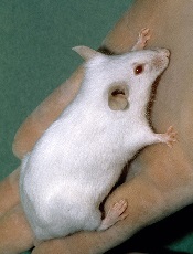
Photo courtesy of NCI
SAN DIEGO—A novel xenograft model of acute myeloid leukemia (AML) demonstrated that the JAK/STAT inhibitor ruxolitinib can prevent severe cytokine release syndrome (CRS) without impairing the anti-tumor effect of chimeric antigen receptor (CAR) T cells, according to research presented at the 2016 ASH Annual Meeting.
Almost all patients responding to CART-cell therapy develop CRS, and up to 60% develop severe CRS.
The research team believes the mouse model and findings with ruxolitinib will provide an important platform for studying CRS prevention and treatment.
At ASH, Saad Kenderian, MD, of the Mayo Clinic in Rochester, Minnesota, explained that CRS produces very high levels of the inflammatory protein IL-6.
Treatment with ruxolitinib in clinical studies has reduced human inflammatory cytokines. Therefore, it made sense to the investigators to study ruxolitinib as a means to prevent CRS after CAR T-cell therapy.
Tocilizumab has been used to treat grade 3 and 4 CRS, but physicians are concerned that earlier introduction during the course of CRS may impair CAR T-cell function.
At present, no relevant preclinical model for CRS after CAR T-cell therapy exists, “which is limiting the development of CRS preventative modalities that could, in turn, enhance the feasibility of CAR T-cell therapy,” Dr Kenderian said.
And so the investigators decided to create an animal model.
Dr Kenderian described the work at the meeting as abstract 652.
Mouse model for human CRS
Using NSG-S mice (non-obese diabetic, SCID ɣ -/- mice additionally transgenic for human stem cell factor, IL-3, and GM-CSF), investigators injected them with blasts from AML patients. After 3 to 4 weeks, investigators treated the mice with 1 x 10⁶ CD123-directed CAR T cells.
Dr Kenderian noted this dose of CART123 was 10 times higher than doses previously used in primary AML xenograft models.
The mice became weak, emaciated, developed hunched bodies, became withdrawn, had poor motor responses, and died in 7 to 10 days. The illness started within 1 week of CAR T-cell injection and correlated with significant expansion of T cells in the peripheral blood of these mice.
The team studied the serum from these mice 7 days after CART123 injection. They found extreme elevation of human IL-6, interferon-γ, tumor necrosis factor-α, and other inflammatory cytokines. This response resembled human CRS after CAR T-cell therapy.
Ruxolitinib treatment
The investigators first studied ruxolitinib activity in vitro with CART123 cells and found that ruxolitinib did not impair CAR T-cell effector functions.
“And also, ruxolitinib was not directly toxic to CAR T cells,” Dr Kenderian added.
But ruxolitinib did slow CAR T-cell proliferation in vitro.
They next tested ruxolitinib and CART123 in the mouse model.
Once the mice experienced high-burden disease, investigators treated them with CART123. That same day, investigators began treating the mice with ruxolitinib for 1 week. The mice were randomized to 30, 60, 90 mg/kg, or vehicle twice a day.
Twenty-nine days after AML injection, the mice treated with CART123 plus 90 mg or 60 mg of ruxolitinib experienced less weight loss than those treated with CART123 plus 30 mg of ruxolitinib or CART123-only.
“And more importantly, all mice had eradication of their disease,” Dr Kenderian said.
Mice treated with CART123 plus 90 mg, 60 mg, or 30 mg of ruxolitinib or CART123 alone had fewer AML blasts at day 28 than mice treated with 60 mg of ruxolitinib alone.
The investigators then analyzed the effect of ruxolitinib on the anti-tumor effect of CART123 and found that ruxolitinib did not impair it.
The attenuation of inflammatory cytokines translated to a survival advantage for mice treated with CART123 and ruxolitinib.
The investigators believe the addition of ruxolitinib to CAR T-cell therapy is a modality that should be investigated in patients at high-risk of developing CRS.
Dr Kenderian disclosed patents, royalties, and research funding from Novartis.
Trial supports early treatment of lower-risk ET

Photo courtesy of CDC
SAN DIEGO—Results from the ARETA trial suggest patients with essential thrombocythemia (ET) can benefit from early treatment even if they are not considered high-risk.
In this phase 3 trial, non-high-risk patients were less likely to experience ET-related cardiovascular events or disease progression if they received extended-release anagrelide rather than placebo.
Patients who received extended-release anagrelide were also less likely to become high-risk over time.
And extended-release anagrelide had a safety profile consistent with conventional anagrelide formulations, according to investigator Heinz Gisslinger, MD, of the Medical University of Vienna in Austria.
Dr Gisslinger reported final results of the ARETA trial at the 2016 ASH Annual Meeting (abstract 476).
The trial was sponsored by AOP Orphan Pharmaceuticals AG, the company developing the extended-release formulation of anagrelide, known as anagrelide retard (AR).
Dr Gisslinger noted that the goals of developing AR are to achieve lower peak plasma concentration of anagrelide, reduce the frequency and intensity of peak concentration related to adverse events (AEs), allow for an easier dosing scheme (once daily vs 2 to 3 times daily), and improve patient compliance.
He also pointed out that results of the phase 3 TEAM-ET trial suggest AR is non-inferior to the standard formulation of anagrelide (Thromboreductin, also a product of AOP Orphan Pharmaceuticals).
So with the ARETA trial, Dr Gisslinger and his colleagues set out to determine if AR would be beneficial as an early intervention in patients with non-high-risk ET.
Patients
The trial enrolled 146 patients who had platelet counts below 1000 G/L and met at least 1 of the following criteria:
- Age 40 to 60 years
- ET duration of more than 3 years
- Any risk factor for thrombotic complications (JAK2 mutation, protein C and/or S deficiency, antithrombin III deficiency, factor V Leiden or prothrombin mutation, or cardiovascular risk factors).
Seventy-seven patients were randomized to AR, and 69 were randomized to placebo. In both treatment arms, 100% of patients were Caucasian, and about 74% were female.
The mean age was 40.9 (range, 20-60) in the AR arm and 45.2 (range, 19-59) in the placebo arm. The median disease duration was 75.0 days (range, 1-2502) and 78.0 days (range, 1-2195), respectively. The mean platelet count was 748.6 G/L and 745.3 G/L, respectively.
A majority of patients in both arms had JAK2 mutations (62.7% in the AR arm and 63.8% in the placebo arm). Fewer had CALR mutations (22.7% and 13.6%, respectively) and MPL mutations (16.7% and 12.5%).
Treatment
Patients were stratified by JAK2 status and randomized to receive AR at 2 to 8 mg/day or placebo.
The dosing of AR started at 1 tablet (2 mg) per day during week 1 and was titrated up according to platelet response to 2 tablets in week 2. Dosing was further increased or decreased according to platelet response in weeks 3 and 4.
The maximum dose was 4 tablets (8 mg) per day. After week 4, the maximum dose to achieve optimal platelet counts (<450 G/L) was maintained, and patients continued with weekly visits through week 6.
After that, patients had visits every 3 months in both the main phase of this study and the extension phase. The main phase lasted 1 year, and the extension phase lasted up to 3 years.
Sixty patients (77.9%) in the AR arm and 52 (75.4%) in the placebo arm completed the main phase of the study.
Fifty-seven patients in the AR arm entered the extension phase, and 44 (57.1%) completed it. Thirty-four patients in the placebo arm entered the extension phase, and 21 (30.4%) completed it.
Efficacy
The primary endpoint was time to ET-related cardiovascular events (as confirmed by a blinded expert panel) or disease progression/worsening (platelet increase >1000 G/L).
The 1-year event-free rate (patients who did not meet criteria for the primary endpoint) was 87% in the AR arm and 69% in the placebo arm (hazard ratio=0.356, P=0.0008).
According to the expert panel, there were 12 ET-related events in 11 patients in the AR arm, as well as 17 such events in 14 patients in the placebo arm. This included major and minor arterial, venous, and bleeding events.
In total, there were 13 patients who had ET-related events or met platelet criteria in the AR arm (13 events) and 26 patients who had ET-related events or met platelet criteria in the placebo arm (30 events).
Nine patients in the AR arm (11.7%) and 18 in the placebo arm (26.1%) changed to high-risk status at some point during the trial (odds ratio=2.67, P=0.033).
Safety
The overall incidence of AEs was 88.3% in the AR arm and 69.6% in the placebo arm. The incidence of treatment-related AEs was 76.6% and 27.5%, respectively.
The incidence of treatment-related serious AEs was 1.3% and 0%, respectively. And the incidence of treatment-related AEs leading to withdrawal was 9.1% and 7.2%, respectively.
Treatment-related AEs occurring in more than 10% of patients in either arm (the AR and placebo arms, respectively) included headache (41.6% and 15.9%), dizziness (35.1% and 14.5%), palpitations (28.6% and 1.4%), and tachycardia (10.4% and 1.4%).
In closing, Dr Gisslinger noted that the primary endpoint of this study was met, and AR allowed for platelet count normalization and delayed progression to high-risk status.
Furthermore, the safety profile of AR is consistent with conventional anagrelide formulations, but AR allows for a more convenient dosing schedule.
Dr Gisslinger concluded, “[T]hese data from the ARETA study support an early treatment concept for all ET patients where platelet count or symptom reduction is a goal and those patients who can be attributed as intermediate-risk patients.”