User login
Study reveals CML patients likely to benefit from HSCT long-term

Photo by Chad McNeeley
SAN DIEGO—Researchers believe they have identified patients with chronic myeloid leukemia (CML) who are likely to derive long-term benefit from allogeneic hematopoietic stem cell transplant (allo-HSCT).
The researchers found that CML patients have a low risk of long-term morbidity if they undergo HSCT before the age of 45, are conditioned with busulfan and cyclophosphamide (Bu/Cy), and receive a graft from a matched, related donor (MRD).
Jessica Wu, of the University of Alabama at Birmingham, presented these findings at the 2016 ASH Annual Meeting (abstract 823*).
Wu noted that allogeneic HSCT is potentially curative for CML, but this method of treatment has been on the decline since the introduction of tyrosine kinase inhibitors (TKIs). And today, few CML patients undergo allo-HSCT.
She said that although TKIs can induce remission in CML patients, the drugs also fail to eradicate leukemia, can produce side effects that impact patients’ quality of life, and come with a significant financial burden (estimated at $92,000 to $138,000 per patient per year).
With this in mind, Wu and her colleagues set out to determine if certain CML patients might benefit from allo-HSCT long-term. The team also wanted to quantify overall and cause-specific late mortality after allo-HSCT and the long-term burden of severe/life-threatening chronic health conditions after allo-HSCT.
Patient population
The researchers studied 637 CML patients treated with allo-HSCT between 1981 and 2010 at City of Hope in Duarte, California, or the University of Minnesota in Minneapolis/Saint Paul. The patients had to have survived at least 2 years post-transplant.
About 60% of patients were male, and 67% were non-Hispanic white. Their median age at HSCT was 36.4 years, and 65% received an MRD graft. Nineteen percent of patients were transplanted in 1980-1989, 52% were transplanted in 1990-1999, and 29% were transplanted in 2000-2010.
Fifty-eight percent of patients received Cy/total body irradiation (TBI), 18% received Bu/Cy, and 3% received reduced-intensity conditioning (RIC).
Sixty-one percent of patients had chronic graft-vs-host disease (cGVHD), and 32% had high-risk disease at the time of HSCT.
Survival
The patients were followed for a median of 16.7 years. Thirty percent (n=192) died after surviving at least 2 years post-HSCT.
The median time to death was 8.3 years (range, 2-29.5), and the median age at death was 49.2 (range, 7.8-69.8). At 20 years from HSCT, the overall survival was 68.6%.
HSCT recipients had a 4.4-fold increased risk of death compared with the age-, sex-, and race-matched general population.
“Non-relapse mortality was the major contributor to late mortality, with infection, second malignancies, and cGVHD being the most common causes of death,” Wu said.
Non-relapse mortality was 20%, and relapse-related mortality was 4%. Eight percent of patients died of infection, 6.3% died of cGVHD, and 3.7% died of second malignancies.
Health outcomes
Patients who were still alive at the time of the study were asked to complete the BMTSS-2 health questionnaire, which was used to examine the risk of grade 3/4 chronic health conditions.
A total of 288 patients completed the questionnaire, as did a sibling comparison group of 404 individuals.
Among the patients, the median age at allo-HSCT was 37.5 (range, 3.6-71.4), and the median duration of follow-up was 13.9 years (range, 2-34.6).
Sixty-two percent of patients received an MRD graft, and 38% had a matched, unrelated donor. Eighty-three percent of patients had TBI-based conditioning, 16% received Bu/Cy, and 2.7% received RIC.
The prevalence of grade 3/4 chronic health conditions was significantly higher among patients than among siblings—38% and 24%, respectively (P<0.0001).
The odds ratio (OR)—adjusted for age, sex, race, and socioeconomic status—was 2.7 (P<0.0001).
The cumulative incidence of any grade 3/4 condition at 20 years after HSCT was 47.2% among patients. Common conditions were diabetes (14.9%), second malignancies (12.6%), and coronary artery disease (10%).
The researchers found the risk of grade 3/4 morbidity was significantly higher for the following patient groups:
- Those age 45 and older (hazard ratio [HR]=3.3, P<0.0001)
- Patients with a matched, unrelated donor (HR=3.0, P<0.0001)
- Those who received peripheral blood or cord blood grafts as opposed to bone marrow (HR=2.7, P=0.006).
(This analysis was adjusted for race/ethnicity, sex, education, household income, insurance, cGVHD, and conditioning regimen).
Lower risk
To identify subpopulations with a reduced risk of long-term morbidity, the researchers calculated the risk in various CML patient groups compared to siblings.
The overall OR for CML patients compared with siblings was 2.7 (P<0.0001).
The OR for patients in first chronic phase who underwent HSCT before the age of 45 and had an MRD was 1.5 (P=0.1).
The OR for CML patients in first chronic phase who underwent HSCT before the age of 45, had an MRD, and received Bu/Cy conditioning was 0.8 (P=0.7).
“[W]e found that patients who received a matched, related donor transplant under the age of 45, with busulfan/cyclophosphamide, carried the same burden of morbidity as the sibling cohort,” Wu said. “These findings could help inform decisions regarding therapeutic options for the management of CML.”
Wu noted that the limited sample size in this study prevented the researchers from examining outcomes with RIC. And a lack of data at analysis prevented them from examining pre-HSCT and post-HSCT management of CML, the interval between diagnosis and HSCT, and the life-long economic burden of allo-HSCT.
However, she said data collection is ongoing, and the researchers hope to address some of these limitations.![]()
*Information presented at the meeting differs from the abstract.
Health Canada approves therapy for hemophilia A

Health Canada has approved the use of lonoctocog alfa (Afstyla), a recombinant factor VIII (FVIII) single-chain therapy, in hemophilia A patients of all ages.
Lonoctocog alfa is indicated for use as routine prophylaxis to prevent or reduce the frequency of bleeding episodes, for on-demand treatment to control bleeding episodes, and for perioperative management of bleeding (surgical prophylaxis).
Lonoctocog alfa is the first and only single-chain recombinant FVIII therapy for hemophilia A specifically designed to provide long-lasting protection from bleeds with 2- to 3-times weekly dosing, according to CSL Behring, the company developing the product.
The company says lonoctocog alfa uses a covalent bond that forms one structural entity—a single polypeptide chain—to improve the stability of FVIII and provide FVIII activity with the option of twice-weekly dosing.
Health Canada’s approval of lonoctocog alfa is based on results from the AFFINITY clinical development program, which includes a trial of children (n=84) and a trial of adolescents and adults (n=175).
Among patients who received lonoctocog alfa prophylactically, the median annualized bleeding rate was 1.14 in the adults/adolescents and 3.69 in children younger than 12.
In all, there were 1195 bleeding events—848 in the adults/adolescents and 347 in the children.
Ninety-four percent of bleeds in adults/adolescents and 96% of bleeds in pediatric patients were effectively controlled with no more than 2 infusions of lonoctocog alfa weekly.
Eighty-one percent of bleeds in adults/adolescents and 86% of bleeds in pediatric patients were controlled by a single infusion.
Researchers assessed safety in 258 patients from both studies. Adverse reactions occurred in 14 patients and included hypersensitivity (n=4), dizziness (n=2), paresthesia (n=1), rash (n=1), erythema (n=1), pruritus (n=1), pyrexia (n=1), injection-site pain (n=1), chills (n=1), and feeling hot (n=1).
One patient withdrew from treatment due to hypersensitivity.
None of the patients developed neutralizing antibodies to FVIII or antibodies to host cell proteins. There were no reports of anaphylaxis or thrombosis.
Results from the trial of adolescents/adults were published in Blood in August. Results from the trial of children were presented at the World Federation of Hemophilia 2016 World Congress in July.* ![]()
Characterizing FL transformation, progression
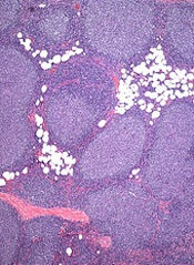
Patients with transformed follicular lymphoma (FL) and FL patients with early progression have “widely divergent patterns of clonal
dynamics,” according to researchers.
The team investigated the molecular events underlying transformation and early progression in FL and found that disparate evolutionary trajectories and mutational profiles drive these 2 distinct clinical endpoints.
Sohrab Shah, PhD, of the University of British Columbia in Vancouver, Canada, and his colleagues reported these findings in PLOS Medicine.
The researchers used whole-genome sequencing to analyze tumor specimens and matched normal specimens from 41 FL patients.
The team then classified the patients according to the following clinical endpoints:
- Patients who presented with transformation (n=15)
- Patients who experienced tumor progression within 2.5 years of starting treatment, without evidence of transformation (n=6)
- Patients who had neither transformation nor progression up to 5 years post-diagnosis (n=20).
The researchers also used targeted capture sequencing of known FL-associated genes in a larger cohort of 277 FL patients (395 samples) to investigate discrete genetic events that drive transformation and early progression.
Results showed that tumors that progress early evolve in different ways from those that transform.
The team found that, for tumors that transform, the cells or clones that constitute the majority of the aggressive tumor were extremely rare at diagnosis, if they were present at all.
In contrast, for early progressive FL, the clonal architecture remained similar from the time of diagnosis to relapse, indicating that the diagnostic tumor may already contain the properties that confer resistance to treatment.
Analysis of the larger cohort revealed genes and biological processes that were associated with transformation and progression.
The researchers identified 12 genes that were more commonly mutated at the time of transformation than the time of diagnosis—TP53, B2M, EZH2, MYC, CCND3, EBF1, PIM1, GNA13, ITPKB, CHD8, S1PR2, and P2RY8.
The team said their findings suggest that defective DNA damage response, increased proliferation, escape from immune surveillance, and loss of confinement within the germinal center are key features that drive histological transformation from indolent to aggressive lymphoma.
The researchers also identified 10 genes that were more commonly mutated in patients with early progression than in patients with late/no progression—B2M, BTG1, FAS, IKZF3, KMT2C, MKI67, MYD88, SOCS1, TP53, and XBP1.
The team noted that most patients with early progression (80%) had mutations in at least 1 of these 10 genes, but none of the genes were mutated at a frequency greater than 27%. This suggests that early progression is related to relatively infrequent genetic alterations.
The researchers said these findings provide a basis for future research on prognostic assay development and potential strategies for monitoring and treatment of patients with FL. ![]()
Platform could optimize treatment of ALL, other diseases

A digital health technology platform may prove useful for optimizing treatment of acute lymphoblastic leukemia (ALL), according to research published in SLAS Technology.
The platform is based on phenotypic personalized medicine (PPM), in which a patient’s response to treatment can be visually represented in the shape of a parabola.
The graph plots the drug dose along the horizontal axis and the patient’s response on the vertical axis.
Researchers said PPM has the ability to accurately identify a person’s optimal drug and dose combinations throughout an entire course of treatment.
“Phenotypic personalized medicine is like turbocharged artificial intelligence,” said study author Dean Ho, PhD, of the University of California, Los Angeles.
“It personalizes combination therapy to optimize efficacy and safety. The ability for our technology to continuously pinpoint the proper dosages of multiple drugs from such a large pool of possible combinations overcomes a challenge that is substantially more difficult than finding a needle in a haystack.”
In this study, Dr Ho and his colleagues used PPM to (retrospectively) individualize drug ratios/dosages in 2 pediatric patients with standard-risk ALL.
The researchers looked at the patients’ records to examine the administration of 4-drug maintenance regimens (dexamethasone, vincristine, mercaptopurine, and methotrexate).
The team said the drug doses served as the inputs, and maintaining absolute neutrophil counts and platelet counts within target ranges served as the outputs for optimization.
Using PPM, the researchers generated individualized 3-dimensional maps to determine the optimal drug ratios.
The team found their technology-suggested drug dosages were as much as 40% lower than clinical chemotherapy dosages, but they still maintained target neutrophil/platelet levels.
The parabolas showed that markedly different dosages of each drug were required to maintain normal cell counts for each patient.
The researchers said their results demonstrate a clear need to personalize ALL treatment and will serve as a foundation for a pending clinical trial to optimize multidrug chemotherapy.
“PPM has the ability to personalize combination therapy for a wide spectrum of diseases, making it a broadly applicable technology,” said study author Chih-Ming Ho, PhD, of the University of California, Los Angeles.
“The fact that we don’t need any information pertaining to a disease’s biological process in order to optimize and personalize treatment is a revolutionary advance. We’re at the interface of digital health and cancer treatment.”
The research team is planning to recruit patients for a prospective trial within the next year. The technology is approved for additional infectious disease and oncology studies. ![]()
‘Unprecedented’ MRD negativity with daratumumab in MM
© Todd Buchanan 2016
SAN DIEGO—Daratumumab added to standard of care regimens drives deep clinical responses beyond complete response (CR), a magnitude that is “unprecedented” in the relapsed/refractory multiple myeloma (MM) setting, according to a speaker at the 2016 ASH Annual Meeting.
Investigators added daratumumab to lenalidomide/dexamethasone in the POLLUX trial and to bortezomib/dexamethasone in the CASTOR trial.
In both phase 3 trials, the addition of daratumumab resulted in significant improvements in progression-free survival (PFS), overall response rate, and minimal residual disease (MRD) negativity when compared to control groups.
“The magnitude of daratumumab-induced MRD negativity in the relapsed setting is unprecedented and, for me, was not expected,” said Hervé Avet-Loiseau, MD, of Centre Hospitalier Universitaire Rangueil, Unité de Genomique du Myelome in Toulouse, France.
Dr Avet-Loiseau presented the MRD findings from CASTOR and POLLUX at the ASH Annual Meeting as abstract 246.*
He noted that, based on these studies, daratumumab received US Food and Drug Administration approvals for use in combination with standard of care regimens for MM patients who had received 1 or more prior lines of treatment.
Daratumumab had been previously approved as monotherapy for relapsed or refractory MM.
Study designs and findings from the POLLUX and CASTOR trials have been described earlier in Hematology Times.
Dr Avet-Loiseau provided updated PFS figures for the 2 studies.
At 18 months’ follow-up in the POLLUX study, the PFS rate for patients treated with daratumumab/lenalidomide/dexamethasone was 76%, compared to 49% in the lenalidomide/dexamethasone arm (P<0.0001).
At 12 months’ follow-up in the CASTOR study, the PFS with daratumumab was 60%, compared to 22% for bortezomib/dexamethasone (P<0.0001).
MRD criteria
In both studies, MRD assessments were conducted at suspected complete response (CR). Assessments were also conducted at 3 months and 6 months after CR in the POLLUX study and at 6 months and 12 months after the first study dose in the CASTOR study.
For the assessment of MRD, investigators used bone marrow aspirate samples and the ClonoSEQTM NGS-based assay.
Investigators evaluated MRD at 3 sensitivity thresholds: 10-4, 10-5, and 10-6.
And they used a stringent, unbiased evaluation, Dr Avet-Loiseau said. Any patient in the intent-to-treat population who was not assessed to be MRD negative was scored as MRD positive.
And the minimum cell input equivalent to the sensitivity threshold was required to determine MRD negativity.
MRD results
In the POLLUX study, 24.8% of patients achieved MRD negativity at the 10-5 cutoff, and 11.9% achieved MRD negativity at the 10-6 cutoff with the daratumumab combination.
This compared to 5.7% and 2.5% MRD negativity at the 10-5 and 10-6 cutoffs, respectively, without daratumumab (P<0.0001).
In the CASTOR study, the daratumumab-treated patients achieved 10.4% and 4.4% MRD negativity at the 10-5 and 10-6 cutoffs, respectively.
This compared to 2.4% and 0.8% MRD negativity in the control arm at the 10-5 and 10-6 cutoffs (P<0.005 and P<0.05), respectively.
“So, definitely, the addition of daratumumab improved the MRD negativity rate in both studies,” Dr Avet-Loiseau said.
“If you just look at the patients who did achieve CR in the POLLUX study, almost 50% of the patients [treated with daratumumab] achieved CR, and half of them were MRD negative at the cutoff of 10-5.”
In the CASTOR study, 25% of the patients treated with daratumumab achieved a CR. The MRD negativity rate was one-third in these patients.
“So again, we have consistently higher MRD negative rates in patients who achieve CR when they were treated in the daratumumab arms,” Dr Avet-Loiseau said.
“What is interesting, I think, is that the achievement of molecular CR was very rapid. [A]t 3 months, some patients did already achieve MRD negativity, and so we continued to see an improvement. [W]e still continue to see some achievement of MRD negativity.”
Investigators continue to follow the patients annually.
The investigators also analyzed MRD at 10-5 by cytogenetic risk and did not observe any MRD negativity in the control arm in either the POLLUX or CASTOR study.
“In contrast, we did observe some significant MRD negativity in the experimental arm with daratumumab—18% (POLLUX) and 14% (CASTOR) in high-risk patients,” Dr Avet-Loiseau said. “The most important prognostic factor is to achieve MRD negativity.”
However, even for patients who did not achieve MRD negativity, the PFS was much better in the experimental arms than in the control arms, he added.
This study, presented as a “Best of ASH” abstract, was funded by Janssen Research & Development, LLC. ![]()
*Information in the abstract differs from that presented at the meeting.
How old is too old to be on a kids’ protocol for ALL?

Photo by Bill Branson
SAN DIEGO—In recent years, pediatric or pediatric-inspired protocols have become the preferred treatment approach for younger adults with acute lymphoblastic leukemia (ALL).
These protocols include higher doses of steroids, vincristine, methotrexate, and L-asparaginase.
However, the upper age limit for this strategy has not been defined.
With the GRAALL-2005 study, investigators set out to determine how old is too old to be treated on pediatric protocols.
Their results suggest 55 is likely the upper age limit for patients with Ph-negative ALL.
The investigators also evaluated a hyper-fractionated (hyper-C) versus standard dose (standard-C) of cyclophosphamide during induction and late intensification.
They found that hyper-C did not provide an event-free survival (EFS) benefit in the overall study population, but patients age 55 and older did appear to benefit from hyper-C.
Françoise Huguet, MD, of the Institut Universitaire du Cancer de Toulouse in Toulouse, France, presented these findings at the 2016 ASH Annual Meeting (abstract 762).
GRAALL investigators had previously evaluated a pediatric-inspired protocol for adult patients in the GRAALL-2003 study, which validated the approach.
Study design
Patients with newly diagnosed, Ph-negative ALL were eligible to enroll if they were 18 to 59 years of age.
Treatment comprised a steroid pre-phase, a 5-drug induction, two 3-block dose-dense consolidation phases, a late intensification, a third consolidation phase, CNS irradiation, and a 2-year maintenance phase.
Patients could proceed to allogeneic transplant in first complete remission (CR) if eligible.
During induction and late intensification, patients received cyclophosphamide at 750 mg/m2 on day 1 and were then randomized to hyper-C (300 mg/m2/every 12 hours on days 15 to 17) or standard-C (750 mg/m2 on day 15).
The primary endpoint was EFS.
Patient population
Investigators randomized 787 evaluable patients—398 in the standard-C arm and 389 in the hyper-C arm.
Their median age was 36 years, 67% of patients had B-cell precursor ALL, and 33% had T-ALL.
Most had high-risk ALL, 72% of them receiving standard-C and 66% receiving hyper-C.
About a third of the patients in each arm proceeded to allogeneic stem cell transplant in first CR.
Results
The CR rate after induction therapy was 90.2% in the standard-C arm and 93.6% in the hyper-C arm, for an overall CR rate of 92%.
Most patients—87.5% in the standard-C arm and 91.8% in the hyper-C arm—achieved a response in 1 course of therapy.
Sixty percent of patients tested in the standard-C arm and 66% of those tested in the hyper-C arm were minimal residual disease negative at less than 10-4.
There were 26 (6.5%) deaths in the standard-C arm and 18 (4.6%) in the hyper-C arm.
The 5-year EFS rate was 52% overall, and hyper-C treatment had no impact on EFS (hazard ratio=0.89 [range, 0.7-1.1]; P=0.26).
Impact of age
Investigators conducted a post-hoc subgroup analysis of 5 age groups—18-24 years (n=200), 25-34 (n=172), 35-44 (n=171), 45-54 (n=151), and 55+ (n=93).
Overall, the CR rate tended to decrease with age. The rates were 98.5% (18-24), 95.3% (25-34), 87.7% (35-44), 89.4% (45-54), and 79.6% (55+).
Induction death rates increased from 0.5% in the youngest group to 18.3% in the oldest, but the rate of cumulative incidences of failure at 5 years was similar among all the age groups.
The cumulative incidence of treatment-related mortality, without censoring for transplant, ranged from 7.6% in the youngest group to 39.7% in the oldest.
And the 5-year EFS for the youngest patients was 60%, while, for the oldest, it was 26%.
“Above 50 years, the increase in age became highly significant,” Dr Huguet emphasized. “There were fewer CRs and lower survival.”
Treatment compliance
In terms of treatment compliance and median dose received in the induction course, patients aged 55-59 received significantly less L-asparaginase than those aged 18-54 (P<0.001).
During all 3 consolidation phases, patients aged 55-59 received significantly lower median doses of all medications—cytarabine, methotrexate, cyclophosphamide—than patients aged 18-54.
And in late intensification, patients aged 55-59 received significantly lower median doses of vincristine, prednisone, daunorubicin, and hyper-C than all other patients. The median doses of L-asparaginase and standard-C received were lower in the older patients but not significantly so.
EFS by age and randomization
The 5-year EFS for patients aged 18-54 was 57% with hyper-C, compared with 55% in the standard-C arm (P=0.66).
However, for older patients, there was a significant advantage for those receiving hyper-C. The 5-year EFS was 38% with hyper-C, compared to 12% with standard-C (P=0.007).
Dr Huguet explained that inferior compliance in patients 55 and older “might explain why a benefit associated with early hyper-C reinforcement became apparent in these older patients only.”
Dr Huguet concluded that the results “suggest that 55 years is likely to be the upper age limit to tolerate a pediatric-like therapy for younger adults with Ph-negative ALL.”
She added that patients over 54 might benefit from alternative front-line strategies.
Accordingly, investigators are planning to use new agents, such as blinatumomab or inotuzumab ozogamicin, in the next European Working Group on Adult ALL studies. ![]()
Two mutations may help drive CBF-AML
2016 ASH Annual Meeting
SAN DIEGO—Researchers have found evidence to suggest that mutations in the CCND1 and CCND2 genes may contribute to the development of core-binding factor acute myeloid leukemia (CBF-AML).
The team noted that CBF-AML is defined by the presence of either t(8;21)(q22;q22)/RUNX1-RUNX1T1 or inv(16)(p13.1q22)/t(16;16)(p13.1;q22)/CBFB-MYH11.
However, the fusion genes alone are not capable of causing CBF-AML.
“The hematology community has long sought to determine what other factors in addition to the fusion genes occur in this special type of leukemia,” said Ann-Kathrin Eisfeld, MD, of The Ohio State University Comprehensive Cancer Center in Columbus.
“We are now the first to describe that mutations in CCND1—and among the first to describe that mutations in the sister gene CCND2—are unique features of CBF-AML with t(8;21). In addition, we have collected the first evidence that mutations in CCND2 lead to more aggressive growth of leukemia cell lines.”
Dr Eisfeld and her colleagues reported these findings in a paper published in Leukemia and in a poster presented at the 2016 ASH Annual Meeting (abstract 2740).
A previous study of genetic mutations in CBF-AML revealed the presence of at least 1 mutation in 85% of patients studied. This meant the remaining 15% of patients harbored other, undiscovered mutations.
For the current study, Dr Eisfeld and her colleagues searched CBF-AML samples for the missing mutations that, together with the fusion genes, might contribute to the leukemia in this subgroup of cases.
The team analyzed pretreatment bone marrow and peripheral blood samples from 177 adult CBF-AML patients who received similar treatment through a clinical trial conducted at multiple centers across the US.
Using a targeted, next-generation sequencing approach, the researchers looked for mutations in 84 leukemia- and/or cancer-associated genes. They also performed tests on blood or bone marrow cells to look for chromosomal irregularities.
The team discovered 2 significant mutations in the CCND1 and CCND2 genes, representing the first dual evidence of these recurrent mutations in patients with t(8;21)-positive CBF-AML.
CCND1 and CCND2 mutations were found in 15% (n=10) of patients with t(8;21)-positive CBF-AML. Two patients had mutations in CCND1, and 8 had mutations in CCND2.
The researchers also found a single CCND2 mutation in 1 (0.9%) patient with inv(16)-positive CBF-AML.
In comparison, the incidence of CCND1 and CCND2 mutations was 0.77% (n=11) in a cohort of 1426 patients with non-CBF-AML.
“This is extremely valuable information that was previously unknown,” Dr Eisfeld said, “and it might help us develop targeted therapies more likely to help patients with [CBF-AML] in the near future.” ![]()

2016 ASH Annual Meeting
SAN DIEGO—Researchers have found evidence to suggest that mutations in the CCND1 and CCND2 genes may contribute to the development of core-binding factor acute myeloid leukemia (CBF-AML).
The team noted that CBF-AML is defined by the presence of either t(8;21)(q22;q22)/RUNX1-RUNX1T1 or inv(16)(p13.1q22)/t(16;16)(p13.1;q22)/CBFB-MYH11.
However, the fusion genes alone are not capable of causing CBF-AML.
“The hematology community has long sought to determine what other factors in addition to the fusion genes occur in this special type of leukemia,” said Ann-Kathrin Eisfeld, MD, of The Ohio State University Comprehensive Cancer Center in Columbus.
“We are now the first to describe that mutations in CCND1—and among the first to describe that mutations in the sister gene CCND2—are unique features of CBF-AML with t(8;21). In addition, we have collected the first evidence that mutations in CCND2 lead to more aggressive growth of leukemia cell lines.”
Dr Eisfeld and her colleagues reported these findings in a paper published in Leukemia and in a poster presented at the 2016 ASH Annual Meeting (abstract 2740).
A previous study of genetic mutations in CBF-AML revealed the presence of at least 1 mutation in 85% of patients studied. This meant the remaining 15% of patients harbored other, undiscovered mutations.
For the current study, Dr Eisfeld and her colleagues searched CBF-AML samples for the missing mutations that, together with the fusion genes, might contribute to the leukemia in this subgroup of cases.
The team analyzed pretreatment bone marrow and peripheral blood samples from 177 adult CBF-AML patients who received similar treatment through a clinical trial conducted at multiple centers across the US.
Using a targeted, next-generation sequencing approach, the researchers looked for mutations in 84 leukemia- and/or cancer-associated genes. They also performed tests on blood or bone marrow cells to look for chromosomal irregularities.
The team discovered 2 significant mutations in the CCND1 and CCND2 genes, representing the first dual evidence of these recurrent mutations in patients with t(8;21)-positive CBF-AML.
CCND1 and CCND2 mutations were found in 15% (n=10) of patients with t(8;21)-positive CBF-AML. Two patients had mutations in CCND1, and 8 had mutations in CCND2.
The researchers also found a single CCND2 mutation in 1 (0.9%) patient with inv(16)-positive CBF-AML.
In comparison, the incidence of CCND1 and CCND2 mutations was 0.77% (n=11) in a cohort of 1426 patients with non-CBF-AML.
“This is extremely valuable information that was previously unknown,” Dr Eisfeld said, “and it might help us develop targeted therapies more likely to help patients with [CBF-AML] in the near future.”
EC authorizes new use for ofatumumab in CLL

Photo courtesy of GSK
The European Commission (EC) has granted marketing authorization for ofatumumab (Arzerra®) to be used in combination with fludarabine and cyclophosphamide (FC) in the treatment of adults with relapsed chronic lymphocytic leukemia (CLL).
Ofatumumab is a monoclonal antibody designed to target CD20. The drug is marketed under a collaboration agreement between Genmab and Novartis.
The EC previously authorized the use of ofatumumab as a single agent to treat CLL patients who are refractory to fludarabine and alemtuzumab.
The agency also authorized the use of ofatumumab in combination with chlorambucil or bendamustine in CLL patients who have not received prior therapy and are not eligible for fludarabine-based therapy.
The EC’s decision to approve the use of ofatumumab in combination with FC was based on results from the phase 3 COMPLEMENT 2 study, which were published in Leukemia & Lymphoma in October.
The trial enrolled 365 patients with relapsed CLL. The patients were randomized 1:1 to receive up to 6 cycles of ofatumumab in combination with FC or up to 6 cycles of FC alone.
The primary endpoint was progression-free survival, as assessed by an independent review committee.
The median progression-free survival was 28.9 months for patients receiving ofatumumab plus FC, compared to 18.8 months for patients receiving FC only (hazard ratio=0.67, P=0.0032).
The incidence of grade 3 or higher adverse events was 74% in the ofatumumab-plus-FC arm and 69% in the FC-only arm. Neutropenia was the most common of these events, occurring in 49% and 36% of patients, respectively.
Another treatment on the horizon for SCD

Photo courtesy of ASH
SAN DIEGO—The first-in-class humanized anti-P-selectin antibody SelG1, also known as crizanlizumab, significantly reduced sickle cell pain crises (SCPC) when compared to placebo in the phase 2 SUSTAIN trial.
The higher dose of SelG1 tested reduced the annual rate of SCPC by 45% (P=0.01) and the annual rate of uncomplicated SCPC by 63% (P=0.015).
Acute painful crises are the primary cause for patients with sickle cell disease (SCD) to seek medical attention.
Kenneth I. Ataga, MD, of the University of North Carolina at Chapel Hill, explained that upregulation of P-selectin on endothelial cells and platelets contributes to the cell-cell interaction involved in the pathogenesis of SCPC. SelG1 binds to P-selectin and inhibits its interaction with P-selectin glycoprotein ligand 1.
Dr Ataga presented results from the SUSTAIN study during the plenary session of the 2016 ASH Annual Meeting (abstract 1).
The study was also published in NEJM. The research was sponsored by Selexys Pharmaceuticals Corporation, which was recently acquired by Novartis.
Patient population
A total of 198 patients were randomized, 67 to high-dose SelG1 (5.0 mg/kg), 66 to low-dose SelG1 (2.5 mg/kg), and 65 to placebo.
They received a loading dose in the first 2 weeks of treatment, followed by monthly dosing for a year.
Patients had to have a diagnosis of SCD, including genotypes HbSS, HbSC, HbSb0-thalassemia, or HbSB+-thalassemia.
They had to have at least 2 but not more than 10 acute sickle-related pain events within 12 months of study entry.
Patients ranged in age from 16 to 57 years, about 70% had HbSS, and 60% were on concomitant hydroxyurea therapy.
Patients not already receiving hydroxyurea were not permitted to start it during the study. And patients could not be on chronic transfusion therapy.
Study endpoints
The primary endpoint was the annual rate of adjudicated SCPC.
“Painful crisis was defined as an active episode of pain, and it was felt to be related to sickle cell disease-specific events and no other medically defined causes,” Dr Ataga explained.
The pain episodes also had to result in a visit to a medical facility and require treatment with parenteral or oral narcotics or parenteral nonsteroidal anti-inflammatory drugs.
The definition of SCPC included not only typical painful episodes but also acute chest syndrome (ACS), hepatic or splenic sequestration, and priapism.
Secondary endpoints included the annual rate of days hospitalized, time to first and second SCPC, the annual rate of uncomplicated pain crises, and the annual rate of ACS, among others.
Efficacy
The median annual rate of SCPC was 1.63 in the high-dose arm, 2.01 in the low-dose arm, and 2.98 in the placebo arm, amounting to a significant 45.3% reduction with the higher dose of SelG1 compared to placebo. The low dose resulted in a reduction of 32.6% compared to placebo, but this was not significant.
Twenty-four patients in the high-dose arm had an SCPC rate of 0, compared with 12 in the low-dose arm and 11 on placebo.
SCD genotype or concomitant hydroxyurea use did not impact these results.
Patients in the high-dose arm had a median annual rate of 4 hospitalization days, compared with 6.87 in both the low-dose and placebo groups. The difference was not significant, although the reduction in the high-dose arm was 41.8%.
The median annual rate of uncomplicated SCPC was 1.08 in the high-dose arm, 2.00 in the low-dose arm, and 2.91 in the placebo arm. Uncomplicated SCPC excluded ACS, splenic or hepatic sequestration, and priapism.
The reduction in the high-dose arm compared to placebo was significant, at 62.9% (P=0.015).
“The rate of ACS was pretty rare,” Dr Ataga said, “so the median rate across the various groups was 0.”
And time to the first SCPC was 4.1 months (P=0.001) in the high-dose group, 2.2 months (P=0.136) in the low-dose group, and 1.4 months in the placebo group.
“The curves separated pretty early,” Dr Ataga noted, “and were maintained throughout the course of the treatment phase, suggesting that the beneficial effect of SelG1 manifested pretty early following initiation of treatment.”
The time to second event was also significant in the high-dose arm compared to placebo, at 10.3 months and 5.1 months, respectively (P=0.022).
Safety
One or more adverse events occurred in over 85% of patients in each group.
Adverse events that occurred in at least 10% of SelG1-treated patients and amounted to at least double the number in the placebo group were arthralgia, diarrhea, pruritus, vomiting, and chest pain.
Five patients died while on study, but none of these deaths were related to the study drug.
Despite the adverse events, Dr Ataga said the drug was, overall, well-tolerated among the patients who received treatment.
Predicting therapy-related myeloid neoplasms

Photo courtesy of
MD Anderson Cancer Center
SAN DIEGO―Clonal hematopoiesis could be used as a predictive marker to identify cancer patients at risk of developing therapy-related myeloid neoplasms (t-MNs), according to researchers.
The team conducted a case-control study, which showed that patients who developed t-MNs—acute myeloid leukemia and myelodysplastic syndromes—were significantly more likely than patients without t-MNs to have clonal hematopoiesis at the time of primary cancer diagnosis.
“Based on these findings, we believe pre-leukemic mutations may function as a new biomarker that would predict t-MN development,” said Andy Futreal, PhD, of The University of Texas MD Anderson Cancer Center in Houston.
Dr Futreal and his colleagues reported these findings in The Lancet Oncology.
Co-author Koichi Takashi, MD, also of MD Anderson, presented the findings at the 2016 ASH Annual Meeting (abstract 38).
Initial cohort
The researchers analyzed data on patients treated at MD Anderson from 1997 to 2015.
The 14 cases the team identified had been treated for a primary cancer and later developed t-MNs. The 54 age-matched control subjects had been treated for lymphoma, received combination chemotherapy, and did not develop t-MNs after at least 5 years of follow-up.
For both cases and controls, the researchers performed gene sequencing on pre-treatment peripheral blood samples. For cases, the researchers also performed targeted gene sequencing on bone marrow samples taken at t-MN diagnosis.
“We found that prevalence of pre-leukemic mutations was significantly higher in patients who developed t-MNs versus those who did not,” Dr Futreal said.
Clonal hematopoiesis was present in 71% of cases (n=10) and 31% of controls (n=17).
“We found genetic mutations that are present in t-MNs leukemia samples actually could be found in blood samples obtained at the time of their original cancer diagnosis,” Dr Takashi noted.
Overall, the cumulative incidence of t-MNs at 5 years was significantly higher in patients with clonal hematopoiesis than in those without it—30% and 7%, respectively (P=0.016).
Validation cohort
The researchers also assessed clonal hematopoiesis in an external cohort of 74 patients with lymphoma who were treated in a trial of front-line chemotherapy with cyclophosphamide, doxorubicin, vincristine, and prednisone, with or without melatonin.
In this cohort, 7% (n=5) of patients developed t-MNs. Eighty percent of these patients (n=4) had clonal hematopoiesis.
Of the 69 patients who did not develop t-MNs, 16% (n=11) had clonal hematopoiesis.
The cumulative incidence of t-MNs at 10 years was significantly higher in patients with clonal hematopoiesis than in those without it—29% and 0%, respectively (P=0.0009).
Multivariate analysis suggested clonal hematopoiesis significantly increased the risk of t-MNs, with a hazard ratio of 13.7 (P=0.013).
“[W]e believe the data suggest potential approaches of screening for clonal hematopoiesis in cancer patients that may identify patients at risk of developing t-MNs, although further studies are needed,” Dr Takashi concluded.