User login
Multiple Sclerosis Hub
David Brandes, MD

IV Immunoglobulin Increases JCV Antibody Levels in Patients With MS
COPENHAGEN—In patients with multiple sclerosis (MS), IV immunoglobulin (IV Ig) infusions increase John Cunningham virus (JCV) antibody levels, which may result in seropositivity, according to a study presented at the 29th Congress of the European Committee for Treatment and Research in MS (ECTRIMS). This finding suggests that risk stratification rules based on JCV antibody status may not apply to patients recently treated with IV Ig.
The concentration of JCV antibodies is likely to decrease over time after IV Ig is discontinued. “The lack of consistent sampling in our patients prevents us from making firm conclusions as to the likelihood of return to innate (pre–IV Ig) JCV antibody levels after stopping IV Ig,” said Ilya Kister, MD, Assistant Professor of Neurology at New York University School of Medicine in New York City. He and his colleagues are investigating whether age, IV Ig therapy duration, JCV antibody concentration during IV Ig therapy, and time from IV Ig administration to JCV antibody testing affect JCV antibody concentration after IV Ig discontinuation.
The STRATIFY Trial
Dr. Kister and colleagues examined data for 102 patients with MS who participated in the STRATIFY-2 trial and had monthly infusions of IV Ig (0.7 g/kg) at any point during the trial. The researchers also analyzed results from the commercial STRATIFY JCV and STRATIFY JCV Dx SELECT assays for participants in STRATIFY-2 who had been exposed to IV Ig. The team calculated the relative concentration of polyclonal JCV antibodies as JCV monoclonal antibody equivalents by interpolating the optical density or index values for each sample against a reference curve prepared using a monoclonal antibody to JCV.
JCV Antibody Levels Decreased Over Time
Of the 1,251 patients enrolled in STRATIFY-2, 1,143 were not exposed to IV Ig, and 58% of these patients were seropositive for JCV antibody. In contrast, all 71 STRATIFY-2 enrollees who were tested for JCV antibodies within 30 days of IV Ig administration were seropositive. Sixteen of 25 (64%) STRATIFY-2 enrollees who were exposed to IV Ig more than 30 days after IV Ig administration were seropositive, which suggests that the level of JCV antibodies in IV Ig–exposed patients declines over time.
The median relative concentration of JCV antibodies was 0 µg/mL for patients not yet exposed to IV Ig. For patients receiving IV Ig, the median relative concentration of JCV antibodies was approximately 2 µg/mL. At 30 days after IV Ig exposure, the median relative concentration of JCV antibodies was less than 1 µg/mL.
The researchers observed a significant decrease or complete elimination of JCV antibody relative concentrations for many samples after discontinuation of IV Ig, which implies a reduction of passively transferred JCV antibody levels over time. “However, without pre–IV Ig samples, it is not possible to determine if subjects return to baseline levels or not,” said Dr. Kister.
—Erik Greb
Suggested Reading
Filippini G, Del Giovane C, Vacchi L, et al. Immunomodulators and immunosuppressants for multiple sclerosis: a network meta-analysis. Cochrane Database Syst Rev. 2013 Jun 6;6:CD008933.
Yeh EA, Weinstock-Guttman B. The management of pediatric multiple sclerosis. J Child Neurol. 2012;27(11):1384-1393.
COPENHAGEN—In patients with multiple sclerosis (MS), IV immunoglobulin (IV Ig) infusions increase John Cunningham virus (JCV) antibody levels, which may result in seropositivity, according to a study presented at the 29th Congress of the European Committee for Treatment and Research in MS (ECTRIMS). This finding suggests that risk stratification rules based on JCV antibody status may not apply to patients recently treated with IV Ig.
The concentration of JCV antibodies is likely to decrease over time after IV Ig is discontinued. “The lack of consistent sampling in our patients prevents us from making firm conclusions as to the likelihood of return to innate (pre–IV Ig) JCV antibody levels after stopping IV Ig,” said Ilya Kister, MD, Assistant Professor of Neurology at New York University School of Medicine in New York City. He and his colleagues are investigating whether age, IV Ig therapy duration, JCV antibody concentration during IV Ig therapy, and time from IV Ig administration to JCV antibody testing affect JCV antibody concentration after IV Ig discontinuation.
The STRATIFY Trial
Dr. Kister and colleagues examined data for 102 patients with MS who participated in the STRATIFY-2 trial and had monthly infusions of IV Ig (0.7 g/kg) at any point during the trial. The researchers also analyzed results from the commercial STRATIFY JCV and STRATIFY JCV Dx SELECT assays for participants in STRATIFY-2 who had been exposed to IV Ig. The team calculated the relative concentration of polyclonal JCV antibodies as JCV monoclonal antibody equivalents by interpolating the optical density or index values for each sample against a reference curve prepared using a monoclonal antibody to JCV.
JCV Antibody Levels Decreased Over Time
Of the 1,251 patients enrolled in STRATIFY-2, 1,143 were not exposed to IV Ig, and 58% of these patients were seropositive for JCV antibody. In contrast, all 71 STRATIFY-2 enrollees who were tested for JCV antibodies within 30 days of IV Ig administration were seropositive. Sixteen of 25 (64%) STRATIFY-2 enrollees who were exposed to IV Ig more than 30 days after IV Ig administration were seropositive, which suggests that the level of JCV antibodies in IV Ig–exposed patients declines over time.
The median relative concentration of JCV antibodies was 0 µg/mL for patients not yet exposed to IV Ig. For patients receiving IV Ig, the median relative concentration of JCV antibodies was approximately 2 µg/mL. At 30 days after IV Ig exposure, the median relative concentration of JCV antibodies was less than 1 µg/mL.
The researchers observed a significant decrease or complete elimination of JCV antibody relative concentrations for many samples after discontinuation of IV Ig, which implies a reduction of passively transferred JCV antibody levels over time. “However, without pre–IV Ig samples, it is not possible to determine if subjects return to baseline levels or not,” said Dr. Kister.
—Erik Greb
Suggested Reading
Filippini G, Del Giovane C, Vacchi L, et al. Immunomodulators and immunosuppressants for multiple sclerosis: a network meta-analysis. Cochrane Database Syst Rev. 2013 Jun 6;6:CD008933.
Yeh EA, Weinstock-Guttman B. The management of pediatric multiple sclerosis. J Child Neurol. 2012;27(11):1384-1393.
Glutamate Level Decreases Significantly in Patients With Secondary Progressive MS
COPENHAGEN—Levels of glutamate may decline almost 10 times faster over time in patients with secondary progressive multiple sclerosis (MS) than in healthy controls, according to a study presented at the 29th Congress of the European Committee for Treatment and Research in MS (ECTRIMS). Levels of glutamine and the combination of glutamate and glutamine also may decrease faster in patients with secondary progressive MS than in healthy controls.
“The combined decline of glutamate and glutamine suggests a loss of synapse function or a loss of synapses over time,” said Erin MacMillan, PhD, postdoctoral research fellow at the University of British Columbia in Vancouver. The two metabolites may offer new biomarkers of progression and feasible outcome measures for multicenter clinical trials in secondary progressive MS.
“Few longitudinal proton magnetic resonance spectroscopy (1H-MRS) studies have been performed in a cohort of progressive MS patients, and, to our knowledge, this is the first with sufficient spectral quality and data analysis methods to reliably detect both glutamate and glutamine,” she added.
Monitoring Metabolite Concentrations in Patients With MS
Dr. MacMillan and colleagues analyzed data for patients with secondary progressive MS who were enrolled in a clinical trial at the University of British Columbia. The investigators focused on 47 subjects who completed MRS scans for at least two time points, including baseline, year one, and year two. Participants (median age, 51.7; median Expanded Disability Status Scale score, 6.0) were not on other disease-modifying therapies within three months before the start of the trial.
The researchers used 1H-MRS to examine concentrations of glutamate, glutamine, the combination of glutamate and glutamine, myo-inositol, choline-containing compounds, and other metabolites in a primarily white-matter region inside the brain. Concentration differences from year one and year two to baseline were tested with the two-sided Wilcoxon signed rank test. Concentrations at all three time points for each metabolite were fit with a linear random effects model to allow for individual offsets but the same slope across subjects.
Concentrations of Most Metabolites Changed Little
Participants’ level of N-acetyl-aspartate + N-acetyl-aspartylglutamate (tNAA) had no significant relationship with time. In contrast, glutamate concentration decreased from approximately 6.5 mM at baseline to approximately 6 mM at year two. Its rate of decrease was 4.9% per year. Glutamine concentration declined from approximately 2.5 mM at baseline to approximately 2 mM at year two. Its rate of decrease was 13.1% per year.
The lack of change in tNAA, creatine and phosphocreatine, and choline-containing compounds over two years is consistent with a previous study of participants with primary progressive MS over three years, said Dr. MacMillan.
“Glutamate and glutamine, as measured by 1H-MRS, are promising new biomarkers of MS disease progression that may be more sensitive than current state-of-the-art techniques, such as brain atrophy,” Dr. MacMillan told Neurology Reviews. “These new biomarkers may prove to be better prognostic measures and determinants of treatment efficacy.
“We hope to repeat this study with two regions of interest—one clearly within the cortical gray matter, and one clearly within the white matter—to distinguish whether the changes we have seen with disease progression are taking place in the gray matter or the white matter,” she concluded.
—Erik Greb
Suggested Reading
Cambron M, D’Haeseleer M, Laureys G, et al. White-matter astrocytes, axonal energy metabolism, and axonal degeneration in multiple sclerosis. J Cereb Blood Flow Metab. 2012;32(3):413-424.
Tisell A, Leinhard OD, Warntjes JB, et al. Increased concentrations of glutamate and glutamine in normal-appearing white matter of patients with multiple sclerosis and normal MR imaging brain scans. PLoS One. 2013;8(4):e61817.
COPENHAGEN—Levels of glutamate may decline almost 10 times faster over time in patients with secondary progressive multiple sclerosis (MS) than in healthy controls, according to a study presented at the 29th Congress of the European Committee for Treatment and Research in MS (ECTRIMS). Levels of glutamine and the combination of glutamate and glutamine also may decrease faster in patients with secondary progressive MS than in healthy controls.
“The combined decline of glutamate and glutamine suggests a loss of synapse function or a loss of synapses over time,” said Erin MacMillan, PhD, postdoctoral research fellow at the University of British Columbia in Vancouver. The two metabolites may offer new biomarkers of progression and feasible outcome measures for multicenter clinical trials in secondary progressive MS.
“Few longitudinal proton magnetic resonance spectroscopy (1H-MRS) studies have been performed in a cohort of progressive MS patients, and, to our knowledge, this is the first with sufficient spectral quality and data analysis methods to reliably detect both glutamate and glutamine,” she added.
Monitoring Metabolite Concentrations in Patients With MS
Dr. MacMillan and colleagues analyzed data for patients with secondary progressive MS who were enrolled in a clinical trial at the University of British Columbia. The investigators focused on 47 subjects who completed MRS scans for at least two time points, including baseline, year one, and year two. Participants (median age, 51.7; median Expanded Disability Status Scale score, 6.0) were not on other disease-modifying therapies within three months before the start of the trial.
The researchers used 1H-MRS to examine concentrations of glutamate, glutamine, the combination of glutamate and glutamine, myo-inositol, choline-containing compounds, and other metabolites in a primarily white-matter region inside the brain. Concentration differences from year one and year two to baseline were tested with the two-sided Wilcoxon signed rank test. Concentrations at all three time points for each metabolite were fit with a linear random effects model to allow for individual offsets but the same slope across subjects.
Concentrations of Most Metabolites Changed Little
Participants’ level of N-acetyl-aspartate + N-acetyl-aspartylglutamate (tNAA) had no significant relationship with time. In contrast, glutamate concentration decreased from approximately 6.5 mM at baseline to approximately 6 mM at year two. Its rate of decrease was 4.9% per year. Glutamine concentration declined from approximately 2.5 mM at baseline to approximately 2 mM at year two. Its rate of decrease was 13.1% per year.
The lack of change in tNAA, creatine and phosphocreatine, and choline-containing compounds over two years is consistent with a previous study of participants with primary progressive MS over three years, said Dr. MacMillan.
“Glutamate and glutamine, as measured by 1H-MRS, are promising new biomarkers of MS disease progression that may be more sensitive than current state-of-the-art techniques, such as brain atrophy,” Dr. MacMillan told Neurology Reviews. “These new biomarkers may prove to be better prognostic measures and determinants of treatment efficacy.
“We hope to repeat this study with two regions of interest—one clearly within the cortical gray matter, and one clearly within the white matter—to distinguish whether the changes we have seen with disease progression are taking place in the gray matter or the white matter,” she concluded.
—Erik Greb
Suggested Reading
Cambron M, D’Haeseleer M, Laureys G, et al. White-matter astrocytes, axonal energy metabolism, and axonal degeneration in multiple sclerosis. J Cereb Blood Flow Metab. 2012;32(3):413-424.
Tisell A, Leinhard OD, Warntjes JB, et al. Increased concentrations of glutamate and glutamine in normal-appearing white matter of patients with multiple sclerosis and normal MR imaging brain scans. PLoS One. 2013;8(4):e61817.

How Do Smoking and Epstein–Barr Virus Affect Risk of MS?
COPENHAGEN—The interaction between smoking and Epstein–Barr virus (EBV), two risk factors for multiple sclerosis (MS), may depend on age, according to research presented at the 29th Congress of the European Committee for Treatment and Research in MS. Smoking and EBV appear to have a negative interaction in adults younger than 26 and a positive interaction in adults older than 26.
“The negative interaction in younger subjects suggests that EBV and smoking as risk factors for MS act independently from an etiologic perspective,” said Jonatan Salzer, MD, PhD, a physician at Umeå University in Sweden. “The positive interaction in older subjects may suggest that the two risk factors share a common pathophysiologic pathway, most obviously that smoking enhances the antibody response against EBV after several years with MS.”
Analysis of Prospectively Collected Blood Samples
In a nested case–control study, Dr. Salzer and colleagues analyzed prospectively collected biobank blood samples. Of the patient population, 192 persons subsequently developed MS and 384 participants served as matched controls. The researchers measured levels of cotinine, a nicotine metabolite, in the samples with an immunoassay. Anti-EBNA-1 IgG antibodies were measured using an enzyme-linked immunosorbent assay (ELISA).
The investigators considered participants with cotinine levels of 10 ng/mL or higher to be smokers. The group also divided anti-EBNA-1 IgG levels at the median among controls. Dr. Salzer and colleagues assessed interaction on the additive and multiplicative scales and estimated the effects of the risk factors across strata of each other.
Smoking and EBV Did Not Interact Significantly
When analyzing the entire cohort, the researchers found no statistically significant interactions between smoking and EBV. The mean level of anti-EBNA-1 IgG was higher among subjects with cotinine levels of 10 ng/mL or higher than among participants with cotinine levels lower than 10 ng/mL. The investigators found no heterogeneity of effects of one risk factor across strata of the other. When the researchers analyzed interaction on the additive and multiplicative scales, they found that the group of patients with both risk factors (ie, high levels of anti-EBNA-1 IgG and cotinine level of 10 ng/mL or greater) had a slightly higher odds ratio of MS than predicted by the independent effects of the risk factors.
An analysis restricted to younger subjects indicated that the odds ratio for the patients with both risk factors was in between that of the additive interaction and that of the multiplicative interaction. The effects of both risk factors showed nonsignificant signs of heterogeneity, however. Among older patients, the pattern of heterogeneity of effects across strata was the opposite of that among younger patients, the researchers reported.
—Erik Greb
Suggested Reading
Salzer J, Hallmans G, Nyström M, et al. Smoking as a risk factor for multiple sclerosis. Mult Scler. 2013;19(8):1022-1027.
Salzer J, Nyström M, Hallmans G, et al. Epstein-Barr virus antibodies and vitamin D in prospective multiple sclerosis biobank samples. Mult Scler. 2013;19(12):1587-1591.
Salzer J, Stenlund H, Sundstrom P. The interaction between smoking and Epstein-Barr virus as multiple sclerosis risk factors may depend on age. Mult Scler. 2013 Oct 9 [Epub ahead of print.
COPENHAGEN—The interaction between smoking and Epstein–Barr virus (EBV), two risk factors for multiple sclerosis (MS), may depend on age, according to research presented at the 29th Congress of the European Committee for Treatment and Research in MS. Smoking and EBV appear to have a negative interaction in adults younger than 26 and a positive interaction in adults older than 26.
“The negative interaction in younger subjects suggests that EBV and smoking as risk factors for MS act independently from an etiologic perspective,” said Jonatan Salzer, MD, PhD, a physician at Umeå University in Sweden. “The positive interaction in older subjects may suggest that the two risk factors share a common pathophysiologic pathway, most obviously that smoking enhances the antibody response against EBV after several years with MS.”
Analysis of Prospectively Collected Blood Samples
In a nested case–control study, Dr. Salzer and colleagues analyzed prospectively collected biobank blood samples. Of the patient population, 192 persons subsequently developed MS and 384 participants served as matched controls. The researchers measured levels of cotinine, a nicotine metabolite, in the samples with an immunoassay. Anti-EBNA-1 IgG antibodies were measured using an enzyme-linked immunosorbent assay (ELISA).
The investigators considered participants with cotinine levels of 10 ng/mL or higher to be smokers. The group also divided anti-EBNA-1 IgG levels at the median among controls. Dr. Salzer and colleagues assessed interaction on the additive and multiplicative scales and estimated the effects of the risk factors across strata of each other.
Smoking and EBV Did Not Interact Significantly
When analyzing the entire cohort, the researchers found no statistically significant interactions between smoking and EBV. The mean level of anti-EBNA-1 IgG was higher among subjects with cotinine levels of 10 ng/mL or higher than among participants with cotinine levels lower than 10 ng/mL. The investigators found no heterogeneity of effects of one risk factor across strata of the other. When the researchers analyzed interaction on the additive and multiplicative scales, they found that the group of patients with both risk factors (ie, high levels of anti-EBNA-1 IgG and cotinine level of 10 ng/mL or greater) had a slightly higher odds ratio of MS than predicted by the independent effects of the risk factors.
An analysis restricted to younger subjects indicated that the odds ratio for the patients with both risk factors was in between that of the additive interaction and that of the multiplicative interaction. The effects of both risk factors showed nonsignificant signs of heterogeneity, however. Among older patients, the pattern of heterogeneity of effects across strata was the opposite of that among younger patients, the researchers reported.
—Erik Greb
Suggested Reading
Salzer J, Hallmans G, Nyström M, et al. Smoking as a risk factor for multiple sclerosis. Mult Scler. 2013;19(8):1022-1027.
Salzer J, Nyström M, Hallmans G, et al. Epstein-Barr virus antibodies and vitamin D in prospective multiple sclerosis biobank samples. Mult Scler. 2013;19(12):1587-1591.
Salzer J, Stenlund H, Sundstrom P. The interaction between smoking and Epstein-Barr virus as multiple sclerosis risk factors may depend on age. Mult Scler. 2013 Oct 9 [Epub ahead of print.

How Early Should MS Treatment Begin?
COPENHAGEN—At the 29th Congress of the European Committee for Treatment and Research in Multiple Sclerosis (ECTRIMS), Sten Fredrikson, MD, Professor of Neurology at the Department of Clinical Neuroscience at the Karolinska Institute in Stockholm, summarized the arguments for and against early MS treatment.
“As it turns out, MS is really a moving target,” he said. “It’s not the same thing now as [it was] 20 or 30 years ago.” Prof. Fredrikson pointed to recent updates to MS diagnostic criteria that allow for immediate diagnosis if the first MRI shows dissemination in space (DIS) and dissemination in time (DIT). “When we talk about early MS, it has probably been ongoing for quite a long time, not only in clinically isolated syndrome (CIS), but also before that, without any clinical symptoms,” he explained. This new diagnostic classification is called radiologically isolated syndrome (RIS).
The Arguments for Early Treatment
A comparison of the 2010 diagnostic criteria with the 2005 criteria showed that 30% of CIS patients can now be diagnosed as having MS after a single MRI. The significance of altering these diagnostic margins is not only that it moves up the diagnostic timeline for individual patients, but that it also reclassifies into a treatment category a large percentage of patients who previously would not have been treated. “The time to diagnosis differed significantly between the 2005 and 2010 criteria, so the conclusion will be that the CIS group will probably be reduced after the introduction of the new MS criteria, and we will have more relapsing-remitting cases,” Prof. Fredrikson explained.
The main presenting complaint for patients referred for an MRI that identifies RIS is headache, he reported. “About two-thirds of the patients with RIS show radiologic progression over the next five years, and about one-third develop neurologic symptoms,” said Prof. Fredrikson.
“The question that we come back to is, of course, should RIS also be treated?” Prof. Fredrikson looked at the current understanding of MS pathogenesis, according to which, inflammation appears to be dominant in early phases of the disease, while degeneration dominates in later, progressive disease. “The drugs we have are anti-inflammatory, which means [that] these drugs are most effective during the anti-inflammatory phase,” he said. The rationale for early treatment (ie, at the point of CIS or RIS) is the prevention of irreversible axonal damage that leads to later disability. Cognitive effects resulting from early gray matter changes also may be averted.
One of the interesting arguments for early treatment in MS comes from a knowledge base borrowed from hypertension studies, Prof. Fredrikson explained, where it has been established that the number of patients with mildly elevated blood pressure who need to be treated to prevent one stroke is 118. In studies of CIS, numbers needed to treat are reported in the BENEFIT and CHAMPS trials as six and seven patients, respectively. “This [result] speaks in favor of the treatments that we can offer the patients having clinical effect,” said Prof. Fredrikson.
Questions of long-term efficacy are beginning to be answered, he said, pointing to recent studies by Trojano et al and Tedeholm et al that indicate a longer time to progression, as well as a study by Goodin et al that showed improved long-term survival with earlier intervention.
The Arguments Against Early Treatment
Still, Prof. Fredrikson said, there are a number of arguments against early treatment, the most significant of which is the potential for misdiagnosis. “Everyone who has been working with the disease for some years has seen so-called typical CIS that has been caused by other reasons,” he explained, emphasizing that it is important when managing patients with CIS to exclude other pathologies. “One way of doing that is by looking for the red flags described by Miller et al,” he said.
Data from the BENEFIT trial show that 85% of CIS patients will develop MS within two years based on MRI findings. Long-term data are not yet available on CIS patients, but benign MS appears to be a rare phenomenon, said Prof. Fredrikson. Therefore, 15 to 20 years of disease-modifying therapy in these few patients may entail more risks than benefits.
Patient Selection Is Key
Prof. Fredrikson offered practical recommendations for early treatment approaches. Initiation of therapy is appropriate for patients whose symptoms have no explanation other than MS, and for CIS with demyelination, he said, noting that the decision to treat or not to treat is less clear in CIS with MRI not showing DIT and DIS. In these cases, he recommended repeating the MRI in three to six months, but noted that starting therapy is also an option. Finally, for patients with RIS who have clinical symptoms with no better explanation, the notion of treatment is more controversial. “There are no studies or evidence to indicate how to handle these patients,” concluded Prof. Fredrikson, suggesting that waiting three to six months before repeating the MRI to decide on treatment is the most reasonable approach.
“Can we select patients that will benefit from early treatment?” asked Prof. Fredrikson. “This is still an ongoing area, but in our practical everyday life, there is not enough evidence on these markers to select individual patients.”
—Linda Peckel
Suggested Reading
Comi G. Early treatment. Neurol Sci. 2006;27 Suppl 1:S8-12.
Galetta SL. The controlled high risk Avonex multiple sclerosis trial (CHAMPS Study). J Neuroophthalmol. 2001;21(4):292-295.
Goodin DS, Reder AT, Ebers GC, et al. Survival in MS: a randomized cohort study 21 years after the start of the pivotal IFNb-1b trial. Neurology. 2012;78(17):1315-1322.
Granberg T, Martola J, Kristoffersen-Wiberg M, et al. Radiologically isolated syndrome—incidental magnetic resonance imaging findings suggestive of multiple sclerosis, a systematic review. Mult Scler. 2013;19(3):271-280.
Kang H, Metz L, Traboulsee A, et al. Application and a proposed modification of the 2010 McDonald criteria for the diagnosis of multiple sclerosis in a Canadian cohort of patients with clinically isolated syndromes. Mult Scler. 2013 Sep 2 [Epub ahead of print].
Miller DH, Weinshenker BG, Filippi M, et al. Differential diagnosis of suspected multiple sclerosis: a consensus approach. Mult Scler. 2008;14(9):1157–1174.
Polman CH, Reingold SC, Banwell B, et al. Diagnostic criteria for multiple sclerosis: 2010 revisions to the McDonald criteria. Ann Neurol. 2011;69(2):292-302.
Tedeholm H, Lycke J, Skoog B, et al. Time to secondary progression in patients with multiple sclerosis who were treated with first generation immunomodulating drugs. Mult Scler. 2013;19(6):765-774.
COPENHAGEN—At the 29th Congress of the European Committee for Treatment and Research in Multiple Sclerosis (ECTRIMS), Sten Fredrikson, MD, Professor of Neurology at the Department of Clinical Neuroscience at the Karolinska Institute in Stockholm, summarized the arguments for and against early MS treatment.
“As it turns out, MS is really a moving target,” he said. “It’s not the same thing now as [it was] 20 or 30 years ago.” Prof. Fredrikson pointed to recent updates to MS diagnostic criteria that allow for immediate diagnosis if the first MRI shows dissemination in space (DIS) and dissemination in time (DIT). “When we talk about early MS, it has probably been ongoing for quite a long time, not only in clinically isolated syndrome (CIS), but also before that, without any clinical symptoms,” he explained. This new diagnostic classification is called radiologically isolated syndrome (RIS).
The Arguments for Early Treatment
A comparison of the 2010 diagnostic criteria with the 2005 criteria showed that 30% of CIS patients can now be diagnosed as having MS after a single MRI. The significance of altering these diagnostic margins is not only that it moves up the diagnostic timeline for individual patients, but that it also reclassifies into a treatment category a large percentage of patients who previously would not have been treated. “The time to diagnosis differed significantly between the 2005 and 2010 criteria, so the conclusion will be that the CIS group will probably be reduced after the introduction of the new MS criteria, and we will have more relapsing-remitting cases,” Prof. Fredrikson explained.
The main presenting complaint for patients referred for an MRI that identifies RIS is headache, he reported. “About two-thirds of the patients with RIS show radiologic progression over the next five years, and about one-third develop neurologic symptoms,” said Prof. Fredrikson.
“The question that we come back to is, of course, should RIS also be treated?” Prof. Fredrikson looked at the current understanding of MS pathogenesis, according to which, inflammation appears to be dominant in early phases of the disease, while degeneration dominates in later, progressive disease. “The drugs we have are anti-inflammatory, which means [that] these drugs are most effective during the anti-inflammatory phase,” he said. The rationale for early treatment (ie, at the point of CIS or RIS) is the prevention of irreversible axonal damage that leads to later disability. Cognitive effects resulting from early gray matter changes also may be averted.
One of the interesting arguments for early treatment in MS comes from a knowledge base borrowed from hypertension studies, Prof. Fredrikson explained, where it has been established that the number of patients with mildly elevated blood pressure who need to be treated to prevent one stroke is 118. In studies of CIS, numbers needed to treat are reported in the BENEFIT and CHAMPS trials as six and seven patients, respectively. “This [result] speaks in favor of the treatments that we can offer the patients having clinical effect,” said Prof. Fredrikson.
Questions of long-term efficacy are beginning to be answered, he said, pointing to recent studies by Trojano et al and Tedeholm et al that indicate a longer time to progression, as well as a study by Goodin et al that showed improved long-term survival with earlier intervention.
The Arguments Against Early Treatment
Still, Prof. Fredrikson said, there are a number of arguments against early treatment, the most significant of which is the potential for misdiagnosis. “Everyone who has been working with the disease for some years has seen so-called typical CIS that has been caused by other reasons,” he explained, emphasizing that it is important when managing patients with CIS to exclude other pathologies. “One way of doing that is by looking for the red flags described by Miller et al,” he said.
Data from the BENEFIT trial show that 85% of CIS patients will develop MS within two years based on MRI findings. Long-term data are not yet available on CIS patients, but benign MS appears to be a rare phenomenon, said Prof. Fredrikson. Therefore, 15 to 20 years of disease-modifying therapy in these few patients may entail more risks than benefits.
Patient Selection Is Key
Prof. Fredrikson offered practical recommendations for early treatment approaches. Initiation of therapy is appropriate for patients whose symptoms have no explanation other than MS, and for CIS with demyelination, he said, noting that the decision to treat or not to treat is less clear in CIS with MRI not showing DIT and DIS. In these cases, he recommended repeating the MRI in three to six months, but noted that starting therapy is also an option. Finally, for patients with RIS who have clinical symptoms with no better explanation, the notion of treatment is more controversial. “There are no studies or evidence to indicate how to handle these patients,” concluded Prof. Fredrikson, suggesting that waiting three to six months before repeating the MRI to decide on treatment is the most reasonable approach.
“Can we select patients that will benefit from early treatment?” asked Prof. Fredrikson. “This is still an ongoing area, but in our practical everyday life, there is not enough evidence on these markers to select individual patients.”
—Linda Peckel
Suggested Reading
Comi G. Early treatment. Neurol Sci. 2006;27 Suppl 1:S8-12.
Galetta SL. The controlled high risk Avonex multiple sclerosis trial (CHAMPS Study). J Neuroophthalmol. 2001;21(4):292-295.
Goodin DS, Reder AT, Ebers GC, et al. Survival in MS: a randomized cohort study 21 years after the start of the pivotal IFNb-1b trial. Neurology. 2012;78(17):1315-1322.
Granberg T, Martola J, Kristoffersen-Wiberg M, et al. Radiologically isolated syndrome—incidental magnetic resonance imaging findings suggestive of multiple sclerosis, a systematic review. Mult Scler. 2013;19(3):271-280.
Kang H, Metz L, Traboulsee A, et al. Application and a proposed modification of the 2010 McDonald criteria for the diagnosis of multiple sclerosis in a Canadian cohort of patients with clinically isolated syndromes. Mult Scler. 2013 Sep 2 [Epub ahead of print].
Miller DH, Weinshenker BG, Filippi M, et al. Differential diagnosis of suspected multiple sclerosis: a consensus approach. Mult Scler. 2008;14(9):1157–1174.
Polman CH, Reingold SC, Banwell B, et al. Diagnostic criteria for multiple sclerosis: 2010 revisions to the McDonald criteria. Ann Neurol. 2011;69(2):292-302.
Tedeholm H, Lycke J, Skoog B, et al. Time to secondary progression in patients with multiple sclerosis who were treated with first generation immunomodulating drugs. Mult Scler. 2013;19(6):765-774.

New and Noteworthy Information—January 2014
The Bacille Calmette-Guérin (BCG) vaccine may benefit patients with clinically isolated syndrome (CIS), according to research published online ahead of print December 4, 2013, in Neurology. A total of 82 participants with CIS were randomized to BCG or placebo and monitored monthly with brain MRI for six months. All patients subsequently received IM interferon β-1a for 12 months. In an open-label extension phase, patients received disease-modifying therapies (DMTs) recommended by their neurologists. During the initial six months, the number of cumulative lesions was significantly lower among vaccinated subjects. The number of total T1-hypointense lesions was lower in the BCG group at months 6, 12, and 18. After 60 months, the probability of clinically definite multiple sclerosis was lower in the BCG plus DMT arm, and more vaccinated people remained DMT-free.
Exercise programs may significantly improve the ability of people with dementia to perform activities of daily living, according to a study published online ahead of print December 4, 2013, in the Cochrane Library. Exercise also may improve cognition in these patients, but may not affect depression. Investigators reviewed randomized controlled trials in which older people diagnosed with dementia were allocated to exercise programs or to control groups, which received standard care or social contact. Sixteen trials with 937 participants met the inclusion criteria. The trials were highly heterogeneous in terms of subtype and severity of participants’ dementia, and type, duration, and frequency of exercise. The researchers found that informal caregivers’ burden may be reduced when the family member with dementia participates in an exercise program.
Thrombin activity may enable neurologists to detect multiple sclerosis (MS) before clinical signs of the disease are present, according to research published online ahead of print November 29, 2013, in Annals of Neurology. Using a novel molecular probe, investigators characterized the activity pattern of thrombin, the central protease of the coagulation cascade, in experimental autoimmune encephalomyelitis. Thrombin activity preceded the onset of neurologic signs; increased at disease peak; and correlated with fibrin deposition, microglial activation, demyelination, axonal damage, and clinical severity. Mice with a genetic deficit in prothrombin confirmed the specificity of the thrombin probe. Scientists may be able to use thrombin activity to develop sensitive probes for the preclinical detection and monitoring of neuroinflammation and MS progression, according to the investigators.
An athlete with concussion symptoms should not be allowed to return to play on the same day, according to the latest consensus statement on sports-related concussion, which was summarized in the December 2013 issue of Neurosurgery. The Concussion in Sport Group (CISG 4) based its recommendations on the advice of an expert panel that was sponsored by five international sports governing bodies. Between 80% and 90% of concussions resolve within seven to 10 days, but recovery may take longer in children and adolescents, according to the consensus statement. The updated statement emphasizes the distinction between concussion and mild traumatic brain injury. The CISG 4 suggests that patients with concussion have normal findings on brain neuroimaging studies (eg, CT scan), but those with traumatic brain injury have abnormal imaging findings.
Vitamin D may prevent multiple sclerosis (MS) by blocking T helper (TH) cells from migrating into the CNS, according to research published online ahead of print December 9, 2013, in Proceedings of the National Academy of Sciences. Investigators administered 1,25-dihydroxyvitamin D3 [1,25(OH)2D3], the bioactive form of vitamin D, to animals with experimental autoimmune encephalomyelitis, a mouse model of MS. Myelin-reactive TH cells were generated in the presence of 1,25(OH)2D3, secreted proinflammatory cytokines, and did not preferentially differentiate into suppressor T cells. The cells left the lymph node, entered the peripheral circulation, and migrated to the immunization sites. TH cells from 1,25(OH)2D3-treated mice were unable to enter the CNS parenchyma, however. Instead, the cells were maintained in the periphery. The mice developed experimental autoimmune encephalomyelitis when treatment ceased.
Among people with type 2 diabetes, dementia incidence may be highest among Native Americans and African Americans and lowest among Asians, according to a study published online ahead of print November 22, 2013, in Diabetes Care. Scientists identified 22,171 patients age 60 or older with diabetes and without preexisting dementia in the Kaiser Permanente Northern California Diabetes Registry. The investigators abstracted prevalent medical history and dementia incidence from medical records and calculated age-adjusted incidence densities. Dementia was diagnosed in 17.1% of patients. Age-adjusted dementia incidence densities were 34/1,000 person-years among Native Americans, 27/1,000 person-years among African Americans, and 19/1,000 person-years among Asians. Hazard ratios (relative to Asians) were 1.64 for Native Americans, 1.44 for African Americans, 1.30 for non-Hispanic whites, and 1.19 for Latinos.
Veterans with blast injuries have changes in brain tissue that may be apparent on imaging years later, according to data presented at the 99th Annual Meeting of the Radiological Society of North America. Researchers compared diffusion tensor imaging (DTI)-derived fractional anisotropy (FA) values in 10 veterans of Operations Iraqi Freedom and Enduring Freedom who had been diagnosed with mild traumatic brain injury with those of 10 healthy controls. The average time elapsed between the blast-induced injury and DTI scan among the patients was 51.3 months. FA values were significantly different between the two groups, and the researchers found significant correlations between FA values and attention, delayed memory, and psychomotor test scores. The results suggest that blast injury may have a long-term impact on the brain.
Among college athletes, head impact exposure may be related to white matter diffusion measures and cognition during the course of one playing season, even in the absence of diagnosed concussion, according to data published online ahead of print December 11, 2013, in Neurology. Researchers prospectively studied 79 noncontact sport athletes and 80 nonconcussed varsity football and ice hockey players who wore helmets that recorded the acceleration-time history of the head following impact. Mean diffusivity (MD) in the corpus callosum was significantly different between groups. Measures of head impact exposure correlated with white matter diffusivity measures in the corpus callosum, amygdala, cerebellar white matter, hippocampus, and thalamus. The magnitude of change in corpus callosum MD postseason was associated with poorer performance on a measure of verbal learning and memory.
Among veterans, traumatic brain injury (TBI) during the most recent deployment is the strongest predictor of postdeployment symptoms of post-traumatic stress disorder (PTSD), even when accounting for predeployment symptoms, prior TBI, and combat intensity, according to research published online ahead of print December 11, 2013, in JAMA Psychiatry. A total of 1,648 active-duty Marine and Navy servicemen underwent clinical interviews and completed self-assessments approximately one month before a seven-month deployment and three to six months after deployment. At the predeployment assessment, 56.8% of participants reported prior TBI. At postdeployment assessment, 19.8% reported sustaining TBI between predeployment and postdeployment assessments. Probability of PTSD was highest for participants with severe predeployment symptoms, high combat intensity, and deployment-related TBI. TBI doubled the PTSD rates for participants with less severe predeployment PTSD symptoms.
Fidgetin inhibition could promote tissue regeneration and repair the broken cell connections that occur in spinal cord injury and other conditions, according to research presented at the 2013 Annual Meeting of the American Society for Cell Biology. Fidgetin prunes unstable microtubule scaffolding in cells, as well as unneeded connections in the neuronal network as the latter grows. Researchers used a novel nanoparticle technology to block fidgetin in the injured nerves of adult rats. The nanoparticles were infused with small interfering RNA that bound the messenger RNA (mRNA) transcribed from the fidgetin gene. The mRNA for fidgetin was not translated, and the cell did not produce fidgetin. Blocking fidgetin restarted tissue growth in the animals. The technique could benefit patients with myocardial infarction or chronic cutaneous wounds.
Deep brain stimulation may improve driving ability for people with Parkinson’s disease, according to a study published online ahead of print December 18, 2013, in Neurology. Investigators studied 23 people who had deep brain stimulators, 21 people with Parkinson’s disease without stimulators, and 21 healthy individuals. Participants were tested with a driving simulator. Individuals with stimulators completed the test once with the stimulator on, once with it off, and once with the stimulator off after receiving levodopa. People with Parkinson’s disease without stimulators performed worse than controls in almost every category. People with stimulators did not perform significantly worse than the controls. Participants with stimulators had an average of 3.8 slight driving errors on the test, compared with 7.5 for the controls and 11.4 for people with Parkinson’s disease without stimulators.
Gadolinium-based contrast medium (Gd-CM) may be associated with abnormalities on brain MRI, according to research published online ahead of print December 17, 2013, in Radiology. Researchers compared unenhanced T1-weighted MR images of 19 patients who had undergone six or more contrast-enhanced brain scans with images of 16 people who had received six or fewer unenhanced scans. The hyperintensity of the dentate nucleus and globus pallidus correlated with the number of Gd-CM administrations. Hyperintensity in the dentate nucleus and globus pallidus on unenhanced MRI may be a consequence of the number of previous Gd-CM administrations, according to the researchers. Because gadolinium has a high signal intensity in the body, the data suggest that the toxic gadolinium component remains in the body in patients with normal renal function.
—Erik Greb

Remyelination May Continue Despite Age in Patients With MS
NEW ORLEANS—Despite a consensus that the repair capacity of the adult human brain decreases with age, cortical remyelination appears to continue in patients with multiple sclerosis (MS) into the eighth decade of life, according to research presented at the 2013 Annual Meeting of the American Neurological Association. Depending on the region of the brain, the environment may affect remyelination, said Bruce Trapp, PhD, Chair of the Department of Neurosciences at the Lerner Research Institute of the Cleveland Clinic. Sulfate proteoglycans inhibit the differentiation of oligodendrocyte progenitor cells (OPCs) and are highly expressed in chronic white-matter lesions, but not in gray-matter lesions.
These findings have “therapeutic implications because we’re at the cusp of designing clinical trials for remyelinating therapies,” said Dr. Trapp. “Therapeutic enhancement of white-matter remyelination may be more efficacious for acute lesions in early stages of MS. Cortical remyelination should be considered as a primary outcome measure in clinical trials that test remyelination because it may be a more amenable target for enhancement,” he added.
Oligodendrocytes Were More Common in Gray Matter
Cortical lesions are not inflamed to the same extent as white-matter lesions and consequently are difficult to detect by standard MRI techniques in living patients. Active white-matter lesions, however, can be identified by an abundance of immune cells within the lesion. Immune cells may be involved in cortical lesions, too, albeit in significantly lower numbers, said Dr. Trapp.
He and his colleagues studied 19 brains from patients with MS to determine whether remyelination occurred in cortical lesions. Patients’ age ranged between 27 and 77, and the investigators examined 147 subpial lesions and 65 leukocortical lesions. Approximately 75% of the subpial lesions had evidence of remyelination, and about 25% of them had no remyelination. “We were really quite surprised by how often remyelination was occurring in these subpial lesions,” said Dr. Trapp.
Confocal microscopy indicated that processes sent by oligodendrocytes attached to the ends of inner nodes, not to the center of the internodes. The finding likely will change myelin biologists’ understanding of how remyelination occurs, said Dr. Trapp.
The researchers also identified 65 leukocortical lesions in the study, seven of which were active and had no remyelination in the white or gray matter. Of 58 chronic lesions, 27 had active remyelination, and 24 of these had significantly greater remyelination in the gray matter than in the white matter.
The investigators found 37 remyelinating oligodendrocytes/mm2 of tissue in the gray-matter portion of the leukocoritcal lesions, compared with four oligodendrocytes/mm2 of tissue in the white-matter portion. The oligodendrocytes in the gray-matter lesions were forming 10 to 30 myelin sheaths each, while the oligodendrocytes in the demyelinated white matter were forming few myelin internodes. Two patients with extensive, ongoing cortical remyelination were age 77. The data provide “compelling evidence that remyelination in the cortex is much more apparent than it is in the white matter in aged patients,” said Dr. Trapp.
Hyaluronan May Inhibit Remyelination in White Matter
One reason that cortical remyelination continues throughout a patient’s life may be that little inflammation occurs during demyelination of gray matter. “This inflammatory response sets up certain aspects of the microenvironment of the brain in white matter, but not in gray matter,” said Dr. Trapp.
Another possible explanation is the fact that cortical lesions have normal densities of OPCs, which make new remyelinating oligodendrocytes. White-matter lesions have reduced densities of OPCs. Investigators also suggest that white-matter lesions have molecules that inhibit remyelination, while gray matter does not have these molecules. One such molecule, hyaluronan, inhibits oligodendrocyte differentiation in vitro and remyelination in vivo.
To test whether these molecules are present in white-matter lesions, Dr. Trapp and colleagues examined leukocortical lesions that contain both gray- and white-matter demyelination. They observed that astrocytes in the white-matter portion of leukocoritcal lesions, but not in gray-matter portions, significantly upregulated CD44, hyaluranon, and versican. The group also found a “significant” increase in glial fibrillary acidic protein in the white-matter lesion, but not in the gray-matter lesion.
“These molecules form complexes that can reduce oligodendrocyte maturation and remyelination,” said Dr. Trapp. “These [molecules] are similar to molecules that inhibit axon regeneration in spinal cord injury, so there’s a huge amount of literature on them. Reducing or disrupting this complex may increase remyelination for white-matter lesions.”
—Erik Greb
Suggested Reading
Chang A, Staugaitis SM, Dutta R, et al. Cortical remyelination: a new target for repair therapies in multiple sclerosis. Ann Neurol. 2012;72(6):918-926.
Fancy SP, Glasgow SM, Finley M, et al. Evidence that nuclear factor IA inhibits repair after white matter injury. Ann Neurol. 2012;72(2):224-233.
Fischer MT, Wimmer I, Höftberger R, et al. Disease-specific molecular events in cortical multiple sclerosis lesions. Brain. 2013;136(pt 6):1799-1815.
NEW ORLEANS—Despite a consensus that the repair capacity of the adult human brain decreases with age, cortical remyelination appears to continue in patients with multiple sclerosis (MS) into the eighth decade of life, according to research presented at the 2013 Annual Meeting of the American Neurological Association. Depending on the region of the brain, the environment may affect remyelination, said Bruce Trapp, PhD, Chair of the Department of Neurosciences at the Lerner Research Institute of the Cleveland Clinic. Sulfate proteoglycans inhibit the differentiation of oligodendrocyte progenitor cells (OPCs) and are highly expressed in chronic white-matter lesions, but not in gray-matter lesions.
These findings have “therapeutic implications because we’re at the cusp of designing clinical trials for remyelinating therapies,” said Dr. Trapp. “Therapeutic enhancement of white-matter remyelination may be more efficacious for acute lesions in early stages of MS. Cortical remyelination should be considered as a primary outcome measure in clinical trials that test remyelination because it may be a more amenable target for enhancement,” he added.
Oligodendrocytes Were More Common in Gray Matter
Cortical lesions are not inflamed to the same extent as white-matter lesions and consequently are difficult to detect by standard MRI techniques in living patients. Active white-matter lesions, however, can be identified by an abundance of immune cells within the lesion. Immune cells may be involved in cortical lesions, too, albeit in significantly lower numbers, said Dr. Trapp.
He and his colleagues studied 19 brains from patients with MS to determine whether remyelination occurred in cortical lesions. Patients’ age ranged between 27 and 77, and the investigators examined 147 subpial lesions and 65 leukocortical lesions. Approximately 75% of the subpial lesions had evidence of remyelination, and about 25% of them had no remyelination. “We were really quite surprised by how often remyelination was occurring in these subpial lesions,” said Dr. Trapp.
Confocal microscopy indicated that processes sent by oligodendrocytes attached to the ends of inner nodes, not to the center of the internodes. The finding likely will change myelin biologists’ understanding of how remyelination occurs, said Dr. Trapp.
The researchers also identified 65 leukocortical lesions in the study, seven of which were active and had no remyelination in the white or gray matter. Of 58 chronic lesions, 27 had active remyelination, and 24 of these had significantly greater remyelination in the gray matter than in the white matter.
The investigators found 37 remyelinating oligodendrocytes/mm2 of tissue in the gray-matter portion of the leukocoritcal lesions, compared with four oligodendrocytes/mm2 of tissue in the white-matter portion. The oligodendrocytes in the gray-matter lesions were forming 10 to 30 myelin sheaths each, while the oligodendrocytes in the demyelinated white matter were forming few myelin internodes. Two patients with extensive, ongoing cortical remyelination were age 77. The data provide “compelling evidence that remyelination in the cortex is much more apparent than it is in the white matter in aged patients,” said Dr. Trapp.
Hyaluronan May Inhibit Remyelination in White Matter
One reason that cortical remyelination continues throughout a patient’s life may be that little inflammation occurs during demyelination of gray matter. “This inflammatory response sets up certain aspects of the microenvironment of the brain in white matter, but not in gray matter,” said Dr. Trapp.
Another possible explanation is the fact that cortical lesions have normal densities of OPCs, which make new remyelinating oligodendrocytes. White-matter lesions have reduced densities of OPCs. Investigators also suggest that white-matter lesions have molecules that inhibit remyelination, while gray matter does not have these molecules. One such molecule, hyaluronan, inhibits oligodendrocyte differentiation in vitro and remyelination in vivo.
To test whether these molecules are present in white-matter lesions, Dr. Trapp and colleagues examined leukocortical lesions that contain both gray- and white-matter demyelination. They observed that astrocytes in the white-matter portion of leukocoritcal lesions, but not in gray-matter portions, significantly upregulated CD44, hyaluranon, and versican. The group also found a “significant” increase in glial fibrillary acidic protein in the white-matter lesion, but not in the gray-matter lesion.
“These molecules form complexes that can reduce oligodendrocyte maturation and remyelination,” said Dr. Trapp. “These [molecules] are similar to molecules that inhibit axon regeneration in spinal cord injury, so there’s a huge amount of literature on them. Reducing or disrupting this complex may increase remyelination for white-matter lesions.”
—Erik Greb
Suggested Reading
Chang A, Staugaitis SM, Dutta R, et al. Cortical remyelination: a new target for repair therapies in multiple sclerosis. Ann Neurol. 2012;72(6):918-926.
Fancy SP, Glasgow SM, Finley M, et al. Evidence that nuclear factor IA inhibits repair after white matter injury. Ann Neurol. 2012;72(2):224-233.
Fischer MT, Wimmer I, Höftberger R, et al. Disease-specific molecular events in cortical multiple sclerosis lesions. Brain. 2013;136(pt 6):1799-1815.
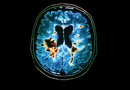
Risk Stratification May Mitigate Adverse Events Associated With Newer MS Drugs
COPENHAGEN—Risk stratification can help neurologists determine whether to prescribe one of the newer multiple sclerosis (MS) drugs for their patients, according to a lecture given at the 29th Congress of the European Committee for Treatment and Research in MS. Natalizumab, fingolimod, and alemtuzumab have potentially serious side effects, but their benefits outweigh their risks, said Per Soelberg Sørensen, MD, Head of the Danish MS Center at Copenhagen University Hospital.
“MS is a serious disease, particularly active MS,” said Dr. Sørensen. “Therefore, people need these drugs to control the disease.”
JCV Antibody Status Influences Treatment With Natalizumab
Before treating a patient with natalizumab, a neurologist should administer a blood test and an MRI, according to the European Medicines Agency. Because natalizumab entails the risk of progressive multifocal leukoencephalopathy (PML), the neurologist should determine whether the patient has antibodies to the John Cunningham virus (JCV). Patients with no JCV antibodies have almost no risk of PML, said Dr. Sørensen. Patients who have the antibodies and have used immunosuppressants are at considerable risk for PML and should not take natalizumab, he added. For patients with JCV antibodies who have not used immunosuppressants, the risk of PML is low within the first 24 months of treatment with natalizumab, but it increases during the following 12 months.
Treatment with natalizumab should be stopped if a patient develops hypersensitivity reactions or antibodies against natalizumab, according to Dr. Sørensen, who added that neurologists should monitor liver function and perform annual MRIs for all patients taking the drug. In addition, he advised neurologists to test for JCV antibodies every six months and noted that JCV antibody titer may increase for patients who begin treatment with a low titer. If the neurologist suspects that the patient is developing PML, he or she should stop natalizumab treatment, perform MRI, and perform CSF tests for JCV, said Dr. Sørensen.
Antiarrhythmics and Antineoplastics May Interfere With Fingolimod
Fingolimod, a sphingosine-1 phosphate receptor modulator, helps patients by acting on lymphocytes but may cause macular edema or restrict respiratory flow. The drug is contraindicated in patients who are taking antiarrhythmic, antineoplastic, or immunosuppressant drugs. Neurologists should be cautious when prescribing fingolimod to patients receiving drugs that lower the heart rate (eg, calcium channel blockers and beta-blockers) or medicines that inhibit CYP3A4-mediated metabolism, said Dr. Sørensen. Fingolimod initially decreases a patient’s white blood cell count, and it takes more than four weeks for the count to return to normal. Patients on fingolimod therefore should not receive attenuated live vaccines. Neurologists also should be vigilant for unknown safety signals, added Dr. Sørensen.
Blood and liver function tests are recommended before initiating treatment with fingolimod, and high-risk patients should undergo ophthalmologic exams. Neurologists should observe all patients for six hours during treatment initiation. Because of a risk of infection, clinicians should monitor patients for two months following treatment initiation, said Dr. Sørensen. During treatment, patients should report symptoms of infection and use contraception, and their blood pressure and liver function should be monitored regularly as well, he added.
Alemtuzumab Entails a Risk of Thyroid Disorder
Alemtuzumab is associated with many side effects, most of which are related to the infusion. Approximately 36% of patients develop a thyroid disorder while taking alemtuzumab, and the peak incidence occurs during the second or third year of treatment. About 1% of patients develop immune thrombocytopenia, which, like thyroid disorder, typically occurs during the second or third year of treatment. Diagnosis is important because immune thrombocytopenia responds to standard medical treatment, said Dr. Sørensen. In addition, some patients have developed neuropathy on alemtuzumab, and early detection can improve the patient’s prognosis, he added.
To maximize safety, neurologists should perform a blood test and screen for tuberculosis or HIV before initiating alemtuzumab. Patients who are anti-thyroid peroxidase positive at baseline are more likely to have thyroid-related adverse events on alemtuzumab, said Dr. Sørensen.
To prevent infusion-related reactions, neurologists can administer corticosteroids, antihistamines, and antipyretics during the first three days of treatment. Patients’ pulse and blood pressure should be monitored, and patients should receive an oral antiherpes medication for at least one month after receiving alemtuzumab, noted Dr. Sørensen. Neurologists should perform blood tests and monitor thyroid function regularly, he added. “This [monitoring] has to continue for four years after the last dose of alemtuzumab,” said Dr. Sørensen.
Monthly tests for blood count and urinalysis and thrice-monthly tests for thyroid function are recommended until four years after the last dose of alemtuzumab, although this regimen may pose logistic problems, he noted. Patients on alemtuzumab also should use contraceptives and report symptoms of infections and signs of bleeding, Dr. Sørensen concluded.
—Erik Greb
Senior Associate Editor
Suggested Reading
Perumal J, Khan O. Emerging disease-modifying therapies in multiple sclerosis. Curr Treat Options Neurol. 2012;14(3):256-263.
Rudick R, Polman C, Clifford D, et al. Natalizumab: bench to bedside and beyond. JAMA Neurol. 2013;70(2):172-182.
COPENHAGEN—Risk stratification can help neurologists determine whether to prescribe one of the newer multiple sclerosis (MS) drugs for their patients, according to a lecture given at the 29th Congress of the European Committee for Treatment and Research in MS. Natalizumab, fingolimod, and alemtuzumab have potentially serious side effects, but their benefits outweigh their risks, said Per Soelberg Sørensen, MD, Head of the Danish MS Center at Copenhagen University Hospital.
“MS is a serious disease, particularly active MS,” said Dr. Sørensen. “Therefore, people need these drugs to control the disease.”
JCV Antibody Status Influences Treatment With Natalizumab
Before treating a patient with natalizumab, a neurologist should administer a blood test and an MRI, according to the European Medicines Agency. Because natalizumab entails the risk of progressive multifocal leukoencephalopathy (PML), the neurologist should determine whether the patient has antibodies to the John Cunningham virus (JCV). Patients with no JCV antibodies have almost no risk of PML, said Dr. Sørensen. Patients who have the antibodies and have used immunosuppressants are at considerable risk for PML and should not take natalizumab, he added. For patients with JCV antibodies who have not used immunosuppressants, the risk of PML is low within the first 24 months of treatment with natalizumab, but it increases during the following 12 months.
Treatment with natalizumab should be stopped if a patient develops hypersensitivity reactions or antibodies against natalizumab, according to Dr. Sørensen, who added that neurologists should monitor liver function and perform annual MRIs for all patients taking the drug. In addition, he advised neurologists to test for JCV antibodies every six months and noted that JCV antibody titer may increase for patients who begin treatment with a low titer. If the neurologist suspects that the patient is developing PML, he or she should stop natalizumab treatment, perform MRI, and perform CSF tests for JCV, said Dr. Sørensen.
Antiarrhythmics and Antineoplastics May Interfere With Fingolimod
Fingolimod, a sphingosine-1 phosphate receptor modulator, helps patients by acting on lymphocytes but may cause macular edema or restrict respiratory flow. The drug is contraindicated in patients who are taking antiarrhythmic, antineoplastic, or immunosuppressant drugs. Neurologists should be cautious when prescribing fingolimod to patients receiving drugs that lower the heart rate (eg, calcium channel blockers and beta-blockers) or medicines that inhibit CYP3A4-mediated metabolism, said Dr. Sørensen. Fingolimod initially decreases a patient’s white blood cell count, and it takes more than four weeks for the count to return to normal. Patients on fingolimod therefore should not receive attenuated live vaccines. Neurologists also should be vigilant for unknown safety signals, added Dr. Sørensen.
Blood and liver function tests are recommended before initiating treatment with fingolimod, and high-risk patients should undergo ophthalmologic exams. Neurologists should observe all patients for six hours during treatment initiation. Because of a risk of infection, clinicians should monitor patients for two months following treatment initiation, said Dr. Sørensen. During treatment, patients should report symptoms of infection and use contraception, and their blood pressure and liver function should be monitored regularly as well, he added.
Alemtuzumab Entails a Risk of Thyroid Disorder
Alemtuzumab is associated with many side effects, most of which are related to the infusion. Approximately 36% of patients develop a thyroid disorder while taking alemtuzumab, and the peak incidence occurs during the second or third year of treatment. About 1% of patients develop immune thrombocytopenia, which, like thyroid disorder, typically occurs during the second or third year of treatment. Diagnosis is important because immune thrombocytopenia responds to standard medical treatment, said Dr. Sørensen. In addition, some patients have developed neuropathy on alemtuzumab, and early detection can improve the patient’s prognosis, he added.
To maximize safety, neurologists should perform a blood test and screen for tuberculosis or HIV before initiating alemtuzumab. Patients who are anti-thyroid peroxidase positive at baseline are more likely to have thyroid-related adverse events on alemtuzumab, said Dr. Sørensen.
To prevent infusion-related reactions, neurologists can administer corticosteroids, antihistamines, and antipyretics during the first three days of treatment. Patients’ pulse and blood pressure should be monitored, and patients should receive an oral antiherpes medication for at least one month after receiving alemtuzumab, noted Dr. Sørensen. Neurologists should perform blood tests and monitor thyroid function regularly, he added. “This [monitoring] has to continue for four years after the last dose of alemtuzumab,” said Dr. Sørensen.
Monthly tests for blood count and urinalysis and thrice-monthly tests for thyroid function are recommended until four years after the last dose of alemtuzumab, although this regimen may pose logistic problems, he noted. Patients on alemtuzumab also should use contraceptives and report symptoms of infections and signs of bleeding, Dr. Sørensen concluded.
—Erik Greb
Senior Associate Editor
Suggested Reading
Perumal J, Khan O. Emerging disease-modifying therapies in multiple sclerosis. Curr Treat Options Neurol. 2012;14(3):256-263.
Rudick R, Polman C, Clifford D, et al. Natalizumab: bench to bedside and beyond. JAMA Neurol. 2013;70(2):172-182.
Suggested Reading
Perumal J, Khan O. Emerging disease-modifying therapies in multiple sclerosis. Curr Treat Options Neurol. 2012;14(3):256-263.
Rudick R, Polman C, Clifford D, et al. Natalizumab: bench to bedside and beyond. JAMA Neurol. 2013;70(2):172-182.

Repetitive TMS May Reduce Depression and Fatigue in Patients With MS
COPENHAGEN—When applied to the motor cortex, repetitive transcranial magnetic stimulation (rTMS) may ameliorate depression and fatigue related to multiple sclerosis (MS), according to a study presented at the 29th Congress of the European Committee for Treatment and Research in MS (ECTRIMS).
The effects of rTMS may persist for as long as six weeks after the end of stimulation. To a lesser extent, rTMS of the left prefrontal cortex also may reduce depression and fatigue for patients with MS, said Sven Schippling, MD, deputy head of the Neuroimmunology and MS Research Section at University Hospital Zurich.
Results from the study of patients with MS suggest that the technique is safe and well tolerated. The most frequent side effects included headache on the day of or the day after stimulation and paresthesia in the lower or upper limbs.
Technicians Administered Stimulation With an H-Coil
Dr. Schippling and Friedemann Paul, MD, consultant neurologist at Charité University Medical Center in Berlin, administered rTMS with an H-coil that was jointly developed by the Weizmann Institute of Science in Rehovot, Israel, the NIH, and the Jerusalem-based company Brainsway. Compared with the conventional figure-eight coil, the H-coil directly stimulates deeper brain regions (ie, more than 1 cm below the scalp) over a less focused area.
The investigators randomly assigned 28 patients with MS (26 with relapsing-remitting MS and two with secondary progressive MS, female-to-male ratio 4:1, median EDSS 3.0) to rTMS of the motor cortex, rTMS of the left prefrontal cortex, or sham stimulation of the left prefrontal cortex. Patients who received treatment of the motor cortex did not know whether stimulation was real or sham, but technicians and investigators were not blinded for this condition. Rating physicians, who had been blinded to patients’ assignment, recorded clinical ratings and administered questionnaires. Stimulation of the left prefrontal cortex was performed in a double-blinded fashion. After randomization, participants underwent three stimulation sessions per week during a six-week treatment period. Follow-up lasted for six additional weeks and consisted of three biweekly visits.
The researchers assessed fatigue with the Fatigue Severity Scale (FSS), the Modified Fatigue Impact Scale, and a visual analog scale that the patient completed. Depression was evaluated using the Beck Depression Inventory (BDI) and the 18-item Hamilton Rating Scale for Depression.
Eligible participants were relapse-free for more than 30 days before inclusion and had taken immunomodulatory or immunosuppressant treatment for more than three months before inclusion. To be included, patients had to have an FSS score of 4 or higher or a BDI score of 12 or higher.
Depression Scores Were Not Equally Distributed at Baseline
Approximately one-third of patients had headache on the day of stimulation, and fewer patients had headache on the day after stimulation. Several patients had paresthesia in the lower or upper limbs. Both headache and paresthesia were similar in frequency in the sham and the true stimulation conditions. The investigators observed no severe events in the patients that received true stimulation, and the types and frequency of adverse events were comparable between the three treatment groups.
Baseline BDI scores were not equally distributed between the groups, said Dr. Schippling. Patients who received rTMS of the prefrontal cortex had higher baseline BDI scores, compared with patients who received rTMS of the motor cortex. FSS and BDI scores decreased significantly in patients who received rTMS of the motor cortex. Motor cortex stimulation significantly lowered the rate of fatigue in all the fatigue and depression scores, including the visual analog scale, said Dr. Schippling.
A clear limitation of the study is its small size, said Dr. Schippling. “However, we find the results promising, and we want to embark on a phase II trial because further evidence is needed to draw firm conclusions about what might be an effective treatment. This would be highly desirable, as treatment options in MS-related fatigue are limited,” he concluded.
—Erik Greb
Senior Associate Editor
Suggested Reading
Speer AM, Wassermann EM, Benson BE, et al. Antidepressant efficacy of high and low frequency rTMS at 110% of motor threshold versus sham stimulation over left prefrontal cortex. Brain Stimul. 2013 Jul 29 [Epub ahead of print].
Yusuf A, Koski L. A qualitative review of the neurophysiological underpinnings of fatigue in multiple sclerosis. J Neurol Sci. 2013;330(1-2):4-9.
COPENHAGEN—When applied to the motor cortex, repetitive transcranial magnetic stimulation (rTMS) may ameliorate depression and fatigue related to multiple sclerosis (MS), according to a study presented at the 29th Congress of the European Committee for Treatment and Research in MS (ECTRIMS).
The effects of rTMS may persist for as long as six weeks after the end of stimulation. To a lesser extent, rTMS of the left prefrontal cortex also may reduce depression and fatigue for patients with MS, said Sven Schippling, MD, deputy head of the Neuroimmunology and MS Research Section at University Hospital Zurich.
Results from the study of patients with MS suggest that the technique is safe and well tolerated. The most frequent side effects included headache on the day of or the day after stimulation and paresthesia in the lower or upper limbs.
Technicians Administered Stimulation With an H-Coil
Dr. Schippling and Friedemann Paul, MD, consultant neurologist at Charité University Medical Center in Berlin, administered rTMS with an H-coil that was jointly developed by the Weizmann Institute of Science in Rehovot, Israel, the NIH, and the Jerusalem-based company Brainsway. Compared with the conventional figure-eight coil, the H-coil directly stimulates deeper brain regions (ie, more than 1 cm below the scalp) over a less focused area.
The investigators randomly assigned 28 patients with MS (26 with relapsing-remitting MS and two with secondary progressive MS, female-to-male ratio 4:1, median EDSS 3.0) to rTMS of the motor cortex, rTMS of the left prefrontal cortex, or sham stimulation of the left prefrontal cortex. Patients who received treatment of the motor cortex did not know whether stimulation was real or sham, but technicians and investigators were not blinded for this condition. Rating physicians, who had been blinded to patients’ assignment, recorded clinical ratings and administered questionnaires. Stimulation of the left prefrontal cortex was performed in a double-blinded fashion. After randomization, participants underwent three stimulation sessions per week during a six-week treatment period. Follow-up lasted for six additional weeks and consisted of three biweekly visits.
The researchers assessed fatigue with the Fatigue Severity Scale (FSS), the Modified Fatigue Impact Scale, and a visual analog scale that the patient completed. Depression was evaluated using the Beck Depression Inventory (BDI) and the 18-item Hamilton Rating Scale for Depression.
Eligible participants were relapse-free for more than 30 days before inclusion and had taken immunomodulatory or immunosuppressant treatment for more than three months before inclusion. To be included, patients had to have an FSS score of 4 or higher or a BDI score of 12 or higher.
Depression Scores Were Not Equally Distributed at Baseline
Approximately one-third of patients had headache on the day of stimulation, and fewer patients had headache on the day after stimulation. Several patients had paresthesia in the lower or upper limbs. Both headache and paresthesia were similar in frequency in the sham and the true stimulation conditions. The investigators observed no severe events in the patients that received true stimulation, and the types and frequency of adverse events were comparable between the three treatment groups.
Baseline BDI scores were not equally distributed between the groups, said Dr. Schippling. Patients who received rTMS of the prefrontal cortex had higher baseline BDI scores, compared with patients who received rTMS of the motor cortex. FSS and BDI scores decreased significantly in patients who received rTMS of the motor cortex. Motor cortex stimulation significantly lowered the rate of fatigue in all the fatigue and depression scores, including the visual analog scale, said Dr. Schippling.
A clear limitation of the study is its small size, said Dr. Schippling. “However, we find the results promising, and we want to embark on a phase II trial because further evidence is needed to draw firm conclusions about what might be an effective treatment. This would be highly desirable, as treatment options in MS-related fatigue are limited,” he concluded.
—Erik Greb
Senior Associate Editor
Suggested Reading
Speer AM, Wassermann EM, Benson BE, et al. Antidepressant efficacy of high and low frequency rTMS at 110% of motor threshold versus sham stimulation over left prefrontal cortex. Brain Stimul. 2013 Jul 29 [Epub ahead of print].
Yusuf A, Koski L. A qualitative review of the neurophysiological underpinnings of fatigue in multiple sclerosis. J Neurol Sci. 2013;330(1-2):4-9.

Teriflunomide Is Safe and Effective for Patients With CIS—Main Results From the TOPIC Study
COPENHAGEN—Teriflunomide (14 mg and 7 mg) is safe and effective in the treatment of patients with clinically isolated syndrome, according to TOPIC trial data presented at the 29th Congress of the European Committee for Treatment and Research in Multiple Sclerosis (ECTRIMS).
“These findings highlight the ability of early intervention with teriflunomide to delay onset of MS symptoms,” said Aaron E. Miller, MD, Professor of Neurology at the Icahn School of Medicine at Mount Sinai, New York, and his study collaborators. “To date, all teriflunomide phase III studies have shown consistent safety and efficacy, and greater efficacy for a 14-mg dose.”
Teriflunomide is a novel, once-daily, oral immunomodulator approved in the United States, Argentina, and Australia for the treatment of relapsing-remitting MS. Prior clinical studies of teriflunomide in patients with relapsing-remitting MS (TEMSO and TOWER) showed consistent efficacy across key clinical measures and a well-characterized safety profile. TOPIC was a phase III clinical trial conducted to assess the efficacy and safety of teriflunomide in patients with a first clinical episode suggestive of MS (clinically isolated syndrome).
In TOPIC, a double-blind, placebo-controlled, parallel-group study, 618 patients with clinically isolated syndrome were enrolled from February 2008 to September 2012 (projected end date, December 2012) and randomized to placebo, teriflunomide 7 mg, or teriflunomide 14 mg. TOPIC ended three months early as revised diagnostic criteria enabled earlier diagnosis of MS. The primary and key secondary end points were conversion to clinically definite MS and occurrence of a new clinical relapse or MRI lesion. Other efficacy end points and safety and tolerability were also assessed.
Baseline characteristics were generally well balanced among the treatment groups. Of the randomized population (n = 618), 59.1% had monofocal and 40.9% had multifocal lesion presentation, and 31.4% had one or more gadolinium-enhancing lesions. Median time since first neurologic event was two months.
Compared with placebo, teriflunomide 14 mg reduced the risk of conversion to clinically definite MS by 42.6%, with a probability of conversion to clinically definite MS at 108 weeks of 24.0% (probability for placebo group, 35.9%). Teriflunomide 14 mg also reduced the risk of occurrence of new relapse or new MRI lesion by 34.9%. As measured by MRI over the two-year study period, patients treated with teriflunomide 14 mg had a 5% increase in total lesion volume, compared with a 28% increase among patients treated with placebo. In addition, patients treated with teriflunomide 14 mg had a 59% reduction in gadolinium-enhancing T1 lesions, compared with patients receiving placebo.
Teriflunomide 7 mg reduced the risk of conversion to clinically definite MS by 37.2% (108-week probability, 27.6%) and the risk of occurrence of new relapse or new MRI lesion by 31.4%. The occurrence of adverse events was similar across treatment groups. The most common adverse events reported more frequently in the teriflunomide arms included alanine aminotransferase elevation, headache, hair thinning (14 mg only), diarrhea, and paresthesia.
—Glenn S. Williams
Vice President/Group Editor