User login
Cyst Removal: Punch Incision Leaves Smaller Scar
RALEIGH, N.C. – Punch incision epidermal inclusion cysts located on the trunk leaves a significantly smaller scar than does elliptical excision with a similarly low recurrence rate, according to the results of a randomized trail.
Procedure time was essentially the same for the two techniques, at around 13 minutes. Although punch incision and its wound closure can be easier, it took a fair amount of time to squeeze the cyst contents through the small punch opening and remove the cyst lining using a curette, Dr. Justin T. Cheeley explained at the annual meeting of the Society for Investigative Dermatology.
He reported on 40 consecutive patients with one or more truncal epidermal inclusion cysts 1-3 cm in diameter who were randomized to elliptical excision or punch incision in a head-to-head comparative trial.
The primary study end point – cyst recurrence during 16 months of prospective follow-up – occurred in three patients in the punch incision group and two in the elliptical excision group. Predictors of cyst recurrence were sought, but none could be identified, according to Dr. Cheeley of Emory University, Atlanta.
Most secondary end points were similar for the two study arms, including early and late complication rates, as well as improvement in skin-specific quality of life and patient satisfaction as measured by change in Skindex-16 scores.

There was, however, a significant difference between the two study groups in terms of average scar length. In the punch incision group, average scar length was 1.1 cm, compared with 1.8 cm in the elliptical excision group.
The investigators employed a 4-mm punch for the most part, although they turned to a 6-mm punch in treating larger cysts. Punch incision wounds were closed with a single nylon suture. Closure of the elliptical excision sites required more extensive suturing.
Audience member Dr. Eric L. Simpson complimented Dr. Cheeley and his coinvestigators for conducting a study with important cost implications given how often epidermal inclusion cysts are encountered in practice.
“The difference between punch incision and elliptical excision with an intermediate-level repair is probably 10-fold in terms of cost,” said Dr. Simpson of Oregon Health and Science University, Portland.
Dr. Cheeley reported having no financial conflicts.
RALEIGH, N.C. – Punch incision epidermal inclusion cysts located on the trunk leaves a significantly smaller scar than does elliptical excision with a similarly low recurrence rate, according to the results of a randomized trail.
Procedure time was essentially the same for the two techniques, at around 13 minutes. Although punch incision and its wound closure can be easier, it took a fair amount of time to squeeze the cyst contents through the small punch opening and remove the cyst lining using a curette, Dr. Justin T. Cheeley explained at the annual meeting of the Society for Investigative Dermatology.
He reported on 40 consecutive patients with one or more truncal epidermal inclusion cysts 1-3 cm in diameter who were randomized to elliptical excision or punch incision in a head-to-head comparative trial.
The primary study end point – cyst recurrence during 16 months of prospective follow-up – occurred in three patients in the punch incision group and two in the elliptical excision group. Predictors of cyst recurrence were sought, but none could be identified, according to Dr. Cheeley of Emory University, Atlanta.
Most secondary end points were similar for the two study arms, including early and late complication rates, as well as improvement in skin-specific quality of life and patient satisfaction as measured by change in Skindex-16 scores.
There was, however, a significant difference between the two study groups in terms of average scar length. In the punch incision group, average scar length was 1.1 cm, compared with 1.8 cm in the elliptical excision group.
The investigators employed a 4-mm punch for the most part, although they turned to a 6-mm punch in treating larger cysts. Punch incision wounds were closed with a single nylon suture. Closure of the elliptical excision sites required more extensive suturing.
Audience member Dr. Eric L. Simpson complimented Dr. Cheeley and his coinvestigators for conducting a study with important cost implications given how often epidermal inclusion cysts are encountered in practice.
“The difference between punch incision and elliptical excision with an intermediate-level repair is probably 10-fold in terms of cost,” said Dr. Simpson of Oregon Health and Science University, Portland.
Dr. Cheeley reported having no financial conflicts.
FROM THE ANNUAL MEETING OF THE SOCIETY FOR INVESTIGATIVE DERMATOLOGY
Major Finding: In the punch incision group, average scar length was 1.1 cm, compared with 1.8 cm in the elliptical excision group.
Data Source: This was a randomized trial of 40 consecutive patients.
Disclosures: Dr. Cheeley reported having no financial conflicts.
Onion Extract Improved Scars by 36%
RALEIGH, N.C. – A new once-daily topical gel containing a proprietary onion extract resulted in a 36% improvement in the appearance of recent postsurgical dermal scars at 8 weeks, according to the results of a randomized, controlled trial.
The over-the-counter product, Merz Pharmaceuticals’ Mederma Advanced Scar Gel, was studied in 44 adults, each of whom underwent surgical shave removal of two similar-size seborrheic keratoses on the chest. At 2 weeks, after the wounds had reepithelialized, patients were randomly assigned to apply the nonprescription onion extract gel once daily to one scar and no treatment to the other.

Blinded investigator assessment was carried out after 2, 4, and 8 weeks of once-daily therapy. Each scar was graded on a 0-3 scale for improvement over baseline for overall appearance and for more specific individual domains of texture, redness, and softness. Patients independently carried out the same assessments, explained Dr. Zoe D. Draelos, a clinical dermatologist and researcher in High Point, N.C.
At week 8, investigators rated the onion extract–treated scars as demonstrating a mean 2.6-point improvement over baseline in terms of overall appearance, with comparable improvements noted in texture, redness, and scar softness. These were significantly better outcomes than was the mean 2.1-point improvement in the overall appearance of untreated control scars, she noted.
The patients rated the onion extract gel–treated scars as showing a mean 2.0-point improvement at week 8, significantly better than the 1.5-point improvement noted in the control scars.
Although optimal results were seen at week 8, the topical gel–treated scars showed a significant advantage in appearance scores, compared with control scars, as early as week 4, with a nonsignificant favorable trend noted at week 2.
The chief advantage that the transparent onion extract gel offers over other scar treatment products is the convenience of once-daily application, noted Dr. Draelos.
Merz announced the launch of Mederma Advanced Scar Gel in the spring. It is available in the first-aid section of pharmacies nationwide at a retail price of about $20 for a 20-g tube and $32 for 50 g, according to the company.
Other Merz products containing Cepalin, the proprietary onion extract, include Mederma Scar Cream plus SPF 30, Mederma for Kids, and Mederma Stretch Marks Therapy.
Dr. Draelos received research funding from Merz to conduct the clinical trial.
RALEIGH, N.C. – A new once-daily topical gel containing a proprietary onion extract resulted in a 36% improvement in the appearance of recent postsurgical dermal scars at 8 weeks, according to the results of a randomized, controlled trial.
The over-the-counter product, Merz Pharmaceuticals’ Mederma Advanced Scar Gel, was studied in 44 adults, each of whom underwent surgical shave removal of two similar-size seborrheic keratoses on the chest. At 2 weeks, after the wounds had reepithelialized, patients were randomly assigned to apply the nonprescription onion extract gel once daily to one scar and no treatment to the other.
Blinded investigator assessment was carried out after 2, 4, and 8 weeks of once-daily therapy. Each scar was graded on a 0-3 scale for improvement over baseline for overall appearance and for more specific individual domains of texture, redness, and softness. Patients independently carried out the same assessments, explained Dr. Zoe D. Draelos, a clinical dermatologist and researcher in High Point, N.C.
At week 8, investigators rated the onion extract–treated scars as demonstrating a mean 2.6-point improvement over baseline in terms of overall appearance, with comparable improvements noted in texture, redness, and scar softness. These were significantly better outcomes than was the mean 2.1-point improvement in the overall appearance of untreated control scars, she noted.
The patients rated the onion extract gel–treated scars as showing a mean 2.0-point improvement at week 8, significantly better than the 1.5-point improvement noted in the control scars.
Although optimal results were seen at week 8, the topical gel–treated scars showed a significant advantage in appearance scores, compared with control scars, as early as week 4, with a nonsignificant favorable trend noted at week 2.
The chief advantage that the transparent onion extract gel offers over other scar treatment products is the convenience of once-daily application, noted Dr. Draelos.
Merz announced the launch of Mederma Advanced Scar Gel in the spring. It is available in the first-aid section of pharmacies nationwide at a retail price of about $20 for a 20-g tube and $32 for 50 g, according to the company.
Other Merz products containing Cepalin, the proprietary onion extract, include Mederma Scar Cream plus SPF 30, Mederma for Kids, and Mederma Stretch Marks Therapy.
Dr. Draelos received research funding from Merz to conduct the clinical trial.
FROM THE ANNUAL MEETING OF THE SOCIETY FOR INVESTIGATIVE DERMATOLOGY
Major Finding: An 8-week regimen of an onion extract–based topical gel led to a 36% greater improvement in the overall appearance of new postsurgical dermal scars, compared with no treatment.
Data Source: A randomized, controlled study of 44 patients who underwent surgical removal of two similar-sized seborrheic keratoses on their chest.
Disclosures: Dr. Draelos received research funding from Merz to conduct the clinical trial.
Skin of Color: Which Butter Is Better?
For many years, cocoa butter has played a major role in the ethnic skin care market. Derived from cocoa beans in tropical regions, it has been used in topicals for moisturization, stretch marks, scars, fade creams, and more. In recent years, shea butter has also become prominent in emollient creams and lotions. Cocoa butter, shea butter, mango seed butter, and cupuacu butter are all found as ingredients in many products.
So which butter is better for your patients? The answer is not a simple one, but here are some fun facts and the latest research about each option.
Cocoa butter
Cocoa butter – also called theobroma oil – is a pale yellow, pure, edible vegetable fat extracted from the cocoa bean. It is used to make chocolate and baked goods, and it is also used in topical preparations, including moisturizers and striae creams.
Cocoa butter is currently available in many skin care brands, but Palmer's is one of the most well-known brands of topical cocoa butter preparations on the market. The brand has been family-owned since the mid-19th century and under current ownership since 1971.
Ex vivo studies demonstrate that cocoa polyphenols improve skin elasticity and skin tone, namely, glycosaminoglycans and collagen I, III, and IV (Int. J. Cosmet. Sci. 2008;30:339-45).
However, one study that examined comedogenicity of ingredients and vehicles in cosmetics, found cocoa butter to be comedogenic in external rabbit ear canals (Cutan. Ocul. Toxicol. 2007;26:287-92).
A randomized, double-blind placebo controlled trial of 300 pregnant Afro-Caribbean women in Jamaica found that cocoa butter cream did not prevent striae gravidarum. This study found that development of striae was related to the young age of the mother and large neonates (Int. J. Gynaecol. Obstet. 2010;108:65-8).
And another randomized placebo controlled trial of 210 nulliparous women in Lebanon also found that topical application of cocoa butter cream did not decrease the likelihood of striae gravidarum, compared with placebo (BJOG 2008;115:1138-42).
Ingestion of cocoa butter in the forms of baked goods or chocolate has some antioxidant value because of the high levels of polyphenols. Oral consumption of cocoa also has anecdotal antimalarial effects through increased availability of antioxidants in plasma, membrane effects in general and erythrocyte membrane in particular, increased plasma levels of nitric oxide, antimalarial activity of cocoa flavanoids and their derivatives, and boosted immune system mediated by components of cocoa, including cocoa butter, polyphenols, magnesium, and zinc.
Shea butter
Shea butter is a slightly yellowish or ivory-colored fat extracted from the nut of the African shea tree (Vitellaria paradoxa). It has been used traditionally throughout Africa as a moisturizer; it has also been used in combination with coconut oil, palm oil, and gobi oil as a natural mosquito repellant (one that also protects against onchocerciasis).
In Africa, shea butter is used as cooking oil, as a waterproofing wax, for hairdressing, for candle-making, and as an ingredient in medicinal ointments. It is also used by makers of traditional African percussion instruments to increase the durability of wood.
Shea butter has been shown to have anti-inflammatory effects in studies through inhibition of iNOS, COX-2, and cytokines via the Nf-κB pathway in LPS-activated J774 macrophage cells (J. Complement. Integr. Med. 2012;9:Article 4).
Like cocoa butter, shea butter contains polyphenols. It also contains exceptionally high levels of triterpenes, indicating that shea nuts and shea fat constitute a significant source of anti-inflammatory and anti-tumor promoting compounds (J. Oleo. Sci. 2010;59:273-80).
Shea butter also contains no IgE-binding soluble proteins, making it of low allergenic potential (J. Allergy Clin. Immunol. 2011;127:680-2).
Mango seed butter
Mango seed butter's solid content profile is very similar to that of cocoa butterexcept it is softer (Bioresour. Technol. 2004;92:71-8). It is rich in beta carotene, essential fatty acids, and vitamins A and E. It is also used in skin creams, but not much has been published about mango seed butter in peer-reviewed journals.
Cupuacu butter
Cupuaçu (Theobroma grandiflorum) is a tropical rainforest tree related to cacao. Common throughout the Amazon basin, it is widely cultivated in the jungles of Colombia, Bolivia, Peru, and in northern Brazil. Some skin care brands, particularly in Brazil, use cupuaçu butter in topical emollient creams.
It has been shown to contain high concentrations of polyphenolic antioxidants, but less caffeine than its cocoa counterpart.
Activity-guided fractionation of cupuacu seeds in one study resulted in the identification of new sulfated flavonoid glycosides, theograndins I and II. In addition, nine flavonoid antioxidants were identified (J. Nat. Prod. 2003;66:1501-4). The theograndins had antioxidant effects and were weakly cytotoxic against human colon cancer cells.
No head-to-head comparative studies have been performed on these butters. Given the research to date, shea butter has a slight edge due to the sheer number of studies that show positive properties.
- Naissan Wesley, M.D.
Do you have questions about treating patients with darker skin? If so, send them to sknews@elsevier.com.
Ajuga turkestanica
A perennial herb found primarily in Central Asia, Ajuga turkestanica is known to contain several bioactive compounds and has been used in traditional medicine to treat heart disease, and stomach and muscle aches (Chem. Nat. Compd. 1998;34:150-4; Chem. Nat. Compd. 2004;40:85-6). It is one of the many species of Ajuga gaining attention for exhibiting medicinal properties with the potential for commercial applications (Phytochem. Lett. 2008;1:81-4).
Clerodane diterpenes, recognized sources of antimicrobial, antiviral, antitumor, antibiotic, and amoebicidal activities (Phytochem. Rev. 2008;7:25-49), are among the three classes of potentially bioactive compounds, along with phytoecdysteroids and iridoid glycosides found in the Ajuga genus (Phytochem. Lett. 2008;1:81-4). Phytoecdysteroids are known to display significant physiological activities in insects and mammals; and iridoid glycosides, especially abundant in A. decumbens, have exhibited anticancer activity (Cancer Lett. 2000;157:87-92).

A. turkestanica reportedly contains several phytoecdysteroids (turkesterone, 20-hydroxyecdysone, cyasterone, cyasterone 22-acetate, ajugalactone, ajugasterone B, alpha-ecdysone, and ecdysone 2,3-monoacetonide), as well as the iridoids harpagide and harpagide 8-acetate (Chem. Nat. Compd. 2005;41:361-9; Chem. Nat. Compd. 1978;14:175-8; Chem. Nat. Compd. 1975;11:484-7; Chem. Nat. Compd. 1973;9:125-6; Chem. Nat. Compd. 1971;7:520; Phytochem. Lett. 2008;1:81-4).
Aquaporins
Aquaporins (AQPs) are integral membrane proteins that facilitate water transport in several organs, including the skin, brain, eyes, and digestive tract, as well as in the renal tubules. Thirteen isoforms of aquaporins (AQPs 0-12) are found in mammals. Of these, there are two functional subtype classifications: AQPs 1, 2, 4, 5, and 8 conduct only water, and AQPs 3, 7, 9, and 10 transport water and other substances including glycerol and urea (Prog. Histochem. Cytochem. 2004;39:1-83). AQP-3, permeable to water and glycerol, is the main water channel in human epidermis. Glycerol acts as an endogenous humectant, thereby facilitating hydration of the stratum corneum (SC) (J. Invest. Dermatol. 2005;125:288-93).
Defects in AQP-3 in mice models have been demonstrated to lead to epidermal xerosis and to reductions in SC hydration and epidermal glycerol content, followed by diminished elasticity and impaired skin barrier recovery (J. Biol. Chem. 2002;277:46616-21; Proc. Natl. Acad. Sci. 2003;100:7360-5). Such findings underscore the important role of glycerol in cutaneous hydration. Significantly, AQPs, particularly AQP-3, contribute to the transport of water, glycerol, and solutes between keratinocytes.
Dumas et al. note that the role of AQPs in hydrating the living layers of the epidermis where keratinocyte differentiation occurs and in barrier development and recovery suggests that they are significant protein targets for improving the quality and resistance of the skin surface, as well as ameliorating aging- and UV-induced xerosis (J. Drugs Dermatol. 2007;6(6 Suppl):s20-4).
A. turkestanica and Aquaporins
Patented extracts of A. turkestanica have been shown to contain sufficient ecdysteroids and other active ingredients to improve the differentiation of keratinocytes, thus facilitating skin hydration and yielding antiaging effects (U.S. Patent 7,060,693 B1, June 13, 2006). The patent inventors Dumas et al. observed that the extracts are especially effective in regulating epidermal water transport, achieving improved hydration of the basal layer by working in concert with or enhancing AQP-3 (Cosmet. Toil. 2008;123:22-7).
In 2007, Dumas et al. conducted in vitro and in vivo studies of active ingredients capable of raising AQP-3 levels to enhance hydration in human skin keratinocytes, with the understanding that improving hydration in keratinocytes would ultimately improve epidermal hydration (Eur. J. Dermatol. 2002;12:XXV-XXVI). They used an ethanol/water (70/30 v/v) extract of A. turkestanica as the hydrating agent (2.5 mcg/mL), and found that after 17 days of in vitro treatment every 2 days in human reconstructed epidermis, AQP-3 expression measured at the protein level was significantly elevated. Increased epidermal proliferation and differentiation were also noted. Electron microscopy showed a significantly thicker, compact SC and more clearly differentiated desmosomes. Constituents of the A. turkestanica extract revealed by chemical analysis included iridoids (chiefly harpagoside and 8-O-acetylharpagoside), ajugasterone B, ajugalactone, turkesterone, 22-acetylcyasterone, and phenols (J. Drugs. Dermatol. 2007;6:s20-4).
The investigators prepared an oil-water emulsion infused with A. turkestanica extract (0.3% w/w) for an in vivo study in which 15 healthy female volunteers (22-56 years old) applied the formulation twice daily to their forearms for 21 days. Significant reductions in transepidermal water loss were seen in the treated area compared with the control area on days 7 and 21. The researchers concluded that the tested A. turkestanica extract formulation enhanced AQP-3 expression and human epidermal differentiation in vitro, and ameliorated epidermal barrier structure and human skin recovery in vivo (J. Drugs. Dermatol. 2007;6:s20-4).
Conclusion
There is a dearth of research on A. turkestanica. But what exists is particularly favorable, insofar as the botanical appears to impact AQP-3, a compound whose significance has been recognized during the past 2 decades. Thus, A. turkestanica appears to have the potential to play an effective role in cosmetic products for treating dry skin. Much more research is necessary, however.
A perennial herb found primarily in Central Asia, Ajuga turkestanica is known to contain several bioactive compounds and has been used in traditional medicine to treat heart disease, and stomach and muscle aches (Chem. Nat. Compd. 1998;34:150-4; Chem. Nat. Compd. 2004;40:85-6). It is one of the many species of Ajuga gaining attention for exhibiting medicinal properties with the potential for commercial applications (Phytochem. Lett. 2008;1:81-4).
Clerodane diterpenes, recognized sources of antimicrobial, antiviral, antitumor, antibiotic, and amoebicidal activities (Phytochem. Rev. 2008;7:25-49), are among the three classes of potentially bioactive compounds, along with phytoecdysteroids and iridoid glycosides found in the Ajuga genus (Phytochem. Lett. 2008;1:81-4). Phytoecdysteroids are known to display significant physiological activities in insects and mammals; and iridoid glycosides, especially abundant in A. decumbens, have exhibited anticancer activity (Cancer Lett. 2000;157:87-92).
A. turkestanica reportedly contains several phytoecdysteroids (turkesterone, 20-hydroxyecdysone, cyasterone, cyasterone 22-acetate, ajugalactone, ajugasterone B, alpha-ecdysone, and ecdysone 2,3-monoacetonide), as well as the iridoids harpagide and harpagide 8-acetate (Chem. Nat. Compd. 2005;41:361-9; Chem. Nat. Compd. 1978;14:175-8; Chem. Nat. Compd. 1975;11:484-7; Chem. Nat. Compd. 1973;9:125-6; Chem. Nat. Compd. 1971;7:520; Phytochem. Lett. 2008;1:81-4).
Aquaporins
Aquaporins (AQPs) are integral membrane proteins that facilitate water transport in several organs, including the skin, brain, eyes, and digestive tract, as well as in the renal tubules. Thirteen isoforms of aquaporins (AQPs 0-12) are found in mammals. Of these, there are two functional subtype classifications: AQPs 1, 2, 4, 5, and 8 conduct only water, and AQPs 3, 7, 9, and 10 transport water and other substances including glycerol and urea (Prog. Histochem. Cytochem. 2004;39:1-83). AQP-3, permeable to water and glycerol, is the main water channel in human epidermis. Glycerol acts as an endogenous humectant, thereby facilitating hydration of the stratum corneum (SC) (J. Invest. Dermatol. 2005;125:288-93).
Defects in AQP-3 in mice models have been demonstrated to lead to epidermal xerosis and to reductions in SC hydration and epidermal glycerol content, followed by diminished elasticity and impaired skin barrier recovery (J. Biol. Chem. 2002;277:46616-21; Proc. Natl. Acad. Sci. 2003;100:7360-5). Such findings underscore the important role of glycerol in cutaneous hydration. Significantly, AQPs, particularly AQP-3, contribute to the transport of water, glycerol, and solutes between keratinocytes.
Dumas et al. note that the role of AQPs in hydrating the living layers of the epidermis where keratinocyte differentiation occurs and in barrier development and recovery suggests that they are significant protein targets for improving the quality and resistance of the skin surface, as well as ameliorating aging- and UV-induced xerosis (J. Drugs Dermatol. 2007;6(6 Suppl):s20-4).
A. turkestanica and Aquaporins
Patented extracts of A. turkestanica have been shown to contain sufficient ecdysteroids and other active ingredients to improve the differentiation of keratinocytes, thus facilitating skin hydration and yielding antiaging effects (U.S. Patent 7,060,693 B1, June 13, 2006). The patent inventors Dumas et al. observed that the extracts are especially effective in regulating epidermal water transport, achieving improved hydration of the basal layer by working in concert with or enhancing AQP-3 (Cosmet. Toil. 2008;123:22-7).
In 2007, Dumas et al. conducted in vitro and in vivo studies of active ingredients capable of raising AQP-3 levels to enhance hydration in human skin keratinocytes, with the understanding that improving hydration in keratinocytes would ultimately improve epidermal hydration (Eur. J. Dermatol. 2002;12:XXV-XXVI). They used an ethanol/water (70/30 v/v) extract of A. turkestanica as the hydrating agent (2.5 mcg/mL), and found that after 17 days of in vitro treatment every 2 days in human reconstructed epidermis, AQP-3 expression measured at the protein level was significantly elevated. Increased epidermal proliferation and differentiation were also noted. Electron microscopy showed a significantly thicker, compact SC and more clearly differentiated desmosomes. Constituents of the A. turkestanica extract revealed by chemical analysis included iridoids (chiefly harpagoside and 8-O-acetylharpagoside), ajugasterone B, ajugalactone, turkesterone, 22-acetylcyasterone, and phenols (J. Drugs. Dermatol. 2007;6:s20-4).
The investigators prepared an oil-water emulsion infused with A. turkestanica extract (0.3% w/w) for an in vivo study in which 15 healthy female volunteers (22-56 years old) applied the formulation twice daily to their forearms for 21 days. Significant reductions in transepidermal water loss were seen in the treated area compared with the control area on days 7 and 21. The researchers concluded that the tested A. turkestanica extract formulation enhanced AQP-3 expression and human epidermal differentiation in vitro, and ameliorated epidermal barrier structure and human skin recovery in vivo (J. Drugs. Dermatol. 2007;6:s20-4).
Conclusion
There is a dearth of research on A. turkestanica. But what exists is particularly favorable, insofar as the botanical appears to impact AQP-3, a compound whose significance has been recognized during the past 2 decades. Thus, A. turkestanica appears to have the potential to play an effective role in cosmetic products for treating dry skin. Much more research is necessary, however.
Experimental Traser Device Could Be Laser's Replacement
DANA POINT, CALIF. – A new therapeutic light source being developed in Israel may ultimately replace many of the aesthetic laser devices currently used.
The device, known as Traser (Total Reflection Amplification of Spontaneous Emission of Radiation), "is not a laser and it’s not an intense pulsed light," Dr. Christopher Zachary said at the SDEF Summit in Aesthetic Medicine sponsored by Skin Disease Education Foundation (SDEF).
"No photons come from the flash lamps. It’s one device with many wavelengths. It’s tunable, has a high peak power, and has variable pulse duration from 0.45 to 100 milliseconds," he said.
The Traser contains a "dye cell" (an internally reflecting body that hosts a fluorescent dye), one rear mirror, flashlamps above and below, an output waveguide, and a reflector cavity that houses the device. The Traser is the brainchild of Morgan Gustavsson, a pioneer of intense pulsed-light technology. It was first described in the peer-reviewed online journal PLoS ONE (2012;7:e35899 [doi: 10.1371/journal.pone.0035899]).
"It’s one device with many wavelengths. It’s tunable, has a high peak power, and has variable pulse duration from 0.45 to 100 milliseconds," Dr. Christopher Zachary said.
Unlike a laser, the Traser has no optical resonator, no output coupler, no stimulated emission, no filters, and no filter technology. "Photons from the flash lamps excite fluorescent dye material and spontaneously emit a different narrow spectrum of light," explained Dr. Zachary, professor and chair of the dermatology department at the University of California, Irvine, who plans to begin studying the device this summer.
"This traps 45%-61% of the light that’s generated, and the photons propagate axially along the length of the dye cell in both directions. The mirror redirects the light forward, and light is passively coupled out at the distal end of the cell," he said.
The dyes used to date are water soluble, "and do not require hazardous solvents or additives," wrote Dr. Zachary and Mr. Gustavsson, who is with Rockport Consulting Services in Newport Beach, Calif., in the PLoS One article. "When required, [the dyes] are reclaimed by a filter loop, which in less than a minute can completely eliminate the dye and purify the circulating water. This water is not only reconstituted with new dyes within the Traser circulation cavity, but is also used to cool the device."
Changing the dye enables the user to produce UVA, blue, green, orange, red, and near-infrared wavelengths, which allows for wide variability in a single device. The fluorescent dyes used and described in the PLoS ONE article include pyrromethene 556, rhodamine 590, and sulforhodamine 640 chloride. "The wavelength that comes out is dependent on the dye that you use and its concentration," Dr. Zachary said at the meeting. "It’s a very manipulable yet sensitive system."
In theory, the Traser "could replace the KTP laser and could be very good for individual blood vessels, for instance, or a rosaceous blush. It could replace the ruby laser as a hair-removal device, or the pulsed dye laser for port wine stains – all from a single device. All you do is change the dye. I love the science of this: It’s simple but has all the characteristics of a device that is going to be very effective. I’d like to see some studies conducted in animals and in humans."
He concluded his remarks by predicting that the TRASER will probably be less costly than a laser, and "highly tunable and with multiple pulse durations, and one that could replace three to five lasers in your office."
Dr. Zachary said that he had no relevant financial conflicts to disclose. SDEF and this news organization are owned by Elsevier.
EXPERT ANALYSIS FROM THE SDEF SUMMIT IN AESTHETIC MEDICINE
Liposonix Slimmed Waists by Almost 3 cm
DANA POINT, CALIF. – Of the 152 patients who have been treated with the Liposonix body sculpting system in controlled clinical trials to date, the average reduction in waist circumference reached 2.8 cm.
The system, cleared by the Food and Drug Administration in September 2011 for noninvasive fat removal, "has a good safety profile, no discernible effect on internal organs or adjacent structures, and minimal adverse events," Dr. Michael A.C. Kane said at the Summit in Aesthetic Medicine sponsored by Skin Disease Education Foundation (SDEF), where he reviewed the evidence on the device.
Solta Medical's Liposonix system uses high-intensity, focused ultrasound to destroy anterior abdominal subcutaneous adipose tissue in a single treatment without harming skin or surrounding tissue. The pattern generator of the device "allows for uniform delivery of ultrasonic energy, analogous to pattern generators used with aesthetic lasers," said Dr. Kane, a plastic surgeon in private practice in New York. The treatment area measures 21 cm2.
Liposonix's mechanism of action is necrosis by a thermal process. "An ultrasound beam that can focus at a specific depth beneath the dermis, combined with proprietary application techniques, results in the intended adipose tissue disruption," Dr. Kane explained. "The pattern generator moves the focus of the ultrasound to successive parts of the adipose tissue. The automated movement of the transducer creates a matrix of lesions produced by highly focused, high-intensity ultrasound."
Macrophage cells, he continued, "are attracted to the treated area to engulf and transport lipids and cellular debris. As the treated area is cleared, the lesion resolves naturally and results in an overall reduction in local adipose tissue volume."
According to data on the 152 patients treated with Liposonix, the most common post-treatment side effects were temporary erythema, mild ecchymosis, discomfort, and edema. The total energy dose ranged from 118 to 148 J/cm2, the treatment depth was 13 mm beneath the skin line, and the average change in waist circumference was a reduction of 2.8 cm, generally within 8-12 weeks post treatment. No significant changes in blood lipid levels, liver function tests, or other blood parameters were observed.
Patients expressed a high degree of satisfaction, Dr. Kane said, with 91.3% saying they were satisfied with the results of treatment and 87% saying they were likely or very likely to have a second treatment.
The bottom line, he concluded, "is that ... ultrasonic energy can be accurately used to kill fat cells."
Dr. Kane disclosed that he received compensation from Medicis, which originally developed the Liposonix system, as a consultant and research investigator.
SDEF and this news organization are owned by Elsevier.
EXPERT ANALYSIS FROM THE SDEF SUMMIT IN AESTHETIC MEDICINE
Frozen Fat Returns Volume to Aging Face
DANA POINT, CALIF. – Fat transfer for facial volume restoration holds certain advantages over off-the-shelf fillers, according to Dr. Lisa M. Donofrio. Chief among them is that the procedure involves adding a biologically identical substance to the targeted treatment area.
Fat transfer "is also capable of dramatic changes [and] has the potential for permanence; it's nonreactive, and it may promote stem cell growth," Dr. Donofrio said at the Summit in Aesthetic Medicine sponsored by Skin Disease Education Foundation (SDEF).

In the upper third of the face, the goal of fat transfer is to achieve a smooth, convex forehead, forehead to brow continuity, full temples, and a "railroad tracking" of the upper lid, "so that the upper lid margin and the sulcus are parallel," said Dr. Donofrio of the department of dermatology at Yale University, New Haven, Conn. Advantages of using fat in the upper face, she said, "are that the results can be dramatic; it’s opaque; it’s structural and can be put in all layers of the skin; and it’s very long lasting."
In the middle third of the face, the goal of fat transfer is to achieve a smooth, convex cheek, lid to cheek continuity, full buccal fat, and a broad cheekbone, said Dr. Donofrio, who is also with the department of dermatology at Tulane University in New Orleans. She characterized fat transfer in the middle third of the face as "the longest lasting and [most economical] large-volume filler. It’s able to achieve three-dimensional volumization and it has very predictable longevity."
In the lower third of the face, the goal of fat transfer is to achieve cheek to prementum continuity, bucca to chin continuity, a continuous jawline sweep, and a full submandibular area. "The strength of fat in this area is its ability to be structural," she explained. "When combined with microsuction, it can redistribute fat to a youthful contour."
Dr. Donofrio routinely freezes fat prior to transfer "because it works," she said. "Why does it work? I have no idea, but maybe there is some microenvironment that is creating growth factors."
She uses the centrifuge minimally, "just to remove some of the tumescent fluid," and leaves triglycerides in the syringe. She places the fat into a freezer at –20° C for up to 12 hours, and then moves it to a plasma freezer at –30° C. She defrosts the fat rapidly prior to transfer.
Of the more than 6,000 fat transfers she has performed, about three-quarters of them have involved frozen fat. When patients return to her for additional volumization, "they will not entertain the idea of doing only a fresh fat transfer, because they think the frozen fat transfer did so well," she noted.
Dr. Donofrio disclosed that she is a member of the scientific advisory board of, a consultant to, and investigator for Allergan and Medicis. She is also an investigator for Cynosure, Kythera, Mentor, and Merz.
SDEF and this news organization are owned by Elsevier.
DANA POINT, CALIF. – Fat transfer for facial volume restoration holds certain advantages over off-the-shelf fillers, according to Dr. Lisa M. Donofrio. Chief among them is that the procedure involves adding a biologically identical substance to the targeted treatment area.
Fat transfer "is also capable of dramatic changes [and] has the potential for permanence; it's nonreactive, and it may promote stem cell growth," Dr. Donofrio said at the Summit in Aesthetic Medicine sponsored by Skin Disease Education Foundation (SDEF).
In the upper third of the face, the goal of fat transfer is to achieve a smooth, convex forehead, forehead to brow continuity, full temples, and a "railroad tracking" of the upper lid, "so that the upper lid margin and the sulcus are parallel," said Dr. Donofrio of the department of dermatology at Yale University, New Haven, Conn. Advantages of using fat in the upper face, she said, "are that the results can be dramatic; it’s opaque; it’s structural and can be put in all layers of the skin; and it’s very long lasting."
In the middle third of the face, the goal of fat transfer is to achieve a smooth, convex cheek, lid to cheek continuity, full buccal fat, and a broad cheekbone, said Dr. Donofrio, who is also with the department of dermatology at Tulane University in New Orleans. She characterized fat transfer in the middle third of the face as "the longest lasting and [most economical] large-volume filler. It’s able to achieve three-dimensional volumization and it has very predictable longevity."
In the lower third of the face, the goal of fat transfer is to achieve cheek to prementum continuity, bucca to chin continuity, a continuous jawline sweep, and a full submandibular area. "The strength of fat in this area is its ability to be structural," she explained. "When combined with microsuction, it can redistribute fat to a youthful contour."
Dr. Donofrio routinely freezes fat prior to transfer "because it works," she said. "Why does it work? I have no idea, but maybe there is some microenvironment that is creating growth factors."
She uses the centrifuge minimally, "just to remove some of the tumescent fluid," and leaves triglycerides in the syringe. She places the fat into a freezer at –20° C for up to 12 hours, and then moves it to a plasma freezer at –30° C. She defrosts the fat rapidly prior to transfer.
Of the more than 6,000 fat transfers she has performed, about three-quarters of them have involved frozen fat. When patients return to her for additional volumization, "they will not entertain the idea of doing only a fresh fat transfer, because they think the frozen fat transfer did so well," she noted.
Dr. Donofrio disclosed that she is a member of the scientific advisory board of, a consultant to, and investigator for Allergan and Medicis. She is also an investigator for Cynosure, Kythera, Mentor, and Merz.
SDEF and this news organization are owned by Elsevier.
EXPERT ANALYSIS FROM THE SDEF SUMMIT IN AESTHETIC MEDICINE
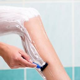
At-Home Hair Removal Device No Better Than Shaving
DANA POINT, CALIF. – An at-home, consumer hot-wire hair removal device worked no better than did standard shaving, according to a recent study.
"Relative to shaving, treatment with the hot-wire device did not produce statistically significant differences in the percentage change from baseline in hair count, duration of hair removal effect, or color and/or thickness of regrowing hair," Dr. Brian S. Biesman said.
There have been no controlled published studies of the no!no! hair removal device (manufactured by Radiancy) in peer-reviewed literature, which led Dr. Biesman to conduct a small study comparing the device’s efficacy with that of standard shaving.
According to information on the no!no! website, the device uses Thermicon technology "to conduct a gentle pulse of heat to the hair," which "instantly removes hair and slows the rate of hair regrowth with no pain." In Dr. Biesman’s study, however, the effectiveness of the hot-wire device, used according to the manufacturer’s recommendations (four passes per session), was found to be equivalent to standard shaving for all study end points.
For instance, active hair follicles and hair regrowth were not affected by a series of treatments with the hair removal device, compared with shaving. Also, hair thickness and color did not change after treatment with the device, Dr. Biesman reported in a poster at the Summit in Aesthetic Medicine sponsored by Skin Disease Education Foundation (SDEF).
A total of 23 patients (7 men, 16 women) aged 18-55 years completed the study; 90% of participants were white. Two sites on one leg of each patient were shaved 4 days before baseline, and then were treated every 3-4 days with the hot-wire device on one site on the leg and by shaving the other site for 8 weeks. The treatment sites were two symmetric 3 × 3 cm areas of the leg that were 3 cm apart and contained at least 15 hairs. The corners of the sites were micro-tattooed with ink, which was visible under black light.
Photographs were taken to measure hair count at baseline, weekly during treatment (before and after), at 4 days following final treatment, and at each follow-up visit (4, 8, and 12 weeks after final treatment), and "blinded visual and digital assessments were made for hair thickness and color," noted Dr. Biesman, who has a private practice in Nashville, Tenn.
The mean baseline hair count of the hot-wire and shaving sites were 86 and 79, respectively, "which remained stable during the 8-week treatment phase. No hair count reduction was seen." At post-treatment follow up, hair counts increased to 95 (treatment site) and 84 (shaving site) at 4 days, 104 and 99 at 1 month, 106 and 100 at 2 months, and 109 and 105 at 3 months. Hair regrowth was noted immediately after ceasing treatment with the hot-wire device.
The mean percent change at 4 days post treatment was 19% with the hot-wire device, compared with 14% with shaving; 41% and 41% at 1 month post treatment; 31% and 28% at 2 months post treatment; and 32% and 37% at 3 months.
A study limitation was that hair removal was evaluated only on the lower leg to sites not randomly assigned.
The study was sponsored by Tria Beauty, for which Dr. Biesman has consulted and from which he has received research support. SDEF and this news organization are owned by Elsevier.
FROM THE SDEF SUMMIT IN AESTHETIC MEDICINE
Major Finding: The mean percent change at 4 days post treatment was 19% with the hot-wire device, compared with 14% with shaving; 41% and 41% after 1 month; 31% and 28% after 2 months; and 32% and 37% after 3 months.
Data Source: Twenty-three patients (7 men, 16 women) aged 18-55 years completed the study; 90% of participants were white.
Disclosures: The study was sponsored by Tria Beauty, for which Dr. Biesman has consulted and from which he has received research support. SDEF and this news organization are owned by Elsevier.
60 Years and Counting: A Celebration of Dermatologic Surgery
It was questionable whether in 1952, when Dr. Norman Orentreich performed the first hair transplant and Dr. George Mackee reported on his 50-year experience with phenol chemical peels, anyone envisioned how far the field of dermatologic surgery would advance, but there’s no question now: The specialty has come a long way.
"Dermatologic surgery has blossomed into a full and diverse specialty with many elements," Dr. C. William Hanke noted in the June issue of Seminars in Cutaneous Medicine and Surgery (2012;31:52-9). He highlighted key events that shaped the burgeoning specialty.

Historical Highlights
From the first report on phenol peels by Dr. Mackee (Br. J. Dermatol. 1952;64:456-9), to the coining of the term "tumescent local anesthesia" by Dr. Hanke and his colleagues in 1998 – and publication of a comprehensive text on the tumescent technique 2 years later by Dr. Jeffery A. Klein ("Tumescent Technique: Tumescent Anesthesia and Microcannular Liposuction," St. Louis, Mosby, 2000) – Dr. Hanke chronicled the specialty’s evolution.
For example, he noted that in 1961, Dr. Leon Goldman became the first physician to treat patients with lasers. "He is acknowledged as the father of lasers in medicine and surgery," Dr. Hanke, a dermatologist in private practice in Carmel, Indiana, wrote. He added that the pioneer's first medicinal use of a laser launched 3 decades of related leadership, practice, and research.
Among other events that stand out in the history of the specialty, according to Dr. Hanke, are the first reports on cryosurgery with liquid nitrogen in 1966 by Dr. Setrag Zacarian ("Cryosurgery of Skin Cancer and Cutaneous Disorders," Mosby, St. Louis, 1985), and on ambulatory phlebectomy by Dr. Robert Muller the same year (Phlebologie 1966;19:227-9).
And, in 1967, Dr. Frederic E. Mohs founded the American College of Chemosurgery, which is today known as the American College of Mohs Surgery. "Mohs surgery continues to be an important part of the dermatologic surgery curriculum," Dr. Hanke said.
Advancements in vein ablation took pace in 1982 when the first report on hypertonic saline injections for treating leg veins, by Bruce Chrisman, was published (Hawaii Med. J. 1982;41:406-8).

In 1986, Dr. Saul Asken published the "Manual of Liposuction Surgery and Autologous Fat Transplantation Under Local Anesthesia" (Terry and Associates, Irvine, Calif.), and the following year, Dr. Jeffery A. Klein published the first report on the tumescent technique for liposuction (Am. J. Cosm. Surg. 1987;4:263-7).
Dr. Alastair Carruthers and Dr. Jean Carruthers revolutionized the field in 1987 when they began using botulinum toxin for cosmetic purposes. "Their joint observation that botulinum toxin can affect the muscles of facial expression began a revolution in cosmetic dermatology," wrote Dr. Hanke.
He also highlighted the launch of numerous societies and publications that helped to advance the specialty, including the founding of the American Society for Dermatologic Surgery in 1970, the launch of the Journal of Dermatologic Surgery in 1975, and the founding of the American Society for Lasers in Medicine and Surgery in 1981. And the specialty has continued to evolve at a rapid pace since that time, he noted.
Work by Dr. R. Rox Anderson and Dr. John A. Parrish on selective photothermolysis, for example, launched research leading to the fractionated laser technology in use today; and ongoing work by the Carruthers on botulinum toxin launched a new era in noninvasive facial rejuvenation, he said.
"Facelift bypass" procedures have satisfied many patients without subjecting them to the expense or morbidity of extensive facial surgery, Dr. Hanke noted, adding that new filler materials can be placed in the subcutaneous or supraperiosteal planes of the face with good results and minimal complications.
"A newly approved hyaluronic acid from Germany will allow treatment of very superficial wrinkles without the risk of the 'Tyndall effect,' " he added.
Also in the last decade, dermatologic surgery-related educational initiatives have been advanced. In 2004, 1-year fellowship training programs were approved by the Accreditation Council for Graduate Medical Education for training in dermatologic surgery; and last year, there were 47 ACGME-accredited procedural dermatology fellowship training programs in place, he said.
"Dermatologic surgery is an important component of nearly all dermatology postgraduate courses," added Dr. Hanke.
The history of fractional laser technology and fillers were addressed in separate articles in the same issue of Seminars in Cutaneous Medicine and Surgery, underscoring the importance of the two recent developments.
Fillers
The next phase of development in injectable fillers has begun, according to Dr. Richard G. Glogau.

"The concept of augmentation has moved from simple lines, scars, and wrinkles to revolumizing the aging face," noted Dr. Glogau, a clinical professor of dermatology at the University of California, San Francisco. "While seeking extended duration of effect balanced against the safety profile of the injectable, our focus is now directed to extending the lifting or volumizing effect that one can achieve with these fillers."
The initial movement into 3-dimensional correction with injectable fillers began with the dramatic improvement seen in lip volume championed in the 1980s, and each of a litany of fillers that followed, including fat, collagen, silicone, hyaluronic acids, poly-l-lactic acids, calcium hydroxylapatite, and polymethylmethacrylate (Sem. Cut. Med. Surg. 2012;31:78-87).
New fillers, some which represent refinements of existing technologies, continue to undergo review. Aquamid a biocompatible, nonabsorbable, permanent injectable hydrogel implant is currently under review by the Food and Drug Administration and is approved for use in Europe.
It is also possible that fillers will move beyond the traditional concept of inert medical devices and into the realm of true biologics – "materials that will improve the texture, elasticity, radiance, and possibly color, of the skin itself," he added.
"Just as the last 40 years has seen the movement from 2 to 3 dimensions, the next 2 decades will see movement form the macro to the micro level, and fillers will become systems for active metabolic manipulation and protection of the aging skin," he concluded.
Fractionation
The development of fractional photothermolysis was "a milestone in the history of laser technology and cutaneous resurfacing," according to Dr. Nazanin Saedi, a dermatologist in private practice in Chestnut Hill, Mass., and her colleagues.
The technology, noted the study authors, builds on the knowledge gained from early CO2 and Er:YAG laser treatment experiences, achieving greater optimization of parameters to induce the types of benefits seen with CO2 resurfacing, but without the significant postoperative morbidity, complications, and discomfort associated with the earlier technologies.
Furthermore, the older therapies destroyed the barrier protection, which "significantly increased the risk of infection throughout the recovery period and required extensive home care. The risk of scarring, delayed-onset permanent hypopigmentation, and demarcation lines was significant even in the hands of an experienced operator," they wrote (Sem. Cut. Med. Surg. 2012;31:105-9).
In an en effort to overcome these problems, what followed was nonablative dermal remodeling (with less than impressive results), and ultimately, the "idea of fractionated laser technology," they continued.
First used in hair transplant surgery, the technology led to development of the 1,550-nm nonablative "Fraxel" laser (now called the Fraxel Re:Store by Solta Medical), which debuted in the literature in 2004. This and other fractional laser technologies are now used to treat photoaging, superficial and deep rhytids, scars (including in patients with darker skin types), and pigmentation.

Both nonablative and ablative fractional resurfacing have proved to be safer than have traditional ablative lasers, Dr. Saedi and her colleagues noted. However, complications can still occur, such as infections (with herpes simplex virus being the most common), acneiform eruptions, prolonged erythema, pigmentary alteration, and scarring (rare, but can also result from infection associated with treatment).
"Counterintuitively, nonablative or ablative fractionated devices at low energies and densities can be useful in the treatment of scarring, including hypertrophic scars" as previously mentioned, they noted.
"While [fractional lasers are] inherently safer due to the pixelated manner of the treatment, complications can be further prevented with attentive surgical technique and judicious use of prophylaxis," they wrote.
The future of fractional laser technology promises to hold exciting developments. For example, since ablative fractional resurfacing creates microscopic vertical holes in tissue, the delivery of topical drugs through these holes is possible. In animal models, the photosensitizer methylaminolevulinic acid has been delivered using this approach, and tests suggest that low density treatment would be sufficient for deep dermal drug delivery.
"Treatment of skin in a porcine model showed enhanced depth of photodynamic therapy following porphyrin application after pretreatment with fractional resurfacing. An in vitro study utilizing low fluence fractionated Erbium:YAG demonstrated upwards of a 125-fold increase in imiquimod delivery," they noted.
Trials in humans are underway to assess the feasibility and safety of enhanced drug delivery using this approach, and there is speculation that ultimately, it could be used for delivery of biologic peptides and vaccines.
Tattoo removal is another promising use for fractional lasers, with early studies demonstrating good results, noted Dr. Saedi and her colleagues, explaining that ablative fractional lasers, when used in conjunction with a Q-switched laser, appear to provide enhanced tattoo removal capabilities.
"With new devices and wavelengths, the applications of this technology continue to grow," they wrote, concluding that "the future remains bright for fractionated laser devices."
Likewise, the future of dermatologic surgery in general remains promising, Dr. Hanke said. "Many new procedures and advances lie ahead."
Neither Dr. Hanke nor Dr. Saedi had disclosures to report. Another author on the article by Dr. Saedi (Dr. Christopher Zachary) reported receiving an honorarium from Solta Medical.
It was questionable whether in 1952, when Dr. Norman Orentreich performed the first hair transplant and Dr. George Mackee reported on his 50-year experience with phenol chemical peels, anyone envisioned how far the field of dermatologic surgery would advance, but there’s no question now: The specialty has come a long way.
"Dermatologic surgery has blossomed into a full and diverse specialty with many elements," Dr. C. William Hanke noted in the June issue of Seminars in Cutaneous Medicine and Surgery (2012;31:52-9). He highlighted key events that shaped the burgeoning specialty.
Historical Highlights
From the first report on phenol peels by Dr. Mackee (Br. J. Dermatol. 1952;64:456-9), to the coining of the term "tumescent local anesthesia" by Dr. Hanke and his colleagues in 1998 – and publication of a comprehensive text on the tumescent technique 2 years later by Dr. Jeffery A. Klein ("Tumescent Technique: Tumescent Anesthesia and Microcannular Liposuction," St. Louis, Mosby, 2000) – Dr. Hanke chronicled the specialty’s evolution.
For example, he noted that in 1961, Dr. Leon Goldman became the first physician to treat patients with lasers. "He is acknowledged as the father of lasers in medicine and surgery," Dr. Hanke, a dermatologist in private practice in Carmel, Indiana, wrote. He added that the pioneer's first medicinal use of a laser launched 3 decades of related leadership, practice, and research.
Among other events that stand out in the history of the specialty, according to Dr. Hanke, are the first reports on cryosurgery with liquid nitrogen in 1966 by Dr. Setrag Zacarian ("Cryosurgery of Skin Cancer and Cutaneous Disorders," Mosby, St. Louis, 1985), and on ambulatory phlebectomy by Dr. Robert Muller the same year (Phlebologie 1966;19:227-9).
And, in 1967, Dr. Frederic E. Mohs founded the American College of Chemosurgery, which is today known as the American College of Mohs Surgery. "Mohs surgery continues to be an important part of the dermatologic surgery curriculum," Dr. Hanke said.
Advancements in vein ablation took pace in 1982 when the first report on hypertonic saline injections for treating leg veins, by Bruce Chrisman, was published (Hawaii Med. J. 1982;41:406-8).
In 1986, Dr. Saul Asken published the "Manual of Liposuction Surgery and Autologous Fat Transplantation Under Local Anesthesia" (Terry and Associates, Irvine, Calif.), and the following year, Dr. Jeffery A. Klein published the first report on the tumescent technique for liposuction (Am. J. Cosm. Surg. 1987;4:263-7).
Dr. Alastair Carruthers and Dr. Jean Carruthers revolutionized the field in 1987 when they began using botulinum toxin for cosmetic purposes. "Their joint observation that botulinum toxin can affect the muscles of facial expression began a revolution in cosmetic dermatology," wrote Dr. Hanke.
He also highlighted the launch of numerous societies and publications that helped to advance the specialty, including the founding of the American Society for Dermatologic Surgery in 1970, the launch of the Journal of Dermatologic Surgery in 1975, and the founding of the American Society for Lasers in Medicine and Surgery in 1981. And the specialty has continued to evolve at a rapid pace since that time, he noted.
Work by Dr. R. Rox Anderson and Dr. John A. Parrish on selective photothermolysis, for example, launched research leading to the fractionated laser technology in use today; and ongoing work by the Carruthers on botulinum toxin launched a new era in noninvasive facial rejuvenation, he said.
"Facelift bypass" procedures have satisfied many patients without subjecting them to the expense or morbidity of extensive facial surgery, Dr. Hanke noted, adding that new filler materials can be placed in the subcutaneous or supraperiosteal planes of the face with good results and minimal complications.
"A newly approved hyaluronic acid from Germany will allow treatment of very superficial wrinkles without the risk of the 'Tyndall effect,' " he added.
Also in the last decade, dermatologic surgery-related educational initiatives have been advanced. In 2004, 1-year fellowship training programs were approved by the Accreditation Council for Graduate Medical Education for training in dermatologic surgery; and last year, there were 47 ACGME-accredited procedural dermatology fellowship training programs in place, he said.
"Dermatologic surgery is an important component of nearly all dermatology postgraduate courses," added Dr. Hanke.
The history of fractional laser technology and fillers were addressed in separate articles in the same issue of Seminars in Cutaneous Medicine and Surgery, underscoring the importance of the two recent developments.
Fillers
The next phase of development in injectable fillers has begun, according to Dr. Richard G. Glogau.
"The concept of augmentation has moved from simple lines, scars, and wrinkles to revolumizing the aging face," noted Dr. Glogau, a clinical professor of dermatology at the University of California, San Francisco. "While seeking extended duration of effect balanced against the safety profile of the injectable, our focus is now directed to extending the lifting or volumizing effect that one can achieve with these fillers."
The initial movement into 3-dimensional correction with injectable fillers began with the dramatic improvement seen in lip volume championed in the 1980s, and each of a litany of fillers that followed, including fat, collagen, silicone, hyaluronic acids, poly-l-lactic acids, calcium hydroxylapatite, and polymethylmethacrylate (Sem. Cut. Med. Surg. 2012;31:78-87).
New fillers, some which represent refinements of existing technologies, continue to undergo review. Aquamid a biocompatible, nonabsorbable, permanent injectable hydrogel implant is currently under review by the Food and Drug Administration and is approved for use in Europe.
It is also possible that fillers will move beyond the traditional concept of inert medical devices and into the realm of true biologics – "materials that will improve the texture, elasticity, radiance, and possibly color, of the skin itself," he added.
"Just as the last 40 years has seen the movement from 2 to 3 dimensions, the next 2 decades will see movement form the macro to the micro level, and fillers will become systems for active metabolic manipulation and protection of the aging skin," he concluded.
Fractionation
The development of fractional photothermolysis was "a milestone in the history of laser technology and cutaneous resurfacing," according to Dr. Nazanin Saedi, a dermatologist in private practice in Chestnut Hill, Mass., and her colleagues.
The technology, noted the study authors, builds on the knowledge gained from early CO2 and Er:YAG laser treatment experiences, achieving greater optimization of parameters to induce the types of benefits seen with CO2 resurfacing, but without the significant postoperative morbidity, complications, and discomfort associated with the earlier technologies.
Furthermore, the older therapies destroyed the barrier protection, which "significantly increased the risk of infection throughout the recovery period and required extensive home care. The risk of scarring, delayed-onset permanent hypopigmentation, and demarcation lines was significant even in the hands of an experienced operator," they wrote (Sem. Cut. Med. Surg. 2012;31:105-9).
In an en effort to overcome these problems, what followed was nonablative dermal remodeling (with less than impressive results), and ultimately, the "idea of fractionated laser technology," they continued.
First used in hair transplant surgery, the technology led to development of the 1,550-nm nonablative "Fraxel" laser (now called the Fraxel Re:Store by Solta Medical), which debuted in the literature in 2004. This and other fractional laser technologies are now used to treat photoaging, superficial and deep rhytids, scars (including in patients with darker skin types), and pigmentation.
Both nonablative and ablative fractional resurfacing have proved to be safer than have traditional ablative lasers, Dr. Saedi and her colleagues noted. However, complications can still occur, such as infections (with herpes simplex virus being the most common), acneiform eruptions, prolonged erythema, pigmentary alteration, and scarring (rare, but can also result from infection associated with treatment).
"Counterintuitively, nonablative or ablative fractionated devices at low energies and densities can be useful in the treatment of scarring, including hypertrophic scars" as previously mentioned, they noted.
"While [fractional lasers are] inherently safer due to the pixelated manner of the treatment, complications can be further prevented with attentive surgical technique and judicious use of prophylaxis," they wrote.
The future of fractional laser technology promises to hold exciting developments. For example, since ablative fractional resurfacing creates microscopic vertical holes in tissue, the delivery of topical drugs through these holes is possible. In animal models, the photosensitizer methylaminolevulinic acid has been delivered using this approach, and tests suggest that low density treatment would be sufficient for deep dermal drug delivery.
"Treatment of skin in a porcine model showed enhanced depth of photodynamic therapy following porphyrin application after pretreatment with fractional resurfacing. An in vitro study utilizing low fluence fractionated Erbium:YAG demonstrated upwards of a 125-fold increase in imiquimod delivery," they noted.
Trials in humans are underway to assess the feasibility and safety of enhanced drug delivery using this approach, and there is speculation that ultimately, it could be used for delivery of biologic peptides and vaccines.
Tattoo removal is another promising use for fractional lasers, with early studies demonstrating good results, noted Dr. Saedi and her colleagues, explaining that ablative fractional lasers, when used in conjunction with a Q-switched laser, appear to provide enhanced tattoo removal capabilities.
"With new devices and wavelengths, the applications of this technology continue to grow," they wrote, concluding that "the future remains bright for fractionated laser devices."
Likewise, the future of dermatologic surgery in general remains promising, Dr. Hanke said. "Many new procedures and advances lie ahead."
Neither Dr. Hanke nor Dr. Saedi had disclosures to report. Another author on the article by Dr. Saedi (Dr. Christopher Zachary) reported receiving an honorarium from Solta Medical.
FROM SEMINARS IN CUTANEOUS MEDICINE AND SURGERY
Dermatologic Surgery Milestones
Dermatologic surgery has come a long way since the 1950s. This look at some of the highlights – from phenol peels and hair transplants to tumescent liposuction and botulinum toxin injections – covers the subspecialty's evolution and influence.
Dermatologic surgery has come a long way since the 1950s. This look at some of the highlights – from phenol peels and hair transplants to tumescent liposuction and botulinum toxin injections – covers the subspecialty's evolution and influence.
Dermatologic surgery has come a long way since the 1950s. This look at some of the highlights – from phenol peels and hair transplants to tumescent liposuction and botulinum toxin injections – covers the subspecialty's evolution and influence.