User login
A talk about, then a plan for, antidepressants in pregnancy
CAS: Depressive disorder, anticipating a pregnancy
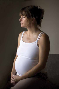
Your patient Megan—well-educated, 29 years old, G0P0—has come to you to discuss her antidepressant (paroxetine [Paxil]) because she is planning her first pregnancy.
Megan has a history of recurrent major depressive disorder (MDD), which is in remission (see “What is MDD?”).
How will you begin the conversation with this patient about keeping MDD in remission during her pregnancy and ensuring the safety of her fetus?
- Major depressive disorder (MDD) is defined by criteria in the fourth edition of the Diagnostic and Statistical Manual of Mental Disorders (DSM-IV)
- The disorder varies in severity, 1) across an affected person’s lifetime and 2) within a depressive episode
- A current or prior episode of depression that includes 1) a significant impact on an individual’s functioning, 2) active suicidality, or 3) hospitalization signals severe MDD
- In women, average age at first episode of depression is 24 years
There is a 20% to 25% lifetime prevalence of depression in women; the disorder peaks during childbearing years, however.1 As of 2003, 13% of pregnant women had taken an antidepressant at some time during their pregnancy, a percentage that has doubled since it was assessed in 1999.2
You are faced with several quandaries in deciding whether to recommend that your patient continue, or discontinue, antidepressant therapy during pregnancy:
- As many as 68% of women who terminate antidepressant treatment before or during pregnancy relapse.
- Even 26% of women who continue antidepressant during pregnancy relapse—requiring a dosage adjustment or change in treatment.3
- Yet the possibly elevated cortisol levels of severe, untreated depression may harm the placenta and fetus.4,5
So, what do you need to know to assess the risks and benefits of “Megan” stopping, or continuing, paroxetine during her anticipated pregnancy? And what are the risks to Megan’s fetus of treating, or not treating, her depression with a serotonin reuptake inhibitor (SRI*)?
“Selective” has been dropped
from “SSRI” to yield simply “SRI.”
Gauging the risks of depression in pregnancy
In any given patient, her history and family history of depression are key to determining the likelihood that she will suffer ongoing or recurrent depression.
CASE continued Repeated treated episodes plus a family history
In obtaining Megan’s history, you learn that she has had three prior episodes of depression, all of which were successfully treated with paroxetine. Megan has been stable on paroxetine for 3 years.
Notably, the second episode of depression was initially treated with a 16-week trial of psychotherapy alone; when depressive symptoms did not remit, paroxetine was added. That episode was considered severe because it included pervasive thoughts of suicide.
You also learn that Megan’s mother suffered from postpartum depression and that her father and paternal grandmother were treated for depression.
Known risk factors for depression during pregnancy include: maternal anxiety; prior diagnosis of depression during pregnancy; history of postpartum anxiety or depression; prior diagnosis of either anxiety or depressive disorder; significant life stress (e.g., divorce, death of a loved one); degree of social support—particularly, intimate social support; “intendedness” of pregnancy; domestic violence; and insurance status.6
You review with Megan her risk factors for depression during pregnancy, namely: three prior episodes of MDD and a strong family history of mood disorder. Her MDD is considered “severe” because she has a history of suicidality. You tell Megan that, given these factors, she is at high risk of a recurrence of her depressive illness during pregnancy.
Megan asks: “Would getting depressed during pregnancy hurt the baby?”
Depression during pregnancy affects both infant and maternal well-being, although studies are in conflict about the extent of that morbidity. Multiple areas of potential risk to mother and infant have been studied, including the effect of depression on:
- maternal well-being
- growth of the infant
- spontaneous abortion
- preterm delivery
- neonatal physiologic and neurobehavioral measures
- long-term considerations for the developing infant and child.
Within these categories of risk, a diagnosis of depression during pregnancy has been associated (in some but not all studies) with a higher risk, or rate, of:
- postpartum depression
- preterm birth
- lower maternal weight gain
- maternal tobacco, alcohol, and other substance use
- lower infant gestational age at birth
- small-for-gestational age infant birth.7-10
In terms of long-term impact on offspring, studies differ in their estimation of risk; however, children exposed to untreated, maternal depression at 18 weeks’ and 32 weeks’ gestation did show a greater degree of developmental delay at 18 months than children who were born to a mother who was not depressed during pregnancy.11
You discuss these risks with Megan. She asks: “What treatment do you recommend for me?” You turn to the 2009 guidelines published jointly by the American Psychiatric Association (APA) and ACOG.
These guidelines recommend that you consider 1) the severity of her current depression, 2) her history of depression severity, and 3) her preference for treatment.12 For mild depression during pregnancy, when there is no history of severe depression, or for a history of depression that responded well to psychotherapy in the past, a trial of psychotherapy without medications is recommended.
But Megan’s history of depression falls into the “severe” category, and a prior episode of depression did not respond well to psychotherapy. Your recommendation to her, therefore, is that she should continue taking an antidepressant—unless she feels strongly that she should discontinue it.
Megan considers what you’ve discussed about her high risk of developing recurrent depression during pregnancy. She decides that she wants to continue taking her antidepressant during pregnancy, but she has concerns—based on what she has been reading on the Internet.
Megan hands you a detailed printout downloaded from a Web site unfamiliar to you and asks about risks to the baby of such medications as paroxetine.
What should you tell Megan about SRIs in pregnancy—paroxetine, specifically?
You preface your remarks to her by noting that the data physicians work with are imperfect—because randomized, controlled clinical trials pose an ethical dilemma as a method of study in pregnant women. You then discuss with her current scientific understanding of potential risks to her fetus.
The difference in the rates of structural malformation among SRI-exposed and SRI-unexposed groups has been studied; most studies have found no increased rate of major or specific cardiac malformations.12 However, first-trimester paroxetine appeared, in some studies, to be associated with an increased rate of cardiac malformations. That led to a category-“D” pregnancy classification in 2005 and an FDA “Public Health Advisory.”
Other large cohort studies have not uncovered such an association. It has been hypothesized that the methodology of data collection may have influenced this finding.13
Other malformations have been implicated in some studies but not others, and have included associations between specific SRIs and cardiac ventricular outflow defects, craniosynostosis, and omphalocele. The absolute risk of these defects remains extremely low, however, and close to the background rate seen in the general population.14
Megan asks: “With that risk-category ‘D’ for paroxetine, do you recommend I continue taking it or should I switch to another medication while I’m pregnant?”
You review again with Megan that, although some studies have linked first-trimester paroxetine to an increased risk of cardiac malformation, that finding has not been replicated in several large cohort studies. You explain that, if she had a history of recurrent depression that had failed to respond to many antidepressants and only paroxetine worked, an attempt at switching the SRI would not be recommended because of the potential for relapse.
Megan tells you that she would feel safer not taking a category-“D” drug. You agree and propose a judicious approach: Because she has come to see you before she became pregnant, with enough time to complete a slow crossover to an alternative SRI, and because she has not had any earlier trials of other SRIs, a slow taper of paroxetine, coupled with a crossover to an alternative SRI, is a reasonable option—with the caution that substitution always carries a risk of relapse.
Problems in newborns
Megan considers the risks you’ve discussed so far. She remembers a recent article in a magazine for pregnant women that described severe “respiratory” and “withdrawal” symptoms in infants who were born to mothers taking an SRI antidepressant. She wonders if she should consider discontinuing her SRI in the third trimester to try to mitigate those risks.
Megan is asking you about an SRI exposure risk that has been fairly consistent across studies, called neonatal abstinence syndrome (NAS) or poor neonatal adaptation.
NAS is a cluster of symptoms that occurs in 15% to 30% of newborns who have been exposed to an SRI during the third trimester of pregnancy.15 Signs include irritability, weak cry, tachypnea, temperature instability, and hypoglycemia—all of which are transient, peak during the first 48 hours after delivery, and resolve in less than 2 weeks.
Multiple hypotheses have been put forward to account for NAS, including the possibilities that it reflects a withdrawal syndrome, pharmacotoxicity, or an underlying gene–SRI interaction. The physiology behind NAS remains unknown, however.12
Megan next asks you about persistent pulmonary hypertension of the newborn (PPHN). You explain that PPHN is of recent concern in women who have been taking an SRI in the latter half of their pregnancy.
The rate of PPHN in the general population is 0.5 to 2 newborns for every 1,000. Associated mortality is approximately 10% to 20%.16-18 This rate is thought to rise to approximately 6 of every 1,000 newborns among those who have been exposed to an SRI in utero—with some evidence of increased risk conferred through SRI exposure during later pregnancy (studies define this as the second half of the pregnancy).15 Although the relative risk of PPHN is increased threefold to sixfold when an SRI is used in pregnancy, absolute risk remains extremely low.
Concerns have been raised over research methodology in the few studies that have looked into SRI exposure and PPHN. Not all such studies found a change in relative risk or absolute risk of PPHN in SRI- exposed infants, compared to what was found in non-SRI–exposed infants.15,19,20
Megan presses you, however, with the understandable question of whether she should taper her SRI during the last trimester (which the Web site she has found recommends). With the above information in mind, you explain that, given current understanding of the low absolute risk of PPHN, and given her illness history and severity of prior depression, you would not recommend that she taper the antidepressant in the third trimester.
Furthermore, the same counsel applies in regard to NAS: Given the risk of psychiatric morbidity caused by discontinuing an SRI during the third trimester, you do not recommend that she taper an SRI during that period to avoid NAS.
You explain that, instead, physicians now counsel women who take an SRI about the signs of NAS so that they can be prepared if they observe any of them in their infant.
Megan has one more question: “Will I be able to breastfeed while I’m taking an antidepressant?”
Given the inherent difficulties and risks of relapse associated with a crossover to an alternative antidepressant postpartum, it makes sense, when possible, for a woman to take an antidepressant during pregnancy that can safely be continued while she is breastfeeding.
You tell Megan that, even though the quality of the data in this area is also thin, SRIs that have a low maternal serum profile are considered safest in breastfeeding.
To date, two SRIs—sertraline and paroxetine—have not been detectable in the breast milk of women taking either of them.21
CASE Appointment concluded, overflowing with information,
advice, and optimism
Megan says that, taking into account all that you and she have talked about, and even though she wants to return with her husband, she would like to switch to sertraline before she becomes pregnant—while she gauges its effectiveness at keeping her disorder in remission.
A good outcome requires you to prevail over obstacles
Because a diagnosis of depression spans a continuum of severity and, often, is not perceived as an acutely life-threatening illness, evaluating the risks and benefits of treatment is a murky undertaking.
Our role as physicians is to, first, educate ourselves and our patients about these variables and, second, support our patients in the decisions that they make. Physicians who care for pregnant women must be aware of the benefits and limitations of treatments as reported in the most current literature if they are going to assist women with decisions about treatment in the best possible way.
Social stigma. There remains the impact of stigma. Depressive and anxiety disorders are often perceived to be either under the control of an affected person’s “free will” or not as serious as other forms of “medical” disease. Consequently, the role that cultural and social pressures play in the risk–benefit analysis conducted by pregnant women and their physicians can’t be discounted.
Customized decision-making. As more data emerge about the treatment of depression in pregnancy, it has become clear: Treatment algorithms meant to simplify our decisions must always be individualized and extended into the postpartum period.
Treatment selection. Management of mild depression during pregnancy does not always require medication. Multiple variables—the list is long, and includes a patient’s psychiatric history, family psychiatric history, response to prior treatment, severity of depression, severity of prior depression, degree of social support, and personal desires—must be considered in determining what treatment is appropriate before, during, and after a pregnancy.
For a woman who suffers mild or moderate depression, with few antenatal depression risk factors, a trial of psychotherapy is recommended as first-line treatment. For a woman suffering from severe depression, or one who has a history of severe depression that has not responded well to psychotherapy alone, continuation or initiation of an SRI antidepressant is the current recommendation.
We want to hear from you! Tell us what you think.
1. Kessler RC, Berglund P, Demler O, et al. National Comorbidity Survey Replication. The epidemiology of major depressive disorder: results from the National Comorbidity Survey Replication (NCS-R). JAMA. 2003;289(23):3095-3105.
2. Cooper W, Willy M, Pont S, Ray W. Increasing use of antidepressants in pregnancy. Am J Obstet Gynecol. 2007;196(6):544.e1-e5.
3. Cohen LS, Altshuler LL, Harlow BL, et al. Relapse of major depression during pregnancy in women who maintain or discontinue antidepressant treatment. JAMA. 2006;295(5):499-507.
4. Kramer MS, Lydon J, Séguin L, et al. Stress pathways to spontaneous preterm birth: the role of stressors, psychological distress, and stress hormones. Am J Epidemiol. 2009;169(11):1319-1326.
5. Ellman LM, Schetter CD, Hobel CJ, Chicz-Demet A, Glynn LM, Sandman CA. Timing of fetal exposure to stress hormones: effects on newborn physical and neuromuscular maturation. Dev Psychobiol. 2008;50(3):232-241.
6. Lancaster CA, Gold KJ, Flynn HA, Yoo H, Marcus SM, Davis MM. Risk factors for depressive symptoms during pregnancy: a systematic review. Am J Obstet Gynecol. 2010;202(1):5-14.
7. Suri R, Altshuler L, Hellemann G, Burt VK, Aquino A, Mintz J. Effects of antenatal depression and antidepressant treatment on gestational age at birth and risk of preterm birth. Am J Psychiatry. 2007;164(8):1206-1213.
8. Wisner KL, Sit DK, Hanusa BH, et al. Major depression and antidepressant treatment: impact no pregnancy and neonatal outcomes.” Am J Psychiatry. 2009;166(5):557-566.
9. Li D, Liu L, Odouli R. Presence of depressive symptoms during early pregnancy and the risk of preterm delivery: a prospective cohort study. Hum Reprod. 2009;24(1):146-153.
10. Zuckerman B, Amaro H, Bauchner H, Cabral H. Depressive symptoms during pregnancy: relationship to poor health behaviors. Am J Obstet Gynecol. 1989;150(5Pt 1):1107-1111.
11. Deave T, Heron J, Evans J, Emond A. The impact of maternal depression in pregnancy on early child development. BJOG. 2008;115(8):1043-1051.
12. Yonkers KA, Wisner KL, Stewart DE, et al. The management of depression during pregnancy: a report from the American Psychiatric Association and the American College of Obstetricians and Gynecologists. Obstet Gynecol. 2009;114(3):703-713.
13. Gentile S, Bellantuono C. Selective serotonin reuptake inhibitor exposure during early pregnancy and the risk of fetal major malformations: focus on paroxetine. J Clin Psychiatry. 2009;70(3):414-422.
14. Louik C, Lin AE, Werler MM, Hernandez-Diaz S, Mitchell AA. First-trimester use of selective serotonin-reuptake inhibitors and the risk of birth defects. New Engl J Med. 2007;356(26):2675-2683.
15. Chambers CD, Hernandez-Diaz S, Marter LJV, et al. Selective seroteonin-reuptake inhibitors and risk of persistent pulmonary hypertension of the newborn. New Engl J Med. 2006;354(6):579-587.
16. Chambers CD, Johnson KA, Dick LM, Felix RJ, Jones KL. Birth outcomes in pregnant women taking fluoxetine. New Engl J Med. 1996;335(14):1010-1015.
17. Hageman JR, Adams MA, Gardner TH. Persistent pulmonary hypertension of the newborn. Trends in incidence, diagnosis and management. Am J Dis Child. 1984;137(6):592-595.
18. Fricker J. Nitric oxide may reduce need for extracorporeal membrane oxygenation. Lancet. 1996;347(9012):1397.-
19. Kallen B, Olausson P. Maternal use of selective serotonin re-uptake inhibitors and persistent pulmonary hypertension of the newborn. Pharmacoepidemiol Drug Saf. 2008;17(8):801-806.
20. Andrade S, McPhillips H, Loren D, et al. Antidepressant medication use and risk of persistent pulmonary hypertension of the newborn. Pharmacoepidemiol Drug Saf. 2009;18(3):246-252.
21. Lanza di Scalea T, Wisner K. Antidepressant medication use during breastfeeding. Clin Obstet Gynecol. 2009;52(3):483-497.
CAS: Depressive disorder, anticipating a pregnancy
Your patient Megan—well-educated, 29 years old, G0P0—has come to you to discuss her antidepressant (paroxetine [Paxil]) because she is planning her first pregnancy.
Megan has a history of recurrent major depressive disorder (MDD), which is in remission (see “What is MDD?”).
How will you begin the conversation with this patient about keeping MDD in remission during her pregnancy and ensuring the safety of her fetus?
- Major depressive disorder (MDD) is defined by criteria in the fourth edition of the Diagnostic and Statistical Manual of Mental Disorders (DSM-IV)
- The disorder varies in severity, 1) across an affected person’s lifetime and 2) within a depressive episode
- A current or prior episode of depression that includes 1) a significant impact on an individual’s functioning, 2) active suicidality, or 3) hospitalization signals severe MDD
- In women, average age at first episode of depression is 24 years
There is a 20% to 25% lifetime prevalence of depression in women; the disorder peaks during childbearing years, however.1 As of 2003, 13% of pregnant women had taken an antidepressant at some time during their pregnancy, a percentage that has doubled since it was assessed in 1999.2
You are faced with several quandaries in deciding whether to recommend that your patient continue, or discontinue, antidepressant therapy during pregnancy:
- As many as 68% of women who terminate antidepressant treatment before or during pregnancy relapse.
- Even 26% of women who continue antidepressant during pregnancy relapse—requiring a dosage adjustment or change in treatment.3
- Yet the possibly elevated cortisol levels of severe, untreated depression may harm the placenta and fetus.4,5
So, what do you need to know to assess the risks and benefits of “Megan” stopping, or continuing, paroxetine during her anticipated pregnancy? And what are the risks to Megan’s fetus of treating, or not treating, her depression with a serotonin reuptake inhibitor (SRI*)?
“Selective” has been dropped
from “SSRI” to yield simply “SRI.”
Gauging the risks of depression in pregnancy
In any given patient, her history and family history of depression are key to determining the likelihood that she will suffer ongoing or recurrent depression.
CASE continued Repeated treated episodes plus a family history
In obtaining Megan’s history, you learn that she has had three prior episodes of depression, all of which were successfully treated with paroxetine. Megan has been stable on paroxetine for 3 years.
Notably, the second episode of depression was initially treated with a 16-week trial of psychotherapy alone; when depressive symptoms did not remit, paroxetine was added. That episode was considered severe because it included pervasive thoughts of suicide.
You also learn that Megan’s mother suffered from postpartum depression and that her father and paternal grandmother were treated for depression.
Known risk factors for depression during pregnancy include: maternal anxiety; prior diagnosis of depression during pregnancy; history of postpartum anxiety or depression; prior diagnosis of either anxiety or depressive disorder; significant life stress (e.g., divorce, death of a loved one); degree of social support—particularly, intimate social support; “intendedness” of pregnancy; domestic violence; and insurance status.6
You review with Megan her risk factors for depression during pregnancy, namely: three prior episodes of MDD and a strong family history of mood disorder. Her MDD is considered “severe” because she has a history of suicidality. You tell Megan that, given these factors, she is at high risk of a recurrence of her depressive illness during pregnancy.
Megan asks: “Would getting depressed during pregnancy hurt the baby?”
Depression during pregnancy affects both infant and maternal well-being, although studies are in conflict about the extent of that morbidity. Multiple areas of potential risk to mother and infant have been studied, including the effect of depression on:
- maternal well-being
- growth of the infant
- spontaneous abortion
- preterm delivery
- neonatal physiologic and neurobehavioral measures
- long-term considerations for the developing infant and child.
Within these categories of risk, a diagnosis of depression during pregnancy has been associated (in some but not all studies) with a higher risk, or rate, of:
- postpartum depression
- preterm birth
- lower maternal weight gain
- maternal tobacco, alcohol, and other substance use
- lower infant gestational age at birth
- small-for-gestational age infant birth.7-10
In terms of long-term impact on offspring, studies differ in their estimation of risk; however, children exposed to untreated, maternal depression at 18 weeks’ and 32 weeks’ gestation did show a greater degree of developmental delay at 18 months than children who were born to a mother who was not depressed during pregnancy.11
You discuss these risks with Megan. She asks: “What treatment do you recommend for me?” You turn to the 2009 guidelines published jointly by the American Psychiatric Association (APA) and ACOG.
These guidelines recommend that you consider 1) the severity of her current depression, 2) her history of depression severity, and 3) her preference for treatment.12 For mild depression during pregnancy, when there is no history of severe depression, or for a history of depression that responded well to psychotherapy in the past, a trial of psychotherapy without medications is recommended.
But Megan’s history of depression falls into the “severe” category, and a prior episode of depression did not respond well to psychotherapy. Your recommendation to her, therefore, is that she should continue taking an antidepressant—unless she feels strongly that she should discontinue it.
Megan considers what you’ve discussed about her high risk of developing recurrent depression during pregnancy. She decides that she wants to continue taking her antidepressant during pregnancy, but she has concerns—based on what she has been reading on the Internet.
Megan hands you a detailed printout downloaded from a Web site unfamiliar to you and asks about risks to the baby of such medications as paroxetine.
What should you tell Megan about SRIs in pregnancy—paroxetine, specifically?
You preface your remarks to her by noting that the data physicians work with are imperfect—because randomized, controlled clinical trials pose an ethical dilemma as a method of study in pregnant women. You then discuss with her current scientific understanding of potential risks to her fetus.
The difference in the rates of structural malformation among SRI-exposed and SRI-unexposed groups has been studied; most studies have found no increased rate of major or specific cardiac malformations.12 However, first-trimester paroxetine appeared, in some studies, to be associated with an increased rate of cardiac malformations. That led to a category-“D” pregnancy classification in 2005 and an FDA “Public Health Advisory.”
Other large cohort studies have not uncovered such an association. It has been hypothesized that the methodology of data collection may have influenced this finding.13
Other malformations have been implicated in some studies but not others, and have included associations between specific SRIs and cardiac ventricular outflow defects, craniosynostosis, and omphalocele. The absolute risk of these defects remains extremely low, however, and close to the background rate seen in the general population.14
Megan asks: “With that risk-category ‘D’ for paroxetine, do you recommend I continue taking it or should I switch to another medication while I’m pregnant?”
You review again with Megan that, although some studies have linked first-trimester paroxetine to an increased risk of cardiac malformation, that finding has not been replicated in several large cohort studies. You explain that, if she had a history of recurrent depression that had failed to respond to many antidepressants and only paroxetine worked, an attempt at switching the SRI would not be recommended because of the potential for relapse.
Megan tells you that she would feel safer not taking a category-“D” drug. You agree and propose a judicious approach: Because she has come to see you before she became pregnant, with enough time to complete a slow crossover to an alternative SRI, and because she has not had any earlier trials of other SRIs, a slow taper of paroxetine, coupled with a crossover to an alternative SRI, is a reasonable option—with the caution that substitution always carries a risk of relapse.
Problems in newborns
Megan considers the risks you’ve discussed so far. She remembers a recent article in a magazine for pregnant women that described severe “respiratory” and “withdrawal” symptoms in infants who were born to mothers taking an SRI antidepressant. She wonders if she should consider discontinuing her SRI in the third trimester to try to mitigate those risks.
Megan is asking you about an SRI exposure risk that has been fairly consistent across studies, called neonatal abstinence syndrome (NAS) or poor neonatal adaptation.
NAS is a cluster of symptoms that occurs in 15% to 30% of newborns who have been exposed to an SRI during the third trimester of pregnancy.15 Signs include irritability, weak cry, tachypnea, temperature instability, and hypoglycemia—all of which are transient, peak during the first 48 hours after delivery, and resolve in less than 2 weeks.
Multiple hypotheses have been put forward to account for NAS, including the possibilities that it reflects a withdrawal syndrome, pharmacotoxicity, or an underlying gene–SRI interaction. The physiology behind NAS remains unknown, however.12
Megan next asks you about persistent pulmonary hypertension of the newborn (PPHN). You explain that PPHN is of recent concern in women who have been taking an SRI in the latter half of their pregnancy.
The rate of PPHN in the general population is 0.5 to 2 newborns for every 1,000. Associated mortality is approximately 10% to 20%.16-18 This rate is thought to rise to approximately 6 of every 1,000 newborns among those who have been exposed to an SRI in utero—with some evidence of increased risk conferred through SRI exposure during later pregnancy (studies define this as the second half of the pregnancy).15 Although the relative risk of PPHN is increased threefold to sixfold when an SRI is used in pregnancy, absolute risk remains extremely low.
Concerns have been raised over research methodology in the few studies that have looked into SRI exposure and PPHN. Not all such studies found a change in relative risk or absolute risk of PPHN in SRI- exposed infants, compared to what was found in non-SRI–exposed infants.15,19,20
Megan presses you, however, with the understandable question of whether she should taper her SRI during the last trimester (which the Web site she has found recommends). With the above information in mind, you explain that, given current understanding of the low absolute risk of PPHN, and given her illness history and severity of prior depression, you would not recommend that she taper the antidepressant in the third trimester.
Furthermore, the same counsel applies in regard to NAS: Given the risk of psychiatric morbidity caused by discontinuing an SRI during the third trimester, you do not recommend that she taper an SRI during that period to avoid NAS.
You explain that, instead, physicians now counsel women who take an SRI about the signs of NAS so that they can be prepared if they observe any of them in their infant.
Megan has one more question: “Will I be able to breastfeed while I’m taking an antidepressant?”
Given the inherent difficulties and risks of relapse associated with a crossover to an alternative antidepressant postpartum, it makes sense, when possible, for a woman to take an antidepressant during pregnancy that can safely be continued while she is breastfeeding.
You tell Megan that, even though the quality of the data in this area is also thin, SRIs that have a low maternal serum profile are considered safest in breastfeeding.
To date, two SRIs—sertraline and paroxetine—have not been detectable in the breast milk of women taking either of them.21
CASE Appointment concluded, overflowing with information,
advice, and optimism
Megan says that, taking into account all that you and she have talked about, and even though she wants to return with her husband, she would like to switch to sertraline before she becomes pregnant—while she gauges its effectiveness at keeping her disorder in remission.
A good outcome requires you to prevail over obstacles
Because a diagnosis of depression spans a continuum of severity and, often, is not perceived as an acutely life-threatening illness, evaluating the risks and benefits of treatment is a murky undertaking.
Our role as physicians is to, first, educate ourselves and our patients about these variables and, second, support our patients in the decisions that they make. Physicians who care for pregnant women must be aware of the benefits and limitations of treatments as reported in the most current literature if they are going to assist women with decisions about treatment in the best possible way.
Social stigma. There remains the impact of stigma. Depressive and anxiety disorders are often perceived to be either under the control of an affected person’s “free will” or not as serious as other forms of “medical” disease. Consequently, the role that cultural and social pressures play in the risk–benefit analysis conducted by pregnant women and their physicians can’t be discounted.
Customized decision-making. As more data emerge about the treatment of depression in pregnancy, it has become clear: Treatment algorithms meant to simplify our decisions must always be individualized and extended into the postpartum period.
Treatment selection. Management of mild depression during pregnancy does not always require medication. Multiple variables—the list is long, and includes a patient’s psychiatric history, family psychiatric history, response to prior treatment, severity of depression, severity of prior depression, degree of social support, and personal desires—must be considered in determining what treatment is appropriate before, during, and after a pregnancy.
For a woman who suffers mild or moderate depression, with few antenatal depression risk factors, a trial of psychotherapy is recommended as first-line treatment. For a woman suffering from severe depression, or one who has a history of severe depression that has not responded well to psychotherapy alone, continuation or initiation of an SRI antidepressant is the current recommendation.
We want to hear from you! Tell us what you think.
1. Kessler RC, Berglund P, Demler O, et al. National Comorbidity Survey Replication. The epidemiology of major depressive disorder: results from the National Comorbidity Survey Replication (NCS-R). JAMA. 2003;289(23):3095-3105.
2. Cooper W, Willy M, Pont S, Ray W. Increasing use of antidepressants in pregnancy. Am J Obstet Gynecol. 2007;196(6):544.e1-e5.
3. Cohen LS, Altshuler LL, Harlow BL, et al. Relapse of major depression during pregnancy in women who maintain or discontinue antidepressant treatment. JAMA. 2006;295(5):499-507.
4. Kramer MS, Lydon J, Séguin L, et al. Stress pathways to spontaneous preterm birth: the role of stressors, psychological distress, and stress hormones. Am J Epidemiol. 2009;169(11):1319-1326.
5. Ellman LM, Schetter CD, Hobel CJ, Chicz-Demet A, Glynn LM, Sandman CA. Timing of fetal exposure to stress hormones: effects on newborn physical and neuromuscular maturation. Dev Psychobiol. 2008;50(3):232-241.
6. Lancaster CA, Gold KJ, Flynn HA, Yoo H, Marcus SM, Davis MM. Risk factors for depressive symptoms during pregnancy: a systematic review. Am J Obstet Gynecol. 2010;202(1):5-14.
7. Suri R, Altshuler L, Hellemann G, Burt VK, Aquino A, Mintz J. Effects of antenatal depression and antidepressant treatment on gestational age at birth and risk of preterm birth. Am J Psychiatry. 2007;164(8):1206-1213.
8. Wisner KL, Sit DK, Hanusa BH, et al. Major depression and antidepressant treatment: impact no pregnancy and neonatal outcomes.” Am J Psychiatry. 2009;166(5):557-566.
9. Li D, Liu L, Odouli R. Presence of depressive symptoms during early pregnancy and the risk of preterm delivery: a prospective cohort study. Hum Reprod. 2009;24(1):146-153.
10. Zuckerman B, Amaro H, Bauchner H, Cabral H. Depressive symptoms during pregnancy: relationship to poor health behaviors. Am J Obstet Gynecol. 1989;150(5Pt 1):1107-1111.
11. Deave T, Heron J, Evans J, Emond A. The impact of maternal depression in pregnancy on early child development. BJOG. 2008;115(8):1043-1051.
12. Yonkers KA, Wisner KL, Stewart DE, et al. The management of depression during pregnancy: a report from the American Psychiatric Association and the American College of Obstetricians and Gynecologists. Obstet Gynecol. 2009;114(3):703-713.
13. Gentile S, Bellantuono C. Selective serotonin reuptake inhibitor exposure during early pregnancy and the risk of fetal major malformations: focus on paroxetine. J Clin Psychiatry. 2009;70(3):414-422.
14. Louik C, Lin AE, Werler MM, Hernandez-Diaz S, Mitchell AA. First-trimester use of selective serotonin-reuptake inhibitors and the risk of birth defects. New Engl J Med. 2007;356(26):2675-2683.
15. Chambers CD, Hernandez-Diaz S, Marter LJV, et al. Selective seroteonin-reuptake inhibitors and risk of persistent pulmonary hypertension of the newborn. New Engl J Med. 2006;354(6):579-587.
16. Chambers CD, Johnson KA, Dick LM, Felix RJ, Jones KL. Birth outcomes in pregnant women taking fluoxetine. New Engl J Med. 1996;335(14):1010-1015.
17. Hageman JR, Adams MA, Gardner TH. Persistent pulmonary hypertension of the newborn. Trends in incidence, diagnosis and management. Am J Dis Child. 1984;137(6):592-595.
18. Fricker J. Nitric oxide may reduce need for extracorporeal membrane oxygenation. Lancet. 1996;347(9012):1397.-
19. Kallen B, Olausson P. Maternal use of selective serotonin re-uptake inhibitors and persistent pulmonary hypertension of the newborn. Pharmacoepidemiol Drug Saf. 2008;17(8):801-806.
20. Andrade S, McPhillips H, Loren D, et al. Antidepressant medication use and risk of persistent pulmonary hypertension of the newborn. Pharmacoepidemiol Drug Saf. 2009;18(3):246-252.
21. Lanza di Scalea T, Wisner K. Antidepressant medication use during breastfeeding. Clin Obstet Gynecol. 2009;52(3):483-497.
Does weekly progesterone prolong gestation in women who have PPROM?
Approximately 13 million preterm births occur annually worldwide.1 Depending on the geographic locale, PPROM is responsible for 16% to 40% of these births.2
The clinical approach to PPROM is one of the most contentious issues in obstetrics, with disagreement on virtually every aspect of it. Under debate are the lower and upper limits of the gestational age range at which intervention is warranted, as well as the use of ancillary interventions such as corticosteroids and antibiotics. Briery and colleagues add to the scientific debate now by asking whether 17P would be effective as cotreatment (with antibiotics) to prolong latency after PPROM.
According to their findings, the answer to this question is “No.”
Details of the trial
Briery and colleagues conducted a placebo-controlled, double-blind, randomized clinical trial of women with a singleton gestation complicated by PPROM. Excluded from the study were women whose pregnancy involved additional fetal or placental complications.
All women included in the study received antibiotics according to a protocol from the National Institutes of Health; they also were given betamethasone for fetal maturation. Tocolytics were not used. Because randomization did not occur until after each woman was transferred from the labor and delivery unit to the high-risk floor, we can assume that no participants were manifesting uterine contractions.
Women received weekly injections of 17P or placebo until 34 weeks’ gestation or delivery. The primary outcome was the interval from study entry to delivery.
One woman had a pregnancy of 23.5 weeks’ duration at randomization; the remainder had gestations that were 24 weeks or older. There were no other differences in demographics, cervical dilatation, gestational age at study entry, or reasons for delivery between the two study groups.
Study design may have been unrealistic
The authors calculated that they needed a sample size of 56 patients to detect a 50% increase in latency, based on population data from their institution showing that 80% of patients who have PPROM deliver within 7 days. Such a calculation may have set an unrealistic—albeit logistically convenient—goal, rendering the study underpowered to detect smaller effects. Note, for example, that when antibiotics are given to women who have PPROM, prolongation of the latency period is only 33% (pooled effect from a recent meta-analysis).3 Even so, given the findings of Briery and colleagues, latency improvement after 17P administration would appear to be unlikely even in a larger study. There was not even a trend toward a longer interval to delivery (mean of 11.2 days with 17P vs 14.5 days with placebo).
Only secondary prevention of preterm birth is effective
The indications for progesterone supplementation in pregnancy are still evolving as part of a sustained scientific effort to prevent preterm labor and delivery. Strategies to prevent preterm delivery can be categorized as primary, secondary, or tertiary, as can strategies for other public health concerns.
Because any number of variables—known and unknown—may trigger preterm labor, identifying them and providing primary preventive strategies in the entire pregnant population remain elusive tasks.
Tertiary prevention—i.e., treatment given to already symptomatic individuals—is also notoriously ineffective. There are no data supporting the use of progesterone as primary prevention (in low-risk women) or tertiary prevention (e.g., tocolytic). ACOG made note of this in 2003, and its conclusions remain valid today.4 According to a 2010 Cochrane review, there is insufficient evidence to advocate progestational agents as tocolytic agents for women who present with threatened or established preterm labor.5
In light of these data, the results reported by Briery and colleagues are hardly surprising. In women who may have already entered the irreversible phase of parturition (manifesting uterine contractions; presenting with advanced, painless cervical dilatation; or after PPROM), progesterone will remain ineffective. The only applicable use of prophylactic progesterone in pregnancy is as secondary prevention.4 In contrast to primary and tertiary prevention, the secondary level of prevention—i.e., an intervention aimed at minimizing the risk of preterm birth in women who are identified as having an elevated risk—is supported by several systematic reviews of randomized, controlled trials.6,7 According to these reviews, progesterone certainly is effective in high-risk pregnant women who have a short cervix or a history of spontaneous preterm birth. The same cannot be said about women who have PPROM.
Based on the evidence, including this study by Briery and colleagues, administration of antibiotics appears to be the only intervention available to delay delivery and reduce neonatal morbidity in the setting of PPROM.8 The use of tocolytics is not supported by the data in the clinical context of PPROM.9—Alex C. Vidaeff, MD, MPH
We want to hear from you! Tell us what you think.
1. Villar J, Abalos E, Carroli G, et al. Heterogeneity of perinatal outcomes in the preterm delivery syndrome. Obstet Gynecol. 2004;104(1):78-87.
2. Simmons LE, Rubens CE, Darmstadt GL, et al. Preventing preterm birth and neonatal mortality: exploring the epidemiology, causes, and interventions. Semin Perinatol. 2010;34(6):408-415.
3. Hutzal CE, Boyle EM, Kenyon SL, et al. Use of antibiotics for the treatment of preterm parturition and prevention of neonatal morbidity: a meta-analysis. Am J Obstet Gynecol. 2008;199(6):620.e1-8.
4. Society for Maternal Fetal Medicine Publications Committee. ACOG Committee Opinion #419. Use of progesterone to reduce preterm birth. Obstet Gynecol. 2008;112(4):963-965.
5. Su L-L, Samuel M, Chong Y-S. Progestational agents for treating threatened or established preterm labor. Cochrane Database Syst Rev. 2010;(1):CD006770.-doi: 10.1002/14651858.CD006770.
6. Dodd JM, Crowther CA. The role of progesterone in prevention of preterm birth. Int J Women Health. 2010;1:73-84.
7. Rode L, Langhoff-Roos J, Andersson C, et al. Systematic review of progesterone for the prevention of preterm birth in singleton pregnancies. Acta Obstet Gynecol Scand. 2009;88(11):1180-1189.
8. Kenyon S, Boulvain M, Neilson JP. Antibiotics for preterm rupture of membranes. Cochrane Database Syst Rev. 2003;(2):CD001058.-doi: 10.1002/14651858.CD001058.
9. Mercer BM. Is there a role for tocolytic therapy during conservative management of preterm premature rupture of the membranes? Clin Obstet Gynecol. 2007;50(2):487-496.
Approximately 13 million preterm births occur annually worldwide.1 Depending on the geographic locale, PPROM is responsible for 16% to 40% of these births.2
The clinical approach to PPROM is one of the most contentious issues in obstetrics, with disagreement on virtually every aspect of it. Under debate are the lower and upper limits of the gestational age range at which intervention is warranted, as well as the use of ancillary interventions such as corticosteroids and antibiotics. Briery and colleagues add to the scientific debate now by asking whether 17P would be effective as cotreatment (with antibiotics) to prolong latency after PPROM.
According to their findings, the answer to this question is “No.”
Details of the trial
Briery and colleagues conducted a placebo-controlled, double-blind, randomized clinical trial of women with a singleton gestation complicated by PPROM. Excluded from the study were women whose pregnancy involved additional fetal or placental complications.
All women included in the study received antibiotics according to a protocol from the National Institutes of Health; they also were given betamethasone for fetal maturation. Tocolytics were not used. Because randomization did not occur until after each woman was transferred from the labor and delivery unit to the high-risk floor, we can assume that no participants were manifesting uterine contractions.
Women received weekly injections of 17P or placebo until 34 weeks’ gestation or delivery. The primary outcome was the interval from study entry to delivery.
One woman had a pregnancy of 23.5 weeks’ duration at randomization; the remainder had gestations that were 24 weeks or older. There were no other differences in demographics, cervical dilatation, gestational age at study entry, or reasons for delivery between the two study groups.
Study design may have been unrealistic
The authors calculated that they needed a sample size of 56 patients to detect a 50% increase in latency, based on population data from their institution showing that 80% of patients who have PPROM deliver within 7 days. Such a calculation may have set an unrealistic—albeit logistically convenient—goal, rendering the study underpowered to detect smaller effects. Note, for example, that when antibiotics are given to women who have PPROM, prolongation of the latency period is only 33% (pooled effect from a recent meta-analysis).3 Even so, given the findings of Briery and colleagues, latency improvement after 17P administration would appear to be unlikely even in a larger study. There was not even a trend toward a longer interval to delivery (mean of 11.2 days with 17P vs 14.5 days with placebo).
Only secondary prevention of preterm birth is effective
The indications for progesterone supplementation in pregnancy are still evolving as part of a sustained scientific effort to prevent preterm labor and delivery. Strategies to prevent preterm delivery can be categorized as primary, secondary, or tertiary, as can strategies for other public health concerns.
Because any number of variables—known and unknown—may trigger preterm labor, identifying them and providing primary preventive strategies in the entire pregnant population remain elusive tasks.
Tertiary prevention—i.e., treatment given to already symptomatic individuals—is also notoriously ineffective. There are no data supporting the use of progesterone as primary prevention (in low-risk women) or tertiary prevention (e.g., tocolytic). ACOG made note of this in 2003, and its conclusions remain valid today.4 According to a 2010 Cochrane review, there is insufficient evidence to advocate progestational agents as tocolytic agents for women who present with threatened or established preterm labor.5
In light of these data, the results reported by Briery and colleagues are hardly surprising. In women who may have already entered the irreversible phase of parturition (manifesting uterine contractions; presenting with advanced, painless cervical dilatation; or after PPROM), progesterone will remain ineffective. The only applicable use of prophylactic progesterone in pregnancy is as secondary prevention.4 In contrast to primary and tertiary prevention, the secondary level of prevention—i.e., an intervention aimed at minimizing the risk of preterm birth in women who are identified as having an elevated risk—is supported by several systematic reviews of randomized, controlled trials.6,7 According to these reviews, progesterone certainly is effective in high-risk pregnant women who have a short cervix or a history of spontaneous preterm birth. The same cannot be said about women who have PPROM.
Based on the evidence, including this study by Briery and colleagues, administration of antibiotics appears to be the only intervention available to delay delivery and reduce neonatal morbidity in the setting of PPROM.8 The use of tocolytics is not supported by the data in the clinical context of PPROM.9—Alex C. Vidaeff, MD, MPH
We want to hear from you! Tell us what you think.
1. Villar J, Abalos E, Carroli G, et al. Heterogeneity of perinatal outcomes in the preterm delivery syndrome. Obstet Gynecol. 2004;104(1):78-87.
2. Simmons LE, Rubens CE, Darmstadt GL, et al. Preventing preterm birth and neonatal mortality: exploring the epidemiology, causes, and interventions. Semin Perinatol. 2010;34(6):408-415.
3. Hutzal CE, Boyle EM, Kenyon SL, et al. Use of antibiotics for the treatment of preterm parturition and prevention of neonatal morbidity: a meta-analysis. Am J Obstet Gynecol. 2008;199(6):620.e1-8.
4. Society for Maternal Fetal Medicine Publications Committee. ACOG Committee Opinion #419. Use of progesterone to reduce preterm birth. Obstet Gynecol. 2008;112(4):963-965.
5. Su L-L, Samuel M, Chong Y-S. Progestational agents for treating threatened or established preterm labor. Cochrane Database Syst Rev. 2010;(1):CD006770.-doi: 10.1002/14651858.CD006770.
6. Dodd JM, Crowther CA. The role of progesterone in prevention of preterm birth. Int J Women Health. 2010;1:73-84.
7. Rode L, Langhoff-Roos J, Andersson C, et al. Systematic review of progesterone for the prevention of preterm birth in singleton pregnancies. Acta Obstet Gynecol Scand. 2009;88(11):1180-1189.
8. Kenyon S, Boulvain M, Neilson JP. Antibiotics for preterm rupture of membranes. Cochrane Database Syst Rev. 2003;(2):CD001058.-doi: 10.1002/14651858.CD001058.
9. Mercer BM. Is there a role for tocolytic therapy during conservative management of preterm premature rupture of the membranes? Clin Obstet Gynecol. 2007;50(2):487-496.
Serum Markers Predict Severe Preeclampsia
Major Finding: The combination of 25-hydroxyvitamin D level and sFlt-1:PlGF ratio early in the second trimester had an area under the ROC curve of 0.834 for predicting severe preeclampsia.
Data Source: A nested, case-control study of 164 pregnant women, one-fourth of whom had developed severe preeclampsia.
Disclosures: Dr. Woodham did not report any relevant financial disclosures.
SAN FRANCISCO – Levels of serum markers measured early in the second trimester of pregnancy may help identify women who are likely to develop severe preeclampsia, the results of a nested case-control study indicated.
In the study, there was no association between the level of vitamin D and levels of two angiogenic factors that have been previously implicated in the development of preeclampsia, soluble FMS-like tyrosine kinase 1 (sFlt-1) and placental growth factor (PlGF). But both the level of vitamin D and the ratio of sFlt-1 to PlGF predicted the development of severe preeclampsia after other risk factors were taken into account. And in ROC (receiver operating characteristic) curve analysis, the combination outperformed either measure individually.
“These results suggest that 25-hydroxyvitamin D and angiogenic factors play independent roles in the pathogenesis of preeclampsia,” said Dr. Padmashree Chaudhury Woodham. “Our findings suggest that the combination of 25-hydroxy-vitamin D level and sFlt-1:PlGF ratio is a better predictor of preeclampsia in midgestation than either marker alone.”
A low 25-hydroxyvitamin D level has previously been shown to be a risk factor for severe preeclampsia, according to Dr. Woodham, who is a fellow in ob.gyn. at the University of North Carolina at Chapel Hill. But its association with angiogenic factors is unclear.
The investigators drew their study patients from a large cohort of pregnant women who gave blood for routine prenatal screening early in the second trimester (gestational age, 15–20 weeks). They restricted analyses to women with a singleton pregnancy who did not have chronic medical illnesses and whose fetuses did not have congenital abnormalities.
Each woman who developed severe preeclampsia (n = 41) was matched by race/ethnicity with three control women who had uncomplicated births at term (n = 123). Banked frozen serum samples were assayed to determine levels of vitamin D (total 25-hydroxyvitamin D) and the angiogenic markers sFlt-1, PlGF, and vascular endothelial growth factor (VEGF).
The severe preeclampsia and control groups were similar in terms of age, parity, and body mass index, Dr. Woodham reported. Overall, 39% were black, 29% were white, 27% were Hispanic, and 5% were Asian. The season and the median gestational age at the time blood was drawn were also similar. But the median gestational age at delivery was younger in the preeclampsia group (32.6 vs. 39.6 weeks; P less than .001).
Relative to their control counterparts, the women who developed preeclampsia had lower levels of vitamin D (P less than .001), VEGF (P less than .001), and PlGF (P = .03), and a higher ratio of sFlt-1 to PlGF (P = .02). Levels of vitamin D were not correlated with levels of any of the angiogenic factors or with the sFlt-1:PlGF ratio, contrary to the findings of in vitro and animal studies. However, in a multivariate model, each 1-nmol/L increase in total vitamin D level was associated with a 5% reduction in the odds of preeclampsia, whereas each 1-unit increase in the sFlt-1:PlGF ratio was associated with an 11% increase in the odds.
ROC curve analysis showed that for predicting preeclampsia, the area under the curve was 0.745 for vitamin D alone and 0.669 for the sFlt-1:PlGF ratio alone. But it was higher with their combination (0.834). There was also a small further improvement when VEGF level was added to the mix, with an area under the curve of 0.851.
From the Annual Meeting of the Society for Maternal-Fetal Medicine
First-Trimester Screening: State of the Art Is Standard of Care
The decades-long shift in our approach to prenatal screening which brought us from a point that maternal age was the main criterion for assessing risk of chromosomal abnormalities to a more precise first-trimester screening approach – one that combines biochemistry and imaging – is continuing to evolve.
Indeed, researchers are honing in on a “platform” of first-trimester assessments that can screen for an even wider array of risks and pregnancy complications than previously envisioned – an array that extends far beyond chromosomal abnormalities. Much of this first-trimester screening platform is currently being applied and is poised to become a new standard of care.
The continued evolution of first-trimester screening is critical, as a massive amount of time and resources is spent trying to identify problem pregnancies. Many of these resources still are used inefficiently because detection is incomplete or too late to make a difference. With an expanded and precise first-trimester platform for assessment, we can offer women and their physicians significantly more information early on. This will enable us to channel our resources to improve decision -making, direct management, and enhance pregnancy outcome.
Early Screening's Development
Prenatal screening used to be all about maternal age. Our early methods were based on the fact that risk increases with age, and then on the idea that particular age cut-offs may signify varying levels of risk.
However, advances in ultrasound, and the identification of four pregnancy-related maternal blood analytes, provided us new and exciting insights on fetal status. These chemicals became part of a second-trimester screening process focused largely on trisomy 21. Although we still used age as a factor to assess risk, we learned that the cut-offs we had identified earlier were arbitrary and that risk could now be individualized.
As research continued, it became apparent that versions of the biochemical tests used in second-trimester screening could be done in the first trimester – even by 12–14 weeks of gestation – and could be used to assess the risks not only of Down syndrome, but of other chromosomal abnormalities and some physical abnormalities. These biochemical tests (free beta-human chorionic gonadotropin and pregnancy-associated plasma protein-A) were combined with first-trimester ultrasound measurements of nuchal translucency into a screening algorithm that took hold more than 5 years ago and has steadily gained acceptance.
Since then, a number of parameters have been added to first-trimester screening to make the prediction of normality, or abnormality, even more precise. Assessment of the nasal bone, of the frontonasal facial angle, and of various structures inside the brain have become part of an anatomic review, for instance, that help us better define which babies we should be most concerned about.
Additionally, Doppler assessment of blood flow measurement – specifically of tricuspid regurgitation and of flow through the ductus venosus, a small fetal blood vessel that leads to the heart – can provide valuable information about fetal cardiac status and can easily be done in the context of the first-trimester ultrasound evaluation. Abnormal first-trimester Doppler findings also appear to predict Down syndrome and other adverse outcomes independently of a normal nuchal translucency measurement.
Combined with additional, early biochemical tests on maternal serum, these imaging advances (for fetal anatomic reviews and blood flow measurements) have led to an improved detection rate as high as 90% for trisomy 21 and other chromosomal abnormalities. More importantly, this detection rate is achieved without invasive testing, enabling us to reserve invasive procedures such as chorionic villus sampling (CVS) or amniocentesis for women with higher identified risks.
A New Cardiac Focus
The nuchal translucency test, which measures levels of fluid in a small area in the back of the fetal neck, has been available in the United States for approximately 15 years. With time, we have come to appreciate that a number of problems, in addition to Down syndrome, are associated with increases in nuchal translucency. We also better understand that abnormal nuchal translucency measurements are not always indicative of a problem, and that when there is a problem, the issue is not always chromosomal in nature.
The quest to detect other kinds of problems (mainly structural abnormalities, and congenital heart defects, in particular) as early as we can detect chromosomal problems has taken on added urgency in recent years.
Indeed, significant improvements in the overall computing capability of modern ultrasound equipment, in three-dimensional color ultrasonography, and in ultrasound image resolution – as well as specific new technologic developments such as tomographic imaging and spatiotemporal image correlation – have opened the door to first-trimester cardiac screening.
In the majority of patients, up to 12 parameters of fetal cardiac structure can be visualized. Each of the three segments of the exam takes only a few seconds to perform, so the actual collection of information is rapid. The technologic advances have also made the acquisition of images easier and less operator dependent. Moreover, the analysis is then performed offline, so the mother can go home afterward. Offline analysis of images also means that the ultrasound scan itself can be performed by trained sonographers at a distance from a cardiac center, with the information transmitted to the center for expert analysis.
It wasn't long ago that second-trimester fetal echocardiography was the gold standard for any prenatal evaluation of fetal cardiac structure and function. Now, with an early and integrated screening approach that utilizes first-trimester fetal cardiac examination, we can in fact diagnose many of the most severe heart defects as early as 12 weeks of gestation. At this stage, the fetal heart is as small as the tip of the little finger.
This component of first-trimester screening is just now coming to the forefront. Its availability can benefit populations at high risk of cardiac anomalies (such as women who have long-standing diabetes). It may be especially beneficial to those who were in poor glycemic control at the beginning of their pregnancy. It appears, though, that the exam can be meaningfully applied in low-risk populations as well. Research is underway to determine the best approaches to counseling and to determine which patients should have subsequent invasive testing.
Other New Frontiers
Another area of interest is the potential ability to predict which women will develop preeclampsia later in pregnancy based on how the fetus and placenta are faring at approximately 12 weeks' gestation.
Doppler investigations have shown us that placental abnormalities are difficult to distinguish from normal placental development early in pregnancy. In the first trimester, therefore, Doppler alone is a fair mechanism for knowing whether placental development is deficient enough to put the mother at high risk for developing preeclampsia or isolated hypertension.
However, when Doppler is combined with measurement of a family of maternal serum analytes – some of them inflammatory substances and some of them chemicals that regulate the formation of blood vessels – it can be employed to predict who will develop early hypertensive complications. And when other factors such as maternal weight and blood pressure at the time of first-trimester assessment are added to the equation, the accuracy of our predictions increases further.
We are proceeding in this area with a bit of caution, as we cannot yet predict the onset of preeclampsia later on in pregnancy. The predictive value of the first-trimester assessment for hypertensive problems that occur closer to term is not very good, so patients with normal early assessments still need careful prenatal care.
Still, in many ways we can tackle the most severe problems through early detection. There is some evidence that the administration of low-dose aspirin can reduce the incidence of hypertension and preeclampsia, as well as complications with the baby's growth, in women with detected placental abnormalities. This means that not only are we able to define and identify those women at highest risk, but we also have the ability to potentially modify the course of placental development and perhaps even eliminate hypertensive complications.
Current research is aimed at defining who will best benefit from this approach, because while low-dose aspirin appears in some research to work when started early in high-risk women, benefits have not been duplicated in other studies.
More broadly, first-trimester assessment of maternal characteristics (such as weight), serum analytes, and ultrasound features set the stage for ongoing maternal evaluation of characteristics such as weight gain during pregnancy to predict her risk of developing preeclampsia, diabetes, and other serious problems, including neonatal concerns requiring specialized newborn care.
The Big Picture
As first-trimester screening evolves with technologic developments to become more comprehensive and precise, one of its ever-important components involves the art of history taking, physician-patient dialogue, and the incorporation of low-tech risk assessments for coping with and possibly preventing preterm labor and delivery.
Measuring the cervix at this very early stage is not a good predictor of its ability to contain the pregnancy for the rest of the gestation or even until a reasonably mature gestation is reached. In the first trimester, the cervix generally is not under enough pressure from the weight of the pregnancy to disclose whether it is a strong or weak cervix or whether it has the potential to shorten in an extreme way or not. This is different from measuring the cervix later in pregnancy when the shortening process has already started, and when intervention is based on proven results.
The first trimester is an excellent time, however, to have the mother recount her history. It is also a good time to make decisions about the use of progesterone, which in weekly injections has been shown to reduce the incidence of preterm delivery, and to institute a serial monitoring program so that any changes may be detected before the patient presents with rapidly advancing preterm labor – i.e., before a clinical emergency.
Such dialogue and interaction emphasizes to me the importance of a team approach to first-trimester screening that involves the ob.gyn. physicians, well-trained sonographers, well-trained perinatal nurses, and perinatologists who specialize in high-risk maternal and fetal complications.
Prenatal screening is no longer an in-and-out assessment of two or three measures. That began to change more than 5 years ago with adoption of the first-trimester screening approach combining biochemistry and imaging. It continues to evolve as prenatal screening provides an even more thorough and comprehensive view of fetal, placental, and maternal function that allows us to thoroughly map out the care of our patients. For women who have normal pregnancies, this is incredibly reassuring. And for those with any kind of outlying results or overt complications, it provides a starting point for making the best of even the most challenging pregnancies.
At left, the narrow nuchal translucency and brightly echogenic nasal bone at 12 weeks' gestation reduce the likelihood of aneuploidy. At right, the fetus has a NT over 4 mm and nonvisualizing nasal bone. CVS on the second fetus revealed Down syndrome.
Source Images courtesy Dr. Christopher R. Harman
In another pair of fetuses appearing at 12 weeks' gestation for nucal translucency screening, tricuspid valve Doppler shows normal flow on the left. The fetus on the right has a large downward jet of tricuspid regurgitation, suggesting possible abnormalities; pulmonary stenosis was later diagnosed.
Source Images courtesy Dr. Christopher R. Harman
3-D blocks analyzed by tomographic section in a systematic approach yield a complete catalogue of anatomic cardiac landmarks in over 80% of fetuses at 12 weeks.
Complete endocardial cushion defect was diagnosed at 12 weeks. First trimester echocardiography was triggered by abnormal ductus venosus alone during routine screening.
Source Elsevier Global Medical News
The Evolution of Prenatal Assessment
It is astonishing how much obstetrics and maternal-fetal medicine have grown. There was a time not too long ago when obstetric care was primarily delivered to the mother, with the fetus being a hopeful beneficiary. We could listen to the fetal heart rate using the fetoscope, but access to the fetus for its early developmental analysis was otherwise off-limits; its growth and development were assumed as part of maternal-focused obstetric care.
The introduction of electronic fetal monitoring gave us the opportunity to see a recording of the fetal heart rate pattern – its rhythm, and its quality – and we used that as an indirect measure of fetal well-being. Subsequently, ultrasound became available, and we could then evaluate the anatomy of the fetus – though usually in the latter part of pregnancy – and appreciate the morphology and overall growth performance.
It was not until relatively recently that the focus of prenatal assessment has shifted to the first trimester. In large measure, this change has been consumer driven. Families have become very interested in the development of their unborn children, and that interest increasingly has centered on obtaining more information earlier on. Such demand has pushed physician scientists working in the field to adapt their technologies to the first trimester. Recent research has, in large measure, advanced in response to parental interests.
Fetal diagnosis in the first trimester was thus born of this great desire and has evolved to the point where, as stated in this month's Master Class, it is becoming the standard of care. The field of first-trimester fetal diagnosis now consists of a series of biochemical and biophysical assessments that can truly evaluate fetal well-being at the current time and can contribute to the prediction of later development and later fetal well-being, or more importantly, the loss of fetal well-being.
It is in light of this burgeoning field of first-trimester evaluation that we decided to develop a Master Class to review this new state of the art. I have invited Dr. Christopher R. Harman, an international expert in the field of ultrasound and Doppler technology, to serve as this month's guest professor.
Dr. Harman is professor and interim chair of the department of obstetrics, gynecology, and reproductive sciences at the University of Maryland, Baltimore, as well as director of the school's maternal-fetal medicine division. He will explain how research is honing in on a first-trimester platform of assessments that holds even more potential for predicting risks and complications than we realized with the first-trimester screening algorithm that took hold more than 5 years ago.
Study: Be Cautious When Using Anti-TNFs in Pregnancy
Pregnant women taking tumor necrosis factor inhibitors at conception experienced a higher rate of spontaneous abortion than did patients who did not.
The data, culled from the British Society for Rheumatology Biologics Registry, are from the “largest detailed prospective collection of pregnancy outcomes in women with arthritis-related diseases exposed to anti-TNF therapy” to date, according to the authors. However, the study was unable to control for the possibility that disease severity itself plays a role in adverse pregnancy outcomes.
Nevertheless, “no firm conclusions can be drawn about the safety of anti-TNF therapy during pregnancy and, without further evidence, guidelines which suggest these drugs should be avoided at the time of conception must remain,” recommended Dr. Suzanne M.M. Verstappen of the University of Manchester's Arthritis Research UK Epidemiology Unit, and her colleagues. They looked at women who received adalimumab, etanercept, or infliximab either at conception or at any time prior to conception. A subset was also exposed to methotrexate and/or leflunomide at time of conception in addition to the anti-TNFs. A fourth cohort with active rheumatoid arthritis had no history of anti-TNF use, but rather received nonbiological disease-modifying antirheumatic drugs (DMARDs), excluding methotrexate and leflunomide (Ann. Rheum. Dis. 2011 [doi:10.1136/ard.2010.140822
Among cohort Ia, which included 20 women (21 pregnancies) who took anti-TNFs plus either methotrexate and/or leflunomide at conception, there were 10 live births, 4 terminations, and 7 (33%) spontaneous abortions (miscarriages occurring prior to 20 weeks or to viability outside the womb). Among cohort Ib, which included 44 women who took anti-TNFs at conception, but not methotrexate or leflunomide, there were 50 pregnancies. They included 32 live births among this cohort, 4 terminations, 12 spontaneous abortions (24%) and 2 intrauterine deaths (occurring post 20 weeks). There was also one neonatal death registered. The women who had taken anti-TNFs in the past, but not at the time of conception (cohort II), did have seemingly better outcomes: the 59 pregnancies (54 women) resulted in live births in 46 cases (including one of two twins), terminations in 2, and spontaneous abortions in 10 (17%). There were two intrauterine deaths, including the twin death. Among the 10 pregnancies in 10 women who had no history of anti-TNF use (cohort III), there were zero terminations and one spontaneous abortion (10%).
The British Society for Rheumatology receives restricted income from Abbott Laboratories, Biovitrum, Shering-Plough, Wyeth Pharmaceuticals, and Roche. The authors added that they had no personal competing interests in relation to this study. The society also receives funding from several pharmaceutical companies, including the makers of anti-TNFs.
From Annals of the Rheumatic Diseases
Treating Mood Disorders: No Easy Decisions
MADRID – There are no easy choices for women who present with mood disorders during pregnancy – or for the physicians who treat them.
The decision to treat prenatal depression or anxiety is not to be taken lightly, but often must be considered, despite a relative dearth of data supporting or refuting drug safety during pregnancy, Dr. Shari I. Lusskin said at the conference.
“There are data, but not great data,” said Dr. Lusskin, director of reproductive psychiatry at the NYU Langone Medical Center, New York. “We will never have 10,000 patients taking medication compared to 10,000 not taking it, compared to a control group that is not depressed or anxious, nor will we have years of follow-up data on their children.”
The data that do exist are mostly comprised of case series, which are small and lack controls. “When you see a negative study [about the effect of treating mental disorders in a pregnant woman], keep in mind that there is often a lack of information about the mother's diagnosis. Reports of smoking and substance abuse are notoriously unreliable,” and can't be accounted for in the results, she said. “We also know nothing about the mother's body mass index, which is associated with many fetal complications that have nothing to do with drug exposure. And different studies use different comparison scales – you can't compare apples and oranges.”
The biggest exception is sodium valproate, an anticonvulsant also used to treat mood disorders, Dr. Lusskin said. The ongoing Neurodevelopmental Effects of Antiepileptic Drugs (NEAD) study has found in utero exposure to be associated with an increase in birth defects, and with cognitive problems in 3-year-olds whose mothers took it during pregnancy.
The uncertainty of treatment leaves pregnant women and their physicians to weigh the risks of psychotropic medications against the risks of untreated mood disorders. “There is no such thing as nonexposure,” Dr. Lusskin said. If the mother is not treated, “the fetus is going to be exposed to the mother's illness, which can create phenotypic changes with lifelong effects. If the mother is treated inadequately, the fetus will be exposed to both the effects of the illness and the medication. And if the mother is treated to remission, with the medication titrated to her response, medication exposure can be at least limited,” while the new mother becomes healthy enough to give her baby the best possible care.
The biggest barrier to treating mental illness during pregnancy is fear of fetal harm: teratogenicity, neonatal complications from drug exposure, and long-term neurodevelopmental effects. But these risks must be considered in light of real world experience – not just based on numbers from a study, Dr. Lusskin said. “Any risk of impairment has to be compared to the background risk of a birth defect, which is is 2%–4%. If a drug increases this risk in a clinically meaningful way, it has to be over and above this background rate.”
Even a substantial increase in a rare birth defect can be misleading. For instance, Ebstein's anomaly is a heart defect that occurs in 1 in 20,000 births. “Lithium is said to increase this rate to about 1 in 2,000 births. But this is still way below the expected background rate of birth defects. Risks are relevant in terms of their relativity – the absolute risk is just quoting a figure,” she said.
But untreated mental illness poses its own risk. Babies exposed in utero to the chronic stress hormones associated with anxiety and depressive disorders might become phenotypically programmed, prone themselves to early childhood behavioral problems and, in later life, to mental illness. “The fetal programming hypothesis is the interaction between the baby's genetics and the environmental exposure in utero, and it's mediated by the sex of the fetus and the timing of exposure,” Dr. Lusskin said.
Dr. Lusskin said she consults for the nonprofit drug safety database www.reprotox.org
Expert Analysis from the World Conference on Women's Mental Health
Tailor Antibiotic Dose to Weight Before Cesarean
SAN FRANCISCO – As women grow heavier, tailoring antibiotic dose to weight or body mass index may be critical for preventing surgical site infections after cesarean deliveries, a study has shown.
In the prospective cohort study, all 29 women undergoing a cesarean were given the same standard fixed dose of cefazolin before their surgery. Concentrations of the antibiotic in adipose tissue collected at the start of the surgery were one-third lower in obese women and one-half lower in extremely obese women than those in their lean counterparts. Perhaps most importantly, some of the heavier women had tissue concentrations below those believed to be necessary for preventing antibiotic resistance.
“Based on the current findings, a considerable proportion of obese women undergoing cesarean delivery do not have adequate antimicrobial protection for the duration of the procedure, following current guidelines,” said Dr. Leo Pevzner, an ob.gyn. at the University of California, Irvine.
The National Surgical Infection Prevention Project has endorsed tailoring antibiotic dose to weight or body mass index (BMI), but data on appropriate prophylactic doses in adults are limited, he noted. Hence, recommendations still call for an intravenous dose of 1–2 g for all adults, large or small.
“Current obesity trends, along with evolutionary changes in bacterial resistance, portend a questionable utility of existing prophylaxis regimens and have the potential to drastically increase the rates of surgical site infections if no attempts are made to address antimicrobial dosing based on patients' weight or BMI,” Dr. Pevzner commented.
The investigators enrolled in the study women with a singleton pregnancy who were scheduled for a cesarean delivery at term (greater than 37 weeks' gestation). Any who had received antibiotics in the previous week or who had chronic hypertension or pregestational diabetes were excluded.
On the basis of their BMI, the women were classified as lean (less than 30 kg/m
All were given 2 g of cefazolin 30–60 minutes before skin incision for the cesarean surgery. The investigators collected adipose tissue at the time of skin incision (initial) and again just before closure (final), as well as myometrial tissue after delivery and blood at the end of the procedure.
The concentration of cefazolin in all samples was assessed in a blinded manner with a microbiologic plate assay performed in triplicate, using plates seeded with Streptococcus sanguis. Zones of inhibition were measured in millimeters.
The study participants were 30 years old, on average. The mean BMI was 27, 34, and 45 kg/m
Regression analysis showed that the higher the women's BMI, the lower the concentration of the antibiotic in their initial adipose tissue sample (r = −0.67, P less than .001). None of the lean women had an adipose concentration of cefazolin below 4 mcg/g, the theoretic breakpoint for preventing resistance, according to Dr. Pevzner.
But eight women – four obese and four extremely obese – had an initial or a final adipose concentration below this breakpoint.
And three women – all extremely obese – had both initial and final adipose concentrations below this breakpoint.
The concentrations of cefazolin in the final adipose tissue, myometrial tissue, and serum also decreased with increasing BMI category, but these differences were not statistically significant, Dr. Pevzner reported.
Among the 25 women with follow-up, 2 developed surgical site infections requiring antibiotic therapy. Both were in the extremely obese group, and both had initial and final adipose cefazolin concentrations below the 4-mcg/g threshold.
The study was small, Dr. Pevzner acknowledged. “As such, there is not enough information to reach a conclusion regarding the weight or BMI above which a higher dose of antibiotics should be used,” he said.
Also, the impact of multiple gestations and maternal diseases, such as hypertension and diabetes, in this setting is unknown.
Dr. Pevzner did not report any relevant financial disclosures.
From the Annual Meeting of the Society for Maternal-Fetal Medicine
Poor Sleep May Drive Antenatal Depression
Major Finding: Pregnant women with severe physical discomfort in the first trimester might experience poor sleep, which could predispose them to antenatal depression.
Data Source: A prospective study of 257 pregnant women who were surveyed about physical symptoms, sleep quality, and mood in early and late pregnancy.
Disclosures: The Australian Research Council supported the study. Dr. Skouteris said she had no relevant financial disclosures.
MADRID – Poor sleep in early pregnancy might be one significant driver of depression in later pregnancy, an Australian prospective study has found.
“This finding suggests the need for clinicians to not just screen for depressive symptoms during pregnancy (which is the trend now), but to consider other potentially important problems, like the quality of sleep and severe physical symptoms during early pregnancy,” Helen Skouteris, Ph.D., said at the conference “These issues may very well contribute to the development of – or maintain – depression throughout pregnancy.”
Dr. Skouteris, an expert in developmental psychology at Deakin University in Burwood, Victoria, presented the results of a prospective study of 257 pregnant women in Australia who were recruited at 15–23 weeks' gestation. The study consisted of two self-reported questionnaires that assessed physical symptoms of pregnancy, sleep quality, and mood during early pregnancy and during the last trimester.
At recruitment, the women's mean age was 32 years. Almost half (48%) were first-time mothers and most (77%) were married. Most of the women (73%) also had at least some college education, and 57% had an annual household income of more than 75,000 Australian dollars ($77,500). “This is typical of the women who volunteer for our research programs,” Dr. Skouteris noted.
At the time of the first survey, the women had been pregnant for a mean of 18 weeks; the mean gestation at the time of the second survey was 34 weeks.
Both packets, which were mailed to the subjects, contained the self-administered Beck Depression Inventory, the Pittsburgh Sleep Quality Index, and a physical symptoms questionnaire that listed 29 common symptoms of pregnancy. Women were asked to rate these symptoms on a 0–3 scale (with 0 as “not at all” and 3 as “very much”) regarding discomfort from the symptoms, their frequency, and their general effect on life. Sleep was reported for the prior 4 weeks, physical symptoms for the prior 8 weeks, and mood at the time of the interview.
The current unpublished data, based on three regression analyses, show that poor sleep is the missing link between early physical symptoms and late depression. Fatigue was the most commonly reported physical symptom at both the early and later interviews (94% and 92%, respectively).
“The path seems to travel from severe physical symptoms early to poor sleep throughout, and then to depression later in pregnancy. Clearly, these results show that physical symptoms early in pregnancy might be a key risk factor for depression later on in pregnancy. Now we must consider other moderators in this relationship; there might be other things that contribute to this equation.”
She also mentioned that 15% of the women in the study rated very high on the anxiety measure.
“These women were very anxious about the delivery and what it might mean to their education, career, and general way of life,” Dr. Skouteris said. “We're now exploring these subjective ways a woman feels about her pregnancy, and their possible relationship between stress, anxiety, and depression throughout pregnancy and into the postnatal period.” This study will provide more data about these links, since the women will be assessed monthly instead of twice.
From the World Conference on Women's Mental Health
Study Finds 1 in 15 Late Preterm Births Could Be Avoidable
One in 15 neonates in a large, retrospective, observational study was delivered at 34–36 weeks' gestation for potentially avoidable or elective precursors for late preterm delivery, and those deliveries were associated with greater risk of neonatal morbidity and mortality than were deliveries at or after 37 weeks for the same indications.
The findings suggest that nearly 7% of late preterm births – and possibly their associated morbidity and mortality – could be avoided, according to Dr. S. Katherine Laughon of the Eunice Kennedy Shriver National Institute of Child Health and Human Development and her colleagues.
The investigators also found that different precursors for late preterm deliveries were associated with differing rates of neonatal morbidity in the study, a factor that has implications for counseling patients about the risks and benefits of late preterm delivery, they reported .
Nearly 66% of preterm deliveries were late preterm deliveries in this study, which compared 15,136 singleton gestations delivered late preterm (between 34 weeks and 36 weeks 6 days) vs. 170,593 gestations delivered between 37 weeks and 41 weeks 6 days. The investigators used data from the Consortium on Safe Labor, a study that included 228,668 deliveries from 12 clinical centers and 19 hospitals representing nine American College of Obstetricians and Gynecologists districts in 2002–2008.
Precursors for late preterm birth included spontaneous labor in 30% of cases, preterm premature rupture of membranes (PPROM) in 32% of cases, and medical indications for an obstetric, maternal, or fetal condition in 32% of cases. The cause of late preterm birth was unknown in 6% of cases, the investigators said (Obstet. Gynecol. 2001;116:1047–55).
The investigators found that among the “indicated” categories, 18% were for soft – or potentially avoidable – precursors. Additionally, in the “unknown” category there were 175 elective deliveries with no other maternal-fetal or obstetric complications, “and together these 1,044 soft or elective precursors made up 6.9%, or approximately 1 in 15, of all late preterm deliveries,” they noted, adding that the “adjusted risk of oxygen use, transient tachypnea of the newborn, mechanical ventilation, respiratory distress syndrome, pneumonia or newborn sepsis, and admission to the NICU all were significantly decreased for neonates with soft or elective precursors delivered at 37, 38, 39, and 40 weeks of gestation compared with late preterm.”
No increase in the risk of stillbirth or neonatal mortality was seen with expectant management of these soft precursors, suggesting that at least 1 in 15 of the deliveries with soft precursors could have been expectantly managed until 39 weeks' gestation, they said.
Furthermore, the differences in neonatal outcomes based on precursor type suggest that “the underlying pathology for precursors is an important determining factor in neonatal morbidity.” Based on these findings, the investigators recommended that elective deliveries be postponed until 39 weeks' gestation. “More prospective data are needed and guidelines should be developed to help providers and women decide which soft precursors can be managed expectantly,” they said.
Dr. Laughton and her associates said they had no relevant financial disclosures.
View on The News
Delay Delivery When Possible
These findings provide important information about the risks of delivery prior to term – including in the late preterm – and particularly in women with “soft” precursors for late preterm delivery, Dr. Erol Amon said in an interview.
Most research on complications associated with preterm delivery involves babies born before 32 weeks' gestation, he said, noting that because babies born in what is now known as the late preterm period (previously known as near term) typically do quite well, there is some complacency when it comes to delivering in this time period.
However, as this well-conducted study demonstrates, they don't always do well, and for that reason it is important to delay delivery when possible, he said.
The take-home message, he said, is that there is a great deal of physician intervention in this category of patients who have soft precursors for late preterm delivery, but that's not to say physicians are doing anything wrong.
“In the vast majority of cases they are doing the right thing,” Dr. Amon said.
The decision not to manage these patients expectantly may be an understandable result of concern regarding stillbirth, and although this study suggested that there was no increased risk of stillbirth with expectant management, it wasn't designed for that purpose, so that finding is not conclusive, he said.
Guideline development, as recommended by the authors, could indeed help with decision making in that small percentage of patients with soft or unknown indications for late preterm birth, in whom expectant management might be the best policy, he said.
DR. AMON is professor of obstetrics and gynecology, and director of maternal-fetal medicine at St. Louis University, Mo. Dr. Amon disclosed that he has received honoraria from Alere for speaking on late preterm birth.
From Obstetrics & Gynecology
U.S. Teen Birth Rates Tumble to Record Low
Teen birth rates in the United States fell by 37% over the past 2 decades, reaching the lowest rate ever recorded, according to data from the Centers for Disease Control and Prevention.
Despite those promising data, “we still have a ways to go to improve our teen birth rate to reflect what is seen in other parts of the world,” Ursula Bauer, Ph.D., director of the National Center for Chronic Disease Prevention and Health Promotion, said in a teleconference.
The birth rate for girls aged 15–19 years in 2008 and 2009 was 39 per 1,000 girls in the United States, Dr. Bauer and Dr. Wanda Barfield, director of the division of reproductive health, National Center for Chronic Disease Prevention and Health Promotion, wrote in the report, which was published in Morbidity and Mortality Weekly Report (2011;60:1–8).
However, teen birth rates in the United States are as much as nine times higher than in many other developed countries. Teen birth rates for 2008–2009 were 27 per 1,000 in Great Britain, 10 per 1,000 in Germany and France, and 6 per 1,000 in Sweden and Denmark.
The researchers, from the Centers for Disease Control and Prevention (CDC), reviewed National Vital Statistics System data from 1991 to 2009 on teen birth rates, as well as National Youth Risk Behavior Survey data on sexual activity and contraceptive use. They also reviewed data on sex education and the use of reproductive health services for teens aged 15–19 years from the 2006–2008 National Survey of Family Growth.
In 2009, teen birth rates were lowest in the Northeastern and upper Midwestern states and highest in the Southern states. The states with the lowest birth rates – Connecticut, Massachusetts, New Hampshire, New Jersey, and Vermont – posted rates ranging from 16 to 23 births per 1,000 girls aged 15–19 years. The states with the highest rates – Arkansas, Mississippi, New Mexico, Oklahoma, and Texas – had rates ranging from 59 to 64 births per 1,000 girls aged 15–19 years.
The number of teens having sex declined across white, black, and Hispanic ethnicities for both boys and girls, but black and Hispanic girls remained at least twice as likely as white girls to become teen mothers. Approximately 46% of teens in the United States have had sexual intercourse – down from 54% in 1991 – and 12% of those teens used no contraception, down from 16% in 1991.
“Health care providers have a key role to play in bringing down teen birth rates and teen pregnancy rates,” said Dr. Bauer. “Talking to teens, both boys and girls, about sexual health and reproductive health, and talking about available contraception, is very important for health care providers in their encounters with teens,” she said.
According to a CDC fact sheet, health care providers can help reduce the teen birth rate in the United States by making more birth control options available to sexually active teens, including long-acting methods such as IUDs, and by educating teens about the proper use of birth control options, including condoms and oral contraceptives.
For the complete MMWR Vital Signs report on teen birth rates, visit www.cdc.gov/vitalsigns
Vitals
Source Elsevier Global Medical News
From Morbidity and Mortality Weekly Report