User login
Targeting inflammation improved survival for some patients with advanced pancreatic cancer
CHICAGO – Adding the kinase inhibitor ruxolitinib to capecitabine improved overall survival of a subset of patients with metastatic pancreatic cancer, compared with capecitabine and placebo.
In a randomized phase II trial in 127 patients with metastatic pancreatic ductal adenocarcinoma, the second-line therapy combination of ruxolitinib (Jakafi) and capecitabine (Xeloda) was not associated with better outcomes than capecitabine and placebo in the overall population.
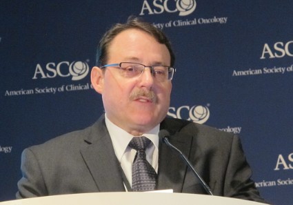
But among 60 patients with high levels of C-reactive protein indicative of systemic inflammation, adding ruxolitinib improved median overall survival to 83 days, compared with 55 days for controls, reported Dr. Herbert I. Hurwitz, professor of medicine in the division of oncology at Duke University, Durham, N.C.

"These data support the role of inflammation and the JAK-STAT pathway in particular for patients with pancreatic cancer," Dr. Hurwitz said at the annual meeting of the American Society of Clinical Oncology.
Ruxolitinib is an inhibitor of the Janus kinases (JAK) 1 and 2, which have been shown to mediate cytokine signaling through activation of STAT transcription factors. In preclinical models, pro-inflammatory cytokine signaling has been shown to contribute to pancreatic cancer initiation and progression.
Systemic inflammation is also associated with weight loss, decreased muscle mass, and poor performance status, all of which can shorten survival in patients with advanced pancreatic cancer.
The investigators hypothesized that ruxolitinib could improve survival of advanced pancreatic cancer patients when added to standard chemotherapy by dampening inflammation and thereby reducing cachexia and its related effects on patients’ overall health.
In tumor xenograft models of pancreatic cancer, the combination of ruxolitinib and capecitabine showed antitumor activity, prompting investigators to evaluate the drugs in human clinical trials.
Recap of RECAP
The phase II RECAP study (A Randomized Phase II Study of Ruxolitinib Efficacy and Safety in Combination With Capecitabine for Subjects With Recurrent or Treatment Refractory Metastatic Pancreatic Cancer) enrolled 127 patients with histologically confirmed metastatic pancreatic ductal adenocarcinoma and a Karnofsky performance score of 60 or greater who had disease progression on gemcitabine (Gemzar).
The patients were randomized to capecitabine 1,000 mg/m2 twice daily for 14 days, plus either ruxolitinib 15 mg twice daily for 21 days, or placebo.
The median overall survival was 136.5 days for patients treated with the combination, and 129.5 days for those treated with capecitabine/placebo. The hazard ratio (HR) was 0.79, but the difference was not statistically significant (P = .25, above the prespecified 2-sided P value of .20).
However, when the authors performed the prespecified analysis of patients with C-reactive protein (CRP) levels above 13 mg/L, they saw a significant difference: 83.0 days for patients on the combination, vs. 55.0 days for capecitabine/placebo (HR, 0.47; 2-sided P = .01).
The 6-month overall survival rate for patients treated with the combination was 42%, compared with 11% for patients treated with capecitabine/placebo.
In a multivariate analysis of patients with high CRP levels – controlling for age, serum lactate dehydrogenase and albumin levels, liver and lung metastases, performance score, prior erlotinib (Tarceva), prior radiation or surgery, and sex – they found that the association of the combination therapy with better overall survival remained (adjusted HR, 0.50; 2-sided P = .037).
In addition, an analysis of survival according to modified Glasgow Prognostic Score (mGPS), a combination of CRP and albumin measures, showed that patients with a higher score indicative of higher degrees of systemic inflammation had better survival when treated with ruxolitinib than with placebo.
The median progression-free survival (PFS), a secondary endpoint, was in the overall population (intention to treat) 51.0 days for combination-treated patients, compared with 46.9 days for those treated with capecitabine/placebo (HR, 0.75; 2-sided P = .14). In the prespecified population with a high CRP level, the respective PFS was 48.0, vs. 41.5 days (HR, 0.62; 2-sided P = .10).
Grade 3 or 4 adverse events occurred in 74.6% of patients on the combination and 81.7% of those on capecitabine/placebo. More patients discontinued the study drug because of adverse events in the placebo group (12 patients, vs. 7 in the ruxolitinib group).
Adverse events were generally lower in frequency among patients treated with the combination, except for pulmonary embolism (a common side effect of JAK/STAT inhibitors), which occurred in seven patients on the combination, compared with three on placebo, and anemia (9 vs. 1 patient, respectively).
Invited discussant Dr. Andrew Ko of the Helen Diller Family Comprehensive Cancer Center at the University of California, San Francisco, applauded the investigators for their "smart trial design and drug development strategy."
He said that the preplanned analysis of a subgroup of interest allowed investigators to better plan for patient selection in a phase III trial, called JANUS.
He noted, however, that the cutoff point for CRP is lower for the phase III study, which could allow for greater enrollment but with the potential trade-off in lower overall efficacy.
In addition, he questioned the wisdom of a capecitabine-only reference arm, "which may be a deterrent to study enrollment and may even be obsolete at some point."
Results of the RECAP trials also suggest that the "survival benefit of ruxolitinib may relate to alleviating cachexia and inanition, as much as reducing tumor burden," he added.
The study was sponsored by Incyte. Dr Hurwitz disclosed serving as a consultant/adviser to, and receiving research funding from, Genentech, maker of capecitabine, and Novartis, which markets ruxolitinib in Europe. Dr. Ko reported no disclosures relevant to the study.
CHICAGO – Adding the kinase inhibitor ruxolitinib to capecitabine improved overall survival of a subset of patients with metastatic pancreatic cancer, compared with capecitabine and placebo.
In a randomized phase II trial in 127 patients with metastatic pancreatic ductal adenocarcinoma, the second-line therapy combination of ruxolitinib (Jakafi) and capecitabine (Xeloda) was not associated with better outcomes than capecitabine and placebo in the overall population.
But among 60 patients with high levels of C-reactive protein indicative of systemic inflammation, adding ruxolitinib improved median overall survival to 83 days, compared with 55 days for controls, reported Dr. Herbert I. Hurwitz, professor of medicine in the division of oncology at Duke University, Durham, N.C.
"These data support the role of inflammation and the JAK-STAT pathway in particular for patients with pancreatic cancer," Dr. Hurwitz said at the annual meeting of the American Society of Clinical Oncology.
Ruxolitinib is an inhibitor of the Janus kinases (JAK) 1 and 2, which have been shown to mediate cytokine signaling through activation of STAT transcription factors. In preclinical models, pro-inflammatory cytokine signaling has been shown to contribute to pancreatic cancer initiation and progression.
Systemic inflammation is also associated with weight loss, decreased muscle mass, and poor performance status, all of which can shorten survival in patients with advanced pancreatic cancer.
The investigators hypothesized that ruxolitinib could improve survival of advanced pancreatic cancer patients when added to standard chemotherapy by dampening inflammation and thereby reducing cachexia and its related effects on patients’ overall health.
In tumor xenograft models of pancreatic cancer, the combination of ruxolitinib and capecitabine showed antitumor activity, prompting investigators to evaluate the drugs in human clinical trials.
Recap of RECAP
The phase II RECAP study (A Randomized Phase II Study of Ruxolitinib Efficacy and Safety in Combination With Capecitabine for Subjects With Recurrent or Treatment Refractory Metastatic Pancreatic Cancer) enrolled 127 patients with histologically confirmed metastatic pancreatic ductal adenocarcinoma and a Karnofsky performance score of 60 or greater who had disease progression on gemcitabine (Gemzar).
The patients were randomized to capecitabine 1,000 mg/m2 twice daily for 14 days, plus either ruxolitinib 15 mg twice daily for 21 days, or placebo.
The median overall survival was 136.5 days for patients treated with the combination, and 129.5 days for those treated with capecitabine/placebo. The hazard ratio (HR) was 0.79, but the difference was not statistically significant (P = .25, above the prespecified 2-sided P value of .20).
However, when the authors performed the prespecified analysis of patients with C-reactive protein (CRP) levels above 13 mg/L, they saw a significant difference: 83.0 days for patients on the combination, vs. 55.0 days for capecitabine/placebo (HR, 0.47; 2-sided P = .01).
The 6-month overall survival rate for patients treated with the combination was 42%, compared with 11% for patients treated with capecitabine/placebo.
In a multivariate analysis of patients with high CRP levels – controlling for age, serum lactate dehydrogenase and albumin levels, liver and lung metastases, performance score, prior erlotinib (Tarceva), prior radiation or surgery, and sex – they found that the association of the combination therapy with better overall survival remained (adjusted HR, 0.50; 2-sided P = .037).
In addition, an analysis of survival according to modified Glasgow Prognostic Score (mGPS), a combination of CRP and albumin measures, showed that patients with a higher score indicative of higher degrees of systemic inflammation had better survival when treated with ruxolitinib than with placebo.
The median progression-free survival (PFS), a secondary endpoint, was in the overall population (intention to treat) 51.0 days for combination-treated patients, compared with 46.9 days for those treated with capecitabine/placebo (HR, 0.75; 2-sided P = .14). In the prespecified population with a high CRP level, the respective PFS was 48.0, vs. 41.5 days (HR, 0.62; 2-sided P = .10).
Grade 3 or 4 adverse events occurred in 74.6% of patients on the combination and 81.7% of those on capecitabine/placebo. More patients discontinued the study drug because of adverse events in the placebo group (12 patients, vs. 7 in the ruxolitinib group).
Adverse events were generally lower in frequency among patients treated with the combination, except for pulmonary embolism (a common side effect of JAK/STAT inhibitors), which occurred in seven patients on the combination, compared with three on placebo, and anemia (9 vs. 1 patient, respectively).
Invited discussant Dr. Andrew Ko of the Helen Diller Family Comprehensive Cancer Center at the University of California, San Francisco, applauded the investigators for their "smart trial design and drug development strategy."
He said that the preplanned analysis of a subgroup of interest allowed investigators to better plan for patient selection in a phase III trial, called JANUS.
He noted, however, that the cutoff point for CRP is lower for the phase III study, which could allow for greater enrollment but with the potential trade-off in lower overall efficacy.
In addition, he questioned the wisdom of a capecitabine-only reference arm, "which may be a deterrent to study enrollment and may even be obsolete at some point."
Results of the RECAP trials also suggest that the "survival benefit of ruxolitinib may relate to alleviating cachexia and inanition, as much as reducing tumor burden," he added.
The study was sponsored by Incyte. Dr Hurwitz disclosed serving as a consultant/adviser to, and receiving research funding from, Genentech, maker of capecitabine, and Novartis, which markets ruxolitinib in Europe. Dr. Ko reported no disclosures relevant to the study.
AT THE ASCO ANNUAL MEETING 2014
Major finding: Median overall survival of patients with metastatic pancreatic cancer with CRP levels above 13 mg/L treated with ruxolitinib and capecitabine was 83 days, vs. 55 days for capecitabine/placebo-treated patients.
Data source: A randomized phase II trial with 127 patients, 60 of whom had serum C-reactive protein levels above 13 mg/L.
Disclosures: The study was sponsored by Incyte. Dr Hurwitz disclosed serving as a consultant/adviser to, and receiving research funding from, Genentech, maker of capecitabine, and Novartis, which markets ruxolitinib in Europe. Dr. Ko reported no disclosures relevant to the study.

Adding age to stage better predicts adrenocortical carcinoma prognosis
BOSTON – A proposed system for staging adrenocortical carcinomas appears to more accurately predict prognoses across all age and stage groups, but the system is not quite ready for prime time, investigators say.
The system combines information on patient age, tumor stage, and nodal and metastatic (TNM) status. In a retrospective study, the TNM-age system was better at predicting 5-year overall survival than was the European Network for the Study of Adrenal Tumors (ENSAT) staging system, which was in turn a modification of another system, said Dr. Elliot Asare, a research resident in the department of surgical education at the Medical College of Wisconsin in Milwaukee.

The improved predictive power of the TNM-age system may be due to differences in tumor biology between older and younger patients, Dr. Asare noted.
"Improved staging allows for a more accurate assessment of the natural history of the disease," he said at the annual meeting of the American Association of Endocrine Surgeons.
The two main staging systems currently used for adrenocortical carcinoma (ACC) are the American Joint Committee on Cancer/International Union Against Cancer (AJCC/UICC) system, and ENSAT, which was proposed in 2009 as a modified version of the AJCC/UICC staging system. Under the ENSAT modification, stage IV disease would be limited to patients with distant metastases.
However, the ENSAT criteria were not good at discriminating between stage I and stage II disease, and failed to show a significant survival difference, Dr. Asare noted.
The investigators undertook to see whether the ENSAT’s prognostic accuracy might improve with a larger data set, and to determine whether adding age as a variable to staging ACC could improve the accuracy of survival predictions.
They drew data on patients with a histologic diagnosis of ACC from 1985 through 2006 in the National Cancer Database, and used Surveillance, Epidemiology and End Results (SEER) summary stage information to establish TNM stage according to ENSAT criteria.
They considered tumor size, resection margin status, histologic grade, lymph node status, SEER summary stage, vital status, and age of diagnosis.
Out of a total of 3,263 patients with ACC, sufficient data were available on 1,597.
When they applied the staging criteria, they were able to validate the ENSAT system for stage III vs. stage IV (P less than .0001), and for stage II vs. state III (P less than .0001), but no significant differences between I and II (P =.04). The 5-year overall survival rates under ENSAT were 68% for stage I and 61% for stage II.
They then developed their alternative staging system by adding age to the mix, as follows:
• Stage 1: T1-T2, N0, M0, age 55 or younger.
• Stage II: T1-T2, N0, M0, age over 55.
• Stage III: T1-T2, N1, M0, any age, or T3-T4, any N, M0, any age.
• Stage IV: any T, any N, M1.
By using this system applied to the same cohort, they found that the respective 5-year overall survival rates (stage I-IV) were 70%, 53%, 37%, and 9.7%, respectively. In addition, the survival rates were significantly different between stages I and II (P less than .0001), stages II and III (P = .0004), and stages III and IV (P less than .0001).
Significant predictors of death under the TNM-age staging were stage II and above, positive tumor resection margins, and grade.
Dr. Asare noted that the study was limited by the lack of some variables in the database and an absence of information on cause-specific mortality, and by the fact that age cannot be used as a continuous variable in a classification system.
The staging system needs to be tested in a validation study with information from an independent database, he added.
Dr. Asare disclosed receiving support from the American College of Surgeons Clinical Scholars in Residence fellowship, partially supported by an unrestricted education grant from Genentech.
BOSTON – A proposed system for staging adrenocortical carcinomas appears to more accurately predict prognoses across all age and stage groups, but the system is not quite ready for prime time, investigators say.
The system combines information on patient age, tumor stage, and nodal and metastatic (TNM) status. In a retrospective study, the TNM-age system was better at predicting 5-year overall survival than was the European Network for the Study of Adrenal Tumors (ENSAT) staging system, which was in turn a modification of another system, said Dr. Elliot Asare, a research resident in the department of surgical education at the Medical College of Wisconsin in Milwaukee.
The improved predictive power of the TNM-age system may be due to differences in tumor biology between older and younger patients, Dr. Asare noted.
"Improved staging allows for a more accurate assessment of the natural history of the disease," he said at the annual meeting of the American Association of Endocrine Surgeons.
The two main staging systems currently used for adrenocortical carcinoma (ACC) are the American Joint Committee on Cancer/International Union Against Cancer (AJCC/UICC) system, and ENSAT, which was proposed in 2009 as a modified version of the AJCC/UICC staging system. Under the ENSAT modification, stage IV disease would be limited to patients with distant metastases.
However, the ENSAT criteria were not good at discriminating between stage I and stage II disease, and failed to show a significant survival difference, Dr. Asare noted.
The investigators undertook to see whether the ENSAT’s prognostic accuracy might improve with a larger data set, and to determine whether adding age as a variable to staging ACC could improve the accuracy of survival predictions.
They drew data on patients with a histologic diagnosis of ACC from 1985 through 2006 in the National Cancer Database, and used Surveillance, Epidemiology and End Results (SEER) summary stage information to establish TNM stage according to ENSAT criteria.
They considered tumor size, resection margin status, histologic grade, lymph node status, SEER summary stage, vital status, and age of diagnosis.
Out of a total of 3,263 patients with ACC, sufficient data were available on 1,597.
When they applied the staging criteria, they were able to validate the ENSAT system for stage III vs. stage IV (P less than .0001), and for stage II vs. state III (P less than .0001), but no significant differences between I and II (P =.04). The 5-year overall survival rates under ENSAT were 68% for stage I and 61% for stage II.
They then developed their alternative staging system by adding age to the mix, as follows:
• Stage 1: T1-T2, N0, M0, age 55 or younger.
• Stage II: T1-T2, N0, M0, age over 55.
• Stage III: T1-T2, N1, M0, any age, or T3-T4, any N, M0, any age.
• Stage IV: any T, any N, M1.
By using this system applied to the same cohort, they found that the respective 5-year overall survival rates (stage I-IV) were 70%, 53%, 37%, and 9.7%, respectively. In addition, the survival rates were significantly different between stages I and II (P less than .0001), stages II and III (P = .0004), and stages III and IV (P less than .0001).
Significant predictors of death under the TNM-age staging were stage II and above, positive tumor resection margins, and grade.
Dr. Asare noted that the study was limited by the lack of some variables in the database and an absence of information on cause-specific mortality, and by the fact that age cannot be used as a continuous variable in a classification system.
The staging system needs to be tested in a validation study with information from an independent database, he added.
Dr. Asare disclosed receiving support from the American College of Surgeons Clinical Scholars in Residence fellowship, partially supported by an unrestricted education grant from Genentech.
AT AAES 2014
Key clinical point: Adding age to the ENSAT staging system improved prediction of adrenocortical carcinoma prognosis.
Major finding: A modified staging system showed significant differences in 5-year overall survival between all stages of adrenocortical carcinoma; the ENSAT staging system did not.
Data source: Retrospective study of 1,597 patients with adrenocortical carcinoma in the National Cancer Database.
Disclosures: Dr. Asare disclosed receiving support from the American College of Surgeons Clinical Scholars in Residence fellowship, partially supported by an unrestricted education grant from Genentech.

Time to look beyond wild-type KRAS in metastatic CRC?
CHICAGO – Molecular testing of patients with metastatic colorectal cancer for activating mutations in KRAS and NRAS oncogenes can help clinicians choose the most appropriate first-line therapy, findings of a mutational analysis study suggest.
In the CRYSTAL study, the addition of cetuximab (Erbitux) to the FOLFIRI regimen (irinotecan, 5-fluorouracil, and leucovorin) improved survival in patients with wild-type KRAS (codons 12 and 13 in exon 2) compared with FOLFIRI alone, but not those with mutations in KRAS exon 2.
The current study, a retrospective analysis of a subgroup of patients with wild-type KRAS who had mutations in other KRAS or NRAS exons, showed that any RAS mutation neutralized the benefit of cetuximab, reported Dr. Eric Van Cutsem of University Hospitals Gasthuisberg Leuven and KU Leuven, Belgium.
"I think it’s fair to say that exclusion of patients with other RAS mutations from the KRAS codon 12/13 wild-type exon 2 treatment population improves the benefit-risk ratio associated with the addition of cetuximab to FOLFIRI. We believe therefore that all patients with colorectal cancer should be treated for a full, extensive RAS analysis from today on, and that this marker testing of tumors for all activating mutations of KRAS and NRAS is therefore essential in selecting the most appropriate first-line treatment in patients with metastatic colorectal cancer," he said at the annual meeting of the American Society of Clinical Oncology.
Dr. Van Cutsem and his colleagues conducted an exploratory analysis looking at the treatment effect of adding cetuximab to FOLFIRI in patients with KRAS codon 12/13 wild-type metastatic colorectal cancer.
BEAMing up RAS
They identified 430 patients with tumor samples evaluable for other RAS mutations using BEAMing (Beads, Emulsions, Amplification, Magnetics) technology. The technique, which some investigators have dubbed "liquid biopsy," involves treating plasma with beads coated with DNA sequences that are complementary to target mutational sequences to create an emulsion polymerase chain reaction (PCR). The PCR amplifies the circulating DNA, which can then be identified with flow cytometry. The test can detect one circulating DNA molecule per 10,000 molecules in plasma, according to the American Association for Cancer Research.
Among all 430 RAS evaluable patients, FOLFIRI plus cetuximab was associated with better progression-free survival (PFS; 11.3 vs. 7.7 months for FOLFIRI alone, hazard ratio [HR] 0.58, P = .0001), overall survival (OS; 26.1 vs. 20.2 months, HR 0.75, P = .0080), and objective response rate (ORR; 61.4% vs. 38.2%, HR 2.64, P less than .0001).
But in a subanalysis of the 63 patients (14.7%) with wild-type KRAS exon 2 but new RAS mutations in KRAS exons 3 and 4, or NRAS exons, 2, 3 and 4, they found that any RAS mutation appeared to nullify the benefit of adding cetuximab, with no significant differences between FOLFIRI with or without cetuximab in either PFS, OS, or ORR.
For example, for patients with RAS wild type, median PFS for those treated with FOLFIRI plus cetuximab was 11.4 months vs. 8.4 months for those treated with FOLFIRI only (HR 0.50, 95% confidence interval [CI] 0.41-0.76). But for patients with any RAS mutation, the PFS was 7.4 and 7.5 months respectively (HR 1.10, CI, 0.85-1.4).
Similarly, median overall survival among all RAS wild-type patients in the subgroup was 28.4 months vs. 20.2 months (HR 0.69, CI 0.54-0.88), but among those with any RAS mutation was 16.4 vs. 17.7 months (HR 1.05, CI, 0.86-1.28)
Although cetuximab did not benefit patients with RAS mutations, neither did it cause harm, as evidenced by a similar safety profile among RAS wild-type and mutant subgroups, Dr Van Cutsem said.
Evidence from this and five other clinical trials suggests that clinicians should not use epidermal growth factor receptor (EGFR) inhibitors in patients with tumors that harbor any RAS mutations, said Dr. Neal J. Meropol, chief of hematology and oncology at the University Hospitals Seidman Cancer Center at Case Western Reserve University, Cleveland.
"I view this as a four-star [out of five] recommendation. As each of the new mutations are rare, and real differences in biologic behavior and response to treatment are still possible within these rare sub [mutations]," he said.
Although the differences in outcomes between various studies may be attributable to the use of different testing platforms, "it seems very unlikely to me that the benefit of treatment in one of these rare [mutations] will be great enough to conclude that this is a high-value treatment for these patients. Further pooled analyses across studies are needed to provide additional insight into this question," he said.
The study was supported by Merck. Dr. Van Cutsem reported receiving research funding from Merck Serono. Dr. Meropol disclosed serving as consultant/adviser to Precision Therapeutics.
AT THE ASCO ANNUAL MEETING 2014
Major finding: Among 63 patients with metastatic colorectal cancer with wild-type KRAS exon 2 but new RAS mutations in other exons, there was no benefit in either progression-free or overall survival from the addition of cetuximab to FOLFIRI.
Data source: Subgroup analysis from the CRYSTAL trial of 430 patients with tumor samples evaluable for RAS mutational status.
Disclosures: The study was supported by Merck. Dr. Van Cutsem reported receiving research funding from Merck Serono. Dr. Meropol disclosed serving as consultant/adviser to Precision Therapeutics.
Maintenance improves PFS in patients with metastatic colorectal cancer
CHICAGO – Patients with metastatic colorectal cancer who had at least stable disease after induction chemotherapy fared better on maintenance therapy with capecitabine and bevacizumab than on observation alone.
That’s the conclusion reached by investigators in a phase III trial conducted in the Netherlands. They found that patients who were randomly assigned to maintenance therapy after six cycles of induction therapy with capecitabine (Xeloda), oxaliplatin (Eloxatin), and bevacizumab (Avastin) – the CAPOX-B regimen – had better progression-free survival (PFS) and time to progression (TTP) than did patients assigned to observation alone.
With maintenance therapy, "the quality of life is maintained and clinically not inferior to observation," said lead author Dr. Miriam Koopman of the University Medical Center Utrecht Cancer Center, the Netherlands. She presented the final results of the CAIRO3 (Maintenance Treatment With Capecitabine and Bevacizumab Versus Observation After Induction Treatment With Chemotherapy and Bevacizumab as First-line Treatment) trial at the annual meeting of the American Society of Clinical Oncology.
The investigators enrolled 588 patients with metastatic colorectal cancer who had a response of stable-disease or better following six cycles of CAPOX-B to either observation or maintenance with oral capecitabine 625 mg/m2 twice daily and intravenous bevacizumab 7.5 mg/kg on day 1 every 3 weeks. The patients were stratified by prior adjuvant therapy, serum lactate dehydrogenase, response to induction therapy, World Health Organization performance status, and institution.
In both arms, CAPOX-B reintroduction was planned at the time of first progression. The primary endpoint was progression-free survival following reintroduction of CAPOX-B (dubbed PFS2). For those patients who for any reason did not receive CAPOX-B after the first PFS, PFS2 was considered to be equal to PFS1, Dr. Koopman explained.
Of the 279 patients assigned to observation, 168 (60%) had CAPOX-B reintroduced. Of the remaining 111 (40%) of patients in this arm, seven had ongoing observation, 31 received no treatment, and 73 received treatment other than CAPOX-B.
Of the 279 assigned to maintenance, 132 (47%) had CAPOX-B reintroduced. Of the remaining 147 patients (53%) in this arm, 13 continued on maintenance, 45 had no further treatment, 88 had other treatments, and 1 withdrew.
The median PFS from the time of randomization (not including induction) to first progression was 4.1 months in the observation arm and 8.5 months in the maintenance arm (stratified hazard ratio 0.43, P less than .0001).
The benefits of maintenance were also seen with the primary endpoint of PFS2, with a median of 8.5 months for observation, compared with 11.7 months for maintenance (stratified HR 0.67, P less than .0001)
The median time to second progression was 11.1 months among patients randomized to observation, vs. 13. 9 months for those randomized to maintenance (stratified HR 0.68, P less than .0001).
There were no significant differences in median overall survival (OS), however, at 18.1 months for the observation arm and 21.6 months for the maintenance arm.
There was a small but significant difference in quality of life scores between the groups (3.9 point difference on a 100-point scale, P = .004), but this difference was too small to be considered clinically meaningful, Dr. Koopman said.
In preplanned subgroup analyses, the authors found evidence to suggest that patients with synchronous metastases who had resection of the primary tumor had a greater OS benefit from maintenance therapy than did patients with metachronous disease, and that those who had complete or partial response as best response to induction therapy seemed to do better than did patients with stable disease after induction.
"We do not have a clear-cut explanation for this subgroup analysis," Dr. Koopman said.
The investigators hypothesize that the differences between patients with synchronous and metachronous disease could be due to differences in sensitivity to systemic treatment, the prognostic role of resection of the primary tumor, dependence of the angiogenic environment in metastases on the resection status of the primary tumor, or co-option of the local vasculature by the tumor, leading to decreased sensitivity of metachronous metastases to bevacizumab.
The CAIRO3 results suggest that "treatment breaks for all patients at 4 months may be too many breaks, and too early," said Dr. Leonard Saltz, chief of the gastrointestinal oncology service at Memorial Sloan-Kettering Cancer Center, New York.
"Responding patients likely benefit from treatment at least until maximal response, and that’s an aspect to consider in terms of figuring out how to utilize a treatment strategy. And treatment-break strategies will need to be individualized, but should not be abandoned," said Dr. Saltz, who was the invited discussant.
He also cautioned that the quality of life results reported by Dr. Koopman and colleagues may not accurately reflect how patients feel.
"In terms of the quality of life, what we have to conclude is that as our instruments can measure it we do not see a detriment in quality of life. But intuitively, we can conjecture that being on chemotherapy has some negative aspects – just ask any of our patients. So the idea that there is no detriment in quality of life when we’re comparing chemotherapy to nonchemotherapy suggests that we need more sensitive and specific tools for the question," he said.
The CAIRO3 study was supported by the Dutch Cancer Foundation and by unrestricted scientific grants from Roche and Sanofi-Aventis. Dr. Koopman disclosed ties with Roche/Genentech and Sanofi. Dr. Saltz disclosed ties with Roche, Genentech, and Sanofi.
AT THE ASCO ANNUAL MEETING 2014
Key clinical point: Patients with metastatic colorectal cancer who have at least stable disease after induction chemotherapy may benefit from maintenance therapy with capecitabine and bevacizumab, though further studies on quality of life are needed.
Major finding: The median time to second progression (PFS2) for patients with metastatic colorectal cancer following induction and retreatment was a median of 8.5 months for observation, compared with 11.7 months for maintenance with capecitabine and bevacizumab.
Data source: Randomized controlled trial of 588 patients from 64 hospitals in the Netherlands.
Disclosures: The CAIRO3 study was supported by the Dutch Cancer Foundation and by unrestricted scientific grants from Roche and Sanofi-Aventis. Dr. Koopman disclosed ties with Roche/Genentech and Sanofi. Dr. Saltz disclosed ties with Roche, Genentech, and Sanofi.
Checkpoint inhibitors produce durable responses in metastatic RCC
CHICAGO – A combination of two immune checkpoint inhibitors – ipilimumab and nivolumab – produced durable responses in patients with metastatic renal cell carcinoma in a phase I trial.
At 40.1 weeks of follow-up, the median duration of response for patients treated with nivolumab 3 mg/kg and ipilimumab (Yervoy) 1 mg/kg for four cycles followed by nivolumab maintenance was 31weeks, and for patients treated with nivolumab 1 mg/kg/ and ipilimumab 3 mg/kg the median duration of response had not been reached, reported Dr. Hans J. Hammers of the Johns Hopkins Sidney Kimmel Comprehensive Cancer Center, Baltimore.
"The objective response rate suggests greater activity than reported previously with nivolumab or ipilimumab monotherapy," Dr. Hammers said at the annual meeting of the American Society of Clinical Oncology.
"Responses appear durable even after discontinuation of study drug," he added.
Nonredundant checkpoints
Both drugs are monoclonal antibodies directed against receptors in immune system checkpoints. Ipilimumab is a cytotoxic T-lymphocyte antigen-4 (CTLA-4) inhibitor that acts as an early brake point in the immune response; nivolumab is a programmed cell death-1 (PD-1) inhibitor that serves as a late brake. By releasing the brakes, the drugs allow the immune system to operate full throttle against cancer.
Ipilimumab is approved by the Food and Drug Administration for the treatment of metastatic melanoma. Nivolumab is an investigational agent that has been shown to have good antitumor activity in monotherapy against melanoma and other malignancies.
At ASCO 2014, investigators reported that the combinations resulted in "unprecedented" 2-year overall survival rates for patients with metastatic melanoma.
Dr. Hammers and his colleagues investigated the combination in a phase I trial comparing two different combinations of the checkpoint inhibitors.
Patients with untreated or previously treated metastatic renal cell carcinoma (mRCC) with clear-cell histology were randomly assigned to receive intravenous induction therapy every 3 weeks for four cycles with either nivolumab 3 mg/kg and ipilimumab 1 mg/kg (N3/I1) or nivolumab 1 mg/kg and ipilimumab 3 mg/kg (N1/I3). Patients received two infusions during each induction, with nivolumab first, followed by ipilimumab started at least 30 minutes after completion of the nivolumab infusion.
Following induction, all patients went on to continuous nivolumab 3 mg/kg every 2 weeks until disease progression.
Adverse events
Treatment-related adverse events, the primary endpoint, occurred in 16 of the 21 patients (76.2%) assigned to N3/I1) and in all 23 patients assigned to N1/I3. Grade 3 or 4 adverse events occurred in 5 of 21 (23.8%) and 14 of 23 (60.9%), respectively. There were no treatment-related deaths.
The grade 3 or 4 adverse events included diarrhea in one patient in N3/I1 and eight patients in N1/I3, increased lipase in three and six patients, respectively, and increased amylase in one and three patients. There were no other grade 3 or 4 adverse events in either study arm. In addition, there were no high-grade pulmonary adverse events or cases of pneumonitis, which are often seen with immunotherapy, Dr. Hammers noted.
The confirmed objective response rate (ORR) was 43% (9 of 21) in N3/I1 and 48% (11 of 23) in N1/I3. As noted before, the median duration of response was 31.1 weeks in N3/I1 and not reached in N1/I3.
Of the patients who had objective response, the responses were ongoing at last follow-up in 7 of 9 on N3/I1 and 9 of 11 on N1/I3.
There was only one complete response, however, occurring in a patient who received N1/I3. Partial responses occurred in nine patients on N3/I1 and 10 in N1/I3.
The respective progression-free survival rates at 24 weeks were 65% and 64%, which "compares favorably with the nivolumab monotherapy experience," Dr. Hammers said.
The majority of patients in each study arm had significant reductions in tumor burden of the target lesions, he added.
Of the patients with ongoing responses, 3 of 9 in the N3/I1 arm and 5 of 11 in the N1/I3 arm continued to have responses after discontinuing therapy for reasons other than disease progression.
"This encouraging antitumor activity reported with this combination is the basis for a planned phase III combination trial in the first-line setting for the treatment of metastatic renal cell carcinoma patients," Dr. Hammers said.
‘Gutsy move’
Dr. Primo Lara, professor of medicine at the University of California, Davis, who was the invited discussant, commented that, "at least for now, I think we could say that combination checkpoint blockade in RCC is at least additive, recalling that the single-agent response rate in this disease for nivo[lumab] is about 20%-29%, and for ipi[limumab] is about 13%, and some of these responses are encouragingly durable."
He cautioned, however, that the toxicities with the combined drugs, primarily driven by ipilimumab, "are not inconsequential."
"We heard today that a phase III trial has been initiated, presumably with a lower-dose ipi[limumab] arm, but I think that’s really a gutsy move, considering that there were only 21 patients in that subset of patients that led to this phase III decision. Just a fair warning to everyone that toxicities observed in a phase I trial tend to magnify in a larger phase III when you have more centers, different eligibility criteria, and less experienced folks administering a pretty toxic combination," he said.
The study was funded by Bristol-Myers Squibb and Ono Pharmaceutical. Dr. Hammers has received research funding from BMS. Dr. Lara disclosed serving as a consultant/advisor, and receiving honoraria and research funding from many companies, but not BMS.
AT THE ASCO ANNUAL MEETING 2014
Major finding: At 40.1 weeks of follow-up, the median duration of response for patients with metastatic renal cell carcinoma treated with nivolumab 3 mg/kg and ipilimumab 1 mg/kg for four cycles followed by nivolumab maintenance was 31 weeks.
Data source: Randomized phase I study comparing two different combinations of nivolumab and ipilimumab in 44 patients with metastatic renal cell carcinoma with a clear cell component.
Disclosures: The study was funded by Bristol-Myers Squibb and Ono Pharmaceutical. Dr. Hammers has received research funding from BMS. Dr. Lara disclosed serving as a consultant/advisor, and receiving honoraria and research funding from many companies, but not BMS.
Tests pinpoint primary sources of neuroendocrine bowel, pancreatic metastases
BOSTON – With a little chemical or genetic snooping, or both, clinicians may be able to pinpoint the source of nearly all metastatic neuroendocrine tumors of the small bowel or pancreas.
By looking at expression patterns of four genes, investigators were able to determine that a surgically obtained metastatic neuroendocrine tumor (NET) originated in the small bowel with more than 96% accuracy, and, with the use of an immunohistochemistry algorithm, they identified the pancreas as the primary source of metastases in 10 of 10 cases.
"All the NETs that were misclassified by one method were correctly identified by the other method," said Dr. Jessica Maxwell of the department of surgery at the University of Iowa Hospitals and Clinics in Iowa City.
In about 15%-20% of cases of metastatic NETs, the primary tumor site is unknown, but is most likely to be in the small bowel or pancreas. Failure to identify the primary tumor site, despite optimal work-up, could delay referral for surgery or complicate choice of systemic medical therapies, she said at the annual meeting of the American Association of Endocrine Surgeons.
The authors tested the mettle of immunohistochemistry and gene expression classification (GEC) methods on 136 metastatic NETs collected intraoperatively from 97 patients with small-bowel NETs (38 with metastases to liver and 59 with metastases to lymph nodes) and 39 with pancreatic NETs (17 liver and 22 lymph node metastases).
The GEC uses quantitative or "real-time" polymerase chain reaction (qPCR) to evaluate expression of four key genes, encoding for the secretin receptor (SCTR), oxytocin receptor (OXTR), bombesin-like receptor-3 (BRS3), and opioid receptor kappa-1 (OPRK1).
The differential patterns of gene expression mark the metastases as originating either in the pancreas or small bowel.
They also tested a two-tiered immunohistochemistry algorithm using the markers CDX2, PAX6, and ISLET1 for tier 1, and PrAP, PRm NESP55, and PDX1 in tier 2. They tested the algorithm on six primary tumors and validated their findings on 37 metastases.
The immunohistochemistry method can identify a primary tumor site with as few as three markers, but if the findings are indeterminate, the addition of the four tier 2 markers can help to nail down the tumor site, Dr. Maxwell said.
They found that the GEC accurately identified 94 of 97 small bowel NETs (96.9%), and 34 of 39 pancreatic NETS (87.2%).
In contrast, the immunohistochemistry algorithm correctly identified the primary site in 23 of 27 small bowel metastases (85.2%), and in 10 of 10 (100%) pancreatic metastases.
When the methods were compared head to head in 27 metastases, GEC had a 96.2% overall accuracy and immunohistochemistry an 85.2% accuracy.
As noted before, the methods were complementary, with all NETs misclassified by one method called accurately by the other.
The investigators suggest that because the methods are highly accurate and complementary, they may best be used sequentially, starting with immunohistochemistry which is both inexpensive and widely available, and if immunohistochemistry fails, moving on to GEC.
"Sequential use allows for identification of nearly all metastatic neuroendocrine tumors from small bowel or pancreatic sites," Dr. Maxwell said.
In the discussion, Dr. Eren Berber of the Center for Endocrine Surgery at the Cleveland Clinic, who was not involved in the study, questioned whether knowing the primary site had any practical implications for surgeons.
Dr. Maxwell noted that some pancreatic NETs are not detected by preoperative studies and that given the risks of pancreatectomy or pancreaticoduodenectomy, accurately identifying the source of an NET may be helpful for patient counseling and preoperative planning.
The study was supported by a grant from the National Institutes of Health. Dr. Maxwell and Dr. Berber reported having no financial disclosures.
AT AAES 2014
Key clinical point: Gene expression can be used to identify the primary source of neuroendocrine small bowel and pancreatic metastases.
Major finding: Gene expression classification accurately identified the primary source of 94 of 97 small bowel neuroendocrine tumor metastases, (96.9%), and 34 of 39 pancreatic metastases (87.2%).
Data source: Retrospective single institution study of metastases from 136 patients with neuroendocrine tumors.
Disclosures: The study was supported by a grant from the National Institutes of Health. Dr. Maxwell and Dr. Berber reported having no financial disclosures.
Swapping bortezomib for vincristine improves PFS in mantle cell lymphoma
CHICAGO – Tweaking the R-CHOP recipe to substitute bortezomib for vincristine may bring clinical benefit to patients with newly diagnosed mantle cell lymphoma who are ineligible for bone marrow transplant.
In a randomized phase III trial in patients with MCL who could not undergo bone marrow transplant due to age or comorbidities, those patients who received a combination of rituximab, doxorubicin, bortezomib (Velcade), cyclophosphamide, and prednisone (the VR-CAP regimen) had significantly better progression-free survival than did patients treated with R-CHOP, the same regimen but with vincristine instead of bortezomib.
The VR-CAP regimen "could be considered a new standard of care for newly diagnosed mantle cell lymphoma patients not considered for intensive treatment and bone marrow transplant," Dr. Franco Cavalli reported at the annual meeting of the American Society of Clinical Oncology.
The bortezomib-containing regimen was associated with more grade 3 or 4 toxicities than standard R-CHOP, but adverse events were manageable, and most patients in each study arm were able to stay on chemotherapy for all prescribed cycles, said Dr. Cavalli of the Oncology Institute of Southern Switzerland, Bellinzona.
R-CHOP is a standard frontline therapy for patients with MCL who are deemed to be ineligible for intensive therapy and/or bone marrow transplant. But the regimen offers only limited progression-free survival (PFS) in this population, Dr. Cavalli said.
Because bortezomib is approved for the treatment of relapsed MCL in the United States and 53 other nations, the authors investigated whether it could improve outcomes when given to patients with newly diagnosed disease.
The LYM-3002 trial was a phase III study conducted at 128 centers in 28 countries in Europe, Asia, the Americas, and Africa. Patients with newly diagnosed MCL stage II-IV, with an Eastern Cooperative Oncology Group (ECOG) performance status of 0-2, and who were ineligible or not considered for bone marrow transplant, were randomized to receive either R-CHOP or VR-CAP. In R-CHOP, vincristine 1.4 mg/m2 was delivered to a maximum of 2 mg intravenously on day 1 of each cycle. In VR-CAP, bortezomib 1.3 mg/m2 was delivered via intravenous infusion on days 1, 4, 8, and 11 of each cycle. Patients were assigned to receive at least six cycles of therapy, with an additional two cycles possible if investigator-assessed responses were first documented at the end of cycle 6.
A total of 487 patients (244 assigned to R-CHOP and 243 to VR-CAP) were included in the intention-to-treat analysis.
At a median follow-up of 40 months, median PFS, the primary endpoint, was 24.7 months in the VR-CAP arm, compared with 14.4 months for R-CHOP (hazard ratio, 0.63; P less than .001), as judged by an independent review committee. Investigator-rated PFS was 30.7 months vs. 16.1 months, respectively (HR, 0.51; P less than .001).
Clinical responses according to International Working Group revised response criteria for malignant lymphoma included overall response rates (complete response, complete unconfirmed response, and partial response) of 90% in the R-CHOP–treated patients and 92% in those who received VR-CAP.
However, there was a higher proportion of combined complete response and complete unconfirmed response in the VR-CAP group: 42% for R-CHOP vs. 53% for VR-CAP (odds ratio, 1.69; P = .007).
Median time to response was also shorter with VR-CAP (1.6 vs. 1.4 months; HR, 1.54; P less than .001).
Independent reviewer-rated median time-to-progression was 16.1 months for R-CHOP vs. 30.5 months for VR-CAP (HR, 0.58; P less than .001). Median time to next therapy was 24.8 vs. 44.5 months, respectively (HR, 0.50; P less than .001), and median treatment-free interval was 20.5 vs. 40.6 months (HR, 0.50; P less than .001).
Median overall survival was 56.3 months among R-CHOP–treated patients, vs. not reached among VR-CAP–treated patients.
Grade 3 or higher drug-related adverse events occurred in 85% and 93% of patients, respectively. The events were considered serious in 21% of R-CHOP–treated patients and in 33% of VR-CAP–treated patients. In all, 7% of patients on R-CHOP and 9% of those on VR-CAP discontinued therapy because of adverse events.
Grade 3 adverse events were more frequent with VR-CAP and included neutropenia, leukopenia, lymphopenia, and thrombocytopenia, the last of which occurred in 6% of patients on R-CHOP, compared with 57% for VR-CAP. Despite this difference, however, rates of grade 3 or higher bleeding were similar between the groups, occurring in 1.2% and 1.7%, respectively.
The invited discussant, Dr. Michael E. Williams, chief of hematology/oncology at the University of Virginia Cancer Center, Charlottesville, commented that the study provides proof of principle "that if you add an active single agent and substitute bortezomib for vincristine, which would appear to be a less active agent, that you can certainly improve PFS significantly."
Dr. Williams said that it remains to be seen, however, whether, as Dr. Cavalli suggested, certain treatment strategies could be used to lower the incidence of drug-related adverse events and improve PFS rates further, such as the use of subcutaneous rather than intravenous bortezomib, different dosing schedules, or rituximab in the maintenance phase.
The study was supported by Janssen Global Services and Millennium. Dr. Cavalli disclosed receiving travel support for attending the ASCO annual meeting, but reported having no other conflicts of interest. Dr. Williams disclosed consulting/advising for Millennium and receiving research funding from Janssen and Millennium.
AT THE ASCO ANNUAL MEETING 2014
Key clinical point: Substituting bortezomib for vincristine may bring clinical benefit to patients with newly diagnosed mantle cell lymphoma who are ineligible for bone marrow transplant.
Major finding: At a median follow-up of 40 months, median progression-free survival was 24.7 months in the bortezomib-containing VR-CAP arm, compared with 14.4 months for R-CHOP, a significant difference.
Data source: Randomized, open-label phase III study in 487 patients with newly diagnosed mantle cell lymphoma.
Disclosures: The study was supported by Janssen Global Services and Millennium. Dr. Cavalli disclosed receiving travel support for attending the ASCO annual meeting, but reported having no other conflicts of interest. Dr. Williams disclosed consulting/advising for Millennium and receiving research funding from Janssen and Millennium.
PET-CT accurately predicts survival of follicular lymphoma patients
CHICAGO – For patients with follicular lymphoma, positron emission tomography–computed tomography performed at the end of induction therapy is strongly predictive of both progression-free and overall survival, a retrospective analysis showed.
The pooled analysis of data on 246 PET-CT scans performed following chemoimmunotherapy in three clinical trials showed that patients with 18-fluorodeoxyglucose (FDG) uptake of 4 or greater on a 5-point scale had a fourfold higher risk for disease progression, compared with patients who became PET negative after induction, reported Dr. Judith Trotman of the University of Sydney (Australia).
At 4.5 years of follow-up, median progression-free survival (PFS) was 16.9 months for patients with a PET uptake of 4 or greater on the 5-point Deauville scale for postinduction response assessment, vs. 74 months for PET-negative patients.
Overall survival at 4.5 years for patients with a higher uptake of FDG PET was 87%, compared with 97% for patients who were PET negative after induction, Dr. Trotman reported at the American Society of Clinical Oncology annual meeting.
"We argue that for the patients who remain PET positive, follicular lymphoma is no longer an indolent histology," Dr. Trotman said.
The study results also showed that conventional CT assessment provides only limited additional value, and that "PET-CT applying the 5-point scale should be the new gold standard for therapeutic response assessment in this lymphoma," she said.
Not so indolent
Although the natural history of follicular lymphoma is a generally indolent course, approximately 15% of patients will die within 5 years of diagnosis, and high-risk scores on the Follicular Lymphoma International Prognostic Index (FLIPI) or its revision (FLIPI2) are not sufficient for predicting which patients are at greatest risk for death, Dr. Trotman said.
Three recent clinical trials reported that positron emission tomography assessment after first-line rituximab-based chemotherapy has good predictive value in patients with high tumor burden follicular lymphoma. Dr. Trotman and his colleagues conducted a pooled analysis of data from the trials with independent review of PET-CT scans to come up with more precise survival estimates and identify the best cutoff for survival using the Deauville scale for response assessment of FDG-avid lymphoma.
The trials were the PRIMA (Primary Rituximab and Maintenance) study of 122 patients, the FOLL05 randomized trial of the Fondazione Italiana Linfomi in 202 patients, and the PET Folliculaire trial in 106 patients.
The Deauville 5-point scale for FDG-avid lymphoma uses the following criteria:
1. No uptake.
2. Uptake less than or equal to mediastinum.
3. Uptake greater than mediastinum but less than or equal to liver.
4. Uptake moderately higher than liver.
5. Uptake markedly higher than liver and/or new lesions.
Dr. Trotman and his colleagues looked at cutoffs of 3 and higher and 4 and higher to see whether they were predictive of prognosis. Reviews of concordance with the trial results, performed by three independent reviewers, showed that a cutoff of 3 or higher had moderate concordance, while a score of 4 or higher had substantial agreement with results.
They then evaluated PET results to see whether they could sharpen the prognostic ability, and found that both cutoffs predicted PFS, but because of the higher concordance score, they chose to focus on the 4+ cutoff.
They found that the hazard ratio (HR) for progression with a score of 4 or greater was 3.9 (P less than .0001), and the hazard ratio for death was 6.7 (P = .0002). Median overall survival in patients with scores of 4 or greater was 79 months, vs. not reached for PET-negative patients.
In multivariate analyses, factors associated with PFS included PET-positive scores of 4 or greater (HR, 3.1; P less than .0001), stable or progressive disease vs. complete responses or complete responses unconfirmed (CR/CRu; HR, 3.7; P = .0013), and partial responses (PRs) vs. CR/CRu (HR, 1.6; P = .04).
Factors associated with OS were PET score and stable/progressive disease vs. CR/CRu (HR, 5.3; P = .05).
"I hope that we can now move on: postinduction PET-CT is a platform for response–adapted therapy, because while achieving PET negativity can better reassure our patients, particularly those otherwise in CRu or PR, the inferior survival of those who remain PET positive compels us to study such PET-response–adapted approaches," Dr. Trotman said.
The invited discussant, Dr. Christopher Flowers of the department of hematology and medical oncology at Emory University, Atlanta, noted that in their analysis, Dr. Trotman and his colleagues included only those scans that were of sufficient quality for central review, and that slightly more than half of all patients had PET scans.
"I think it’s important to try and understand how the PET-available cohort compared to the other clinical trial characteristics of patients to understand whether or not this PET population is a unique population, and [whether] the behavior characteristics may be different from what you might expect from a broader population of patients," he said.
It will be important to see whether the findings will hold up in patients treated with emerging regimens, such as rituximab and bendamustine, and other combinations now in clinical trials, he said.
Dr. Trotman reported having no relevant relationships to disclose. Dr. Flowers disclosed uncompensated consultation from several companies, and receiving research funding from Gilead Sciences, Janssen Pharmaceuticals, Millennium, and Spectrum Pharmaceuticals.
AT THE ASCO ANNUAL MEETING 2014
Key clinical point: For patients with follicular lymphoma, a PET-CT performed at the end of induction therapy is predictive of survival.
Major finding: A PET-CT cutoff score of 4 out of 5 on the Deauville lymphoma-response assessment scale was strongly predictive of both progression-free and overall survival of follicular lymphoma.
Data source: A retrospective analysis of prospectively collected data on 246 patients in three clinical trials.
Disclosures: Dr. Trotman reported having no relevant relationships to disclose. Dr. Flowers disclosed uncompensated consultation from several companies, and receiving research funding from Gilead Sciences, Janssen Pharmaceuticals, Millennium, and Spectrum Pharmaceuticals.
Long-term follow-up shows 22% survival rate for advanced GIST
CHICAGO – More than 10 years on, nearly one-fourth of patients with gastrointestinal stromal tumors treated initially with imatinib are still alive, according to results from a collaborative trial reported at the annual meeting of the American Society of Clinical Oncology.
"A significant fraction of patients can survive for more than 10 years with imatinib [Gleevec] as their initial therapy for advanced GIST; and for almost half as their only systemic therapy for advanced GIST, understanding the pathobiology of these exceptional outcomes will be important to understanding the disease better," said Dr. George Demetri, director of the Center for Sarcoma and Bone Oncology, Dana-Farber Cancer Institute, Boston.

In the 14 years that have ensued since the first patient with GIST received imatinib and had an "extraordinary" response, new drugs in the tyrosine kinase inhibitor (TKI) class have become available for patients whose disease has progressed on the TKI imatinib. The evolution in the treatment of GIST emphasizes the fact that overall survival as a trial endpoint is really "a composite endpoint of on-study and poststudy interventions," Dr. Demetri said at meeting.
In the phase III Southwest Oncology Group (SWOG) Intergroup S0033 trial, initiated in 2000, 746 patients with metastatic or unresectable GIST were randomly assigned to receive daily imatinib at a dose of either 400 mg or 800 mg (400 mg twice daily). Patients in the 400-mg qd group had the option of crossing over to the 800-mg dose at the time of disease progression, and 130 patients chose to do so.
The trial was sparked by the discovery by Japanese investigators in 1998 of gain-of-function mutations in the gene encoding for KIT kinase.
As the SWOG S0033 investigators reported in 2008, median progression-free survival at a median follow-up of 4.5 years was 18 months for patients on the 400-mg dose and 20 months for those receiving 800 mg. The median overall survival was 55 months for patients in the 400-mg arm and 51 months for those in the 800-mg arm.
"A question we ask ourselves is, what accounts for this 33-month survival median difference after objective disease progression? It seems like a long time, which is why we wanted to know what happened to these patients after progression," Dr. Demetri said.
The investigators looked at survival by mutational status. Data on the GIST genotype were available on 395 patients, 282 of whom (71%) had KIT exon 11 mutations, 32 of whom (8%) had KIT exon 9 mutations, and 14 (4%) had other KIT or PDGFRA (platelet-derived growth factor receptor–alpha) mutations. Another 67 patients (17%) had no detectable KIT or PDGFRA mutations.
An analysis of data on these patients at 4.5 months’ median follow-up showed that patients with KIT exon 11 mutations had significantly better overall survival than patients with either wild-type (no mutation) KIT (P = .0011) or exon 9 mutations (P = .049).
Updated data with follow-up out to 10 years shows that patients with the KIT exon 9 mutation had significantly worse overall survival than patients with either exon 11 mutations (P = .0001) or no mutations (P = .047). There was no significant difference between patients with exon 11 mutations and no KIT or PDGFRA mutations.
Other factors significantly associated with overall survival in multivariate analysis included age by decade, male sex, performance status, maximum tumor diameter, and serum albumin (3.5 g/dL or less vs. more than 3.5 g/dL).
An analysis of on-study and postprogression therapies among 137 of 180 long-term survivors (8 years and longer) showed that 67 of the 137 (49%) had taken imatinib continuously as the only long-term therapy.
The remaining 70 patients (51%) had some additional therapy, including systemic therapies such as sunitinib (Sutent; 30% of the 137 patients), sorafenib (Nexavar; 12%), and other agents (31%).
In addition, 41 patients (30%) had metastasectomy or other type of surgery, 10 (7%) had radiofrequency ablation of tumors, and 6 (4%) had radiation therapy.
"We know that the landscape of therapeutic options for GIST has evolved greatly since this early large-scale study. We have new TKI therapies like sunitinib, sorafenib, and regorafenib [Stivarga], which has been approved for patients following progression of first-line imatinib, and we now accept the fact that multidisciplinary management of GIST, with resection of limited sites of oligoclonal resistant disease, is a standard option, with continuation of TKI therapy to control residual, unresectable disease," he said.
"Nonetheless , what the survival curves show us is that new options for management of KIT exon 9 mutant and other resistant genotypes are still needed," he concluded.
Dr. Jon Trent, director of the bone and soft-tissue program at the University of Miami Sylvester Cancer Center, the invited discussant, commented that "molecular subtyping should be required for all GIST patients."
"Most of all, I think we really need a tool other than the current version of RECIST [Response Evaluation Criteria in Solid Tumors], in order to really identify early makers of response and early markers of progression in our GIST patients," he said.
RECIST criteria often fail to provide useful information about early responses to therapy in GIST, he said.
The study was supported by the National Cancer Institute. Dr. Demetri disclosed serving as a consultant or adviser to ARIAD, Bayer, Novartis, and Pfizer; receiving research funding from Bayer, Novartis, and Pfizer; and receiving other remuneration from Novartis. Dr. Trent disclosed consulting/advising for Ariad, Bayer/Onyx, Novartis, and Pfizer, and receiving honoraria from Pfizer.
CHICAGO – More than 10 years on, nearly one-fourth of patients with gastrointestinal stromal tumors treated initially with imatinib are still alive, according to results from a collaborative trial reported at the annual meeting of the American Society of Clinical Oncology.
"A significant fraction of patients can survive for more than 10 years with imatinib [Gleevec] as their initial therapy for advanced GIST; and for almost half as their only systemic therapy for advanced GIST, understanding the pathobiology of these exceptional outcomes will be important to understanding the disease better," said Dr. George Demetri, director of the Center for Sarcoma and Bone Oncology, Dana-Farber Cancer Institute, Boston.
In the 14 years that have ensued since the first patient with GIST received imatinib and had an "extraordinary" response, new drugs in the tyrosine kinase inhibitor (TKI) class have become available for patients whose disease has progressed on the TKI imatinib. The evolution in the treatment of GIST emphasizes the fact that overall survival as a trial endpoint is really "a composite endpoint of on-study and poststudy interventions," Dr. Demetri said at meeting.
In the phase III Southwest Oncology Group (SWOG) Intergroup S0033 trial, initiated in 2000, 746 patients with metastatic or unresectable GIST were randomly assigned to receive daily imatinib at a dose of either 400 mg or 800 mg (400 mg twice daily). Patients in the 400-mg qd group had the option of crossing over to the 800-mg dose at the time of disease progression, and 130 patients chose to do so.
The trial was sparked by the discovery by Japanese investigators in 1998 of gain-of-function mutations in the gene encoding for KIT kinase.
As the SWOG S0033 investigators reported in 2008, median progression-free survival at a median follow-up of 4.5 years was 18 months for patients on the 400-mg dose and 20 months for those receiving 800 mg. The median overall survival was 55 months for patients in the 400-mg arm and 51 months for those in the 800-mg arm.
"A question we ask ourselves is, what accounts for this 33-month survival median difference after objective disease progression? It seems like a long time, which is why we wanted to know what happened to these patients after progression," Dr. Demetri said.
The investigators looked at survival by mutational status. Data on the GIST genotype were available on 395 patients, 282 of whom (71%) had KIT exon 11 mutations, 32 of whom (8%) had KIT exon 9 mutations, and 14 (4%) had other KIT or PDGFRA (platelet-derived growth factor receptor–alpha) mutations. Another 67 patients (17%) had no detectable KIT or PDGFRA mutations.
An analysis of data on these patients at 4.5 months’ median follow-up showed that patients with KIT exon 11 mutations had significantly better overall survival than patients with either wild-type (no mutation) KIT (P = .0011) or exon 9 mutations (P = .049).
Updated data with follow-up out to 10 years shows that patients with the KIT exon 9 mutation had significantly worse overall survival than patients with either exon 11 mutations (P = .0001) or no mutations (P = .047). There was no significant difference between patients with exon 11 mutations and no KIT or PDGFRA mutations.
Other factors significantly associated with overall survival in multivariate analysis included age by decade, male sex, performance status, maximum tumor diameter, and serum albumin (3.5 g/dL or less vs. more than 3.5 g/dL).
An analysis of on-study and postprogression therapies among 137 of 180 long-term survivors (8 years and longer) showed that 67 of the 137 (49%) had taken imatinib continuously as the only long-term therapy.
The remaining 70 patients (51%) had some additional therapy, including systemic therapies such as sunitinib (Sutent; 30% of the 137 patients), sorafenib (Nexavar; 12%), and other agents (31%).
In addition, 41 patients (30%) had metastasectomy or other type of surgery, 10 (7%) had radiofrequency ablation of tumors, and 6 (4%) had radiation therapy.
"We know that the landscape of therapeutic options for GIST has evolved greatly since this early large-scale study. We have new TKI therapies like sunitinib, sorafenib, and regorafenib [Stivarga], which has been approved for patients following progression of first-line imatinib, and we now accept the fact that multidisciplinary management of GIST, with resection of limited sites of oligoclonal resistant disease, is a standard option, with continuation of TKI therapy to control residual, unresectable disease," he said.
"Nonetheless , what the survival curves show us is that new options for management of KIT exon 9 mutant and other resistant genotypes are still needed," he concluded.
Dr. Jon Trent, director of the bone and soft-tissue program at the University of Miami Sylvester Cancer Center, the invited discussant, commented that "molecular subtyping should be required for all GIST patients."
"Most of all, I think we really need a tool other than the current version of RECIST [Response Evaluation Criteria in Solid Tumors], in order to really identify early makers of response and early markers of progression in our GIST patients," he said.
RECIST criteria often fail to provide useful information about early responses to therapy in GIST, he said.
The study was supported by the National Cancer Institute. Dr. Demetri disclosed serving as a consultant or adviser to ARIAD, Bayer, Novartis, and Pfizer; receiving research funding from Bayer, Novartis, and Pfizer; and receiving other remuneration from Novartis. Dr. Trent disclosed consulting/advising for Ariad, Bayer/Onyx, Novartis, and Pfizer, and receiving honoraria from Pfizer.
AT THE ASCO ANNUAL MEETING 2014
Major finding: Ten-year overall survival for patients with metastatic or unresectable gastrointestinal tumors (GIST) treated with imatinib was 22%.
Data source: Long-term follow-up of data on 746 patients enrolled in the randomized phase III SWOG Intergroup S0033 trial.
Disclosures: The study was supported by the National Cancer Institute. Dr. Demetri disclosed serving as a consultant or adviser to ARIAD, Bayer, Novartis, and Pfizer; receiving research funding from Bayer, Novartis, and Pfizer; and receiving other remuneration from Novartis. Dr. Trent disclosed consulting/advising for Ariad, Bayer/Onyx, Novartis, and Pfizer, and receiving honoraria from Pfizer.

Mix and match: Chemo, antiangiogenic combos equal in metastatic CRC
CHICAGO – Choose one from column A – FOLFOX or FOLFIRI – and one from column B – cetuximab or bevacizumab – and the outcomes will be virtually identical, say investigators reporting on a phase III trial.
Patients with untreated KRAS wild-type metastatic adenocarcinoma of the colon or rectum who were assigned to either the FOLFOX or FOLFIRI chemotherapy regimens at their doctors’ discretion and were then randomized to receive either cetuximab (Erbitux) or bevacizumab (Avastin) had statistically indistinguishable overall survival rates out to 84 months of follow-up, said Dr. Alan Venook at the annual meeting of the American Society of Clinical Oncology, who is a professor of medical oncology, University of California, San Francisco.
Median overall survival (OS) was 29.0 months in the bevacizumab plus chemotherapy group, and 29.9 months in the cetuximab plus chemotherapy group. Median progression-free survival was 10.8 months and 10.4 months, respectively, Dr. Venook reported in a plenary session at the annual meeting of the American Society of Clinical Oncology.
The findings of the CALGB/SWOG 80450 study suggest that any combination of one of the two chemotherapy regimens and one of the two angiogenesis inhibitors can be considered as equally efficacious options for first-line therapy of patients with KRAS wild-type metastatic colorectal cancer.
"Overall survival exceeding 29 months in both arms establishes a new benchmark for treatment of these patients. This was accomplished across a broad clinical trials network and suggests that the results apply in a variety of practice settings," he said.
Dr. Venook noted that a 1992 meta-analysis based on clinical trials conducted in the 1980s showed a median overall survival of 8 months, highlighting the significant progress made in the field over the last 2 decades.
Take your pick
The investigators enrolled patients with KRAS wild-type metastatic CRC (codons 12 and 13) and Eastern Cooperative Oncology Group performance status of 0 or 1 to, at the treating physician’s discretion, either FOLFOX (oxaliplatin, 5-fluoauracil [5-FU] and leucovorin, the mFOLOX6 modification), or to FOLFIRI (irinotecan, 5-FU, and leucovorin). Treatment could be either for palliative intent or as part of a strategy to resect all metastases. Approximately 75% of the patients received FOLFOX, Dr. Venook said.
The patients were then randomized to receive either bevacizumab or cetuximab. The study, which initially enrolled patients without regard to mutational status and included an arm in which patients received both antiangiogenic agents, was amended following accrual of the first 1420 patients to include only patients with wild-type KRAS, and the combination bevacizumab-cetuximab arm was dropped.
For the current, 11th interim analysis, the investigators compared outcomes for 578 patients treated with cetuximab, and 559 treated with bevacizumab.
As noted before, overall survival (OS) in an intention-to-treat population, the primary endpoint was virtually identical when comparing chemotherapy plus cetuximab or bevacizumab. The hazard ratio (HR) for bevacizumab was 0.925 (P = .34).
Similarly, although PFS was .4 months longer in bevacizumab-treated patients, the difference was not significant (HR, 1.04; P = .55).
Likewise, there were no differences looking at either of the antiangiogenics plus FOLFOX (median OS was 30.1 months with cetuximab vs. 26.9 with bevacizumab, HR , .9; P = .09), or FOLFIRI (28.9 months with cetuximab vs. 33.4 months with bevacizumab, HR, 1.2; P = .28).
Median overall survival among patients determined to be disease free after therapy was 66.3 months.
The only marked differences between the combinations came in grade 3 or 4 toxicities, with grade 3 rash seen with cetuximab but not with bevacizumab, and grade 3 or 4 hypertension and gastrointestinal events occurring more frequently with bevacizumab.
There were similar numbers of study withdrawals and deaths on study.
Dr. Venook noted that data on response rates, duration of therapy by dose intensity, and other parameters are pending and will be reported at a later date.
The results show that "for the time being, the cetuximab added to standard chemotherapy, mainly FOLFOX is not superior to bevacizumab in the first-line treatment of patients with KRAS exon 2 wild-type metastatic colorectal cancer. Expanded RAS analysis may change this statement in the near future," commented Dr. Josep Tabernero, head of medical oncology at Vall d’Hebron University Hospital in Barcelona, the invited discussant.
"The treatment choice for each patient should consider efficacy, safety, cost, drug availability and some other important parameters," he said.
Dr. Clifford Hudis, president of ASCO and chief of the breast cancer service at Memorial Sloan–Kettering Cancer Center in New York, commented that the study may give clinicians the confidence to expand treatment options for patients with metastatic CRC.
"Chemotherapy choice is a choice that is very much market driven in the United States: 75% of patients received FOLFOX. That’s based on sort of a habit as much as anything else, and I hope that this study might raise the other possibility with FOLFIRI," he said at a media briefing earlier in the day.
The study was funded by the National Cancer Institute. Dr. Venook disclosed serving as an uncompensated advisor to several companies including Bristol-Myers Squibb, maker of cetuximab, and Roche/Genetech, maker of bevacizumab. Dr. Tabernero has disclosed serving on advisory boards for Sanofi-Aventis, Pfizer, Roche and Merck. Dr. Hudis served as a data and safety management board chair for Genentech and received research grants from Onyx Pharmaceuticals and Merck.
AT THE ASCO ANNUAL MEETING 2014
Key clinical point: Any combination of one of the two chemotherapy regimens (FOLFOX or FOLFIRI) and one of the two angiogenesis inhibitors (cetuximab or bevacizumab) can be considered as equally efficacious options for first-line therapy of patients with KRAS wild type metastatic colorectal cancer.
Major finding: Median overall survival was 29.0 months for patients with metastatic colorectal cancer treated with first-line bevacizumab plus chemotherapy group, and 29.9 months for those treated with cetuximab plus chemotherapy.
Data source: Randomized multicenter U.S. and Canadian trial in 1,137 patients with untreated metastatic colorectal cancer.
Disclosures: The study was funded by the National Cancer Institute. Dr. Venook disclosed serving as an uncompensated advisor to several companies including Bristol-Myers Squibb, maker of cetuximab, and Roche/Genetech, maker of bevacizumab. Dr. Tabernero has disclosed serving on advisory boards for Sanofi-Aventis, Pfizer, Roche, and Merck. Dr. Hudis served as a DSMB chair for Genentech and received research grants from Onyx Pharmaceuticals and Merck.