User login
FDA approves daratumumab-POM-Dex combo for MM
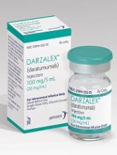
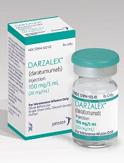
The US Food and Drug Administration (FDA) has approved daratumumab (Darzalex®) in combination with pomalidomide (POM) and dexamethasone (dex) for the treatment of multiple myeloma (MM) in patients who have received at least 2 prior therapies, including lenalidomide and a proteasome inhibitor (PI).
The FDA previously approved daratumumab in combination with lenalidomide and dexamethasone, or bortezomib and dexamethasone, for the treatment of patients with MM who had at least 1 prior therapy.
Daratumumab is also approved by the FDA as monotherapy in MM patients who had at least 3 prior lines of therapy, including a PI and an immunomodulatory (IMiD) agent, or who are double refractory to a PI and an IMiD.
The latest indication is based on the results of the phase 1b MMY1001 EQUULEUS study, which demonstrated that daratumumab produced an overall response (OR) rate of 59% in combination with pomalidomide and dexamethasone, and a very good partial response (VGPR) in 28% of patients.
EQUULEUS study
The daratumumab-POM-Dex arm of the phase 1 open-label EQUULEUS study included 103 MM patients who had received prior treatment with a PI and an immunomodulatory agent.
Patients were a median age of 64 years, and 8% were older than 75.
They had received a median of 4 prior lines of therapy, and 74% had received prior autologous stem cell transplant.
Most (89%) were refractory to lenalidomide and 71% were refractory to bortezomib. Almost two thirds (64%) were refractory to bortezomib and lenalidomide.
Patients were treated with 16 mg/kg of daratumumab in combination with POM and Dex, and 6% achieved a complete response (CR) and 8% achieved a stringent CR.
The median time to response was 1 month (range, 0.9 to 2.8), and the median duration of response was 13.6 months (range, 0.9+ to 14.6+ months).
The most frequent adverse events (AEs) reported in more than 20% of patients were infusion reactions, fatigue, and upper respiratory tract infections (50% each), cough (43%), diarrhea (38%), dyspnea (33%), nausea (30%), muscle spasms (26%), pyrexia (25%), and vomiting (21%).
The overall incidence of serious adverse reactions was 49%.
Grade 3/4 serious AEs reported in 5% of patients or more included pneumonia (7%).
The most common treatment-emergent hematologic laboratory abnormalities included lymphopenia (94%), neutropenia (95%), thrombocytopenia (75%), and anemia (57%).
And the most common grade 3/4 treatment-emergent hematology laboratory abnormalities were neutropenia (82%), lymphopenia (71%), anemia (30%), and thrombocytopenia (20%).
Daratumumab is being developed by Janssen Biotech, Inc., under an exclusive worldwide license to develop, manufacture, and commercialize daratumumab from Genmab.
See the package insert for full prescribing information. ![]()
Large MM trial finds denosumab non-inferior to ZA for SRE
CHICAGO—The largest international multiple myeloma (MM) trial ever conducted, according to the trial sponsor, met its primary endpoint, demonstrating that denosumab is non-inferior to zoledronic acid (ZA) in delaying the time to first on-study skeletal-related event (SRE) in patients with MM.
In addition to bone-specific benefits, denosumab-treated patients had significantly fewer renal adverse events and possible prolongation of progression-free survival.
Denosumab “may in fact be a new standard of care for multiple myeloma-related bone disease,” according to one of the investigators.
“The other important thing to note,” Noopur S. Raje, MD, said during her presentation at the ASCO 2017 Annual Meeting, “is denosumab can be administered despite renal function in patients with myeloma.” It does not need to be dose-adjusted, unlike bisphosphonates.
Dr Raje, of the Massachusetts General Hospital Cancer Center in Boston, Massachusetts, presented the study results as abstract 8005.
Study design
The international, phase 3, randomized, double-blind study is evaluating the safety of denosumab compared with ZA in newly diagnosed MM patients.
Investigators enrolled 1718 patients from 259 sites and 29 countries.
They randomized 859 patients to receive denosumab 120 mg subcutaneously every 4 weeks plus intravenous placebo every 4 weeks, and 859 patients to the standard ZA dose of 4 mg intravenously plus subcutaneous placebo every 4 weeks.
Patients were stratified by whether they were on novel-based anti-myeloma therapy, whether they planned to have an autologous peripheral blood stem cell (PBSC) transplant, disease stage, and previous SRE.
“We were looking for 676 on-study SREs, and if we saw a benefit, patients would be offered open-label denosumab for up to 2 years after this,” Dr Raje said.
“Patients had to have radiographic evidence of bone disease, and this is different from some of the other bone disease studies that you’ve seen in the recent past,” she added.
In addition to documented evidence of MM, patients had to be 18 years or older, be ECOG status of 2 or better, have adequate organ function, and plan to receive or be receiving primary frontline anti-myeloma therapy.
Patients were excluded if they had nonsecretory MM, more than 30 days of previous treatment with anti-myeloma therapy prior to screening, prior use of denosumab, use of oral bisphosphonates with a cumulative dose of more than 1 year, more than 1 previous dose of intravenous bisphosphonate, or prior history or current evidence of osteonecrosis/osteomyelitis of the jaw.
The primary endpoint was time to first on-study SRE, “and the idea here was to look for non-inferiority,” Dr Raje explained.
Secondary endpoints included time to the first on-study SRE (superiority), time to the first-and-subsequent on-study SRE (superiority), and overall survival.
Investigators also included the exploratory objective of progression-free survival (PFS).
Patient demographics
Patients were well balanced across the 2 arms, Dr Raje noted, and the breakdown of myeloma disease stage at diagnosis was comparable between the ZA and denosumab arms.
About 32% of patients were stage I, 37% stage II, and 29% stage III. Stage was not available for 49 patients.
A little more than half (54%) were male, mean age was 63 years, and 82% were white.
Two thirds had prior SRE history, and 54% of patients intended to undergo autologous PBSC transplant.
Enrollment began May 2012 and continued through the end of March 2016. The primary analysis cutoff was July 19, 2016.
Results
The primary endpoint for non-inferiority for time to first on-study SRE was met by denosumab (HR=0.98, 95%CI: 0.85, 1.14; P=0.01).
“When we looked at the secondary endpoints for superiority, we were not able to confirm superiority in this analysis, either for time to first SRE or time to first-and subsequent SRE on this study,” Dr Raje said.
The investigators also did not observe a survival difference between denosumab and ZA, with a hazard ratio (HR) (95% CI) of 0.90 (0.70, 1.16), P=0.41.
“Importantly, we had an exploratory endpoint where we looked at progression-free survival in this newly diagnosed patient population,” she added, “and we saw an interestingly increased or prolonged progression-free survival in patients getting denosumab.”
“And that survival difference was more than 10 months between denosumab and zoledronic acid, favoring the denosumab arm,” she affirmed. The HR was 0.82, 95% CI: 0.68, 0.99, P=0.036 (descriptive).
Safety
“[I]f you look at all treatment-emergent adverse events between denosumab and zoledronic acid, we really could not find a big difference in either of these 2 groups of patients,” Dr Raje said.
“We saw that in general both denosumab and zoledronic acid were extremely well tolerated between the 2 groups of patients.”
The investigators “drilled down” on certain toxicity issues of interest and examined events such as atypical stress fractures, hypersensitivity reactions, musculoskeletal pain, infections and infestations, new primary malignancies, and acute phase reactions.
They observed no atypical femur fractures on the study, nor did they see any big differences with respect to hypersensitivity or acute phase reactions.
The investigators examined closely any renal issues because dosing of ZA specifically is impacted by renal function.
The data showed that treatment-emergent adverse event (TEAE) renal toxicity was significantly higher in the ZA group compared to the denosumab group, 17% and 10%, respectively (P<0.001).
“When you look at patients who had a creatinine clearance less than 60 mL per minute,” Dr Raje emphasized, “we saw an almost doubling of renal toxicity in the zoledronic acid arm (26.4%) compared to the denosumab arm (12.9%).”
Patients with a creatinine level greater than 2 mg/dL had a significant increase in creatinine in the ZA arm (P=0.010), which was also significantly increased if their creatinine clearance was less than 60 mL/minute (P=0.054).
“There was a doubling of creatinine from baseline, more so in the zoledronic acid arm compared to the patients with denosumab,” Dr Raje said. “And this was again more pronounced if you had a creatinine clearance of less than 60.”
Hypocalcemia was “not surprisingly” more common in the denosumab arm than the ZA arm (P=0.009) for all patients, and osteonecrosis of the jaw was equal in both arms (P=0.147), although numerically slightly higher with denosumab treatment.
Dr Raje summarized that there was no difference in overall survival at the time of this analysis, “but I will say that the follow-up for a newly diagnosed patient population is fairly short right now.”
“Progression-free survival, which we saw [cut] off 10.7 months, was actually quite striking when denosumab was compared to zoledronic acid, and this was statistically highly significant.”
“The bone-specific benefits in combination with significantly fewer renal adverse events and possible prolongation of PFS with denosumab therapy we do think is very promising,” she said, “and may in fact be a new standard of care for multiple myeloma-related bone disease.”
The study was funded by Amgen Inc.
Denosumab (XGEVA®) is indicated by the US Food and Drug Administration for the prevention of fractures and other SREs in patients with bone metastases from solid tumors. It is currently not indicated for the prevention of SREs in patients with MM. ![]()
Neurotoxicity needs separate treatment from CRS in CAR T-cell therapy
CHICAGO – The neurotoxicity in adult patients with relapsed or refractory B-cell acute lymphocytic leukemia (B-ALL) treated with CD19 CAR T cells is a separate process from cytokine release syndrome (CRS) and needs to be treated separately, according to a new study presented at the American Society of Clinical Oncology annual meeting (abstract 3019).
CD19-specific CAR-modified T cells produce high, durable anti-tumor activity, but can be associated with treatment-related toxicities, including CRS and neurotoxicity.
Neurotoxicity is poorly understood and it hasn't been clear where to focus further research, said Bianca Santomasso, MD, of Memorial Sloan Kettering Cancer Center in New York, New York.
Dr Santomasso and colleagues had conducted a phase 1 study using autologous 19-CAR T cells in adult patients with relapsing/refractory B-ALL, with high response rates.
To gain a better understanding of CD19 CAR T-cell-associated neurotoxicity, they analyzed clinical and research parameters after stratifying patients by neurotoxicity grade.
At ASCO, she reported neurologic symptom presentation in 51 adult patients with relapsed/refractory B-ALL who were treated with CAR T cells following conditioning chemotherapy, along with cerebrospinal fluid (CSF) data and neuroimaging findings associated with neurotoxicity.
Of the 51 patients treated, 10 patients (20%) developed mild neurologic symptoms (grade 1 or 2) and 21 patients (41%) developed severe neurotoxicity (grade 3 or 4).
No grade 5 neurotoxicity or diffuse cerebral edema occurred and, in all but one case, neurologic symptoms fully resolved.
Fourteen patients (27.4%) developed severe CRS; 6 patients received tocilizumab alone, 13 patients tocilizumab plus steroids, 4 patients steroids alone, and 29 patients supportive care.
The cytokines IL-6, IL-8, IL-10, interferon gamma, and granulocyte-colony stimulating factor were elevated in CSF over serum at the time of neurotoxicity and correlated with CSF protein levels.
“We found no significant correlation between neurotoxicity grade and the CAR T-cell concentration in the CSF during neurotoxicity,” Dr Santomasso said. “Instead, CSF protein level was correlated with neurotoxicity grade.”
Neurotoxicity was associated with peak CAR T-cell expansion in the blood and peak serum levels of several cytokines associated with T-cell activation or proliferation, she said.
“CAR T cells traffic to the CSF of patients with all grades of neurotoxicity, including grade 0, and there is no significant correlation between CAR T cells in CSF at the time of acute neurotoxicity and grade of neurotoxicity,” Dr Santomasso reported. “This suggests that neurotoxicity is not directly mediated by CAR T cells, which cross into the spinal fluid.”
Some chemokines/cytokines are elevated in CSF relative to serum, suggesting CNS-specific production of these factors, she said.
“Clinicians treating these patients tend to lump CRS and neurotoxicity together. Now we have an increasing understanding that these adverse events are separated in time and possibly in underlying pathology,” said Dr Santomasso.
“Even when patients recover from CRS, they could still be at risk for neurotoxicity.”
She noted that the neurotoxicity symptoms that B-ALL patients develop in the setting of CAR T cells are manageable.
In a subset of patients with severe neurotoxicity, T2/FLAIR changes were observed, which resolved with steroids and neurologic symptom resolution. ![]()
CAR T cells plus ibrutinib induce CLL remissions
CHICAGO—Chimeric antigen receptor (CAR) T cells combined with ibrutinib enhance T-cell function and can induce complete remission (CR) in patients with chronic lymphocytic leukemia (CLL), researchers report.
Many CLL patients receive ibrutinib treatment, which is well tolerated, but few patients achieve CR.
Immunotherapy with anti-CD19 CAR T cells has induced CR in 25% - 45% of patients with CLL, Saar Gill, MD, of the University of Pennsylvania in Philadelphia, told Hematology Times, and these CRs tend to be durable.
So investigators conducted a pilot trial in 10 patients to test whether combining anti-CD19 CAR T cells with ibrutinib would enhance the CR rate.
Dr Gill reported the findings of the pilot trial at the ASCO 2017 Annual Meeting (abstract 7509).
The patients must have failed at least 1 regimen before ibrutinib, unless they had del(17)(p13.1) or a TP53 mutation.
T cells were lentivirally transduced to express a CAR that included humanized anti-CD19.
Patients were lymphodepleted 1 week before infusion, and ibrutinib was continued throughout the trial.
After a median follow-up of 6 months, 8 of the 9 evaluable patients show absence of CLL in the bone marrow by flow cytometry or minimal residual disease (MRD) negative, and all remain in marrow CR at last follow-up, Dr Gill said.
Radiologic responses are less clear-cut and may require longer follow-up.
“All but 1 patient achieved MRD with deep sequencing. We have deep response in the bone marrow,” Dr Gill said. He also noted that the treatment was well tolerated.
Cytokine release syndrome (CRS) developed in 9 patients: grade 1 in 2 patients, grade 2 in 6 patients, and grade 3 in 1 patient. One patient developed grade 4 tumor lysis syndrome. Treatment of CRS with the IL-6 receptor antagonist tocilizumab was not required.
There was modest residual splenomegaly in 3 of 5 patients, and adenopathy resolved in 4 of 6 patients, with progression in 1 patient.
Ibrutinib reduced CRS apparently by blocking cytokine production by T cells, said Dr Gill, adding, “The combination led to improved efficacy without increased toxicity.”
Ibrutinib may make CAR T-cell therapy more feasible.
Patients who receive ibrutinib for 6 months have a better T-cell response.
“This opens up future discussions of bringing CAR T-cell therapy earlier into CLL treatment,” said Dr Gill.
He envisions patients receiving ibrutinib for 6 months, which would allow time to manufacture T cells, and then have a T-cell infusion.
“Once patients achieve MRD, then we can discuss the possibility of stopping ibrutinib therapy,” he said.
“Most patients remain on ibrutinib, but longer follow-up may show whether remissions are sustained off ibrutinib.”
The researchers have ongoing plans to treat 25 patients with CTL19 plus ibrutinib in a continuation of this trial.
Dr Gill said longer follow-up will reveal the durability of these results “and could support evaluation of a first-line combination approach in an attempt to obviate the need for chronic therapy.” ![]()
‘Admirable’ overall survival attainable in AML with enasidenib
CHICAGO—The experimental mutant IDH2 (mIDH2) inhibitor enasidenib has produced “admirable” overall survival in patients with mIDH2 relapsed or refractory acute myeloid leukemia (AML), according to Eytan M. Stein, MD, an investigator on the phase 1 dose escalation and expansion study.
Patients who achieved a complete remission (CR) had a median overall survival (OS) of 19.7 months and non-CR responders, 13.8 months.
“I really want to make the point,” Dr Stein said, “this is a group of patients that are highly refractory, either refractory to induction chemotherapy, refractory to standard of care approaches for patients who are unable to get induction chemotherapy, so refractory to hypomethylating agents or low-dose cytarabine.”
Mutations in IDH2 occur in approximately 12% of AML patients.
Dr Stein explained that the mutant protein converts alpha ketoglutarate to beta hydroxyglutarate (2-HG). And increased levels of intracellular 2-HG lead to methylation changes in the cell that cause a block in myeloid differentiation.
Enasidenib, also known as AG-221, is a selective, oral, potent inhibitor of the mIDH2 enzyyme.
Dr Stein, of Memorial Sloan Kettering Cancer Center in New York, New York, presented the results during the ASCO 2017 Annual Meeting (abstract 7004).
The clinical and translational papers were published simultaneously in Blood.
Study design
The phase 1/2 study had a large dose-escalation component, with 113 patients enrolled. Patients had to have an advanced hematologic malignancy with an IDH2 mutation.
Patients received cumulative daily doses of 50 mg – 650 mg of enasidenib in continuous 28-day cycles.
Four expansion arms were added, with 126 patients.
Two expansion arms were in relapsed/refractory AML patients: one in patients 60 years or older or any age if they had relapsed after bone marrow transplant (BMT), and the other in patients younger than 60 excluding those relapsed after BMT.
The other 2 expansion arms were in untreated AML patients and in patients with any hematologic malignancy ineligible for the other arms.
Dr Stein presented results for the relapsed/refractory AML patients in the dose escalation and expansion phases of the study.
The key endpoints were safety, tolerability, maximum tolerated dose (MTD), and dose-limiting toxicities; response rates as assessed by the local investigator according to IWG criteria; and assessment of clinical activity.
Dr Stein noted the phase 2 study is now completely accrued (n=91) and the recommended enasidenib dose is 100 mg/day in relapsed/refractory AML.
The MTD was not reached at doses up to 650 mg/day.
Baseline characteristics
Median age of all 239 phase 1 patients was 70 years (range, 19-100), 57% were male, and almost all patients had intermediate- or poor-risk disease.
The investigators were also interested in the co-occurring mutations in patients on screening and whether there were differences between patients with mIDH2 at R172 and R140.
Seventy-five percent of the patients (n=179) had R140 and 24% had R172 (n=57).
There was a statistically significant difference in the number of co-occurring mutations in the R140 and R172 patients, with the R140 patients having a higher co-mutation burden compared with the R172 patients, (P=0.020).
The most frequent mutations co-occurring in R140 patients were SRSF2, followed by, in descending order of frequency, DNMT3A, RUNX1, ASXL1, and 24 others.
SFSR2 does not occur in R172 patients. DNMT3A was the most frequently co-occurring mutation in R172, followed by ASXL1, BCOR, NRAS, RUNX1, KMT2A, KRAS, and STAG2.
Safety
The most common treatment-emergent adverse events (TEAE) that occurred in 20% or more of all patients of any grade included nausea (46%), hyperbilirubinemia (45%), diarrhea and fatigue (40% each), decreased appetite (38%), vomiting (32%), dyspnea (31%), cough (29%), pyrexia and febrile neutropenia (28% each), thrombocytopenia, anemia, constipation, hypokalemia, and peripheral edema (27% each), pneumonia (21%), and hyperuricemia (20%).
The only 2 grade 3/4 TEAEs that rose above the level of 5% were hyperbilirubinemia (12%) and thrombocytopenia (6%).
“The hyperbilirubinemia, as I’ve mentioned in a number of meetings before this,” Dr Stein clarified, “is one that occurs because the enzyme is an off-target effect of inhibiting the UGT1A1 enzyme, which conjugates bilirubin.”
“So a patient who goes on this study who has a defect in bilirubin conjugation because they have Gilbert’s disease, they will have a higher level of bilirubin compared to a patient who doesn’t have Gilbert’s disease. This does not appear to have any clinical sequelae. You’ll also notice AST, ALT, alkaline phosphatase or any liver failure is not on this [TEAE] list.”
Response
The overall response rate for the patients who received enasidenib 100 mg/day was 38.5% (42/109) and for all doses 40.3% (71/176).
The true CR rate was 20.2% (100 mg/day) and 19.3% for all doses.
An additional 20% achieved a CR with incomplete hematologic recovery, CR with incomplete platelet recovery, partial response (PR), and morphologic leukemia-free state with either 100 mg enasidenib daily or all doses.
“Time to first response is not immediate,” Dr Stein pointed out. “It takes a median of 1.9 months to get there, and the time to complete remission takes even longer, a median of 3.7 months in the 100-mg experience, 3.8 months in all doses, to get to that best response.”
“I think the clinical importance of this is,” he added, “for a patient that one might have who is on this drug, it is important to keep them on the drug for a prolonged period of time so that they have the opportunity to have that response.”
Hematologic parameters also improved gradually.
Increases in platelet count, absolute neutrophil count, and hemoglobin level did not rise exponentially upon administration of study drug, but rather they slowly rose, “again getting to this point, that the drug takes time to work,” Dr Stein emphasized.
Patients in CR had very high transfusion independence rates, “which is what I would expect,” Dr Stein said. “If you are in complete remission, you should be transfusion independent.”
“What’s a little bit more interesting, though,” Dr Stein added, “is those patients who are non-CR responders. [I]n those patients who have responded but have less than a complete remission, 50% of them are independent of red cell transfusions and 50% of them are independent of platelet transfusions.”
Survival
The CR data and transfusion independence data translated into a median OS in these relapsed and refractory AML patients of 9.3 months.
And about 10% - 15% of the patients had prolonged survival up to 2 years and longer on the single agent.
Analysis of OS by best response revealed that for patients with a CR, “they really have an admirable overall survival of 19.7 months, almost 20 months,” Dr Stein said.
Patients who had a non-CR response had a median OS of 13.8 months, and non-responders had a median OS of 7.0 months.
And there was a qualitative improvement in response over time: the number of patients with CRs and PRs increased, while the number with stable disease decreased.
“Again, I think getting at the point it takes time for these responses to occur,” Dr Stein iterated.
Over the course of therapy, some responders had a differentiation of myeloblasts, so that by cycle 3, the marrow looked largely normal.
The investigators did not observe any morphological evidence of cytotoxicity or cellular aplasia.
But they did observe myeloid differentiation using FISH.
Trisomy 8 that was evident at the time of screening in responders’ myeloblasts, persisted in the promyelocytes and mature granulocyte population, and was no longer evident in the lymphoid compartment.
Baseline 2-HG levels and mIDH2 variant allele frequency were similar for responding and non-responding patients.
The investigators believe that differentiation of myeloblsts, not cytotoxicity, may drive the clinical efficacy of enasidenib.
A phase 3 trial of enasidenib monotherapy versus conventional care regimens is underway in older patients with late-stage AML, and phase 1/2 studies of enasidenib combinations are ongoing in newly diagnosed AML patients.
Enasidenib, which also has efficacy in myelodysplastic syndromes, has been granted priority review for relapsed/refractory AML by the US Food and Drug Administration. ![]()
Ferrous sulfate bests iron complex in treating IDA in infants, young kids
A trial comparing ferrous sulfate with iron polysaccharide complex to treat infants and young children with nutritional iron-deficiency anemia (IDA) has shown ferrous sulfate to be more effective at raising hemoglobin levels in this population, according to researchers.
Dozens of oral iron supplements exist for IDA treatment, ferrous sulfate being the most commonly used. Iron polysaccharide complex, however, may be better tolerated.
Investigators undertook the BESTIRON study (NCT01904864) to determine whether the iron complex was more efficacious than ferrous sulfate in increasing hemoglobin concentrations in infants and young children aged 9 to 48 months.
Up to 3% of children aged 1 to 2 years in the United States have IDA, as do millions worldwide. IDA is associated with impaired neurodevelopment in the young.
Inadequate dietary iron intake in this group is the most common cause of IDA. It most often results from excessive cow milk consumption and/or prolonged breastfeeding without appropriate iron supplementation.
For this study, investigators randomized 80 infants and young children with nutritional IDA to receive 3 mg/kg of ferrous sulfate (n=40) or iron complex (n=40) drops once daily for 12 weeks.
Patients had to have hemoglobin concentrations of 10 g/dL or less, mean corpuscular volumes of 70 fL or less, reticulocyte hemoglobin equivalents of 25 pg or less, and either serum ferritin level of 15 ng/mL or less or total iron-binding capacity of 425 μg/dL or greater.
And they could have no clinical or laboratory evidence of other causes of anemia.
All 80 patients were included in the primary analysis evaluating change in hemoglobin concentration during the 12 weeks after starting oral iron therapy.
Patient characteristics
Patient characteristics were similar between the groups. The mean age was 23 months and 55% were male.
Most patients (61%) were Hispanic white, 9% were non-Hispanic white, and 11% were black.
Ten patients in the ferrous sulfate group and 8 in the iron complex group had received a packed red blood cell transfusion prior to study enrollment.
Results
Fifty-nine patients completed all study visits, 28 in the ferrous sulfate group and 31 in the iron complex group.
Patients’ mean hemoglobin level in the ferrous sulfate group increased from 7.9 g/dL to 11.9 g/dL over the 12 weeks. In the iron complex group, the patients’ hemoglobin level increased from 7.7 g/dL to 11.1 g/dL.
Using a linear mixed model, the primary outcome demonstrated a significant difference in the change in hemoglobin concentration of 1.0 g/dL (95% CI, 0.4-1.6; P < .001) between the groups, favoring ferrous sulfate.
IDA completely resolved in 8 of 28 (29%) patients in the ferrous sulfate group and 2 of 31 (6%) in the iron complex group (P=0.04).
However, successful administration of the supplement—meaning he child did not spit out the medication—was higher in the iron complex group (94%) than the iron sulfate group (82%), P=0.009.
The median serum ferritin level increased from 3.0 ng/mL to 15.6 ng/mL in the ferrous sulfate arm, which was significantly better than in the iron complex arm, which increased from 2.0 ng/mL to 7.5 ng/mL, P<0.001.
And the mean total iron binding capacity significantly increased in the ferrous sulfate group compared with the iron oxide group (P<0.001).
Safety
The investigators reported that patients treated with iron complex had significantly more diahrrea, while patients treated with ferrous sulfate had more vomiting, although the latter was not statistically significant.
A gastrointestinal adverse effect profile created at the end of the study showed no significant differences between the groups.
The investigators noted a few limitations of the study.
First, it was conducted in a single tertiary-care children’s hospital, the Children’s Medical Center in Dallas, Texas.
Second, a disproportionate number of patients were from lower income and minority families and frequently had severe anemia, with approximately 23% requiring blood transfusion prior to study start.
And third, the trial had a high lost-to-follow-up rate of 25% at the final visit.
So the results may not be generalizable to the general pediatric population.
Nevertheless, the investigators concluded, “Once daily, low-dose ferrous sulfate should be considered for children with nutritional iron-deficiency anemia.”
The team reported their findings in JAMA.
The study was an investigator-initiated trial with sponsorship from Gensavis Pharmaceuticals LLC, the manufacturer of the iron polysaccharide complex used in the trial. The company provided funding for both trial drugs.
The study received additional grant support from the National Center for Advancing Translational Sciences and the National Heart, Lung, and Blood Institute. ![]()
Company pauses enrollment on 2 trials of pembrolizumab in MM
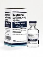
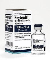
Merck announced that it is pausing enrollment onto 2 phase 3 trials of pembrolizumab (Keytruda®) in combination with other agents to treat multiple myeloma (MM).
An external Data Monitoring Committee recommended the trial be interrupted “to allow for additional information be collected to better understand more reports of death” in the pembrolizumab groups in the KEYNOTE-183 and KEYNOTE-185 trials.
Patients currently enrolled on the trials can continue to receive treatment. Other pembrolizumab trials are continuing without changes.
Merck in its statement did not disclose the number of deaths nor provide any other details on the deaths.
Pembrolizumab is a humanized monoclonal antibody that blocks interaction between the programmed cell death protein 1 (PD-1) and its receptor ligands, PD-L1 and PD-L2.
The US Food & Drug Administration approved pembrolizumab to treat unresectable or metastatic melanoma after ipilimumab treatment.
Pembrolizumab has also been approved to treat non-small cell lung cancer, head and neck squamous cell cancer, classical Hodgkin lymphoma, urothelial carcinoma, and microsatellite instability-high solid tumors.
KEYNOTE-183 (NCT02576977), which has an estimated enrollment of 300 patients, is comparing the combination of pembrolizumab, pomalidomide, and low-dose dexamethasone to pomalidomide and low-dose dexamethasone alone in patients with relapsed or refractory MM who have undergone at least 2 lines of prior therapy.
KEYNOTE-185 (NCT02579863), which has an estimated enrollment of 640 patients, is comparing the combination of pembrolizumab, lenalidomide, and low-dose dexamethasone to lenalidomide and low-dose desamethasone alone in patients with newly diagnosed and treatment-native MM who are ineligible for autologous stem cell transplant.
The comparator agents pomalidomide (Pomalyst®) and lenalidomide (Revlimid®) are products of Celgene Corporation. ![]()
BM-MSCs may be an option for AA patients refractory to IST
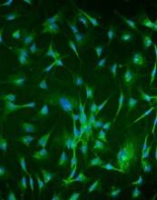
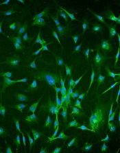
Researchers report that infusions of bone marrow-derived mesenchymal stromal cells (BM-MSCs) may be a treatment option for patients with aplastic anemia (AA) who are refractory to immunosuppressive therapy (IST).
They conducted a phase 2, non-comparative multicenter study to assess the safety and efficacy of this approach and found that after 12 months, 28.4% of patients responded, with 6.8% achieving a complete response (CR) and 21.6% a partial response (PR).
The trial involved 74 patients at 7 centers in China. The research team reported its findings in Stem Cells Translational Medicine.
About 30% to 40% of patients with severe AA (sAA) don’t respond well to IST and continue to have abnormally low levels of red blood cells, white blood cells, and platelets.
The benefit of treatment with BM-MSCs is they support hematopoiesis, express low levels of major histocompatibility (MHC)-I, and lack expression of MHC-II surface molecules.
And BM-MSCs have been reported to cure diseases, including graft-versus-host disease, arthritis, lupus, and other immune and non-immune disorders.
The current study, led by Yan Pang, MD, and Yang Xiao, MD, PhD, of Guangzhou General Hospital of Guangzhou Military Command in China, is based on their previous data evaluating intravenous administration of MSCs from a related donor in 18 patients with refractory AA.
This earlier data showed that 33% of patients with AA refractory to IST achieved a CR or PR to BM-MSC treatment.
So the team undertook to further investigate the use of BM-MSCs in AA.
Study design
Each of the 74 patients received 4 doses of BM-MSCs over a period of 4 weeks. If patients responded after the first month, they continued to receive 4 doses.
Investigators obtained the BM-MSCs from 74 healthy donors—48 males and 26 females. Forty were related donors, 27 haploidentical donors, and 7 unrelated donors.
Patients with AA by standard criteria had to be 16 years or older, had an incomplete response to antithymocyte globulin (ATG) and cyclosporine for at leas 6 months or cyclosporine alone for at least 12 months, did not have a donor available for bone marrow transplantation, and had at least one of the following: hemoglobin <70 g/L, neutrophilic granulocytes <1 × 10⁹/L, or platelet count <30 × 109/L.
Almost half the patients (47%) were between 20 and 40 years, 54% were male, and 32% had severe aplastic anemia.
Patients’ previous therapy for aplastic anemia included cyclosporine and andriol (65%) cyclosporine and ATG (19%), and cyclosporine (16%).
Patients could continue cyclosporine, but no immunosuppressive agents were permitted.
Fifty-three patients (71.6%) completed 1 course of therapy, and 21 patients (18.4%) completed 2 courses.
Response
At 1 year, the overall response rate was 28.4% (n=21) and the PR rate was 21.6% (n=16).
The median time to a leukocyte response was 19 days (range, 11 – 29), to an erythrocyte response 17 days (range, 12 – 25), and to a megakaryocyte response 31 days (range, 26 – 84).
Ten of the patients with hematologic response had normalization of cellularity for more than 1 year.
The median follow-up among survivors was 17 months (range, 3 – 24). The 2-year overall survival was 87.8%.
Three patients progressed to myelodysplasia, 1 with refractory anemia with excess blasts (RAEB)-I and 2 with RAEB-II.
The median time to progression was 11 months (range, 8 – 12).
Nine patients died, all of whom had severe AA. One patient with RAEB-II died of disease progression, two patients died of intracranial hemorrhage, and six patients died of serious infection.
Safety
Adverse events included grade 1 (n=5) and grade 2 (n=2) fever. Two of these patients also had grade 1 headache.
The investigators observed no other adverse events.
Response predictors
The investigators determined that 2 factors predicted response in patients: prior treatment with antithymocyte globulin (ATG) and absence of infection throughout the treatment.
The odds ratio for patients treated with ATG was 1.41 (95% CI: -0.50, 3.31) and for patients without infection, 2.19 (95% CI: 0.50, 3.87).
“Our study strongly indicates that MSC infusion is a promising therapy for severe AA," Dr Pang said, “but improved MSC cultures in vitro and the MSC doses need further study to maximize their therapeutic potential." ![]()
Researchers report that infusions of bone marrow-derived mesenchymal stromal cells (BM-MSCs) may be a treatment option for patients with aplastic anemia (AA) who are refractory to immunosuppressive therapy (IST).
They conducted a phase 2, non-comparative multicenter study to assess the safety and efficacy of this approach and found that after 12 months, 28.4% of patients responded, with 6.8% achieving a complete response (CR) and 21.6% a partial response (PR).
The trial involved 74 patients at 7 centers in China. The research team reported its findings in Stem Cells Translational Medicine.
About 30% to 40% of patients with severe AA (sAA) don’t respond well to IST and continue to have abnormally low levels of red blood cells, white blood cells, and platelets.
The benefit of treatment with BM-MSCs is they support hematopoiesis, express low levels of major histocompatibility (MHC)-I, and lack expression of MHC-II surface molecules.
And BM-MSCs have been reported to cure diseases, including graft-versus-host disease, arthritis, lupus, and other immune and non-immune disorders.
The current study, led by Yan Pang, MD, and Yang Xiao, MD, PhD, of Guangzhou General Hospital of Guangzhou Military Command in China, is based on their previous data evaluating intravenous administration of MSCs from a related donor in 18 patients with refractory AA.
This earlier data showed that 33% of patients with AA refractory to IST achieved a CR or PR to BM-MSC treatment.
So the team undertook to further investigate the use of BM-MSCs in AA.
Study design
Each of the 74 patients received 4 doses of BM-MSCs over a period of 4 weeks. If patients responded after the first month, they continued to receive 4 doses.
Investigators obtained the BM-MSCs from 74 healthy donors—48 males and 26 females. Forty were related donors, 27 haploidentical donors, and 7 unrelated donors.
Patients with AA by standard criteria had to be 16 years or older, had an incomplete response to antithymocyte globulin (ATG) and cyclosporine for at leas 6 months or cyclosporine alone for at least 12 months, did not have a donor available for bone marrow transplantation, and had at least one of the following: hemoglobin <70 g/L, neutrophilic granulocytes <1 × 10⁹/L, or platelet count <30 × 109/L.
Almost half the patients (47%) were between 20 and 40 years, 54% were male, and 32% had severe aplastic anemia.
Patients’ previous therapy for aplastic anemia included cyclosporine and andriol (65%) cyclosporine and ATG (19%), and cyclosporine (16%).
Patients could continue cyclosporine, but no immunosuppressive agents were permitted.
Fifty-three patients (71.6%) completed 1 course of therapy, and 21 patients (18.4%) completed 2 courses.
Response
At 1 year, the overall response rate was 28.4% (n=21) and the PR rate was 21.6% (n=16).
The median time to a leukocyte response was 19 days (range, 11 – 29), to an erythrocyte response 17 days (range, 12 – 25), and to a megakaryocyte response 31 days (range, 26 – 84).
Ten of the patients with hematologic response had normalization of cellularity for more than 1 year.
The median follow-up among survivors was 17 months (range, 3 – 24). The 2-year overall survival was 87.8%.
Three patients progressed to myelodysplasia, 1 with refractory anemia with excess blasts (RAEB)-I and 2 with RAEB-II.
The median time to progression was 11 months (range, 8 – 12).
Nine patients died, all of whom had severe AA. One patient with RAEB-II died of disease progression, two patients died of intracranial hemorrhage, and six patients died of serious infection.
Safety
Adverse events included grade 1 (n=5) and grade 2 (n=2) fever. Two of these patients also had grade 1 headache.
The investigators observed no other adverse events.
Response predictors
The investigators determined that 2 factors predicted response in patients: prior treatment with antithymocyte globulin (ATG) and absence of infection throughout the treatment.
The odds ratio for patients treated with ATG was 1.41 (95% CI: -0.50, 3.31) and for patients without infection, 2.19 (95% CI: 0.50, 3.87).
“Our study strongly indicates that MSC infusion is a promising therapy for severe AA," Dr Pang said, “but improved MSC cultures in vitro and the MSC doses need further study to maximize their therapeutic potential."
Combo with daratumumab could be alternative to ASCT in MM
CHICAGO—Results of an open-label phase 1b study of daratumumab combined with carfilzomib, lenalidomide, and dexamethasone (KRd) in newly diagnosed multiple myeloma (MM) patients have shown the combination to be highly effective, with an overall response rate of 100%.
Ninety-one percent of patients achieved a very good partial response (VGPR) or better, and 43% achieved a complete response (CR) or better.
Investigators had hypothesized that rather than using autologous stem cell transplant (ASCT) to improve results of treatment with KRd, the combination could alternatively be improved by incorporating daratumumab into a KRd regimen.
Andrzej Jakubowiak, MD, of the University of Chicago Medical Center in Illinois, presented the findings of the MMY1001 study at the 2017 ASCO Annual Meeting (abstract 8000*).
“I think what was one of the more important developments in myeloma last year,” Dr Jakubowiak said, “was data from randomized studies showing that adding daratumumab to either lenalidomide and dexamethasone in the POLLUX study or bortezomib and dexamethasone, a proteasome inhibitor, in the CASTOR study, improves responses, depth of response, and . . . dramatically improved progression-free survival.”
“[W]e have now the rationale to potentially combine daratumumab with both an IMiD and proteasome inhibitor,” he explained, “which led to the development of this phase 1b study in which we combined daratumumab with KRd and evaluated tolerability and efficacy.”
Study design
Twenty-two transplant-eligible or -ineligible newly diagnosed MM patients were enrolled on the study.
Treatment duration was planned to be 13 cycles or less and patients had the option to move to transplant after 4 cycles.
They could have no clinically significant cardiac disease and echocardiogram was required prior to transplant.
The dosing schedule was the established dosing schema for daratumumab and KRd with 2 notable differences in the 28-day cycles.
First, the daratumumab dose was a split dose. So patients received 8 mg/kg on days 1-2 of cycle 1, 16 mg/kg a week on cycle 2, 16 mg/kg every 2 weeks on cycles 3 – 6, and every 4th week thereafter.
The second difference was carfilzomib dosing was a weekly regimen with escalation from 20 mg/m2 on day 1, cycle 1 to 70 mg/m2 on day 8 of cycle 1.
Lenalidomide (25 mg on days 1-21 of each cycle) and dexamethasone (40 mg/week) were the standard regimens for these drugs.
The primary endpoint was safety and tolerability. The secondary endpoint was overall response rate (ORR), duration of response, time to response, and infusion-related reactions (IRR).
The study also had an exploratory endpoint of progression-free survival (PFS).
Baseline characteristics
Patients were a median age of 59.5 years (range 34 – 74). About two thirds were younger than 65 and one third were between 65 and 75.
A little over half were male and most (86%) were white.
A little more than half (55%) had an ECOG score of 0, 41% were ECOG 1, and 5% were ECOG 2.
Patient disposition
As of the cutoff date of March 24, 8 of the 22 patients enrolled (36%) discontinued treatment: 1 due to an adverse event (AE), 1 due to progressive disease, and 6 patients (27%) proceeded to ASCT.
Dr Jakubowiak pointed out that response was censored at this point for patients who proceeded to transplant.
The median follow-up was 10.8 months (range, 4.0 – 12.5) and the median number of treatment cycles was 11.5 (range, 1.0 – 13.0).
“What is of interest to many of us,” Dr Jakubowiak said, “is that patients were escalated to the planned dose of 70 mg/m2 by cycle 2 except for 3 patients.”
Of the 3, 1 discontinued before day 1 of the second cycle due to toxicity, 1 had a dose reduction to 56 mg/m2 at day of the second cycle, and 1 escalated to 70 mg/m2 at day 8 of cycle 3.
Ultimately, all patients who remained on study were able to escalate to 70 mg/m2.
Safety
The hematologic treatment-emergent adverse events (TEAE) generally followed what has been observed in similar studies before, Dr Jakubowiak noted.
Hematologic TEAEs of all grades occurring in 30% or more of patients were lymphopenia (68%), thrombocytopenia (55%), anemia (46%), leukopenia (41%), and neutropenia (32%).
The most common non-hematologic TEAEs of all grades occurring in 30% of patients or more were diarrhea (73%), upper respiratory infection (59%) cough, constipation, and fatigue (50% each), dyspnea and insomnia (46%), nausea, rash, and back pain (41%), muscle spasm (36%), and vomiting, pain in extremity, hyperglycemia, and increased ALT (32%).
The most common grade 3/4 TEAEs were infrequent and many events had none of grade 3/4 severity.
The safety profile is consistent with what was previously reported for daratumumab or KRd, Dr Jakubowiak affirmed.
Serious TEAEs
Serious TEAEs occurred in 10 patients (46%), with many occurring in just 1 patient. Pulmonary embolism (PE) was the most frequent, occurring in 3 patients.
All patients were required to be on aspirin prophylaxis and 1 of the patients who had a PE discontinued therapy.
The number of patients with a serious TEAE reasonably related to an individual study drug were 3 (14%) for daratumumab, 5 (23%) for carfilzomib, 5 (23%) for lenalidomide, and 2 (9%) for dexamethasone.
The TEAEs of interest—tachycardia, congestive heart failure, and hypertension—occurred in a single patient each.
Overall, serious TEAEs were consistent with previous reports from KRd studies.
Echocardiogram assessment
Investigators conducted 30 systemic evaluations on the impact of this regimen on heart function. The investigators observed no change from baseline through the duration of treatment in patients’ left ventricular ejection fractions.
One patient developed congestive heart failure, possibly related to daratumumab or carfilzomib. This patient resumed treatment with a reduced carfilzomib dose, elected ASCT on study day 113, and ended treatment with a VGPR.
“In all,” Dr Jakubowiak said, “we feel that there is no apparent signal of adverse impact of the addition of daratumumab on cardiac function.”
Infusion times and reactions
Overall, IRRs occurred in 27% of the patients, “which appears lower than with previous daratumumab studies,” Dr Jakubowiak noted. And IRRs occurred more frequently during the first infusion than subsequent infusions.
The split-dose infusion time was very similar to that of second and subsequent cycles.
There were limited events related to infusions. All were grade 1 or 2 and most occurred in only a single patient.
Response rate
The median number of treatment cycles administered was 11.5 (range, 2.0 – 13.0). The best response was 100% PR or better, 91% achieved VGPR or better, 42% CR or better, and 29% a stringent CR.
The depth of response improved with duration of treatment. For example, the sCR rate increased from 5% after 4 cycles to 29% at the end of treatment.
PFS was an exploratory endpoint. One patient progressed at 10.8 months and the 12-month PFS rate was 94% with all patients alive.
Stem cell harvest and ASCT
“For many of us,” Dr Jakubowiak commented, “it’s also of interest how this regimen will impact stem cell harvest.”
Nineteen of 22 patients were deemed to be transplant eligible, and the median number of CD34+ cells collected from them was 10.4 x 10⁶ cells/kg.
Patients had a median of 5 treatment cycles prior to stem cell harvest, and 14 (74%) had a VGPR or better prior to harvest.
The investigators believe stem cell yield was consistent with previous KRd studies.
Dr Jakubowiak commented that the deepening of response over time “is a phenomenon we think is important. . . . In all, the data from this small phase 1b study provide support for further evaluation of this regimen in newly diagnosed myeloma."
The study was funded by Janssen Research and Development, LLC. ![]()
*Data presented during the meeting differ from the abstract.
Deep molecular responses achievable in AML pts treated with gilteritinib
CHICAGO—Next generation sequencing (NGS) has shown that the FLT3 inhibitor gilteritinib can produce deep molecular responses in a subset of patients with acute myeloid leukemia (AML), according to new research.
Gilteritinib is a highly selective FLT3/AXL inhibitor that is active against FLT3-ITD and FLT3-D835 mutations, but minimal residual disease (MRD) had not systematically been assessed previously in AML patients treated with potent FLT3 inhibitors.
Investigators believed that MRD evaluation in these patients could serve as a useful marker of FLT3 inhibitor efficacy. They therefore conducted an exploratory analysis of a subset of AML patients treated with gilteritinib on the Chrysalis study.
Jessica K. Altman, MD, of the Robert H. Lurie Cancer Center of Northwestern University in Chicago, Illinois, presented the findings at the ASCO 2017 Annual Meeting (abstract 7003).
Chrysalis study: Efficacy and survival
The phase 1/2 Chrysalis study examined the tolerability and antileukemic activity of once daily gilteritinib in a FLT3-ITD-enriched relapsed/refractory AML population of approximately 250 patients.
Overall, gilteritinib was well tolerated and had consistency and potent FLT3 inhibition at doses of >80 mg/day.
The maximum tolerated dose was 300 mg/day. Dose-limiting toxicities were diarrhea and liver function abnormalities.
The greatest overall response rate was 52% and the longest median overall survival (OS) duration was 31 weeks, observed in patients at doses >80 mg/day.
The composite complete remission (CR) rate, comprised of CR, CR with incomplete count recovery (Cri), and CR with incomplete platelet recovery (CRp), was 41%.
The median OS was 31 weeks, and median duration of response 20 weeks.
“Survival probabilities demonstrated that the overall survival for patients who received 80 mg of gilteritinib was higher than those who received less than 80 mg,” Dr Altman said.
Molecular response assessment
Dr Altman then presented the molecular response assessment.
The investigators included all FLT3-ITD mutated patients enrolled in the gilteritinib 120 and 200 mg/day dose cohorts and had bone marrow aspirates available at baseline and at 1 or more additional time points.
“The group I’m reporting on,” Dr Altman explained, “comprises 51% of all FLT3-ITD mutated patients treated at these 2 dose levels.”
FLT3-ITD and total FLT3 alleles were quantified by a novel NGS assay using an Illumina® sequencing platform. Read depth of at least 100,000 reads per sample were implemented.
“Evaluation of MRD was exploratory and it was not prespecified in the study,” Dr Altman noted.
Hence, the investigators defined a molecular response as an ITD signal ratio—FLT3-ITD : FLT3 total—of <10-2.
They defined major molecular response (MMR) as an ITD signal ratio of <10-3, and negative MRD status as <10-4.
Patient characteristics
Baseline characteristics of the 80 patients in the MRD analysis group were similar to those of the entire Chrysalis study population.
Median age was 61 years (range, 23 – 86) and the patients were heavily pretreated: 35% had 3 or more prior lines of AML therapy, and 28% had received a prior FLT3 inhibitor. About a third had prior allogeneic hematopoietic stem cell transplant.
Molecular response
Median OS in this cohort was 32.6 weeks, very similar to the entire study population.
Twenty patients (25%) achieved a molecular response, 18 (23%) an MMR, and 13 (16%) were MRD negative.
The median time to achieve a minimum ITD signal ratio was 8.2 weeks (range, 3.7 – 64).
And molecular response correlated with improved OS.
“The 20 patients who achieved a molecular response had a median overall survival of 59.6 weeks,” Dr Altman said, “which is statistically significantly different and I think clinically different than those who did not attain a molecular response.”
Patients who did not achieve a molecular response had a median overall survival of 28.4 weeks.
“As you could predict,” she added, “the molecular response was greater in those who attained a complete remission than those who had a CRp or Cri.”
Investigators observed similar results in patients who achieved an MMR, using the the cutoff point of 10-3.
“When we stratified by MRD negative status,” she said, “which was an ITD signal ratio of 10-4 or better, there’s clear separation of the Kaplan Meier curves for OS in this cohort again.”
Dr Altman pointed out that this was the first clinical trial to demonstrate that patients with AML treated with a FLT3 inhibitor can attain a molecular response.
“Also, and importantly, there is now a sensitive and specific assay for the detection of minimal residual disease in FLT3-ITD mutated patients and it has the potential to be widely adopted across trials and in clinical practice.”
MRD is prospectively being evaluated in 2 gilteritinib phase 3 maintenance studies.
The trial was sponsored by Astellas Pharma Global Development, Inc. ![]()
