User login
ADCs could treat myeloma, other malignancies
A class of antibody-drug conjugates (ADCs) have shown promise for treating hematologic and solid tumor malignancies, according to research published in Cell Chemical Biology.
The ADCs, known as selenomab-drug conjugates, demonstrated in vitro activity against breast cancer and multiple myeloma (MM).
The ADCs also inhibited tumor growth and prolonged survival in mouse models of both malignancies.
“We’ve been working on this technology for some time,” said study author Christoph Rader, PhD, of The Scripps Research Institute (TSRI) in Jupiter, Florida.
“It’s based on the rarely used natural amino acid selenocysteine, which we insert into our antibodies. We refer to these engineered antibodies as selenomabs.”
He then explained that selenomab-drug conjugates are ADCs that “utilize the unique reactivity of selenocysteine for drug attachment.”
For this study, Dr Rader and his colleagues generated selective selenomab-drug conjugates and tested them in vitro and in vivo.
The team found that CD138-targeting selenomab-drug conjugates were effective against MM cell lines (U266 and H929), and HER2-targeting selenomab-drug conjugates were effective against breast cancer cell lines.
Both types of ADCs demonstrated efficacy in mouse models as well.
One of the CD138-targeting selenomab-drug conjugates, known as CN29, was tested in a mouse model of MM.
One group of mice received CN29 at 3 mg/kg every 4 days for a total of 4 cycles, another group received unconjugated selenomab, and a third received vehicle control.
CN29 significantly inhibited tumor growth (P=0.000085) and extended survival time (P=0.0083) in the mice.
Based on these results, Dr Rader said selenomab-drug conjugates “promise broad utility for cancer therapy.” ![]()
Funders could do more to reduce research waste, team says
A study published in The Lancet suggests agencies that distribute public funds for research could do more to reduce waste.
Investigators evaluated 11 agencies from various countries and found the agencies aren’t always transparent about what they are doing to prevent waste in research.
The investigators also found evidence to suggest that some agencies are not taking certain steps that could reduce waste, and the governments responsible for the public money these agencies distribute are not holding them to account.
“Our investigation has shown that, on the whole, information about the policies and processes used by national funding agencies across the funding landscape are not transparent or readily available,” said Mona Nasser, DDS, of Plymouth University in the UK.
“It would appear that governments around the world often do not hold these agencies accountable for adding value to research and reducing research waste. This is not a call for governments to reduce spending on medical research, but, rather, as public funds become increasingly squeezed, there is no better time for funding agencies and governments to work together to ensure that we will all get the best ‘bang for the buck.’”
For this study, Dr Nasser and her colleagues investigated how research funders monitor and take steps to reduce waste in the research they support. The team also examined how funders support methodology research and the development of research infrastructures to reduce waste.
The investigators looked through the websites of 11 national research funders that distribute public funds in the US, UK, Australia, Canada, Germany, France, The Netherlands, Denmark, and Norway.
The team looked for information on how the agencies decide what to fund and how they ensure what they fund is not wasteful. The investigators also contacted these agencies to verify their findings.
The team found that approaches vary among the funders, but there are weaknesses that are applicable across all funding bodies.
One weakness is that grant committees tend to be dominated by academics and clinicians, which is a problem because patients’ interests may be overlooked.
The funders with the “most extensive” involvement of the general public are the National Institute of Health Research (NIHR) in the UK and ZonMW in The Netherlands.
Another weakness is the fact that practice and policy decisions are often made without the systematic assessment of existing research evidence.
The only funder to require reference to relevant systematic reviews in all funding applications is NIHR.
Yet another weakness is that only 6 of the 11 funding agencies require the publication of full reports of the research they have funded. And none of the funders have a comprehensive strategy to make data from all research projects freely available.
Based on these results, Dr Nasser and her colleagues concluded that more should be done to ensure transparency and accountability.
“In simple terms, there is a 2-pronged requirement for medical research funding bodies which distribute public funds,” Dr Nasser said. “The first is that they need to be fully responsible for how and why those funds are distributed because they are ultimately answerable to every tax payer in their home countries.”
“The second is that they need to ensure that public funds are not only invested wisely in research projects which represent both good value and waste-limited practice, but also to ensure that the results of these studies are made available in a usable format to the people who need them.” ![]()
Computerized systems reduce risk of VTE, analysis suggests
The use of computerized clinical decision support systems can reduce the risk of venous thromboembolism (VTE) among surgical patients, according to new research.
Results of a review and meta-analysis showed that use of these computerized systems was associated with a significant increase in the proportion of surgical patients with adequate VTE prophylaxis and a significant decrease in the patients’ risk of developing VTE.
Zachary M. Borab, of the New York University School of Medicine in New York, New York, and his colleagues reported these findings in JAMA Surgery.
A computerized clinical decision support system is rule or algorithm-based software that can be integrated into an electronic health record and uses data to present evidence-based knowledge at the individual patient level.
Borab and his colleagues conducted a review and meta-analysis to assess the effect of such systems on increasing adherence to VTE prophylaxis guidelines and decreasing post-operative VTEs, when compared with routine care.
The researchers combed through several databases looking for studies of surgical patients in which investigators compared routine care to computerized clinical decision support systems with VTE risk stratification and assistance in ordering VTE prophylaxis.
The team found 11 studies that were eligible for meta-analysis—9 prospective and 2 retrospective trials. The trials included a total of 156,366 patients—104,241 in the computerized clinical decision support systems group and 52,125 in the control group.
Analysis of these data revealed that using the computerized systems was associated with a significant increase in the rate of appropriate ordering of VTE prophylaxis. The odds ratio was 2.35 (95% CI, 1.78-3.10; P<0.001).
Use of the computerized systems was also associated with a significant decrease in the risk of VTE. The risk ratio was 0.78 (95% CI, 0.72-0.85; P<0.001).
Based on these results, Borab and his colleagues concluded that computerized clinical decision support systems should be used to help clinicians assess the risk of VTE and provide the appropriate prophylaxis in surgical patients. ![]()
Team develops paper-based test for blood typing
Researchers say they have created a paper-based assay that provides “rapid and reliable” blood typing.
The team used this test to analyze 3550 blood samples and observed a more than 99.9% accuracy rate.
The test was able to classify samples into the common ABO and Rh blood groups in less than 30 seconds.
With slightly more time (but still in less than 2 minutes), the assay was able to identify multiple rare blood types.
Hong Zhang, of Southwest Hospital, Third Military Medical University in Chongqing, China, and colleagues described this test in Science Translational Medicine.
To create the test, the researchers took advantage of chemical reactions between blood serum proteins and the dye bromocreosol green.
The team applied a small sample of whole blood onto a test-strip containing antibodies that recognized different blood group antigens.
The results appeared as visual color changes—teal if a blood group antigen was present in a sample and brown if not.
The researchers also incorporated a separation membrane to isolate plasma from whole blood, which allowed them to simultaneously identify specific blood cell antigens and detect antibodies in plasma based on how the blood cells clumped together (also known as forward and reverse typing), without a centrifuge.
The team said the rapid turnaround time of this test could be ideal for resource-limited situations, such as war zones, remote areas, and during emergencies. ![]()
Death risks associated with long-term DAPT
A new analysis suggests that patients who receive dual antiplatelet therapy (DAPT) for at least 1 year after coronary stenting are more likely to experience ischemic events than bleeding events, but both types of events are associated with a high risk of death.
Researchers performed a secondary analysis of data from the DAPT study and found that 4% of patients had ischemic events and 2% had bleeding events between 12 and 33 months after stenting.
Both types of events incurred a serious mortality risk—an 18-fold increase after any bleeding event and a 13-fold increase after any ischemic event.
These findings were published in JAMA Cardiology.
“We know from previous trials that continuing dual antiplatelet therapy longer than 12 months after coronary stenting is associated with both decreased ischemia and increased bleeding risk, so these findings reinforce the need to identify individuals who are likely to experience more benefit than harm from continued dual antiplatelet therapy,” said study author Eric Secemsky, MD, of Massachusetts General Hospital in Boston.
For this study, Dr Secemsky and his colleagues analyzed data collected in the DAPT trial, which was designed to determine the benefits and risks of continuing DAPT for more than a year.
The trial enrolled 25,682 patients who were set to receive a drug-eluting or bare-metal stent. After stent placement, they received DAPT—aspirin plus thienopyridine (clopidogrel or prasugrel)—for at least 12 months.
After 12 months of therapy, patients who were treatment-compliant and event-free (no myocardial infarction, stroke, or moderate or severe bleeding) were randomized to continued DAPT or aspirin alone for an additional 18 months. At month 30, patients discontinued randomized treatment but remained on aspirin and were followed for 3 months.
For the present secondary analysis, Dr Secemsky and his colleagues examined data from all 11,648 randomized patients.
Ischemic events
During the study period, 478 patients (4.1%) had 502 ischemic events, including 306 myocardial infarctions, 113 cases of stent thrombosis, and 83 ischemic strokes.
The death rate among patients with ischemic events was 10.9% (n=52), and 78.8% of these deaths (n=41) were attributable to cardiovascular causes. The death rate was 0.7% among patients without a cardiovascular event (82/11,082, P<0.001).
The cumulative incidence of death after ischemic events was 0.5% (0.3% with myocardial infarction, 0.1% with stent thrombosis, and 0.1% with ischemic stroke) among the more than 11,600 randomized patients.
The unadjusted annualized mortality rate after an ischemic event was 27.2 per 100 person-years.
When the researchers controlled for demographic characteristics, comorbid conditions, and procedural factors, having an ischemic event was associated with a 12.6-fold increased risk of death (hazard ratio=14.6 for stent thrombosis, 13.1 for ischemic stroke, and 9.1 for myocardial infarction).
Deaths after ischemic stroke or stent thrombosis usually occurred soon after the event, but the increased risk of death from a myocardial infarction persisted throughout the study period.
Bleeding events
A total of 232 patients (2.0%) had 235 bleeding events—155 moderate and 80 severe bleeds.
The death rate among patients with bleeding events was 17.7% (n=41), compared to 1.6% among patients without a bleed (181/11,416, P<0.001). However, more than half of the deaths occurring after a bleeding event were attributable to cardiovascular causes (53.7%, n=22).
The cumulative incidence of death after a bleeding event was 0.3% (0.1% with moderate and 0.2% with severe bleeding) in the randomized study population.
The unadjusted annualized mortality rate after a bleeding event was 21.5 per 100 person-years.
When the researchers controlled for demographic characteristics, comorbid conditions, and procedural factors, a bleeding event was associated with an 18.1-fold increased risk of death (hazard ratio=36.3 for a severe bleed and 8.0 for a moderate bleed).
Deaths following bleeding events primarily occurred within 30 days of the event.
“Since our analysis found that the development of both ischemic and bleeding events portend a particularly poor overall prognosis, we conclude that we must be thoughtful when prescribing any treatment, such as dual antiplatelet therapy, that may include bleeding risk,” Dr Secemsky said.
“In order to understand the implications of therapies that have potentially conflicting effects—such as decreasing ischemic risk while increasing bleeding risk—we must understand the prognostic factors related to these events. Our efforts now need to be focused on individualizing treatment and identifying those who are at the greatest risk of developing recurrent ischemia and at the lowest risk of developing a bleed.”
In a previous study, Dr Secemsky and his colleagues developed a risk score using DAPT data that can help determine whether or not DAPT should continue past the 1-year mark.
The tool has recently been included in American College of Cardiology(ACC)/American Heart Association guidelines on the duration of DAPT and is available on the ACC website. ![]()
Veterans don’t have higher risk of leukemia, lymphoma
People who have served in the Armed Forces do not have an increased risk of leukemia or lymphoma, according to research published in Cancer Epidemiology.
Researchers analyzed the long-term risks of developing leukemia, Hodgkin lymphoma (HL), and non-Hodgkin lymphoma (NHL) in veterans living in Scotland.
At a mean 30 years of follow-up, there were no significant differences in the risk of the aforementioned malignancies between veterans and non-veterans in Scotland.
This retrospective study included 56,205 veterans and 172,741 non-veterans.
The veterans’ earliest date of entering service was January 1960, and the latest date of leaving service was December 2012.
At a mean follow-up of 29.3 years, 294 (0.52%) veterans and 974 (0.56%) non-veterans were diagnosed with leukemia, HL, or NHL.
There were 125 (0.22%) cases of leukemia in veterans and 365 (0.21%) in non-veterans. There were 59 (0.10%) cases of HL in veterans and 182 (0.11%) in non-veterans. And there were 144 (0.26%) cases of NHL in veterans and 538 (0.31%) in non-veterans.
There was no significant difference in the risk of all 3 cancer types between the veterans and non-veterans. The unadjusted hazard ratio (HR) was 0.96 (P=0.541).
There were no significant differences in an adjusted analysis either. (The analysis was adjusted for regional deprivation, which takes into account information on income, employment, health, education, housing, crime, and access to services.)
The adjusted HR was 1.03 (P=0.773) for leukemias, 1.19 (P=0.272) for HL, and 0.86 (P=0.110) for NHL.
“This is an important study which provides reassurance that military service in the last 50 years does not increase people’s risk of leukemia overall,” said study author Beverly Bergman, PhD, of the University of Glasgow in the UK.
“The Armed Forces comply with all relevant health and safety legislation and regulations, and we can now see that their risk is no different from the general population.” ![]()
‘Inadequate’ anticoagulation found in AFib patients with stroke
A large study has revealed an association between stroke and “inadequate” anticoagulation among patients with atrial fibrillation.
More than 80% of the patients studied did not receive guideline-recommended anticoagulant therapy prior to having a stroke.
The study also showed that when patients did receive recommended anticoagulants, they had less severe stroke outcomes and a lower risk of death.
These findings were published in JAMA.
“Atrial fibrillation is very common, and people with the condition are at a much higher risk of having stroke,” said study author Ying Xian, MD, PhD, of the Duke University Medical Center in Durham, North Carolina.
“Treatment guidelines call for these patients to receive an anticoagulant such as warfarin at a therapeutic dose or a non-vitamin K antagonist oral anticoagulant (NOAC), so it’s surprising that this is not occurring in the vast majority of cases that occur in community settings.”
The study included 94,474 patients who had an acute ischemic stroke and known history of atrial fibrillation.
The patients were admitted from October 2012 through March 2015 to 1622 hospitals participating in the “Get with the Guidelines-Stroke” program, a national stroke registry sponsored by the American Heart Association and American Stroke Association.
In analyzing data from these patients, the researchers found the following:
- 83.6% of patients were not receiving therapeutic anticoagulation prior to stroke
- 30.3% were not receiving any antithrombotic treatment
- 39.9% were receiving antiplatelet therapy only
- 13.5% were receiving sub-therapeutic warfarin
- 7.6% were receiving therapeutic warfarin
- 8.8% were receiving NOACs.
“While some of these patients may have had reasons for not being anticoagulated, such as high bleeding or fall risk, more than two-thirds had no documented reason for receiving inadequate stroke prevention therapy,” Dr Xian said.
Dr Xian added that, in those cases where anticoagulation failed to prevent a stroke, patients who were on anticoagulant therapy tended to have less severe strokes, with less disability and death.
The researchers said the unadjusted rates of moderate or severe stroke were lower among patients receiving therapeutic warfarin (15.8%) or NOACs (17.5%) than among patients receiving no antithrombotic therapy (27.1%), antiplatelet therapy alone (24.8%), or sub-therapeutic warfarin (25.8%).
The same was true for the unadjusted rates of in-hospital mortality. Patients receiving therapeutic warfarin (6.4%) or NOACs (6.3%) had lower rates than patients receiving no antithrombotic therapy (9.3%), antiplatelet therapy alone (8.1%), or sub-therapeutic warfarin (8.8%).
In an adjusted analysis, the use of therapeutic warfarin, NOACs, or antiplatelet therapy was associated with lower odds of moderate or severe stroke (adjusted odds ratio=0.56, 0.65, and 0.88, respectively) and in-hospital mortality (adjusted odds ratio=0.75, 0.79, and 0.83, respectively), when compared to no antithrombotic treatment.
“These findings highlight the human costs of atrial fibrillation and the importance of appropriate anticoagulation,” Dr Xian said. “Broader adherence to these atrial fibrillation treatment guidelines could substantially reduce both the number and severity of strokes in the US. We estimate that between 58,000 to 88,000 strokes might be preventable per year if the treatment guidelines are followed appropriately.” ![]()
A large study has revealed an association between stroke and “inadequate” anticoagulation among patients with atrial fibrillation.
More than 80% of the patients studied did not receive guideline-recommended anticoagulant therapy prior to having a stroke.
The study also showed that when patients did receive recommended anticoagulants, they had less severe stroke outcomes and a lower risk of death.
These findings were published in JAMA.
“Atrial fibrillation is very common, and people with the condition are at a much higher risk of having stroke,” said study author Ying Xian, MD, PhD, of the Duke University Medical Center in Durham, North Carolina.
“Treatment guidelines call for these patients to receive an anticoagulant such as warfarin at a therapeutic dose or a non-vitamin K antagonist oral anticoagulant (NOAC), so it’s surprising that this is not occurring in the vast majority of cases that occur in community settings.”
The study included 94,474 patients who had an acute ischemic stroke and known history of atrial fibrillation.
The patients were admitted from October 2012 through March 2015 to 1622 hospitals participating in the “Get with the Guidelines-Stroke” program, a national stroke registry sponsored by the American Heart Association and American Stroke Association.
In analyzing data from these patients, the researchers found the following:
- 83.6% of patients were not receiving therapeutic anticoagulation prior to stroke
- 30.3% were not receiving any antithrombotic treatment
- 39.9% were receiving antiplatelet therapy only
- 13.5% were receiving sub-therapeutic warfarin
- 7.6% were receiving therapeutic warfarin
- 8.8% were receiving NOACs.
“While some of these patients may have had reasons for not being anticoagulated, such as high bleeding or fall risk, more than two-thirds had no documented reason for receiving inadequate stroke prevention therapy,” Dr Xian said.
Dr Xian added that, in those cases where anticoagulation failed to prevent a stroke, patients who were on anticoagulant therapy tended to have less severe strokes, with less disability and death.
The researchers said the unadjusted rates of moderate or severe stroke were lower among patients receiving therapeutic warfarin (15.8%) or NOACs (17.5%) than among patients receiving no antithrombotic therapy (27.1%), antiplatelet therapy alone (24.8%), or sub-therapeutic warfarin (25.8%).
The same was true for the unadjusted rates of in-hospital mortality. Patients receiving therapeutic warfarin (6.4%) or NOACs (6.3%) had lower rates than patients receiving no antithrombotic therapy (9.3%), antiplatelet therapy alone (8.1%), or sub-therapeutic warfarin (8.8%).
In an adjusted analysis, the use of therapeutic warfarin, NOACs, or antiplatelet therapy was associated with lower odds of moderate or severe stroke (adjusted odds ratio=0.56, 0.65, and 0.88, respectively) and in-hospital mortality (adjusted odds ratio=0.75, 0.79, and 0.83, respectively), when compared to no antithrombotic treatment.
“These findings highlight the human costs of atrial fibrillation and the importance of appropriate anticoagulation,” Dr Xian said. “Broader adherence to these atrial fibrillation treatment guidelines could substantially reduce both the number and severity of strokes in the US. We estimate that between 58,000 to 88,000 strokes might be preventable per year if the treatment guidelines are followed appropriately.”
FDA approves pembrolizumab to treat cHL
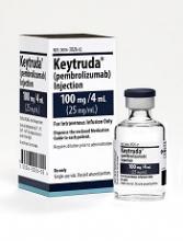
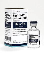
The US Food and Drug Administration (FDA) has granted accelerated approval for pembrolizumab (Keytruda) as a treatment for adult and pediatric patients with relapsed or refractory classical Hodgkin lymphoma (cHL).
Pembrolizumab is a monoclonal antibody that binds to the PD-1 receptor and blocks its interaction with PD-L1 and PD-L2, releasing PD-1 pathway-mediated inhibition of the immune response, including the antitumor immune response.
The drug, which is being developed by Merck, previously received FDA approval as a treatment for melanoma, lung cancer, and head and neck cancer.
Now, pembrolizumab has received accelerated approval to treat adult and pediatric patients with refractory cHL or those with cHL who have relapsed after 3 or more prior lines of therapy.
The accelerated approval was based on tumor response rate and durability of response. Continued approval of pembrolizumab for cHL patients may be contingent upon the verification and description of clinical benefit in confirmatory trials.
In adults with cHL, pembrolizumab is administered at a fixed dose of 200 mg every 3 weeks until disease progression or unacceptable toxicity, or up to 24 months in patients without disease progression.
In pediatric patients with cHL, pembrolizumab is administered at a dose of 2 mg/kg (up to a maximum of 200 mg) every 3 weeks until disease progression or unacceptable toxicity, or up to 24 months in patients without disease progression.
Pembrolizumab trials
The FDA’s approval of pembrolizumab in adults with cHL is based on data from the phase 2 KEYNOTE-087 trial. (The following data were provided by Merck.)
The trial enrolled 210 patients who received pembrolizumab at a dose of 200 mg every 3 weeks until unacceptable toxicity or documented disease progression, or for up to 24 months in patients who did not progress.
Fifty-eight percent of patients were refractory to their last prior therapy, including 35% with primary refractory disease and 14% whose disease was refractory to all prior regimens.
Sixty-one percent of patients had undergone prior autologous hematopoietic stem cell transplant, 83% had prior brentuximab use, and 36% had prior radiation therapy.
At a median follow-up of 9.4 months, the overall response rate was 69%, and the complete response rate was 22%. The median duration of response was 11.1 months (range, 0.0+ to 11.1 months).
Five percent of patients discontinued pembrolizumab due to adverse events (AEs), and 26% had dose interruptions due to AEs. Fifteen percent of patients had an AE requiring systemic corticosteroid therapy.
The most common AEs (occurring in ≥20% of patients) were fatigue (26%), pyrexia (24%), cough (24%), musculoskeletal pain (21%), diarrhea (20%), and rash (20%).
Serious AEs occurred in 16% of patients. The most frequent serious AEs (≥1%) were pneumonia, pneumonitis, pyrexia, dyspnea, graft-vs-host disease, and herpes zoster.
Two patients died from causes other than disease progression. One death was a result of graft-vs-host disease after subsequent allogeneic transplant, and the other was from septic shock.
There is limited experience with pembrolizumab in pediatric patients. The efficacy of the drug for pediatric patients was extrapolated from the results in the adult cHL population.
However, there is safety data on pembrolizumab in pediatric patients enrolled in the phase 1/2 KEYNOTE-051 trial. (These data were also provided by Merck.)
The trial included 40 pediatric patients with advanced melanoma or PD-L1–positive advanced, relapsed, or refractory solid tumors or lymphoma. Patients in this trial received pembrolizumab for a median of 43 days (range, 1-414 days).
The safety profile in these patients was similar to the profile in adults. Toxicities that occurred at a higher rate (≥15% difference) in pediatric patients than in adults under age 65 were fatigue (45%), vomiting (38%), abdominal pain (28%), hypertransaminasemia (28%), and hyponatremia (18%).
HDAC6 inhibitors reverse CIPN in rodents
Histone deacetylase (HDAC) inhibitors can treat chemotherapy-induced peripheral neuropathy (CIPN) in mice and rats, according to research published in PAIN.
The animals developed CIPN after treatment with cisplatin or paclitaxel.
But treatment with an HDAC6 inhibitor—ACY-1083 or ricolinostat (ACY-1215)—was able to reverse and sometimes prevent the symptoms of CIPN in the animals.
The researchers said these findings are “especially promising” because one of the HDAC6 inhibitors, ricolinostat, is already being tested in clinical trials.
This research was funded, in part, by Acetylon Pharmaceuticals Inc., and employees of the company were involved in the research.
The HDAC6 inhibitors are being developed by Regenacy Pharmaceuticals, LLC, a spinout of Acetylon Pharmaceuticals.
The researchers found that ACY-1083 was able to reverse cisplatin-induced mechanical allodynia (pain in response to touch) in mice.
ACY-1083 also prevented mechanical allodynia when given an hour prior to treatment with cisplatin.
The team noted that ricolinostat, which is less selective for HDAC6 than ACY-1083, also reversed cisplatin-induced mechanical allodynia.
The beneficial effect of ricolinostat was still seen a week after treatment, which was the last time point tested.
The researchers also found that ACY-1083 was able to reverse paclitaxel-induced mechanical allodynia in rats.
And ACY-1083 could reverse cisplatin-induced spontaneous pain and numbness in mice.
“These data demonstrate that HDAC6 is a novel therapeutic target for the treatment of chemotherapy-induced neuropathies, for which there are currently no FDA-approved drugs,” said Matthew B. Jarpe, PhD, associate vice-president of biology at Regenacy Pharmaceuticals.
Dr Jarpe said this research suggests an HDAC6 inhibitor could potentially be used in conjunction with platinum- or taxane-based chemotherapy to allow for higher and/or longer chemotherapy dosing regimens.
“Additionally, the reversal of both pain and numbness in this preclinical model suggests that an HDAC6 inhibitor could be used after a course of chemotherapy is completed to treat and potentially reverse resultant debilitating CIPN symptoms that impact function and quality of life for many patients,” he said.
“Preclinical results seen with the clinical candidate ricolinostat highlight the translational potential of these findings to the clinic.”
Family history impacts risk of second cancer after HL
A new study suggests Hodgkin lymphoma (HL) survivors have a high risk of developing a second malignancy, particularly if they have a family history of that malignancy.
The research showed that HL survivors in Sweden were roughly 2.4 times more likely than individuals in the country’s general population to develop a second cancer.
The risk for HL survivors remained high 30 years after treatment, and the risk was even greater in HL survivors who had a family history of specific cancers.
“The vast majority of patients with Hodgkin lymphoma are cured with a combination of chemotherapy and radiotherapy,” said study author Amit Sud, MBChB, of The Institute of Cancer Research, London in the UK.
“Our research has shown that these patients are at substantially increased risk of a second cancer later in life and particularly if they have a family history of cancer.”
Dr Sud and his colleagues described this research in the Journal of Clinical Oncology.
The team analyzed data from the Swedish Family-Cancer Project Database. They identified 9522 HL patients diagnosed between 1965 and 2013. During a median follow-up of 12.6 years, there were 1215 second cancers in 1121 HL patients (12%).
Compared to the general population, the HL patients had a significantly higher risk of all second malignancies, with a standardized incident ratio (SIR) of 2.39 and an absolute excess risk of 71.2 cases per 10,000 person-years.
Cancer types
HL patients had a significantly increased risk of several malignancies. The overall SIRs were as follows:
- NHL—7.99
- Leukemia—6.46
- Connective tissue cancer—5.73
- Thyroid cancer—5.13
- Squamous cell carcinoma—4.44
- Lung cancer—3.61
- Pharyngeal cancer—3.52
- Esophageal cancer—2.62
- Brain cancer—2.58
- Breast cancer—2.52
- Colon cancer—2.21
- Pancreatic cancer—2.09
- Melanoma—2.08
- Colorectal cancer—1.85
- Stomach cancer—1.78
- Bladder cancer—1.57
- Prostate cancer—1.21.
The researchers calculated SIRs over time and found the risk for many of the cancers remained high over 30 years following HL treatment.
Family history
The researchers identified 28,277 first-degree relatives of the HL survivors. Thirty percent of HL survivors (n=2785) had 1 or more first-degree relatives with a family history of cancer.
The SIR for cancers was 1.02 in the relatives. The SIR for second cancers was 2.83 for HL survivors who had first-degree relatives with cancer and 2.16 for HL survivors who did not have any first-degree relatives with cancer.
The researchers said the increased risk of second malignancy was correlated with the number of first-degree relatives with cancer.
The SIR was 2.67 for HL patients who had a single first-degree relative with cancer and 3.40 for HL patients who had 2 or more first-degree relatives with cancer.
The SIRs for different cancer types (for HL patients with at least 1 first-degree relative with cancer and no first-degree relatives with cancer, respectively) were as follows:
- NHL—14.43 vs 7.83
- Leukemia—14.31 vs 6.37
- Squamous cell carcinoma—10.85 vs 4.30
- Lung cancer—11.24 vs 3.39
- Breast cancer—4.36 vs 2.36
- Colorectal cancer—3.71 vs 1.76.
Sex and age
The researchers found significant differences in the SIRs for second cancers between HL patients diagnosed before the age of 35 and those diagnosed after age 35.
For men, the SIRs were:
- All cancers—4.26 for <35, 2.08 for ≥ 35
- Colorectal cancer—4.07 for < 35, 1.73 for ≥35
- Lung cancer—6.16 for < 35, 3.20 for ≥35
- Breast cancer—12.60 for < 35, 4.58 for ≥35
- Squamous cell carcinoma—5.89 for < 35, 3.96 for ≥35
- NHL—15.9 for < 35, 6.93 for ≥35
- Leukemia—12.15 for < 35, 5.57 for ≥35.
For women, the SIRs were:
- All cancers—4.61 for <35, 1.73 for ≥ 35
- Colorectal cancer—1.31 for < 35, 1.65 for ≥35
- Lung cancer—8.84 for < 35, 2.50 for ≥35
- Breast cancer—6.00 for < 35, 1.14 for ≥35
- Squamous cell carcinoma—6.37 for < 35, 4.87 for ≥35
- NHL—6.23 for < 35, 6.55 for ≥35
- Leukemia—10.36 for < 35, 4.51 for ≥35.
“Younger women who have been treated with radiotherapy to the chest for Hodgkin lymphoma are already screened for breast cancer, but our study suggests that we should be looking at ways of monitoring survivors for other forms of cancer too, and potentially offering preventative interventions,” Dr Sud said.
“After patients are cured, they no longer encounter oncologists, so it’s important that other healthcare providers are aware of the increased risk to Hodgkin lymphoma survivors to improve early diagnosis of second cancers.” ![]()


