User login
Study reveals higher rate of early death in kids with cancer
New research suggests that, in the US, early deaths from childhood cancer may be more common than clinical trials suggest.
Researchers found the rate of death within 1 month of diagnosis was higher among patients included in a large national database than among patients enrolled in phase 3 trials.
The data also indicated that early death is more likely in cancer patients under the age of 1 and those belonging to minority racial and ethnic groups.
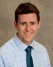
Adam Green, MD, of the University of Colorado Anschutz Medical Campus in Aurora, and his colleagues conducted this research and reported the results in the Journal of Clinical Oncology.
The researchers analyzed data from the Surveillance, Epidemiology and End Results (SEER) database, which collects about 15% of all cancer outcomes across the US (representing a geographic and socioeconomic cross-section).
The team identified 36,337 patients with pediatric cancer (ages 0 to 19) diagnosed between 1992 and 2011. Of these patients, 555 (1.5%) died within 1 month of diagnosis.
Young age
Overall, the strongest predictor of early death was age below 1 year. The odds ratio (OR) was 4.36 for these patients (P<0.001), 0.75 for patients age 1 to 4, 0.78 for patients age 5 to 9, 1.00 (reference) for those age 10 to 14, and 1.69 for those age 15 to 19.
For hematologic malignancies, the adjusted OR (adjusted by poverty, unemployment, education, and year of diagnosis) was 4.32 for patients younger than 1 (P<0.001), 0.76 for patients age 1 to 4, 0.79 for patients age 5 to 9, 1.00 for those age 10 to 14, and 1.76 for those age 15 to 19 (P=0.002).
“In general, babies are just challenging, clinically, because they can’t tell you what they’re feeling,” Dr Green noted. “Parents and physicians have to pick the ones with cancer from the ones with a cold, without the patient being able to tell you about symptoms that could be diagnostic.”
“Babies tend to get aggressive cancers, it’s hard to tell when they’re getting sick, and some are even born with cancers that have already progressed. These factors combine to make very young age the strongest predictor of early death in our study.”
Race/ethnicity
Black race and Hispanic ethnicity also predicted early death. The OR was 1.48 for black race, 1.00 for white race (reference), and 1.09 for “other” races (P=0.102). The OR was 1.39 for Hispanic patients and 1.00 (reference) for non-Hispanic patients (P=0.007).
For hematologic malignancies, the adjusted OR was 1.68 for black patients (P=0.01) and 1.44 for patients of other, non-white races. The adjusted OR was 1.48 for Hispanic patients (P=0.009).
Dr Green said he hopes future studies will be able to determine the factors responsible for these disparities.
Higher death rates
Dr Green and his colleagues also found the rate of early death due to pediatric cancers is higher than reported in clinical trials, and this was true for all cancer subtypes assessed.
“Most of what we know about outcomes for cancer patients come from clinical trials, which have much more thorough reporting rules than cancer treated outside trials,” Dr Green said. “However, these kids in our study aren’t surviving long enough to join clinical trials.”
The researchers looked at a phase 3 trial (COG AAML0531) of pediatric patients with acute myeloid leukemia (AML) who were studied from August 2006 to June 2010. The early death rate in this trial was 1.6% (16/1022).
In contrast, the SEER database showed an early death rate for pediatric AML patients of 5.9% (15/256) during the same period as the trial and 6.2% (106/1698) for the period from 1992 to 2011. This is almost 4 times as high as the death rate in the trial.
The same effect was observed for acute lymphoblastic leukemia (ALL).
The early death rate for non-infant ALL was 0.7% (13/1790) in a trial (POG 9900) conducted from April 2000 to April 2005, 1.6% (30/1823) in the SEER data covering the same time period, and 1.3% (94/7353) in the SEER data from 1992 to 2011.
For infant ALL, the rates were 2.0% (3/149) in a trial (COG AALL0631) conducted from January 2008 to June 2014, 5.0% (2/40) in the SEER data during the trial period, and 5.4% (12/223) in the SEER data from 1992 to 2011.
“I had a hunch this was a bigger problem than we thought,” Dr Green said. “Now we see that is indeed the case.” ![]()
Gene therapy proves effective in SCD patient
Researchers have reported a favorable outcome in the first patient with severe sickle cell disease (SCD) to receive gene therapy in the HGB-205 study.
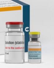
The subject, known as Patient 1204, was treated with LentiGlobin BB305, a product consisting of his own manipulated hematopoietic stem cells (HSCs).
A functional human β-globin gene was inserted into the patient’s HSCs ex vivo, and the cells were returned to him via transplant.
Fifteen months after receiving this treatment, Patient 1204 had high levels of anti-sickling hemoglobin (HbAT87Q), and there were no adverse events thought to be related to LentiGlobin BB305.
These results were published in NEJM. The research was supported by bluebird bio, the company developing LentiGlobin BB305.
Patient 1204 is a male with βS/βS genotype. In May 2014, at 13 years of age, the patient was enrolled in the HGB-205 study at Hôpital Necker-Enfants Malades in Paris, France.
The patient had received hydroxyurea from age 2 to 9 and had both a cholecystectomy and a splenectomy. He received regular transfusions (plus iron chelation with deferasirox) for 4 years prior to this study.
The patient had an average of 1.6 SCD-related events annually in the 9 years prior to starting transfusions. His complications from SCD included vaso-occlusive crises, acute-chest syndrome, bilateral hip osteonecrosis, and cerebral vasculopathy.
The patient underwent 2 bone marrow harvests to collect HSCs for gene transfer and back-up (6.2×10⁸ and 5.4×10⁸ total nucleated cells/kg harvested).
CD34+ cells were enriched from the harvested marrow and then transduced with LentiGlobin BB305 lentiviral vector.
The patient underwent myeloablation with intravenous busulfan (2.3 to 4.8 mg/kg per day for 4 days) with daily pharmacokinetic studies and dose adjustment. Total busulfan area under the curve was 19,363 μmol/min.
After a 2-day washout, the patient received LentiGlobin BB305 in October 2014 at a post-thaw total dose of 5.6×10⁶ CD34+ cells/kg. Neutrophil and platelet engraftment were achieved on day 38 and day 91 post-transplant, respectively.
Red blood cell transfusions were to be continued after transplant until a sufficient proportion of HbAT87Q (25% to 30% of total hemoglobin) was detected. Transfusions were discontinued after day 88 post-transplant.
HbAT87Q reached 5.5 g/dL (46% of total hemoglobin) at month 9 and continued to increase to 5.7 g/dL at month 15 (48%). Hemoglobin S levels were 5.5 g/dL (46%) at month 9 and 5.8 g/dL (49%) at month 15.
Total hemoglobin levels were stable, between 10.6 and 12.0 g/dL, from months 6 to 15. Fetal hemoglobin levels remained below 1.0 g/dL.
No adverse events related to LentiGlobin BB305 were reported. There were, however, adverse events related to busulfan conditioning (grade 3 anemia, thrombocytopenia, and infection; grade 4 neutropenia).
During 15 months of follow-up, there were no SCD-related clinical events or hospitalizations. The patient was able to stop all medications, including pain medication.
The patient resumed regular school attendance and reported full participation in normal physical activities.
“We have managed this patient at Necker for more than 10 years, and standard treatments were not able to control his SCD symptoms,” said Marina Cavazzana, MD, PhD, of Hôpital Necker-Enfants Malades.
“He had to receive blood transfusions every month to prevent severe pain crises. Since receiving the autologous stem cell transplant with LentiGlobin, he has been free from severe symptoms and has resumed normal activities, without the need for further transfusions.” ![]()
Hemophilia B therapy granted access to PRIME program
SPK-9001, an investigational therapy intended to treat hemophilia B, has been granted access to the European Medicines Agency’s (EMA) PRIority MEdicines (PRIME) program.
The goal of the EMA’s PRIME program is to accelerate the development of therapies that may offer a major advantage over existing treatments or benefit patients with no treatment options.
Through PRIME, the EMA offers early and enhanced support to developers in order to optimize development plans and speed regulatory evaluations to potentially bring therapies to patients more quickly.
To be accepted for PRIME, a therapy must demonstrate the potential to benefit patients with unmet medical need through early clinical or nonclinical data.
About SPK-9001
SPK-9001 is a bio-engineered adeno-associated virus capsid expressing a codon-optimized, high-activity human factor IX variant enabling endogenous production of factor IX. The therapy is being developed by Spark Therapeutics and Pfizer Inc.
SPK-9001 is under investigation in a phase 1/2 trial. Results from this trial were presented at the 2016 ASH Annual Meeting.
The presentation included data on 9 patients who received a single intravenous infusion of SPK-9001 at 5 x 10¹¹ vg/kg. The patients were followed for 52 weeks, with a long-term follow-up of 14 years.
Before their SPK-9001 infusion, the patients required anywhere from 0 to 159 factor IX infusions each year. Three patients had annualized bleeding rates (ABRs) of 0, and the remaining 6 patients had ABRs of 1, 2, 4, 10, 12, and 49.
After SPK-9001, 8 patients were able to stop receiving factor IX infusions and achieved an ABR of 0. One patient had an ABR of 2 and required on-demand factor IX therapy. (All patients stopped receiving factor IX prophylaxis).
The researchers said there were no unexpected vector or procedure-related adverse events, and none of the patients developed factor IX alloinhibitory antibodies. ![]()
Combo prevents GVHD, prolongs survival in monkeys
ORLANDO, FL—A 2-drug combination is “an exceptional candidate for clinical translation” as prophylaxis for graft-vs-host disease (GVHD), according to a presenter at the 2017 BMT Tandem Meetings.
The combination consists of sirolimus and KY1005, a monoclonal antibody that binds to OX40L and stops it from activating OX40, a protein that induces prolonged responses in T cells.
Experiments in rhesus macaques showed that KY1005 alone can have a modest effect on GVHD, but the combination of KY1005 and sirolimus can provide long-term, GVHD-free survival.
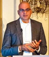
Victor Tkachev, PhD, of Seattle Children’s Research Institute in Washington, presented these results as one of the “Best Abstracts” at the recent BMT Tandem Meetings (abstract 3). This research was supported by Kymab, the company developing KY1005.
Dr Tkachev and his colleagues tested KY1005 alone and in combination with sirolimus in a previously described model of GVHD. In this model, rhesus macaques that do not receive prophylaxis develop severe GVHD after haploidentical hematopoietic stem cell transplant (HSCT).
For the current study, the animals received no prophylaxis, KY1005 alone, sirolimus alone, or KY1005 plus sirolimus.
When compared to no prophylaxis, KY1005 delayed the progression of acute GVHD and significantly prolonged the survival of HSCT recipients. However, all KY1005-treated animals eventually developed lethal GVHD.
Dr Tkachev noted that KY1005 provided partial control of T-cell activation, decreasing CD4 T-cell proliferation but having no significant effect on CD8 T-cell expansion.
As with KY1005 alone, sirolimus alone delayed GVHD progression and prolonged survival when compared to no GVHD prophylaxis.
However, all animals treated with sirolimus monotherapy eventually developed GVHD and died, and sirolimus alone wasn’t able to control T-cell proliferation.
On the other hand, the combination of KY1005 and sirolimus provided long-term, GVHD-free survival. All of the animals that received this combination survived, without developing GVHD, through day 100 after HSCT.
Dr Tkachev noted that, when given together, KY1005 and sirolimus synergistically controlled both CD4 and CD8 T-cell proliferation. However, this effect did not result in a lack of engraftment. In fact, animals that received the combination “displayed robust hematopoietic reconstitution” and maintained a high number of donor T cells.
Further investigation revealed that combination treatment with KY1005 and sirolimus preserves the reconstitution of regulatory T cells after HSCT and prevents both Th1- and Th17-driven alloimmunity.
Dr Tkachev and his colleagues also found that KY1005 plus sirolimus demonstrates an “unprecedented capacity” to protect against acute GVHD. Results with this combination were superior to those observed with tacrolimus and methotrexate in combination as well as abatacept and sirolimus in combination.
“Taken together, these data suggest that combined prophylaxis with KY1005 plus sirolimus represents an exceptional candidate for clinical translation,” Dr Tkachev concluded.
Kymab has said it will begin testing KY1005 in clinical trials this year. ![]()
Study sheds light on genetic landscape of HSTL
SAN FRANCISCO—Researchers say they have identified new driver genes and oncogenic pathways in hepatosplenic T-cell lymphoma (HSTL).
The team found that SETD2, a known tumor suppressor, was the most frequently silenced gene in HSTL.
The researchers also found evidence suggesting the JAK-STAT and PI3K pathways could be therapeutic targets in HSTL.
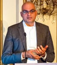
Sandeep Dave, MD, of Duke University in Durham, North Carolina, presented these findings at the 9th Annual T-cell Lymphoma Forum. Results from this research were also published in Cancer Discovery.
The researchers collected complete clinical data on 68 HSTL cases, including 20 with normal DNA. The team performed whole-genome sequencing, bioinformatics analysis, and biological characterization of these cases.
“This is the largest group of HSTL cases ever described, and the data implicate new driver genes and oncogenic pathways for the first time in HSTL,” Dr Dave said.
The data revealed that the most commonly mutated group of genes in HSTL are chromatin modifiers (SETD2, INO80, ARID1B, TET3, and SMARCA2) and signaling pathway genes (STAT5B, STAT3, and PIK3CD). Among these, STAT3, PIK3CD, and SETD2 showed the highest proportion of clonal events.
On the other hand, mutations in EZH2, KRAS, and TP53 were less frequently observed.
Common genetic abnormalities in HSTL include copy number alterations in chromosome 7, trisomy 8, loss of 10p, and gain of 1q.
A comparison of the frequencies of recurrently mutated genes in HSTL with other lymphomas demonstrated the genetically distinct profile of HSTL, wherein mutations in SETD2, INO80, TET3, and STAT5B occurred exclusively in HSTL.
SETD2, a histone lysine methyltransferase and a known tumor suppressor, was identified as the most frequently silenced gene in HSTL.
So the researchers investigated the biological effects of SETD2 loss in HSTL cells and a knockout mouse model.
While loss of SETD2 in HSTL cells resulted in increased cell proliferation, in vivo knockdown of SETD2 led to expansion of γ-δ T cells and a reduction in α-β T cells. A majority of HSTLs are known to arise predominantly from γ-δ T cells.
“These results implicate SETD2 in HSTL oncogenesis and T-cell development,” Dr Dave said.
He and his colleagues also found that constitutive activation of the JAK-STAT and PI3K pathways in HSTL cells was associated with increased proliferation, and inhibition of these pathways led to reduced survival of HSTL cells. This suggests that agents targeting these pathways might be effective in treating HSTL. ![]()
Blinatumomab bests chemo in rel/ref B-ALL
Blinatumomab proved more effective than chemotherapy in a phase 3 trial of adults with Ph-negative, relapsed/refractory B-cell precursor acute lymphoblastic leukemia (B-ALL).
Blinatumomab produced higher remission rates and nearly doubled overall survival (OS) when compared to standard care, which encompassed 4 different chemotherapy regimens (investigator’s choice).
The incidence of grade 3 or higher adverse events (AEs) was higher for patients who received chemotherapy, but the incidence of serious AEs was higher in the blinatumomab arm.
These results were published in NEJM. The trial, known as TOWER, was sponsored by Amgen, the company developing blinatumomab.
“Results from the TOWER study reinforce the potential of this single-agent, bispecific T-cell engager immunotherapy, which helped a higher percentage of patients achieve minimal residual disease response versus standard of care chemotherapy, highlighting the depth and quality of remissions achieved,” said study author Hagop M. Kantarjian, MD, of The University of Texas MD Anderson Cancer Center in Houston.
Treatment
TOWER enrolled 405 patients with Ph-negative, relapsed/refractory B-ALL, 376 of whom ultimately received treatment.
The patients received blinatumomab (n=267) or investigator’s choice of 1 of 4 protocol-defined standard chemotherapy regimens (n=109):
- FLAG (fludarabine, high-dose cytarabine arabinoside, and granulocyte-colony stimulating factor), with or without an anthracycline (n=49, 45%)
- A high-dose cytarabine arabinoside-based regimen (n=19, 17%)
- A high-dose methotrexate-based regimen (n=22, 20%)
- A clofarabine-based regimen (n=19, 17%).
Patients who received blinatumomab received it as a continuous infusion, 4 weeks on and 2 weeks off, at 9 µg/day for 7 days, then 28 µg/day on weeks 2-4. They received 2 cycles of induction, which was followed by 3 cycles of consolidation if they had ≤5% blasts.
If patients still had ≤5% blasts after consolidation, they received up to 12 months of blinatumomab maintenance. Maintenance was a continuous infusion, 4 weeks on and 8 weeks off, at 28 µg/day.
Patients in the blinatumomab arm received a median of 2 cycles of therapy (range, 1-9). And patients in the chemotherapy arm received a median of 1 cycle (range, 1 to 4).
Thirty-two percent of patients in the blinatumomab arm received consolidation, as did 3% of patients in the chemotherapy arm.
Patients
Patient characteristics were similar between the treatment arms. The mean age was 41 in both arms (range, 18-80). Nearly 60% of patients in both arms were male.
About 40% of patients in the blinatumomab arm and 50% in the chemotherapy arm had not received any prior salvage regimens.
Thirty-five percent of patients in the blinatumomab arm and 34% in the chemotherapy arm had an allogeneic hematopoietic stem cell transplant (allo-HSCT) prior to study enrollment. Seventeen percent and 20%, respectively, relapsed after HSCT.
Forty-two percent and 40%, respectively, were refractory to primary or salvage therapy. Twenty-eight percent of patients in both arms were in their first relapse, and their first remission had lasted less than 12 months. Twelve percent of patients in both arms had an untreated second or greater relapse.
Remission
Within 12 weeks of treatment initiation, complete remission (CR) rates were significantly higher in the blinatumomab arm than the chemotherapy arm. (This was in the intent-to-treat population, which included 271 patients in the blinatumomab arm and 134 patients in the chemotherapy arm.)
The rate of CR with full hematologic recovery was 34% in the blinatumomab arm and 16% in the chemotherapy arm (P<0.001). The rate of CR with full, partial, or incomplete hematologic recovery was 44% and 25%, respectively (P<0.001).
Among patients who achieved a CR with full, partial, or incomplete hematologic recovery, 76% of those in the blinatumomab arm and 48% of those in the chemotherapy arm were negative for minimal residual disease.
Survival
At a median follow-up of 11.7 months for the blinatumomab arm and 11.8 months for the chemotherapy arm, the OS was significantly longer in the blinatumomab arm.
The median OS was 7.7 months and 4.0 months, respectively (hazard ratio for death=0.71, P=0.01).
The improvement in OS with blinatumomab was consistent across subgroups, regardless of age, prior salvage therapy, or prior allo-HSCT.
The investigators also considered the effect that post-treatment allo-HSCT might have on OS. Sixty-five patients in the blinatumomab arm and 32 in the chemotherapy arm went on to receive an allo-HSCT (24% of patients in both arms).
When the investigators censored for post-treatment allo-HSCT, the median OS was 6.9 months in the blinatumomab arm and 3.9 months in the chemotherapy arm (hazard ratio=0.66, P=0.004).
Safety
Nearly all patients in both arms (99%) experienced AEs. Grade 3 or higher AEs occurred in 87% of patients in the blinatumomab arm and 92% of those in the chemotherapy arm. Serious AEs occurred in 62% and 45%, respectively.
Grade 3 or higher AEs of interest, according to the researchers, were infection (34% with blinatumomab and 52% with chemotherapy), neutropenia (38% and 58%, respectively), elevated liver enzymes (13% and 15%, respectively), neurologic events (9% and 8%, respectively), cytokine release syndrome (5% and 0%, respectively), infusion reactions (3% and 1%, respectively), and lymphopenia (2% and 4%, respectively).
Fatal AEs occurred in 19% of patients in the blinatumomab arm and 17% of those in the chemotherapy arm.
Fatal AEs that occurred in at least 1% of patients in either arm (blinatumomab and chemotherapy, respectively) were sepsis (3% and 4%), septic shock (2% and 0%), multiorgan failure (1% and 0%), respiratory failure (<1% and 2%), and bacteremia (0% and 2%).
Study advances precision opioid dosing for mucositis
ORLANDO, FL—A pilot study to determine the burden of mucositis in pediatric patients undergoing hematopoietic stem cell transplant (HSCT) showed that more than 50% of patients required a change in their opioids either for toxicity or lack of efficacy.
Investigators also observed that patients’ genotypes were associated with time to optimal pain control, although this needs to be defined more clearly in larger prospective studies.
“Pain from mucositis is a major problem during the early post-transplant period in pediatric patients,” said M. Christa Krupski, DO, of Cincinnati Children’s Hospital Medical Center in Ohio.
The pain frequently requires intravenous (IV) pain medication, but adequate pain management is often delayed by the trial and error of finding the right agent or the right dose, Dr Krupski added.
She and her colleagues tried to find predictors of mucositis that would help optimize pain control and minimize adverse effects of pain medication.
Dr Krupski presented the group’s findings at the 2017 BMT Tandem Meetings as abstract 50*.
Based on the investigators’ previous experience using a pain chip, they hypothesized that host genetic polymorphisms would predict perception of mucositis pain, opioid efficacy, and opioid-induced adverse effects in pediatric patients undergoing HSCT.
The pain chip was comprised of a panel of 46 single-nucleotide polymorphisms (SNPs) in a set of candidate genes known to influence opioid effect.
Study design
In this single-institution, retrospective pilot study, investigators genotyped 100 consecutive HSCT patients using pre-transplant samples.
The team collected demographic and transplant data, information on the utilization of pain medication, and mucositis data according to the standard CTCAE guidelines.
“And it must be noted,” Dr Krupski said, “that many of our patients required total parenteral nutrition during the transplant process, which automatically made them a grade 3 for mucositis.”
The investigators assessed pain using 2 scales, the Face, Legs, Activity, Cry, Consolability (FLACC) Scale, which is an objective measurement, and the more subjective Numeric Rating Scores (NRS).
Demographics
Patients were a median age of 9.9 years (range, 0.5–32.8), 65% were male, 87% were Caucasian, and 13% non-Caucasian.
The main indications for transplant were malignancy (45%), immune deficiency/dysregulation (30%), and bone marrow failure syndrome (19%).
More than two-thirds (68%) of patients had received a myeloablative conditioning regimen.
Results—mucositis
Seventy-six patients experienced mucositis, three-quarters of whom (78%) had received a myeloablative conditioning regimen.
The majority of patients (57%) had severe mucositis, which developed a median of 3 days after transplant (range, -2 to 17).
Regarding treatment, 13 (17%) had medication as needed or no medication, 5 (7%) had scheduled IV opioid, and 58 (76%) had patient-controlled analgesia (PCA).
For analysis purposes, the investigators grouped together the scheduled IV opioid and PCA treatment groups.
Results—opioid efficacy
The opioid efficacy analysis was based on 63 patients.
Time to optimal pain control was a median of 7 days (range, 0–22), and the morphine dose at the time of optimal pain control was a median of 1.5 mg/kg (range, 0.2–15.7).
“You will note, though, the wide inter-patient variability,” Dr Krupski pointed out, “with some of our patients immediately achieving optimal pain control the day the medication was started and others taking over 3 weeks to reach optimal pain control.”
Investigators observed similar inter-patient variability in morphine equivalent use at the time of optimal pain control and total morphine equivalent use.
The total time patients were on PCA was a median of 16 days (range, 1–32), and the median total morphine equivalent use was 0.99 mg/kg/day (range, 0.10–8.07).
“Most interesting, though, was that 18 patients, or nearly one-third of the patients requiring IV opioids, required a change in this opioid due to poor efficacy,” Dr Krupski said.
Results—opioid toxicity
Thirty-two (51%) patients experienced at least 1 adverse effect from their pain medication.
Specific toxicities, based on 32 patients, included pruritus (53%), sedation (16%), and nausea/vomiting (9%). Six patients (19%) had more than one adverse event.
“Similar to what we observed with respect to opioid efficacy,” Dr Krupski said, “another one-third of our patients with mucositis required a change in opioid due to toxicity.”
Results—impact of race
Non-Caucasians patients (n=13) had a significantly higher incidence of mucositis (100%) than Caucasians (n=87, 72%, P=0.03).
Non-Caucasian patients also experienced significantly more pain with mucositis (P=0.03), even though the severity of mucositis did not differ between the 2 groups.
The total equivalent dose of morphine used also did not differ between the groups.
“This raises the question of whether there are factors other than race that may be contributing to this difference,” Dr Krupski said.
Genetic findings
The UGT2B7 gene encodes the main enzyme metabolizer of morphine, and SNPs of this gene (rs7668258 and rs7439366) vary by race.
Non-Caucasian patients had significantly more wild-type SNPs than Caucasian patients (P=0.001). And patients with the wild-type UGT2B7 genotype spent more total days on IV opioids than patients with variant alleles (P=0.03).
On examination of rs4633, a SNP of the COMT gene, which is a key regulator of pain perception, the investigators observed some different findings from what had previously been reported.
There was no difference in mucositis severity between patients with the wild-type and variant allele (P=0.3).
However, patients with the variant allele required more days to optimal pain control than patients with the wild-type allele (P=0.04). This finding confirmed increased pain sensitivity associated with the genotype, irrespective of race.
“[I]f this association holds true in future studies,” Dr Krupski explained, “one may be more aggressive in the initial opioid titration to optimize pain control.”
Despite limitations of sample size, especially with respect to non-Caucasian patients, the pilot study showed association, but not causation, with respect to genetic variants.
“Racial differences affect mucositis pain perception and opioid requirement,” Dr Krupski said. “If genotyping is not feasible, it is important to pay particular attention to this difference while managing patients’ pain from mucositis.”
“We have an opportunity here to improve our care. Therefore, our plan is to validate these findings in additional patients before we use them to achieve our ultimate goal: precision dosing of opioids to individual patients.” ![]()
*Data in the abstract differ slightly from the presentation.
FDA approves new unit for clot-dissolving device
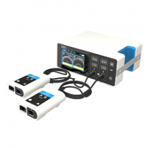
The US Food and Drug Administration (FDA) has granted 510(k) clearance for the EKOS® Control Unit 4.0, a new unit that can control the EKOS® System.
The EKOS System includes an ultrasonic device that uses acoustic pulses to enhance the effects of thrombolytic drugs and dissolve blood clots in patients with deep vein thrombosis, pulmonary embolism, and peripheral arterial occlusions.
With this system, acoustic pulses unwind and thin fibrin to expose drug receptor sites.
This allows thrombolytic drugs to reach deeper into clots, accelerating absorption and helping to dissolve clots faster and with less drug, according to BTG International LTD, the company that markets the EKOS System.
The EKOS Control Unit 4.0 replaces the EKOS System’s old control unit. The new unit can support 2 EKOS devices, allowing physicians to use a single control unit to treat both pulmonary arteries, with the goal of simplifying bilateral pulmonary embolism treatment.
The EKOS System (formerly known as the EkoSonic Endovascular System) was approved by the FDA in 2014.
In clinical studies, the system has been shown to speed time to clot dissolution, increase clot removal, and enhance clinical improvement compared to either standard catheter-directed drug therapy or thrombectomy.1,2
Research has also shown that the EKOS System requires significantly shorter treatment times and less thrombolytic compared to standard catheter-directed drug therapy3, 4,5, lowering the risk of bleeding and other complications.1,5,6 ![]()
1. Lin, P, et al, “Comparison of Percutaneous Ultrasound-Accelerated Thrombolysis versus Catheter-Directed Thrombolysis in Patients with Acute Massive Pulmonary Embolism.” Vascular, Vol. 17, Suppl. 3, 2009, S137–S147.
2. Schrijver, AM, et al, “Dutch Randomized Trial Comparing Standard Catheter-Directed Thrombolysis and Ultrasound-Accelerated Thrombolysis for Arterial Thromboembolic Infrainguinal Disease (DUET)." Journal of Endovascular Therapy 2015, Vol. 22(1):87-95.
3. Litzendorf, ME, et al, “Ultrasound-accelerated thrombolysis is superior to catheter-directed thrombolysis for the treatment of acute limb ischemia.” Journal of Vascular Surgery, Jun 2011; 53(Suppl S), p106S-107S.
4. Lin, P, et al, “Catheter-Directed Thrombectomy and Thrombolysis for Symptomatic Lower-Extremity Deep Vein Thrombosis: Review of Current Interventional Treatment Strategies.” Perspectives in Vascular Surgery and Endovascular Therapy, 2010, 22(3): 152–163.
5. Parikh, S, et al, “Ultrasound-Accelerated Thrombolysis for the Treatment of Deep Vein Thrombosis: Initial Clinical Experience.” Journal of Vascular and Interventional Radiology, Vol. 19, Issue 4, April 2008, 521–528.
6. Kucher, N, et al, “Randomized, Controlled Trial of Ultrasound-Assisted Catheter-Directed Thrombolysis for Acute Intermediate-Risk Pulmonary Embolism.” Circulation, Vol. 129, No. 4, 2014, 479–486.
The US Food and Drug Administration (FDA) has granted 510(k) clearance for the EKOS® Control Unit 4.0, a new unit that can control the EKOS® System.
The EKOS System includes an ultrasonic device that uses acoustic pulses to enhance the effects of thrombolytic drugs and dissolve blood clots in patients with deep vein thrombosis, pulmonary embolism, and peripheral arterial occlusions.
With this system, acoustic pulses unwind and thin fibrin to expose drug receptor sites.
This allows thrombolytic drugs to reach deeper into clots, accelerating absorption and helping to dissolve clots faster and with less drug, according to BTG International LTD, the company that markets the EKOS System.
The EKOS Control Unit 4.0 replaces the EKOS System’s old control unit. The new unit can support 2 EKOS devices, allowing physicians to use a single control unit to treat both pulmonary arteries, with the goal of simplifying bilateral pulmonary embolism treatment.
The EKOS System (formerly known as the EkoSonic Endovascular System) was approved by the FDA in 2014.
In clinical studies, the system has been shown to speed time to clot dissolution, increase clot removal, and enhance clinical improvement compared to either standard catheter-directed drug therapy or thrombectomy.1,2
Research has also shown that the EKOS System requires significantly shorter treatment times and less thrombolytic compared to standard catheter-directed drug therapy3, 4,5, lowering the risk of bleeding and other complications.1,5,6
1. Lin, P, et al, “Comparison of Percutaneous Ultrasound-Accelerated Thrombolysis versus Catheter-Directed Thrombolysis in Patients with Acute Massive Pulmonary Embolism.” Vascular, Vol. 17, Suppl. 3, 2009, S137–S147.
2. Schrijver, AM, et al, “Dutch Randomized Trial Comparing Standard Catheter-Directed Thrombolysis and Ultrasound-Accelerated Thrombolysis for Arterial Thromboembolic Infrainguinal Disease (DUET)." Journal of Endovascular Therapy 2015, Vol. 22(1):87-95.
3. Litzendorf, ME, et al, “Ultrasound-accelerated thrombolysis is superior to catheter-directed thrombolysis for the treatment of acute limb ischemia.” Journal of Vascular Surgery, Jun 2011; 53(Suppl S), p106S-107S.
4. Lin, P, et al, “Catheter-Directed Thrombectomy and Thrombolysis for Symptomatic Lower-Extremity Deep Vein Thrombosis: Review of Current Interventional Treatment Strategies.” Perspectives in Vascular Surgery and Endovascular Therapy, 2010, 22(3): 152–163.
5. Parikh, S, et al, “Ultrasound-Accelerated Thrombolysis for the Treatment of Deep Vein Thrombosis: Initial Clinical Experience.” Journal of Vascular and Interventional Radiology, Vol. 19, Issue 4, April 2008, 521–528.
6. Kucher, N, et al, “Randomized, Controlled Trial of Ultrasound-Assisted Catheter-Directed Thrombolysis for Acute Intermediate-Risk Pulmonary Embolism.” Circulation, Vol. 129, No. 4, 2014, 479–486.
CAR T-cell trial in adult ALL shut down
After 2 clinical holds in 2016 and 5 patient deaths, the Seattle biotech Juno Therapeutics is shutting down the phase 2 ROCKET trial of JCAR015.
The chimeric antigen receptor (CAR) T-cell therapy JCAR015 was being tested in adults with relapsed or refractory B-cell acute lymphoblastic leukemia (ALL).
“We have decided not to move forward . . . at this time,” CEO Hans Bishop said in a statement, “even though it generated important learnings for us and the immunotherapy field.”
He said the company remains “committed to developing better treatment for patients battling ALL.”
The first clinical hold of the ROCKET trial occurred in July after 2 patients died. The company attributed the deaths primarily to the addition of fludarabine to the regimen.
Juno removed fludarabine from the treatment protocol, the clinical hold was lifted, and the trial resumed.
Then, in November, 2 more patients died from cerebral edema, and the trial was put on hold once again.
One patient had died earlier in 2016, totaling 5 patient deaths from cerebral edema, although the earliest death was not necessarily related to treatment, the company stated.
Juno attributed the deaths to multiple factors, including the patients’ treatment history and treatment received at the beginning of the trial.
Juno plans to start a new adult ALL trial in 2018. The therapy, they say, is more similar to JCAR017, which is being tested in pediatric patients.
ROCKET is not the first trial of JCAR015 to be placed on hold.
In 2014, after 2 patients died of cytokine release syndrome, the phase 1 trial was placed on clinical hold.
Juno made changes to the enrollment criteria and dosing, and the hold was lifted. Results from this trial were presented at ASCO 2015 and ASCO 2016.
Exercise better than meds to reduce fatigue in cancer patients
Exercise and/or psychological therapy work better than medications to reduce cancer-related fatigue, according to research published in JAMA Oncology.
Researchers conducted a review and meta-analysis of more than 113 studies and found that exercise and psychological interventions, as well as a combination of both, were associated with reduced fatigue during and after cancer treatment.
However, pharmaceutical interventions were not associated with the same magnitude of improvement.
The researchers therefore concluded that exercise and psychological therapy should be recommended over medications.
“If a cancer patient is having trouble with fatigue, rather than looking for extra cups of coffee, a nap, or a pharmaceutical solution, consider a 15-minute walk,” said study author Karen Mustian, PhD, of the University of Rochester Medical Center in Rochester, New York.
“It’s a really simple concept, but it’s very hard for patients and the medical community to wrap their heads around it because these interventions have not been front-and-center in the past. Our research gives clinicians a valuable asset to alleviate cancer-related fatigue.”
Dr Mustian and her colleagues reached their conclusions after analyzing data from 113 randomized clinical trials testing various treatments for cancer-related fatigue.
There were 11,525 patients enrolled in these studies. Nearly half (46.9%) were women with breast cancer. Ten studies focused on other types of cancer and enrolled only men.
Dr Mustian and her colleagues performed a meta-analysis to establish and compare the mean weighted effect sizes (WESs) of the fatigue treatments.
The team found that exercise alone—whether aerobic or anaerobic—reduced cancer-related fatigue most significantly. The WES was 0.30 (95% CI, 0.25-0.36; P<0.001).
Psychological interventions—such as therapy designed to provide education, change personal behavior, and adapt the way a person thinks about his or her circumstances—also improved fatigue. The WES was 0.27 (95% CI, 0.21-0.330.30; P<0.001).
A combination of psychological interventions and exercise had a significant improvement on fatigue as well. The WES was 0.26 (95% CI, 0.13-0.38; P<0.001).
However, the drugs tested for treating cancer-related fatigue—paroxetine hydrochloride, modafinil, armodafinil, methylphenidate hydrochloride, dexymethylphenidate, dexamphetamine, and methylprednisolone—were not as effective as the other interventions. The WES was 0.09 (95% CI, 0.00-0.19; P=0.05).
“The literature bears out that these drugs don’t work very well, although they are continually prescribed,” Dr Mustian said. “Cancer patients already take a lot of medications, and they all come with risks and side effects. So any time you can subtract a pharmaceutical from the picture it usually benefits patients.”

