User login
Obesity linked to pain, fatigue in SLE
SAN DIEGO – A new study offers a double message about the potential impact of obesity on systemic lupus erythematosus (SLE) in women: Excess pounds are linked to a higher risk of patient-reported outcomes such as pain and fatigue, and body mass index may be an appropriate tool to study weight issues in this population.
Researchers found “a strong relationship between body composition and worse outcomes,” Sarah Patterson, MD, a fellow in rheumatology at the University of California, San Francisco, and the lead study author, said at the annual meeting of the American College of Rheumatology.
For the new study, Dr. Patterson and her colleagues analyzed findings from surveys of 148 participants in the Arthritis Body Composition and Disability study. All participants were women with a verified SLE diagnosis.
About two-thirds of the sample were white, 14% were Asian, and 13% were African American. The average age was 48 years, the average disease duration was 16 years, and 45% took glucocorticoids.
Researchers used two measurements of obesity: BMI of 30 kg/m2 or greater and fat mass index (FMI) of 13 kg/m2 or greater.
They calculated FMI with data collected via whole dual x-ray absorptiometry. Of the participants, 32% and 30% met criteria for obesity under FMI and BMI definitions, respectively.
Researchers also collected survey data regarding measurements of disease activity, depressive symptoms, pain and fatigue.
The study authors controlled their results to account for factors such as age, race, and prednisone use. They found that those defined as obese via FMI had more disease activity and depression than did nonobese women: 14.8 versus 11.5, P = .010, on the Systemic Lupus Activity Questionnaire scale, and 19.8 versus 13.1, P = .004, on the Center for Epidemiologic Studies Depression scale.
On two other scales of pain and fatigue, obese patients scored lower – a sign of worse status – compared with nonobese women: 38.7 versus 44.2, P = .004, on the Short Form 36 (SF-36) Health Survey pain subscale and 39.6 versus 45.2, P = .010, on the SF-36 vitality subscale. The researchers reported similar findings when using BMI to assess obesity.
It’s not clear why obesity and lupus may be linked, Dr. Patterson said, though she noted that inflammation is a shared factor. “People with lupus have arthritis and chronic pain, so there may be this vicious feedback cycle with hindrances to be able to live healthy lifestyles,” she added.
The study has limitations, including that the sample is largely white, while lupus is more common among minority women. In addition, the study does not include underweight patients or track patients over time. “It will be important to look at obesity and patient-reported outcomes to determine whether weight loss results in better outcomes,” Dr. Patterson said.
The study does provide an extra benefit by suggesting that BMI is not an inferior tool to measure the effects of obesity in the SLE population, Dr. Patterson said. BMI has been criticized as a misleading measurement of obesity. But the BMI and FMI measures produced similar results in this study. “That’s really good news in a way for the practicalities of using this information,” she said.
But FMI may still be a better measurement of obesity in the general population, where BMI may be more likely to be thrown off by high muscle mass.
It may seem obvious that obesity is linked to worse lupus outcomes, but rheumatologist Bryant England, MD, of the University of Nebraska, Omaha, said that this research is noteworthy because it highlights the importance of focusing on obesity in the clinic.
Rheumatologists shouldn’t leave obesity to primary care physicians but instead confront it themselves, said Dr. England, who moderated a discussion of new research at an ACR annual meeting press conference. But he cautioned that prudence is especially important when talking about obesity with lupus patients because they may be sensitive about medication-related weight gain.
Dr. Patterson and the other study authors reported having no relevant disclosures. Dr. England also reported no relevant disclosures. The study was funded by the National Institute of Arthritis and Musculoskeletal and Skin Diseases.
AT ACR 2017
Key clinical point:
Major finding: Obese women with SLE had more disease activity than did nonobese women (14.8 versus 11.5, P = .010).
Data source: An analysis of 148 SLE patients (65% white, mean age 48, about 31% obese) with obesity measured by body mass index or fat mass index.
Disclosures: The study authors reported having no relevant disclosures. The National Institute of Arthritis and Musculoskeletal and Skin Diseases funded the study.
Obesity linked to RA disease activity, disability
SAN DIEGO – In what may be the largest study of its kind, British researchers have linked obesity to significantly higher odds of rheumatoid arthritis disease activity and disability.
“This study emphasizes that obesity can have a profound effect on treatment goals in rheumatoid arthritis,” says Elena Nikiphorou, MD, of King’s College London, who presented the study findings at the annual meeting of the American College of Rheumatology. “Obesity reduced the odds of achieving remission or low disease activity by around 30%. And the odds of having disability were increased by 63%. This confirms what’s been shown in other, smaller studies.”

Despite obesity having been tied to decreased joint damage in established RA, Eric L. Matteson, MD, noted in an interview, that“the biomechanical effect of [being] overweight, especially on the weight-bearing joints” is one of the two “especially important” mechanisms explaining the link between RA and obesity. “The other is that fat cells produce inflammatory proteins, which contribute to the disease process and make it more difficult to treat,” said Dr. Matteson, a rheumatologist at the Mayo Clinic, Rochester, Minn.
“In my view the mechanical risk to the joint outweighs any possible ‘protective’ effect of RA,” Dr. Matteson added in an interview.
For the new study, Dr. Nikiphorou and colleagues compiled statistics from two consecutive United Kingdom RA inception cohorts. One tracked 1,465 patients for up to 25 years (median follow-up, 10 years), and the other tracked 1,236 patients for as many as 10 years (median follow-up, 6 years).
At baseline, 37.2% of 2,420 patients (90% of total) were overweight, and 21.3% were obese. Average body mass index (BMI) rose between the two consecutive studies from 25.5 to 27.6.
The researchers found that obesity was linked to lower likelihoods of remission and low disease activity status (odds ratio, 0.71; 95% confidence interval, 0.55-0.93 and OR, 0.69; 95% CI, 0.55-0.87, respectively.) After controlling for factors such as age and gender, they also saw slightly lower odds of remission in those with higher BMI (OR, 0.97; 95% CI, 0.95-0.99). But there was no statistically significant link between higher BMI and low disease activity status.
The study also connected obesity to higher odds of disability (OR, 1.63; 95% CI, 1.20-2.23). Furthermore, higher BMI was linked to higher odds of disability (OR, 1.04; 95% CI, 1.01-1.06).
Study lead author Dr. Nikiphorou, who spoke in an interview and in comments at an ACR press conference, said “this study emphasizes that obesity can have a profound effect on treatment goals in rheumatoid arthritis. It creates a strong case for addressing BMI and addressing obesity, flagging it up to primary care.”
She added that rheumatologists too often focus on only rheumatoid conditions. “We place so much evidence on disease activity scores,” she said. “How often do we really address the patient in terms of other things going on, including obesity? What we can do is include discussion of BMI, exercise, nutrition.”
Dr. Nikiphorou and Dr. Matteson report no relevant disclosures. No specific study funding is reported.
This article was updated 11/10/17.
SAN DIEGO – In what may be the largest study of its kind, British researchers have linked obesity to significantly higher odds of rheumatoid arthritis disease activity and disability.
“This study emphasizes that obesity can have a profound effect on treatment goals in rheumatoid arthritis,” says Elena Nikiphorou, MD, of King’s College London, who presented the study findings at the annual meeting of the American College of Rheumatology. “Obesity reduced the odds of achieving remission or low disease activity by around 30%. And the odds of having disability were increased by 63%. This confirms what’s been shown in other, smaller studies.”
Despite obesity having been tied to decreased joint damage in established RA, Eric L. Matteson, MD, noted in an interview, that“the biomechanical effect of [being] overweight, especially on the weight-bearing joints” is one of the two “especially important” mechanisms explaining the link between RA and obesity. “The other is that fat cells produce inflammatory proteins, which contribute to the disease process and make it more difficult to treat,” said Dr. Matteson, a rheumatologist at the Mayo Clinic, Rochester, Minn.
“In my view the mechanical risk to the joint outweighs any possible ‘protective’ effect of RA,” Dr. Matteson added in an interview.
For the new study, Dr. Nikiphorou and colleagues compiled statistics from two consecutive United Kingdom RA inception cohorts. One tracked 1,465 patients for up to 25 years (median follow-up, 10 years), and the other tracked 1,236 patients for as many as 10 years (median follow-up, 6 years).
At baseline, 37.2% of 2,420 patients (90% of total) were overweight, and 21.3% were obese. Average body mass index (BMI) rose between the two consecutive studies from 25.5 to 27.6.
The researchers found that obesity was linked to lower likelihoods of remission and low disease activity status (odds ratio, 0.71; 95% confidence interval, 0.55-0.93 and OR, 0.69; 95% CI, 0.55-0.87, respectively.) After controlling for factors such as age and gender, they also saw slightly lower odds of remission in those with higher BMI (OR, 0.97; 95% CI, 0.95-0.99). But there was no statistically significant link between higher BMI and low disease activity status.
The study also connected obesity to higher odds of disability (OR, 1.63; 95% CI, 1.20-2.23). Furthermore, higher BMI was linked to higher odds of disability (OR, 1.04; 95% CI, 1.01-1.06).
Study lead author Dr. Nikiphorou, who spoke in an interview and in comments at an ACR press conference, said “this study emphasizes that obesity can have a profound effect on treatment goals in rheumatoid arthritis. It creates a strong case for addressing BMI and addressing obesity, flagging it up to primary care.”
She added that rheumatologists too often focus on only rheumatoid conditions. “We place so much evidence on disease activity scores,” she said. “How often do we really address the patient in terms of other things going on, including obesity? What we can do is include discussion of BMI, exercise, nutrition.”
Dr. Nikiphorou and Dr. Matteson report no relevant disclosures. No specific study funding is reported.
This article was updated 11/10/17.
AT ACR 2017
Key clinical point: Obesity may worsen the risk of disease activity and disability in rheumatoid arthritis.
Major finding: In an adjusted analysis, obese patients with RA were less likely to reach remission and low disease activity status (OR, 0.71; 95% CI, 0.55-0.93 and OR, 0.69; 95% CI, 0.55-0.87, respectively).
Data source: Two consecutive inception cohorts with a total of 1,236 RA patients followed for up to 25 years.
Disclosures: The lead study author reports no disclosures, and no other disclosures are reported. No specific study funding is reported.
Inside the Las Vegas crisis: Surgeons answered the call
SAN DIEGO– Long before the horrific night of Oct. 1, the three trauma centers in the Las Vegas region were ready for a mass casualty event. It was understood among hospital leaders that the city could be the scene of a disaster that would demand a coordinated response from the city’s health care centers.
Then came the deadliest mass shooting in modern American history, and the extensive preparation turned out to have been well worth the time and effort, according to four trauma surgeons who spoke about the medical response to the massacre during a session at the annual clinical congress of the American College of Surgeons.

The killing spree was unusual in a variety of ways, including the fact that it occurred at a site “that’s almost strategically surrounded by trauma centers,” Dr. Fildes said.
UMC is Nevada’s only level I trauma center, while Sunrise is a level II. St. Rose Dominican, in the neighboring city of Henderson is a level III. Only one other Nevada hospital, in Reno, is a verified trauma center.
While the trauma centers received hundreds of patients, “every hospital in the valley saw patients from this event,” Dr. Fildes said. “There were 22,000 people on scene, and when the shooting started, they extricated themselves and went to safety by one means or another. Some drove home to their neighborhood and sought care there. Some drove until they found an acute care facility, whether it was a trauma center or not. Others were transported by Uber or taxi. The drivers knew where the trauma centers were, and decided where to go based on how the patients looked.”
According to Dr. Fildes, Las Vegas–area hospitals kept in touch with each other by phone, and UMC accepted some transfers from other hospitals. “We were ready for transfers,” he said, “and we expected more than we got.”
The trauma centers faced a variety of challenges from confusion and false reports to overcrowding and a media onslaught.
“We knew there was a strong possibility this would happen where we live, so we practiced this,” said Sean Dort, MD, medical director of the hospital’s trauma center. “We have talked and walked through it.”
Indeed, all hospitals in the Las Vegas area take part in regional disaster drills twice a year, and UMC runs other drills during the year such as an active shooter drill, Dr. Fildes said in an interview.
Together, the three hospitals treated hundreds of patients. Three weeks later, a handful were still inpatients.
In the aftermath, Las Vegas trauma surgeons are focusing on missed opportunities and lessons learned.
Dr. Fildes said more attention needs to be paid to how to handle situations when tides of patients bring themselves to the emergency department. “The issue of self-delivery has to be reconsidered, restudied,” he said, and he suggested that it may be a good idea to equip taxis with bleeding control kits.
He said his hospital heard from a doctor who’d treated patients during the Pulse nightclub massacre in Orlando last year. “One of their lessons learned was to position all gurneys and wheelchairs near the intake triage area,” he said. “We did that, and it improved the movement of patients to areas of the hospital that were matched to the intensity of care that they required.”
At Sunrise, the flood of unidentified patients overwhelmed the hospital’s trauma patient alias system, and some names were repeated. “In the future, I think a better naming system should be employed,” said trauma surgeon Matthew S. Johnson, MD.
To that end, he said, the hospital has begun examining how hurricanes are named.

And when it comes to planning, he said, there’s no room for excuses or resistance. “Everyone knew their role,” he said. “You can’t start figuring this out when it happens. You have to push people through it when they don’t want to do it, and they’re busy.”
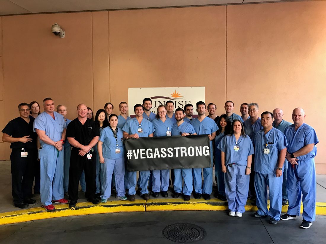
Dr. Fildes said that the UMC staff were physically and emotionally exhausted by the ordeal, but proud of what they were able to do for these patients, and that pride carried them through the experience. “We had support from all over the country; people sent banners with hundreds of signatures. Something like 1,100 pizzas were sent to the UMC staff, and dozens and dozens of surgeons from all over the country offered to come help us.”
Dr. Fildes noted that he is not easily surprised given his daily work, but he was impressed by the generosity and courage of the patients in this crisis situation.
He concluded that, “This was all made possible because of planning, training, commitment by staff and ultimately, the bravery of the patients.”
Dr. Dort, Dr. Fildes, Dr. Kuhls, and Dr. Johnson had no relevant financial disclosures.
I was at home and in bed with a book when my phone went off at 10:22 p.m. on that Sunday. It was a text message from one of my fellow residents who was on call at Sunrise: She wrote: “Mass casualty incident. Shooting on the Strip. You have to come now.”

There were multiple blood trails tracking from various parts of the ambulance bay into the ED. Medics were walking from bedside to bedside putting in lines. Two anesthesia attendings were frantically intubating patients. Two nurses were performing chest compressions.
I picked the nearest bed and started assessing patients. I placed 2 endotracheal tubes and black tagged 4 more patients within minutes of my arrival.
In the initial moments in the ER and in the OR, I focused on caring for the patient and blocked out any other thoughts or emotions. There was no time and no room for my horror or my tears.
As I went bedside to bedside in the ER, I was practically chanting in my head “airway, breathing, circulation, vital signs, other injuries.”
In the OR, I was working on controlling intra-abdominal bleeding from multiple sources, and again, my training became something of a mantra in my head. “Pack, control bleeding, assess injuries, repair.”
We saw well over 200 patients from the Route 91 shooting and operated on 95 of them within the first 24 hours.
Dylan Davey, MD, PhD, General Surgery Resident, PGY-4, Sunrise Hospital & Medical Center.
I was at home and in bed with a book when my phone went off at 10:22 p.m. on that Sunday. It was a text message from one of my fellow residents who was on call at Sunrise: She wrote: “Mass casualty incident. Shooting on the Strip. You have to come now.”
There were multiple blood trails tracking from various parts of the ambulance bay into the ED. Medics were walking from bedside to bedside putting in lines. Two anesthesia attendings were frantically intubating patients. Two nurses were performing chest compressions.
I picked the nearest bed and started assessing patients. I placed 2 endotracheal tubes and black tagged 4 more patients within minutes of my arrival.
In the initial moments in the ER and in the OR, I focused on caring for the patient and blocked out any other thoughts or emotions. There was no time and no room for my horror or my tears.
As I went bedside to bedside in the ER, I was practically chanting in my head “airway, breathing, circulation, vital signs, other injuries.”
In the OR, I was working on controlling intra-abdominal bleeding from multiple sources, and again, my training became something of a mantra in my head. “Pack, control bleeding, assess injuries, repair.”
We saw well over 200 patients from the Route 91 shooting and operated on 95 of them within the first 24 hours.
Dylan Davey, MD, PhD, General Surgery Resident, PGY-4, Sunrise Hospital & Medical Center.
SAN DIEGO– Long before the horrific night of Oct. 1, the three trauma centers in the Las Vegas region were ready for a mass casualty event. It was understood among hospital leaders that the city could be the scene of a disaster that would demand a coordinated response from the city’s health care centers.
Then came the deadliest mass shooting in modern American history, and the extensive preparation turned out to have been well worth the time and effort, according to four trauma surgeons who spoke about the medical response to the massacre during a session at the annual clinical congress of the American College of Surgeons.
The killing spree was unusual in a variety of ways, including the fact that it occurred at a site “that’s almost strategically surrounded by trauma centers,” Dr. Fildes said.
UMC is Nevada’s only level I trauma center, while Sunrise is a level II. St. Rose Dominican, in the neighboring city of Henderson is a level III. Only one other Nevada hospital, in Reno, is a verified trauma center.
While the trauma centers received hundreds of patients, “every hospital in the valley saw patients from this event,” Dr. Fildes said. “There were 22,000 people on scene, and when the shooting started, they extricated themselves and went to safety by one means or another. Some drove home to their neighborhood and sought care there. Some drove until they found an acute care facility, whether it was a trauma center or not. Others were transported by Uber or taxi. The drivers knew where the trauma centers were, and decided where to go based on how the patients looked.”
According to Dr. Fildes, Las Vegas–area hospitals kept in touch with each other by phone, and UMC accepted some transfers from other hospitals. “We were ready for transfers,” he said, “and we expected more than we got.”
The trauma centers faced a variety of challenges from confusion and false reports to overcrowding and a media onslaught.
“We knew there was a strong possibility this would happen where we live, so we practiced this,” said Sean Dort, MD, medical director of the hospital’s trauma center. “We have talked and walked through it.”
Indeed, all hospitals in the Las Vegas area take part in regional disaster drills twice a year, and UMC runs other drills during the year such as an active shooter drill, Dr. Fildes said in an interview.
Together, the three hospitals treated hundreds of patients. Three weeks later, a handful were still inpatients.
In the aftermath, Las Vegas trauma surgeons are focusing on missed opportunities and lessons learned.
Dr. Fildes said more attention needs to be paid to how to handle situations when tides of patients bring themselves to the emergency department. “The issue of self-delivery has to be reconsidered, restudied,” he said, and he suggested that it may be a good idea to equip taxis with bleeding control kits.
He said his hospital heard from a doctor who’d treated patients during the Pulse nightclub massacre in Orlando last year. “One of their lessons learned was to position all gurneys and wheelchairs near the intake triage area,” he said. “We did that, and it improved the movement of patients to areas of the hospital that were matched to the intensity of care that they required.”
At Sunrise, the flood of unidentified patients overwhelmed the hospital’s trauma patient alias system, and some names were repeated. “In the future, I think a better naming system should be employed,” said trauma surgeon Matthew S. Johnson, MD.
To that end, he said, the hospital has begun examining how hurricanes are named.
And when it comes to planning, he said, there’s no room for excuses or resistance. “Everyone knew their role,” he said. “You can’t start figuring this out when it happens. You have to push people through it when they don’t want to do it, and they’re busy.”
Dr. Fildes said that the UMC staff were physically and emotionally exhausted by the ordeal, but proud of what they were able to do for these patients, and that pride carried them through the experience. “We had support from all over the country; people sent banners with hundreds of signatures. Something like 1,100 pizzas were sent to the UMC staff, and dozens and dozens of surgeons from all over the country offered to come help us.”
Dr. Fildes noted that he is not easily surprised given his daily work, but he was impressed by the generosity and courage of the patients in this crisis situation.
He concluded that, “This was all made possible because of planning, training, commitment by staff and ultimately, the bravery of the patients.”
Dr. Dort, Dr. Fildes, Dr. Kuhls, and Dr. Johnson had no relevant financial disclosures.
AT THE ACS CLINICAL CONGRESS
Methotrexate holiday linked to better flu vaccine immunogenicity
SAN DIEGO – Patients with well-controlled rheumatoid arthritis (RA) fared well during a 2-week holiday from methotrexate after flu vaccination and later showed signs of boosted immunity against the flu in comparison with patients who had not stopped the drug, according to results from a randomized controlled trial.
The research doesn’t confirm that vaccinated patients who take a break from methotrexate actually have lower rates of flu. Still, the findings suggest that brief holidays from methotrexate could be feasible in a variety of situations, such as after vaccinations and prior to surgery, said Jin Kyun Park, MD, of Seoul (South Korea) National University Hospital, lead author of the study presented at the annual meeting of the American College of Rheumatology.
The study notes that RA patients are especially prone to infections for two reasons: dysfunctional immune systems and immunity-weakening treatments. According to Dr. Park, methotrexate reduces the effectiveness of flu vaccines by 15%-20%.
In a previous study, Dr. Park and his colleagues found no statistically significant sign of increased flares in patients who went without methotrexate for 2 weeks before and 2 weeks after vaccination, 4 weeks after vaccination, and 4 weeks before vaccination (Ann Rheum Dis. 2017 Sep;76[9]:1559-65).
The earlier findings also suggested that flu vaccine uptake is highest in those who stop methotrexate after vaccination.
For the new study, a randomized controlled trial, researchers recruited patients with well-controlled RA. They assigned 159 to continue weekly doses of methotrexate after flu vaccination and 161 to stop it for 2 weeks.
The groups in the final analysis (156 and 160 subjects, respectively) were similar – about 85% women, average age of 52-53 years, and about half took glucocorticoids. Their methotrexate dose per week was about 13 mg.
At 4 weeks, just over three-quarters of the patients who had briefly stopped methotrexate showed at least a fourfold increase in hemagglutination inhibition antibody titer against two or more vaccine strains. Of those who continued the medication, just 54.5% showed this level of response, which the researchers considered to be satisfactory.
The researchers reported that there was no appreciable increase in RA disease activity.
Dr. Park cautioned that vaccine titers don’t directly reflect immunoprotection levels. Patients who took a break from methotrexate were less likely to develop a flulike illness, but the difference wasn’t statistically significant.
The research raises questions about whether methotrexate could be stopped a week or two before surgery to lower the risk of infections, Dr. Park said.
Dr. Park said that future research should focus on whether stopping methotrexate briefly affects whether patients go on to develop the flu. He would also like to look at whether a break from the medication will boost the immune response in RA patients who get herpes zoster (shingles) vaccines.
Paul Sufka, MD, of HealthPartners and Regions Hospital in St. Paul, Minn., praised the research. The 2-week break from methotrexate is “a fairly pragmatic approach,” said Dr. Sufka, who moderated a press conference where Dr. Park presented his research.
“You can actually pull this off,” he said, versus telling patients to stop the medication for the 2 weeks before they get vaccinated. He cautioned, however, that “these people have a fairly low disease activity. You may not be able to pull this off with those who have high disease activity.”
Dr. Park and Dr. Sufka reported no relevant disclosures. A study author reported consulting for Pfizer and receiving research grants from Green Cross Corp. and Hanmi Pharmaceutical. The study was funded by Green Cross.
AT ACR 2017
Key clinical point:
Major finding: More than three-quarters of patients who had briefly stopped methotrexate and 54.5% of patients who kept using methotrexate showed at least a fourfold increase in hemagglutination inhibition antibody titer against two or more vaccine strains at 4 weeks.
Data source: A randomized controlled trial of 320 patients with RA who were taking methotrexate.
Disclosures: The study was funded by Green Cross Corp. The presenter reported no relevant disclosures. A study author reported consulting for Pfizer and receiving research grants from Green Cross and Hanmi Pharmaceutical.
New, persistent opioid use more common after bariatric surgery
SAN DIEGO – Bariatric patients are nearly 50% more likely than general surgery patients to start using opioids after their procedures and continue taking the painkillers for a year, a new study finds.
It’s not clear why bariatric patients are at higher risk of continued opioid use, nor whether they are more likely to become addicted. Still, bariatric patients are a target for “intervention, enhanced education, early referral to specialists, protocols minimizing inpatient and outpatient narcotics, opioid-free operations, system-based interventions and prescribing guidelines,” says study lead author Sanjay Mohanty, MD, a surgery resident with the Henry Ford Health System, who spoke in a presentation at the annual clinical congress of the American College of Surgeons.
There’s been little research into opioid use among bariatric patients, said Dr. Mohanty. In 2013, a retrospective study found that 8% of 11,719 bariatric patients were chronic opioid users, and more than three-quarters of those remained so after 1 year. However, that study was completed in 2010 before the height of the opioid epidemic (JAMA. 2013;310(13):1369-76).
More recently, a 2017 study found that opioid use among 1,892 bariatric patients who weren’t using at baseline grew from 5.8% at 6 months to 14.2% at 7 years. The study tracked patients until January 2015 (Surg Obes Relat Dis. 2017 Aug;13 (8):1337-46).

For the new study, researchers tracked 14,063 bariatric patients in the Michigan Bariatric Surgery Collaborative, a group of Michigan hospitals and health systems, from 2006-2017.
Of the patients, 73% were opioid-naive at baseline and 27% were users. At 1 year after procedure, overall use dropped slightly to 24%. However, 905 patients – 8.8% of the initial opioid-native group – were new and persistent opioid users.
According to Dr. Carlin, this is almost 50% higher than in patients after general surgical procedures.
These users were significantly more likely to be black (OR 1.67), less likely to have private insurance (0.76 OR), more likely to have income under $25,000 (OR 1.43), and more likely to have a mobility limitation (OR 1.78).
The researchers also found evidence linking a higher risk of new and persistent opioid use to lack of unemployment, depression, musculoskeletal disorders, tobacco use and gastric bypass procedures.
Why might bariatric patients in general be more susceptible to new and persistent opioid use? “We don’t know that answer,” Dr. Carlin said. “Maybe there’s some addiction transfer. Or maybe it’s something physiologic. We’re doing an operation on the gut, and that could have an impact on absorption.”
As for solutions, Dr. Carlin says “prescribe less, prescribe differently, be more patient-specific. We’re looking at different modalities to treat the pain such as nerve blocks during surgery, anti-inflammatories and muscle relaxants.”
And if patients aren’t using opioids in the hospital and not having that much pain, he said, physicians don’t send any pills home with them.
The next steps should include research into links between opioids and perioperative complications and surgical outcomes, the researchers suggested.
Dr. Carlin and Dr. Mohanty report no relevant disclosures.
SAN DIEGO – Bariatric patients are nearly 50% more likely than general surgery patients to start using opioids after their procedures and continue taking the painkillers for a year, a new study finds.
It’s not clear why bariatric patients are at higher risk of continued opioid use, nor whether they are more likely to become addicted. Still, bariatric patients are a target for “intervention, enhanced education, early referral to specialists, protocols minimizing inpatient and outpatient narcotics, opioid-free operations, system-based interventions and prescribing guidelines,” says study lead author Sanjay Mohanty, MD, a surgery resident with the Henry Ford Health System, who spoke in a presentation at the annual clinical congress of the American College of Surgeons.
There’s been little research into opioid use among bariatric patients, said Dr. Mohanty. In 2013, a retrospective study found that 8% of 11,719 bariatric patients were chronic opioid users, and more than three-quarters of those remained so after 1 year. However, that study was completed in 2010 before the height of the opioid epidemic (JAMA. 2013;310(13):1369-76).
More recently, a 2017 study found that opioid use among 1,892 bariatric patients who weren’t using at baseline grew from 5.8% at 6 months to 14.2% at 7 years. The study tracked patients until January 2015 (Surg Obes Relat Dis. 2017 Aug;13 (8):1337-46).
For the new study, researchers tracked 14,063 bariatric patients in the Michigan Bariatric Surgery Collaborative, a group of Michigan hospitals and health systems, from 2006-2017.
Of the patients, 73% were opioid-naive at baseline and 27% were users. At 1 year after procedure, overall use dropped slightly to 24%. However, 905 patients – 8.8% of the initial opioid-native group – were new and persistent opioid users.
According to Dr. Carlin, this is almost 50% higher than in patients after general surgical procedures.
These users were significantly more likely to be black (OR 1.67), less likely to have private insurance (0.76 OR), more likely to have income under $25,000 (OR 1.43), and more likely to have a mobility limitation (OR 1.78).
The researchers also found evidence linking a higher risk of new and persistent opioid use to lack of unemployment, depression, musculoskeletal disorders, tobacco use and gastric bypass procedures.
Why might bariatric patients in general be more susceptible to new and persistent opioid use? “We don’t know that answer,” Dr. Carlin said. “Maybe there’s some addiction transfer. Or maybe it’s something physiologic. We’re doing an operation on the gut, and that could have an impact on absorption.”
As for solutions, Dr. Carlin says “prescribe less, prescribe differently, be more patient-specific. We’re looking at different modalities to treat the pain such as nerve blocks during surgery, anti-inflammatories and muscle relaxants.”
And if patients aren’t using opioids in the hospital and not having that much pain, he said, physicians don’t send any pills home with them.
The next steps should include research into links between opioids and perioperative complications and surgical outcomes, the researchers suggested.
Dr. Carlin and Dr. Mohanty report no relevant disclosures.
AT THE ACS CLINICAL CONGRESS
Key clinical point: It’s common for opioid-naive bariatric patients to begin using opioids after surgery and continuing at 1 year.
Major finding: At 1 year after surgery, 8.8% of the 73% of bariatric patients who were opioid-naive were new and persistent opioid users.
Data source: 14,063 Michigan bariatric patients tracked from 2006-2017.
Disclosures: The study authors report no relevant disclosures.
Junior surgical trainees hew closer to surgery risk calculators than do faculty members
SAN DIEGO – Researchers say that they’ve developed an easy and inexpensive way to instantly track divergences in thinking by faculty and students as they ponder cases presented in Mortality and Morbidity (M&M) conferences. They’ve already produced an intriguing early finding: Interns and junior residents hew more closely than do their elders to estimates provided by a surgical risk calculator.
The research has the potential to shed light on problems in the much-maligned M&M, says study leader Ira Leeds, MD, of Johns Hopkins University, Baltimore. He presented the study findings at the annual Clinical Congress of the American College of Surgeons.
“This project demonstrates that educational technologies can reveal important gaps in surgical education,” said Dr. Leeds, who made comments during his presentation and in an interview.
At issue: The M&M conference, a mainstay of medical education. “This has been defined as the ‘golden hour’ of surgical education,” Dr. Leeds said. “By discussing someone else’s complications, you can learn how to handle your own in the future.”
However, he added, “there’s very little evidence that we’re currently learning from M&M.”
Dr. Leeds and his colleagues are studying the M&M’s role in medical education to see if it can be improved. The new study, a prospective time-series analysis of weekly M&M conferences, aims to understand the potential value of a real-time feedback system. The idea is to develop a way to alert participants to discrepancies in their perceptions about cases.
The researchers turned to a company called Poll Everywhere, whose technology allowed them to collect instant opinions about M&M cases from those in attendance. During 2016-2017, 110 faculty, residents, and interns used Poll Everywhere’s smartphone app to do two things – make guesses about the root causes of adverse events and estimate the risk of complications from surgical procedures over the next 30 days.
“We can see all the results streaming in real time,” said Dr. Leeds, noting that the service cost $600 per year.
The participants, about two-thirds of whom were male, included faculty (35%), fellows and senior residents (28%), and interns and junior residents (37%). They’d been trained an average of 9 years.
The 34 M&M cases represented a mixture of surgical specialties, including oncology, trauma, transplant, and others.
In terms of the root cause analysis, the technology allowed researchers to instantly detect if the guesses of faculty and students were far apart.
The researchers also compared the risk estimates from the participants to those provided by the NSQIP Risk Calculator. They found that the participants tended to boost their estimate of risk, compared with the calculator, by an absolute mean difference of 7.7 percentage points.
“They were overestimating risk by nearly 8 percentage points,” Dr. Leeds said. This isn’t surprising, since other research has revealed a trend toward overestimation of risk by physicians, compared with calculators, he added.
There wasn’t a major difference between the general level of higher estimation of risk among faculty and senior residents (mean of 8.6 and 7.2 percentage points higher than the calculator, respectively). But interns and junior residents estimated risk higher than the calculator by a mean of 4.9 percentage points.
What’s going on? Are the less experienced staff members outperforming their teachers? Another possibility, Dr. Leeds said, is that “the senior surgeons are better picking up on nuances that aren’t being captured by predictive models or the underdeveloped intuition of a junior trainee.”
Rachel Dawn Aufforth, MD, of Johns Hopkins Medicine, who served as discussant for the presentation by Dr. Leeds, said she looks forward to seeing if this technology can improve resident education. She also wondered why estimates via the risk calculator were chosen as a baseline, especially considering that surgeons tend to estimate higher levels of risk.
“One of the things we’ve been trying to do is look at time-series differences,” Dr. Leeds said. “Are they getting better over an academic year? And does that vary by faculty, especially for interns? The calculator isn’t changing or learning on its own.”
In the big picture, the study shows that “collecting real-time risk estimates and root cause assignment is feasible and can be performed as part of routine M&M conferences,” he said.
The study was funded in part by Johns Hopkins University School of Medicine Institute for Excellence in Education. Dr. Leeds reports no relevant disclosures.
AT THE ACS CLINICAL CONGRESS
Surgeons paid a price for presidential procedures
SAN DIEGO – A surgical team was forced to perform a delicate oral procedure on a rocking yacht while making sure to preserve presidential whiskers. A domineering doctor ignored fellow physicians while a president spent months dying in agony. And, after helping to save the leader of the free world, the leader of the American College of Surgeons found himself viciously attacked by his own colleagues.
When a quartet of ill U.S. presidents developed major medical problems, an audience at the annual clinical congress of the American College of Surgeons learned, their treating physicians ended up with major headaches of their own.
President Grover Cleveland, for example, required his surgical team to remove an oral tumor in total secrecy in 1893, robbing him of a big chunk of his upper palate. “The president had a mustache, and the mustache had to be left alone, and there could be no scars,” said the Hospital for Special Surgery’s J. Patrick O’Leary, MD, FACS, who spoke in a session focused on the history of presidential medicine.
The only light came from a single incandescent bulb, and the procedure was performed at sea, on a yacht anchored off Long Island, N.Y.
“If you were presented with these parameters as a surgeon today, my guess is that you would have demurred on taking on this project,” Dr. O’Leary said. “It was a prescription for a disaster.”
President Cleveland survived for another 15 years. James Garfield, a fellow Civil War veteran, wasn’t so fortunate. In 1881, he was astonishingly unlucky, the unwitting victim of a fumbling physician who dominated his care after an assassin shot him in the chest.
That physician, Willard Bliss, MD, dismissed other doctors who knew the president well and isolated this gregarious man from friends and family. He also ignored emerging knowledge about germ control. And he fed Garfield a heavy diet that the digestively sensitive president probably couldn’t have tolerated in the best of times. The result: endless vomiting, the loss of almost 80 pounds, and an unsuccessful rectal feeding regimen.
Toward the end of the president’s gruesome summer-long decline, Dr. Bliss told all but two doctors to stay away, John B. Hanks, MD, of the University of Virginia, Charlottesville, said in his presentation. Then the president died of a wound that Dr. Hanks said would have been survivable with proper care even in the 1880s.
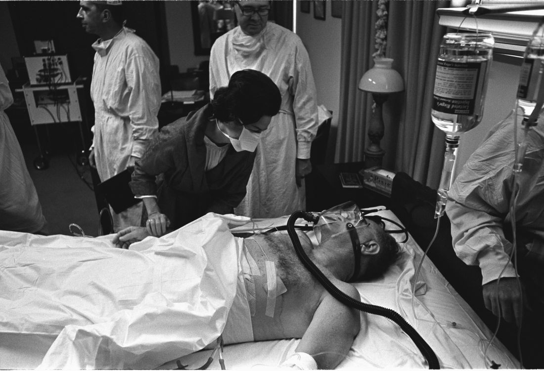
History has been unkind to Dr. Bliss, in part because his patient died. But another presidential physician faced bizarre post surgery scorn from his ACS colleagues, even though his patient lived, according to Justin Barr, MD, PhD, of Duke University, Durham, N.C.
In 1956, surgeon Isidor Ravdin, MD, of the University of Pennsylvania, Philadelphia, was called in when President Dwight D. Eisenhower needed surgery to eliminate a bowel obstruction.
A team of physicians agreed that the president needed surgery. “They felt they were dealing with an elderly, sick patient who’d been in shock during his illness and had recently suffered a myocardial infarction,” Dr. Barr said. “They unanimously decided to proceed with a bypass over resection.”
It’s clear today that the physicians made the correct choice, Dr. Barr said. But his colleagues attacked Dr. Ravdin, who later complained that criticisms multiplied in direct ratio to distance from the operating room.
At the time, Dr. Ravdin was chair of the ACS Board of Regents. The entire board accused him of violating college policies regarding “ghost surgery” (performing procedures without the patient’s knowledge) and “itinerant surgery” (traveling to perform a procedure and then leaving).
Dr. Ravdin acknowledged that he had performed itinerant surgery to some extent, but he denied the ghost surgery charge. In fact, he and the president became friends.
His colleagues also attacked him over his decision to not perform a resection procedure. “They were accusing him of not only being an unethical surgeon, but also an incompetent one,” said Dr. Barr, who calls the letters about the allegations “truly bewildering.”
Also bewildering: Lyndon B. Johnson’s choice to display his gallbladder surgery scar to the press in 1965, spawning one of the most infamous photos of his presidency.
Few surgeons see their handiwork so prominently displayed. Fortunately for them, the operating theater was in a naval hospital, not on a boat. And, as far as we know, no one fretted over the fate of a single facial hair.
SAN DIEGO – A surgical team was forced to perform a delicate oral procedure on a rocking yacht while making sure to preserve presidential whiskers. A domineering doctor ignored fellow physicians while a president spent months dying in agony. And, after helping to save the leader of the free world, the leader of the American College of Surgeons found himself viciously attacked by his own colleagues.
When a quartet of ill U.S. presidents developed major medical problems, an audience at the annual clinical congress of the American College of Surgeons learned, their treating physicians ended up with major headaches of their own.
President Grover Cleveland, for example, required his surgical team to remove an oral tumor in total secrecy in 1893, robbing him of a big chunk of his upper palate. “The president had a mustache, and the mustache had to be left alone, and there could be no scars,” said the Hospital for Special Surgery’s J. Patrick O’Leary, MD, FACS, who spoke in a session focused on the history of presidential medicine.
The only light came from a single incandescent bulb, and the procedure was performed at sea, on a yacht anchored off Long Island, N.Y.
“If you were presented with these parameters as a surgeon today, my guess is that you would have demurred on taking on this project,” Dr. O’Leary said. “It was a prescription for a disaster.”
President Cleveland survived for another 15 years. James Garfield, a fellow Civil War veteran, wasn’t so fortunate. In 1881, he was astonishingly unlucky, the unwitting victim of a fumbling physician who dominated his care after an assassin shot him in the chest.
That physician, Willard Bliss, MD, dismissed other doctors who knew the president well and isolated this gregarious man from friends and family. He also ignored emerging knowledge about germ control. And he fed Garfield a heavy diet that the digestively sensitive president probably couldn’t have tolerated in the best of times. The result: endless vomiting, the loss of almost 80 pounds, and an unsuccessful rectal feeding regimen.
Toward the end of the president’s gruesome summer-long decline, Dr. Bliss told all but two doctors to stay away, John B. Hanks, MD, of the University of Virginia, Charlottesville, said in his presentation. Then the president died of a wound that Dr. Hanks said would have been survivable with proper care even in the 1880s.
History has been unkind to Dr. Bliss, in part because his patient died. But another presidential physician faced bizarre post surgery scorn from his ACS colleagues, even though his patient lived, according to Justin Barr, MD, PhD, of Duke University, Durham, N.C.
In 1956, surgeon Isidor Ravdin, MD, of the University of Pennsylvania, Philadelphia, was called in when President Dwight D. Eisenhower needed surgery to eliminate a bowel obstruction.
A team of physicians agreed that the president needed surgery. “They felt they were dealing with an elderly, sick patient who’d been in shock during his illness and had recently suffered a myocardial infarction,” Dr. Barr said. “They unanimously decided to proceed with a bypass over resection.”
It’s clear today that the physicians made the correct choice, Dr. Barr said. But his colleagues attacked Dr. Ravdin, who later complained that criticisms multiplied in direct ratio to distance from the operating room.
At the time, Dr. Ravdin was chair of the ACS Board of Regents. The entire board accused him of violating college policies regarding “ghost surgery” (performing procedures without the patient’s knowledge) and “itinerant surgery” (traveling to perform a procedure and then leaving).
Dr. Ravdin acknowledged that he had performed itinerant surgery to some extent, but he denied the ghost surgery charge. In fact, he and the president became friends.
His colleagues also attacked him over his decision to not perform a resection procedure. “They were accusing him of not only being an unethical surgeon, but also an incompetent one,” said Dr. Barr, who calls the letters about the allegations “truly bewildering.”
Also bewildering: Lyndon B. Johnson’s choice to display his gallbladder surgery scar to the press in 1965, spawning one of the most infamous photos of his presidency.
Few surgeons see their handiwork so prominently displayed. Fortunately for them, the operating theater was in a naval hospital, not on a boat. And, as far as we know, no one fretted over the fate of a single facial hair.
AT THE ACS CLINICAL CONGRESS
Firearms’ injury toll of $3 billion just ‘a drop in the bucket’
SAN DIEGO – The true impact of firearms injuries may be greatly underestimated, according to a study presented at the American College of Surgeons Clinical Congress.
An analysis released earlier this month estimated that firearms injuries cost nearly $3 billion a year in emergency department and inpatient treatment costs. The real cost is likely to be 10-20 times higher, said the lead author of the study, Faiz Gani, MD, a research fellow with the Johns Hopkins Surgery Center for Outcomes Research, Baltimore.
“This is just a drop in the bucket,” Dr. Gani said in an interview at the annual clinical congress of the American College of Surgeons.

Dr. Gani and his colleagues launched their study (Health Affairs 2017;36[10]:1729-38) to better understand the cost of firearms injuries, including nonfatal and accidental injuries.
Most estimates of the cost of firearm injuries are outdated or focused on states or single trauma centers, he said. “Contemporary [costs] for emergency rooms are unknown,” he said. “Also, the numbers come down and shoot up. It’s important to continually study this.”
The statistics are especially important to surgeons, who handle these injuries. “A lot of times the surgeon is the primary health care provider if the patient is injured severely. It’s important that we as surgeons know what’s going on.”
The researchers retrospectively analyzed data from the Nationwide Emergency Department Sample of the Healthcare Cost and Utilization Project for the years 2006-2014. They identified 150,930 patients who appeared alive in emergency departments over that period with firearms injuries, and they estimated the total weighted number at 704,916.
They found that the incidence of firearms injury admissions actually fell during 2006-2013 (from 27.9 visits per 100,000 people to 21.5, P < .001) but bumped up by 23.7% to 26.6 during 2013-2014 (P < .001).
Not surprisingly, more men were injured than women: 45.8 firearms-injured men per 100,000 patients presenting at emergency departments, compared with 5.5 firearms-injured women. Assaults (49.5%) and accidents (35.3%) accounted for most cases, followed by attempted suicides (5.3%) and legal intervention (2.4%).
Those who were assaulted had a higher likelihood of being poor, while those who tried to kill themselves were more likely to have the highest incomes among firearms-injured patients.
The average costs of emergency and inpatient care for patients injured by firearms were $5,254 and $95,887, respectively, collectively amounting to about $2.8 billion each year.
Dr. Gani mentioned that the estimation of the cost and impact of firearms injuries don’t account for people who died of firearms injuries before reaching the emergency department, he says, including patients who committed suicide and died at home.
The cost estimates also don’t take follow-up care, rehabilitation, and lifelong disability into account. The surgical portion of the cost is likely to be much higher because the study doesn’t take future surgical procedures into account, he said.
Based on estimates by the Centers for Disease Control and Prevention of the impact of the injuries, Dr. Gani argued that the true annual cost could be 10 or 20 times the nearly $3 billion estimated by the study.
Discussant Elliott R. Haut, MD, FACS, a trauma surgeon at Johns Hopkins Medicine in Baltimore, agreed that the study estimates of cost and impact estimated in the study represent a small part of a larger toll. Some families and individuals can pay those costs more than once. He recalls hearing from family members of firearm victims who recognize him because they’ve been at the hospital for other shooting incidents. “We’ve all heard someone say, ‘You were here the last time when my brother/cousin/uncle was shot,’ ” he said.
Future research should focus on better understanding the long-term cost of firearm injuries and the influence of socioeconomics and demographics, Dr. Gani said.
Dr. Gani and Dr. Haut reported no relevant disclosures.
SAN DIEGO – The true impact of firearms injuries may be greatly underestimated, according to a study presented at the American College of Surgeons Clinical Congress.
An analysis released earlier this month estimated that firearms injuries cost nearly $3 billion a year in emergency department and inpatient treatment costs. The real cost is likely to be 10-20 times higher, said the lead author of the study, Faiz Gani, MD, a research fellow with the Johns Hopkins Surgery Center for Outcomes Research, Baltimore.
“This is just a drop in the bucket,” Dr. Gani said in an interview at the annual clinical congress of the American College of Surgeons.
Dr. Gani and his colleagues launched their study (Health Affairs 2017;36[10]:1729-38) to better understand the cost of firearms injuries, including nonfatal and accidental injuries.
Most estimates of the cost of firearm injuries are outdated or focused on states or single trauma centers, he said. “Contemporary [costs] for emergency rooms are unknown,” he said. “Also, the numbers come down and shoot up. It’s important to continually study this.”
The statistics are especially important to surgeons, who handle these injuries. “A lot of times the surgeon is the primary health care provider if the patient is injured severely. It’s important that we as surgeons know what’s going on.”
The researchers retrospectively analyzed data from the Nationwide Emergency Department Sample of the Healthcare Cost and Utilization Project for the years 2006-2014. They identified 150,930 patients who appeared alive in emergency departments over that period with firearms injuries, and they estimated the total weighted number at 704,916.
They found that the incidence of firearms injury admissions actually fell during 2006-2013 (from 27.9 visits per 100,000 people to 21.5, P < .001) but bumped up by 23.7% to 26.6 during 2013-2014 (P < .001).
Not surprisingly, more men were injured than women: 45.8 firearms-injured men per 100,000 patients presenting at emergency departments, compared with 5.5 firearms-injured women. Assaults (49.5%) and accidents (35.3%) accounted for most cases, followed by attempted suicides (5.3%) and legal intervention (2.4%).
Those who were assaulted had a higher likelihood of being poor, while those who tried to kill themselves were more likely to have the highest incomes among firearms-injured patients.
The average costs of emergency and inpatient care for patients injured by firearms were $5,254 and $95,887, respectively, collectively amounting to about $2.8 billion each year.
Dr. Gani mentioned that the estimation of the cost and impact of firearms injuries don’t account for people who died of firearms injuries before reaching the emergency department, he says, including patients who committed suicide and died at home.
The cost estimates also don’t take follow-up care, rehabilitation, and lifelong disability into account. The surgical portion of the cost is likely to be much higher because the study doesn’t take future surgical procedures into account, he said.
Based on estimates by the Centers for Disease Control and Prevention of the impact of the injuries, Dr. Gani argued that the true annual cost could be 10 or 20 times the nearly $3 billion estimated by the study.
Discussant Elliott R. Haut, MD, FACS, a trauma surgeon at Johns Hopkins Medicine in Baltimore, agreed that the study estimates of cost and impact estimated in the study represent a small part of a larger toll. Some families and individuals can pay those costs more than once. He recalls hearing from family members of firearm victims who recognize him because they’ve been at the hospital for other shooting incidents. “We’ve all heard someone say, ‘You were here the last time when my brother/cousin/uncle was shot,’ ” he said.
Future research should focus on better understanding the long-term cost of firearm injuries and the influence of socioeconomics and demographics, Dr. Gani said.
Dr. Gani and Dr. Haut reported no relevant disclosures.
AT THE ACS CLINICAL CONGRESS
Many women have unprotected sex in year after bariatric surgery
More than 40% of reproductive-age women reported having unprotected sex in the year after undergoing bariatric surgery, despite recommendations to avoid pregnancy for at least a year, a new study finds. Another 4% of women reported trying to conceive in the 12 months after surgery.
“We were surprised to find such a large percentage of women were not using contraception, and we were also surprised to find so many were actively trying to conceive,” the study’s lead author Marie N. Menke, MD, MPH, of the University of Pittsburgh, said in an interview. “Reproductive-age women who are considering bariatric surgery should be counseled that they should plan to use contraceptives after surgery for about 12-18 months.”
Pregnancy isn’t recommended over that period mainly because of the risks to the fetus, Dr. Menke said. “These risks are different from obesity in general. We don’t know exactly why surgery causes these risks, but ideally, patients would be weight-stable prior to conception.”
In 2013, the American Association of Clinical Endocrinologists, the Obesity Society, and the American Society for Metabolic & Bariatric Surgery provided a Grade D recommendation for a 12-18 month delay in conception (Obesity [Silver Spring] 2013;21[suppl 1]:S1-27). A recent study provided more insight into the potential risks of pregnancy soon after weight-loss procedures. It reported that, compared with those who gave birth more than 4 years later, women who gave birth within 2 years of bariatric surgery had higher risks of premature birth, admission to neonatal intensive care units, and small-for-gestational-age infants (JAMA Surg. 2017 Feb 1;152[2]:1-8).
The current prospective cohort study included women who underwent bariatric surgery for the first time during 2005-2009 at 10 U.S. hospitals. The women, all aged 18-44 years, answered questions for as long as 7 years, until January 2015 (Obstet Gynecol. 2017 Oct 6. doi: 10.1097/AOG.0000000000002323).
The analysis included 710 women who provided conception data. The median body mass index of the women was 46.3 kg/m2. Most patients underwent Roux-en-Y gastric bypass (73%), followed by laparoscopic adjustable gastric banding (23%).
Researchers found that 4.3% of the women tried to conceive in the first year after surgery (95% confidence interval, 2.4-6.3), and 13.1% did so in the second year (95% CI, 9.3-17.0; P less than .001).
During the first year after surgery, 12.7% of women had no intercourse (95% CI, 9.4-16.0), 40.5% had only protected intercourse (95% CI, 35.6-45.4), and 41.5% (95% CI, 36.4-46.6) had unprotected intercourse while not trying to conceive.
Why are the unprotected sex numbers so high? “We wonder if some women simply feel that they cannot get pregnant,” Dr. Menke said. “Our group has previously reported that 42% of women who had attempted to conceive prior to bariatric surgery had a history of infertility. Some of these women went on to deliver a live birth, but many did not.”
The study reports that 183 of the 710 women did ultimately conceive and that the number may be as high as 237 because data were missing for 54 women who may have not wanted to report a nonlive birth. Of the women who reported conceiving, 68.9% had live births, 1.1% had stillbirths, 1.1% had ectopic pregnancies, 21.9% miscarried and 7.1% had abortions.
“We’d really like this research to be a reason to consider presurgical contraceptive counseling as one of the steps prior to bariatric surgery,” Dr. Menke said. “This may include a referral by the bariatric surgeon to a physician would who provide counseling, prescribe, or both.”
The study was funded by the National Institute of Diabetes and Digestive and Kidney Diseases. Dr. Menke reported having no relevant disclosures. Her coauthors reported financial relationships with pharmaceutical and medical device companies with products to treat metabolic diseases.
FROM OBSTETRICS & GYNECOLOGY
Key clinical point:
Major finding: In the first year after surgery, 4.3% of women surveyed tried to conceive (95% CI, 2.4-6.3), and another 41.5% had unprotected intercourse (95% CI, 36.4-46.6).
Data source: Prospective cohort study of 710 women, aged 18-44 years, who underwent bariatric surgery for the first time during 2005-2009 at 10 U.S. hospitals.
Disclosures: The study was funded by the National Institute of Diabetes and Digestive and Kidney Diseases. Dr. Menke reported having no relevant disclosures. Her coauthors reported financial relationships with pharmaceutical and medical device companies with products to treat metabolic diseases.
Chronic passive exposure to cannabis smoke linked to dependence
Secondhand exposure to cannabis smoke appears linked to signs of cannabis dependence, a researcher said at a Drug Enforcement Agency (DEA) Museum forum on marijuana.
The researcher, Adriaan W. Bruijnzeel, PhD, said at the forum that his team’s work with rats raises the prospect of a link between chronic passive exposure and addiction.

“Immediately after the smoke exposure, you can detect high levels of THC [tetrahydrocannabinol], and then there’s a very quick drop,” Dr. Bruijnzeel, of the department of psychiatry at the University of Florida, Gainesville, said at the Oct. 5 forum at the DEA Museum in Arlington, Va. “Negative mood state associated with the cessation of drug intake helps to maintain the drug addiction.”
In an interview, Mark S. Gold, MD, an expert in addiction who serves as the chair of RiverMend Health’s scientific advisory boards, said Dr. Bruijnzeel’s findings offer lessons for clinicians, particularly for those who treat children.
“Like tobacco smoke, marijuana smoke is an environmental toxin that causes brain changes and addiction,” said Dr. Gold, the 17th Distinguished Alumni Professor at the University of Florida who is also with Washington University in St. Louis. “Second and thirdhand effects of cannabis are important new risks to consider when evaluating the children of marijuana smokers.”
Meanwhile, presenters at the forum, which was webcast, also described marijuana misuse as a threat to general mental health and the adolescent brain, as well as a danger to drivers.
Can preventing cannabis use reduce mental illness? “I think the answer is yes, but the pathway is likely to be fairly complicated and not as straightforward as causation,” said Arpana Agrawal, PhD, a professor of psychiatry at Washington University in St. Louis. “Without a doubt, reduction of cannabis, particularly heavy and persistent use, will likely assist in recovery from psychiatric illness.”
In regard to marijuana’s link to psychosis, in particular, the picture is complex, she said. “There does not appear to be much evidence for a straightforward causal model,” she said. “There’s some evidence of risk in genetically vulnerable individuals, there’s overwhelming support for shared biology; factors other than shared biology are likely to be important. And there’s also some support for increased correlation in the context of high potency use.”
Also, evidence suggests that cannabis is a “reverse gateway” drug among U.S. youth, as well as young people in Australia. “The idea is that youth actually initiate their substance use trajectories with marijuana and then work their way back to nicotine,” Dr. Agrawal said.
A stark warning about marijuana use came from Bertha K. Madras, PhD, professor of psychobiology in the department of psychiatry at Harvard Medical School, Boston; a former deputy director of demand reduction at the Office of National Drug Control Policy; and a member of the President’s Commission on Combating Drug Addiction and the Opioid Crisis, chaired by Gov. Chris Christie (R-N.J.). “We’re not waging a war on drugs,” she said. “We are, in fact, defending our brain, which is the repository of our humanity. And supply reduction, which is what the DEA focuses on to some extent, is a form of prevention. ”
She encouraged adopting a prevention message to discourage all drug use by youth.
Robert L. DuPont, MD, who moderated the forum, said heavy, chronic cannabis users show impairment of psychomotor skills linked to driving for as long as 3 weeks after last use. He also pointed to 2010-2014 data from Washington state that showed increases in the percentages of drivers involved in fatal crashes who tested positive for THC, mainly in addition to alcohol and/or other drugs.
There’s hope that a test will be developed to determine the cannabis tissue level that causes the equivalent impairment as the 0.08 g/dL blood alcohol content standard used to define drunken driving because there is no consistent relationship between THC levels and impairment, said Dr. DuPont, who is the first director of the National Institute on Drug Abuse, the second drug czar, and the president of the Institute for Behavior and Health in Rockville, Md.
Still, several states make driving illegal for drivers with any level of THC and/or THC metabolites, according to the Governors Highway Safety Association. THC can stay in the body for days after marijuana use, while metabolites can remain for weeks, according to the Marijuana Policy Project. An additional 16 states outlaw driving with specific THC levels.
For his part, Dr. DuPont said that, although addressing the opioid epidemic is a top national priority, the legalization of marijuana may be the more enduring threat to the nation’s public health; legalization would make it the third legal drug, joining alcohol and tobacco, which are the two leading causes of illness and death in the country.
“This is the third time in the last 45 years that drugs are front and center” in the United States, Dr. DuPont said. “The first one was heroin addiction related to crime in the early 1970s; the second was the crack epidemic in the late 1980s. In many ways, I think [marijuana is] the more important issue for us.”
Dr. Brijnzeel had no disclosures. Dr. Gold serves as chairman of the scientific advisory boards for RiverMend Health. Dr. Agrawal disclosed NIDA grants. Dr. Madras reported serving on the RiverMend advisory board and working with several organizations, such as the U.S. Department of Justice and the American Bar Association. Dr. DuPont also serves on the RiverMend advisory board.
Secondhand exposure to cannabis smoke appears linked to signs of cannabis dependence, a researcher said at a Drug Enforcement Agency (DEA) Museum forum on marijuana.
The researcher, Adriaan W. Bruijnzeel, PhD, said at the forum that his team’s work with rats raises the prospect of a link between chronic passive exposure and addiction.
“Immediately after the smoke exposure, you can detect high levels of THC [tetrahydrocannabinol], and then there’s a very quick drop,” Dr. Bruijnzeel, of the department of psychiatry at the University of Florida, Gainesville, said at the Oct. 5 forum at the DEA Museum in Arlington, Va. “Negative mood state associated with the cessation of drug intake helps to maintain the drug addiction.”
In an interview, Mark S. Gold, MD, an expert in addiction who serves as the chair of RiverMend Health’s scientific advisory boards, said Dr. Bruijnzeel’s findings offer lessons for clinicians, particularly for those who treat children.
“Like tobacco smoke, marijuana smoke is an environmental toxin that causes brain changes and addiction,” said Dr. Gold, the 17th Distinguished Alumni Professor at the University of Florida who is also with Washington University in St. Louis. “Second and thirdhand effects of cannabis are important new risks to consider when evaluating the children of marijuana smokers.”
Meanwhile, presenters at the forum, which was webcast, also described marijuana misuse as a threat to general mental health and the adolescent brain, as well as a danger to drivers.
Can preventing cannabis use reduce mental illness? “I think the answer is yes, but the pathway is likely to be fairly complicated and not as straightforward as causation,” said Arpana Agrawal, PhD, a professor of psychiatry at Washington University in St. Louis. “Without a doubt, reduction of cannabis, particularly heavy and persistent use, will likely assist in recovery from psychiatric illness.”
In regard to marijuana’s link to psychosis, in particular, the picture is complex, she said. “There does not appear to be much evidence for a straightforward causal model,” she said. “There’s some evidence of risk in genetically vulnerable individuals, there’s overwhelming support for shared biology; factors other than shared biology are likely to be important. And there’s also some support for increased correlation in the context of high potency use.”
Also, evidence suggests that cannabis is a “reverse gateway” drug among U.S. youth, as well as young people in Australia. “The idea is that youth actually initiate their substance use trajectories with marijuana and then work their way back to nicotine,” Dr. Agrawal said.
A stark warning about marijuana use came from Bertha K. Madras, PhD, professor of psychobiology in the department of psychiatry at Harvard Medical School, Boston; a former deputy director of demand reduction at the Office of National Drug Control Policy; and a member of the President’s Commission on Combating Drug Addiction and the Opioid Crisis, chaired by Gov. Chris Christie (R-N.J.). “We’re not waging a war on drugs,” she said. “We are, in fact, defending our brain, which is the repository of our humanity. And supply reduction, which is what the DEA focuses on to some extent, is a form of prevention. ”
She encouraged adopting a prevention message to discourage all drug use by youth.
Robert L. DuPont, MD, who moderated the forum, said heavy, chronic cannabis users show impairment of psychomotor skills linked to driving for as long as 3 weeks after last use. He also pointed to 2010-2014 data from Washington state that showed increases in the percentages of drivers involved in fatal crashes who tested positive for THC, mainly in addition to alcohol and/or other drugs.
There’s hope that a test will be developed to determine the cannabis tissue level that causes the equivalent impairment as the 0.08 g/dL blood alcohol content standard used to define drunken driving because there is no consistent relationship between THC levels and impairment, said Dr. DuPont, who is the first director of the National Institute on Drug Abuse, the second drug czar, and the president of the Institute for Behavior and Health in Rockville, Md.
Still, several states make driving illegal for drivers with any level of THC and/or THC metabolites, according to the Governors Highway Safety Association. THC can stay in the body for days after marijuana use, while metabolites can remain for weeks, according to the Marijuana Policy Project. An additional 16 states outlaw driving with specific THC levels.
For his part, Dr. DuPont said that, although addressing the opioid epidemic is a top national priority, the legalization of marijuana may be the more enduring threat to the nation’s public health; legalization would make it the third legal drug, joining alcohol and tobacco, which are the two leading causes of illness and death in the country.
“This is the third time in the last 45 years that drugs are front and center” in the United States, Dr. DuPont said. “The first one was heroin addiction related to crime in the early 1970s; the second was the crack epidemic in the late 1980s. In many ways, I think [marijuana is] the more important issue for us.”
Dr. Brijnzeel had no disclosures. Dr. Gold serves as chairman of the scientific advisory boards for RiverMend Health. Dr. Agrawal disclosed NIDA grants. Dr. Madras reported serving on the RiverMend advisory board and working with several organizations, such as the U.S. Department of Justice and the American Bar Association. Dr. DuPont also serves on the RiverMend advisory board.