User login
Macitentan boosts quality of life in PAH patients
Macitentan, a recent addition to the drugs that treat pulmonary arterial hypertension (PAH), improves and stabilizes quality of life for patients with the condition, according to an industry-funded study.
Macitentan (Opsumit) remains tremendously expensive, costing as much as $100,000 per year in the United States, and the study provides little in the way of direct comparison to other drugs in its class. Still, the drug’s effects on quality of life are dramatic, said study lead author Sanjay Mehta, MD, FRCPC, FCCP, professor of medicine at the University of Western Ontario and director of the Southwest Ontario Pulmonary Hypertension Clinic at the London Health Sciences Center in London, Ont.
Researchers found that those who took the 10-mg dose, versus placebo, reported significant improvement in seven of eight quality-of-life domains, and in physical and mental components scores, as measured by the 36-item Short Form Health Survey (SF-36). In addition, the study linked 10-mg doses, versus placebo, to a lower risk of a decline of three points or more in the physical component score (hazard ratio [HR], 0.60; 95% CI, 0.47-0.76; P less than .0001] and the mental component scores (HR, 0.76; 95% CI, 0.61-0.95; P = .0173) until end of treatment.
“The drug has shown stability in patients’ quality of life over 6 months and 12 months,” Dr. Mehta said in an interview. “I can’t cure anybody, and they’ll get worse at some point, but I can improve them. They physically feel better, they’re less short of breath with less body pain, and they feel better psychologically.”
Macitentan, an endothelin receptor antagonist, received Food and Drug Administration approval in 2013 following a study that year (N Engl J Med. 2013 Aug 29;369[9]:809-18) that linked 10-mg doses to a significantly lower risk of death and various complications, compared with placebo and the 3-mg dose. The new study (Chest. 2017 Jan;151[1]:106-18), is an analysis of data from the 2013 study.
The PAH patients were randomly assigned to one of three groups: macitentan 10 mg once daily (234), macitentan 3 mg (237), and placebo (239). The study examined responses from 710 patients (76.9% were female, 55.2% were white, mean age was 45.5) to the SF-36 at baseline, 6 months, 12 months, and end of treatment.
Dr. Mehta noted that macitentan has not been clinically compared to the other drugs. The study, however, notes that it is the first PAH treatment to show improvement in seven of eight domains in the quality-of-life survey.
The new study was funded by Actelion Pharmaceuticals, maker of macitentan. Dr. Mehta has received consulting and speaking fees and institutional support for clinical trials from Actelion, among other drug companies. The other authors report various disclosures, including relationships with Actelion.
FROM CHEST
Key clinical point: Macitentan improves and stabilizes quality of life in patients with pulmonary arterial hypertension.
Major finding: Patients who took 10 mg daily macitentan improved in seven of eight quality-of-life domains and in combined physical and mental health measures.
Data source: Multicenter, double-blind, placebo-controlled, randomized phase III study of 710 patients (76.9% female, 55.2% white, mean age 45.5) assigned to placebo, macitentan 3 mg, or macitentan 10 mg once daily.
Disclosures: Actelion Pharmaceuticals, maker of macitentan, funded the study. The authors disclosed ties with Actelion.
Endocrinologists report little training in transgender care
Although endocrinologists are often the go-to specialists for hormone therapy, more than 80% of those surveyed had no training in how to treat transgender patients, a study showed.
Meanwhile, more than a quarter of endocrinology fellowship directors surveyed said their programs don’t offer education in transgender care.
According to a June 2016 report by the Williams Institute at the University of California, Los Angeles, an estimated 0.58% of adults in the United States identify as transgender, with numbers reaching as high as 2.77% in Washington, D.C., 0.78% in Hawaii, 0.76% in California, and 0.75% in New Mexico and Georgia.
Endocrinologists are especially likely to see transgender patients in order to assist with hormone therapy. Of course, they also see them for general endocrinology needs, said lead study author Caroline Davidge-Pitts, MBBCh, of the Mayo Clinic’s department of endocrinology in Rochester, Minn.
For the current study of transgender care, “we wanted to assess what the current knowledge and practice is among practicing physicians as well as the state of education for our endocrinology fellows,” she said in an interview.
Dr. Davidge-Pitts and her colleagues sent an anonymous Internet survey to 104 endocrinology fellowship program directors (PDs) and 6,992 physician members of the Endocrine Society in the United States. Fifty-four of the program directors responded (51.9%), as did 411 of the clinicians (5.9%) (J Clin Endocrinol Metab. 2017 Jan 10. doi: 10.1210/jc.2016-3007).
The program directors represent 54 programs, of which 35 (72.2%) provide instruction regarding transgender care. All offer instruction about hormone therapy in this context.
Almost 94% of the program directors described education about transgender care as important. Forty-two respondents said challenges to better education regarding transgender care include lack of faculty interest or experience (59.5%), lack of resources for training (47.6%), and lack of money (40.5%).
Of 46 respondents, 91% said online training modules for students would be helpful; 71.7% pointed to modules for professors, and 71.7% mentioned lectures from visiting faculty.
Among clinicians, 79.8% said they had ever treated a transgender patient, and 55% reported treating more than five transgender patients a year. But 80.6% of 382 responders said they’d never had training in the treatment of these patients.
Most of the responding endocrinologists said they felt confident regarding definitions, taking a history, and prescribing hormones, but 42.4% or less felt that way about sex change operations, organ-specific screening guidelines, and psychosocial/legal issues.
Dr. Davidge-Pitts said the study indicates more education in transgender care is needed in fellowship programs: “We envision a more structured approach ... with an introductory curriculum in the first year of fellowship aligned to specific competencies, followed by a more advanced curriculum in the second or third year.”
Overall, “we need to allocate resources to develop online training modules to help our endocrine fellows and practitioners too, to give them the ability to get education in their office,” Dr. Davidge-Pitts said.
As for the clinical setting, the study supports changes to make transgender patients more comfortable, such as gender-neutral restrooms and training for staff about how to treat transgender patients with respect, she said.
Dr. Safer, medical director of the center for transgender medicine and surgery at Boston University and Boston Medical Center, said things have changed since he graduated from medical school in 1990. “I didn’t even hear the word transgender in medical school, residency, or fellowship,” he said, “and I don’t think much changed in the next 10-20 years.”
Now, there’s more focus on treating these patients sensitively, but “they still don’t still teach the underlying medicine, so the physicians are still not equipped with the basic knowledge they need to take care of the patients,” Dr. Safer said.
He suggested that endocrinologists who are interested in learning more about transgender care get in touch with the World Professional Association for Transgender Health at wpath.org.
FROM THE JOURNAL OF CLINICAL ENDOCRINOLOGY & METABOLISM
Key clinical point:
Major finding: Of endocrinologists surveyed, 80.6% said they’d never received training in transgender care, while 72.2% of fellowship program directors reported offering instruction in how to treat these patients.
Data source: Responses to surveys from 54 endocrinology fellowship program directors and 411 clinical endocrinologists.
Disclosures: The study authors reported no relevant financial disclosures.

Four-part ‘safety bundle’ targets gynecologic surgery infections
In a new consensus safety bundle designed to reduce the frequency of infections related to gynecologic surgery, an expert panel is calling for preoperative evaluation of infection risk in every patient and surgical timeouts in every case.
The bundle, from the Council on Patient Safety in Women & Mother’s Health Care, seeks to compile existing guidelines and evidence-based recommendations in a way that can be easily implemented with whatever resources an individual institution has available.
The safety bundle covers four domains:
1. Readiness (every facility). The statement calls for standardized preoperative and postoperative care instructions and clearly defined roles for each surgical team member.
Standards should also be established regarding skin preparation, use of prophylactic antibiotics (terminate them within 24 hours after surgery completion unless medical indications are present), and temperature, such as ambient operating room temperature.
“Although the effect of temperature maintenance on surgical site infection is not definitive,” the consensus statement says, “there is no denying other benefits of normothermia; foremost among these is overall patient satisfaction and comfort.”
2. Recognition and prevention (every patient). Every patient should undergo a preoperative evaluation of infection risk based on blood glucose level, body mass index, immunodeficiency, methicillin-resistant Staphylococcus aureus (MRSA) status, nutritional status, and smoking status.
3. Response (every case). Develop “timeouts” during operations, as mandated by the Joint Commission, to address antibiotic dosage and timing, and reassess risk for infection following the procedure based on the length of surgery and blood loss.
4. Reporting and systems learning (every facility). Develop “huddles” – brief team meetings of less than 15 minutes – for high-risk patients. Surgeons and hospital officials should also create a system to report and analyze data about surgical site infections and share physician-specific infection data with all surgeons as part of ongoing professional practice evaluation.
The statement appeared in Obstetrics & Gynecology (2017;129:50-61) and was published concurrently in Anesthesia & Analgesia, the Journal of Obstetric, Gynecologic & Neonatal Nursing, and the AANA Journal.
The authors reported having no potential conflicts of interest.
FROM OBSTETRICS & GYNECOLOGY
Prescribing the landmark hemangioma drug: The challenges and the benefits
For Beth Drolet, MD, a pediatric dermatologist in Wisconsin, the tremendous impact oral propranolol has had on the treatment of severe infantile hemangioma is written on the faces of children diagnosed with the condition in recent years.
“You can tell which drugs the kids were on by their age,” said Dr. Drolet, professor of dermatology and pediatrics at the Medical College of Wisconsin, Milwaukee. “If they were born before 2008, before we used this medication, those kids have had multiple surgeries and are still not looking that good. But we rarely see that in the kids born after.”
Still, it is possible for dermatologists to successfully treat their smallest patients with oral propranolol, according to Dr. Drolet and Ilona J. Frieden, MD, professor of dermatology and pediatrics at the University of California, San Francisco.
In interviews, the two pediatric dermatologists spoke about the challenges and benefits of treating hemangioma patients with oral propranolol solution, which was approved by the Food and Drug Administration in 2014 for “proliferating infantile hemangioma requiring systemic therapy.” It is the only FDA-approved systemic treatment for this indication.
The oral form of the drug was used off label to treat patients with hemangioma after a French dermatologist discovered in 2007 that it could effectively treat the condition. A topical form of propranolol is also used for hemangiomas that do not require systemic treatment.
Prior to about a decade ago, Dr. Drolet said, steroids were used to treat severe hemangiomas with limited success.
In general, infantile hemangiomas “have a natural course of gradually involuting even without treatment,” Dr. Frieden noted. But the most severe cases can produce functional impairment, scarring, and anatomic distortion.
Dr. Drolet said she considers treatment if hemangioma threatens a vital function (hearing, sight, breathing) or can lead to pain, infection, or scarring.
One challenge for dermatologists is that standard of care treatment with oral propranolol requires in-office cardiac monitoring, especially as the dose is increased over the first week or two of treatment.
“I don’t think most dermatologists are comfortable taking a heart rate and blood pressure in an infant,” said Dr. Drolet, who is director of the birthmarks and vascular anomalies section at Children’s Hospital of Wisconsin, Milwaukee. Instead, they tend to refer patients to a pediatrician or pediatric cardiologist.
Her clinic hired a cardiac nurse to train the staff in how to take heart rate and blood pressure in babies. “Partnering with cardiology was really important for us,” she commented. “We worked really closely with our pediatric cardiology team to gain that expertise for our staff to assess that. You have to be pretty comfortable with it. If you’re not, you’re going to have to find someone else.”
Another option for dermatologists, Dr. Frieden said, is to focus on heart rate alone since blood pressure in infants is difficult to measure. “It’s not FDA sanctioned, but many people seem to do that and it’s OK,” she said.
Dr. Frieden and Dr. Drolet provided the following recommendations about treating babies with oral propranolol:
• Caution parents about side effects. Cardiac side effects have been “extraordinarily rare,” Dr. Drolet said. “We have seen problems with wheezing and, very rarely, severe hypoglycemia,” which can be prevented by educating the family. While it’s uncommon for the medication alone to produce wheezing, this may occur when a respiratory infection and propranolol combine to stress the body, she noted.
In some cases, physicians prescribe albuterol for wheezing without realizing that it will interact with propranolol, she added. “One is a beta-blocker, and the other is a beta-antagonist. They completely cancel each other out.”
To prevent hypoglycemia, Dr. Frieden said she recommends that children be fed every 6 hours if they’re under 6 months old or every 8 hours if they’re over 6 months of age. And Dr. Drolet said she advises parents to stop propranolol when their infants are sick.
A major focus of an educational video provided by Dr. Drolet’s clinic is advising parents “to stop the medication if the infant is not eating regularly, vomiting, or has diarrhea. It interferes with how you respond to low blood sugar if you’re not eating,” she said. “That surprised us. Now that we’ve been teaching parents about when to call us, that’s been pretty preventable.”
Minor side effects include cold hands and feet and sleep disturbances such as sleepiness and apparent nightmares, Dr. Frieden pointed out.
• Monitor guidelines regarding safety and protocols. “Over time, we’re getting more and more expertise,” Dr. Drolet said. For example, her clinic no longer performs ECGs on babies who take the medication because research has suggested they are not needed.
• Spend time developing an education program for parents. Dr. Drolet’s clinic provides the educational video to teach parents about how oral propranolol is used. “We haven’t done that for any other drugs,” she said. “But we want to make sure we aren’t overdosing it. We’ve been very careful about our parent education to prevent that.”
Guidelines on the diagnosis and management of infantile hemangioma were published in 2015 in Pediatrics (2015 Oct;136[4]:e1060-104).
Dr. Frieden has consulted for Pierre Fabre Dermatologie, the manufacturer of the oral propranolol product, marketed as Hemangeol. Dr. Drolet has received an investigator-initiated grant from the company.
Study: Pretreatment ECG not always needed in babies with hemangiomas
Routine ECG screening in infants before they receive propranolol for hemangiomas is “not likely to be an effective screening tool in patients with otherwise normal physical examination and family history” and may even cause harmful delays in treatment, study authors concluded.
“As previously published guidelines suggest, it is likely that an indication-driven ECG strategy is a better approach, because there is a low incidence of ECG abnormalities that would limit propranolol use in children,” wrote Kevin B. Yarbrough, MD, a dermatologist at Phoenix Children’s Hospital, and his associates. The results “support published guidelines for propranolol initiation and are congruent with findings from other investigators” (Pediatr Dermatol. 2016 Nov;33[6]:615-20).
In the retrospective study, Dr. Yarbrough and his associates tracked 162 patients (median age, 5.2 months) who underwent routine ECG screening at several clinics before propranolol treatment for hemangiomas from 2008 to 2013. The ECGs were read as abnormal in 69 cases (43%); the most common abnormality was left ventricular hypertrophy (16 patients), followed by right ventricular hypertrophy (8), sinus bradycardia (6), and sinus tachycardia (5).
Cardiologists cleared all 69 patients for propranolol treatment, which they received. “No patients in our cohort experienced an adverse effect during treatment that could have been predicted or prevented by ECG before initiation of the propranolol,” the authors wrote.
“Routine ECG adds to the cost of treating hemangiomas and leads to unnecessary consultations and testing. Even more importantly, abnormalities detected on ECG can lead to delays in treatment initiation, which in turn can lead to greater patient morbidity, as seen in the case of our patient whose hemangioma ulcerated while awaiting cardiology consultation,” they added.
Still, they noted that ECG tests should still be performed on “infants with bradycardia or cardiac arrhythmia found during initial physical examination, a family history of congenital heart disease or arrhythmias, and a maternal history of connective tissue disease.”
Study funding information was not provided. One of the study authors reported that he was a clinical investigator for Pierre Fabre Dermatologie, the manufacturer of the oral propranolol product Hemangeol.
FROM PEDIATRIC DERMATOLOGY
Key clinical point: While it’s appropriate in some cases, routine ECG screening appears to be unnecessary before administering propranolol to infants to treat hemangiomas.
Major finding: All 69 infants whose screening ECGs turned up abnormalities were subsequently cleared by cardiologists.
Data source: A retrospective analysis of 162 patients with infantile hemangiomas seen at various clinics from 2008 to 2013.
Disclosures: Study funding information was not provided. One of the study authors, Alfons L. Krol, MD, reported being a clinical investigator for Pierre Fabre Dermatologie, the manufacturer of the oral propranolol product Hemangeol.
Experts share tips on minimizing the trauma of skin biopsy in children
DVDs, iPads, and toys. “Sweeties” to suck on. Buffered lidocaine, soothing talk, and a distracting “angel’s pinch.”
These are just a few of the strategies that dermatologists can use to calm children during a skin biopsy, which can be traumatic for everyone in the room. “This procedure, while minor, can be a big deal to kids,” said Robert Sidbury, MD, MPH, chief of the division of dermatology at Seattle Children’s Hospital and professor, department of pediatrics, University of Washington, Seattle. “It’s invasive. And it involves a shot and blood and discomfort, albeit relatively mild – all things that are frightening for anyone, but more so for kids.”

When a biopsy is performed in a child, “the anxiety that they bring to the situation is as much an issue as the pain,” Lawrence Eichenfield, MD, chief of pediatric and adolescent dermatology at Rady Children’s Hospital–San Diego, and professor of dermatology and pediatrics, University of California, San Diego, said in an interview.
But there are ways to lessen the intensity of the procedures for children, their parents, and medical staff, according to the two pediatric dermatologists. Here are their tips for various age groups:
Infants
Dr. Sidbury is a big fan of papooses or wraps, as long as they are not obstructive. “Babies are used to being wrapped, and it can be an atraumatic way to restrain,” he said. “If parents are comfortable, I will have them present, talking and cooing to the baby throughout. Their voices are soothing.”

Indeed, Dr. Eichenfield says he breaks his rule about allowing parents in the room for biopsies when the children are under age 7 to 8 months. “It’s more unnerving for them to be in the room, and they’re not that calming to the baby.”
Food can be another soothing strategy. Infants may suck on “sweeties,” a glucose-rich solution known as TootSweet sucrose solution, prior to and during the procedure, Dr. Sidbury said. “EMLA cream or some form of topical anesthetic can be helpful, but the provider must remain mindful of the maximum safe amounts to apply as outlined in the package insert.”
He also advises colleagues to remember the thinness of infant skin. “Biopsying ‘down to the hub,’ as one will often do in an adult with a punch biopsy, can be too deep in some places,” he said.
Toddlers and younger children
“Two-to-six-year-olds are the toughest group,” Dr. Eichenfield noted. “They’re afraid of needles, they don’t understand why they have to have the procedure, and they don’t understand that once it’s done, it’s not going to hurt.”
Shifting away their focus is ideal, he said. “Distraction is always great. They’ll sense less pain and have less anxiety if they’re busy.” Distractions like a video on DVD can be helpful, he said, as can a “counterstimulation” technique, like a firm “angel’s pinch” that prevents them from noticing an injection. “Kids are comfortable getting pinched,” he said. “Many times I’ll block their view of the procedure, too.”
Older children
If a child is over age 6 years, Dr. Eichenfield recommends asking parents about whether the child has had any difficulty while undergoing anesthesia for dental procedures. If they don’t, “you know that they’re not coming with a history of anxiety or pain that can definitely amplify their perception and concern about the procedure.”
Dr. Sidbury also recommended distractions like iPads, movie players, video games, and music. Prizes may also help: They can be given as rewards at the end of procedures.
“Try not to show the needle,” he advised. “But this does not mean surprising kids or not letting them know a shot will be involved.”
And be aware that the numbing in older children is often the hardest part. “They will realize once it stops hurting they are OK,” he pointed out. “Hence, this part should be relatively fast. Don’t linger over the child, needle in hand, explaining things. Keep the needle and sharp, scary-looking instruments covered until needed, and then keep the needle itself covered as long as possible. Just the sight of it can be a deal breaker.”
Anesthesia tips
Regardless of the age of the child, careful use of anesthesia is recommended. “I often have the parents apply a topical anesthetic at home for a few hours before their arrival,” said Bernard Cohen, MD, professor of dermatology, Johns Hopkins University, Baltimore. “I inject deeper in the subcutaneous fat first before injecting more superficially, and I try to extend the anesthetic from the first area of injection to minimize the pain.”

For his part, Dr. Sidbury recommends using EMLA or LMX cream, in advance of 1% lidocaine with buffered epinephrine injected locally. Topical EMLA works better if used liberally – albeit within specified safe limits, he said. So instead of applying a small amount and rubbing it in, a thicker layer can be applied without rubbing it in, and when possible, the area can be occluded with a dressing or other type of covering, “while you are waiting the 30-plus minutes for it to work.” Occluding the area with something like “Press ’N’ Seal” wrap that comes off easily, instead of adhesive, is a good idea, he added, since removing an adhesive dressing can be as painful as the procedure.
Like Dr. Cohen and Dr. Eichenfield, Dr. Sidbury also supports physical distraction when the lidocaine is injected, like “having the patient cough if the movement is not problematic. Or rubbing or scratching the adjacent skin to the site of shot, or the opposite arm.”
Dr. Eichenfield, Dr. Sidbury, and Dr. Cohen reported no relevant disclosures.
DVDs, iPads, and toys. “Sweeties” to suck on. Buffered lidocaine, soothing talk, and a distracting “angel’s pinch.”
These are just a few of the strategies that dermatologists can use to calm children during a skin biopsy, which can be traumatic for everyone in the room. “This procedure, while minor, can be a big deal to kids,” said Robert Sidbury, MD, MPH, chief of the division of dermatology at Seattle Children’s Hospital and professor, department of pediatrics, University of Washington, Seattle. “It’s invasive. And it involves a shot and blood and discomfort, albeit relatively mild – all things that are frightening for anyone, but more so for kids.”
When a biopsy is performed in a child, “the anxiety that they bring to the situation is as much an issue as the pain,” Lawrence Eichenfield, MD, chief of pediatric and adolescent dermatology at Rady Children’s Hospital–San Diego, and professor of dermatology and pediatrics, University of California, San Diego, said in an interview.
But there are ways to lessen the intensity of the procedures for children, their parents, and medical staff, according to the two pediatric dermatologists. Here are their tips for various age groups:
Infants
Dr. Sidbury is a big fan of papooses or wraps, as long as they are not obstructive. “Babies are used to being wrapped, and it can be an atraumatic way to restrain,” he said. “If parents are comfortable, I will have them present, talking and cooing to the baby throughout. Their voices are soothing.”
Indeed, Dr. Eichenfield says he breaks his rule about allowing parents in the room for biopsies when the children are under age 7 to 8 months. “It’s more unnerving for them to be in the room, and they’re not that calming to the baby.”
Food can be another soothing strategy. Infants may suck on “sweeties,” a glucose-rich solution known as TootSweet sucrose solution, prior to and during the procedure, Dr. Sidbury said. “EMLA cream or some form of topical anesthetic can be helpful, but the provider must remain mindful of the maximum safe amounts to apply as outlined in the package insert.”
He also advises colleagues to remember the thinness of infant skin. “Biopsying ‘down to the hub,’ as one will often do in an adult with a punch biopsy, can be too deep in some places,” he said.
Toddlers and younger children
“Two-to-six-year-olds are the toughest group,” Dr. Eichenfield noted. “They’re afraid of needles, they don’t understand why they have to have the procedure, and they don’t understand that once it’s done, it’s not going to hurt.”
Shifting away their focus is ideal, he said. “Distraction is always great. They’ll sense less pain and have less anxiety if they’re busy.” Distractions like a video on DVD can be helpful, he said, as can a “counterstimulation” technique, like a firm “angel’s pinch” that prevents them from noticing an injection. “Kids are comfortable getting pinched,” he said. “Many times I’ll block their view of the procedure, too.”
Older children
If a child is over age 6 years, Dr. Eichenfield recommends asking parents about whether the child has had any difficulty while undergoing anesthesia for dental procedures. If they don’t, “you know that they’re not coming with a history of anxiety or pain that can definitely amplify their perception and concern about the procedure.”
Dr. Sidbury also recommended distractions like iPads, movie players, video games, and music. Prizes may also help: They can be given as rewards at the end of procedures.
“Try not to show the needle,” he advised. “But this does not mean surprising kids or not letting them know a shot will be involved.”
And be aware that the numbing in older children is often the hardest part. “They will realize once it stops hurting they are OK,” he pointed out. “Hence, this part should be relatively fast. Don’t linger over the child, needle in hand, explaining things. Keep the needle and sharp, scary-looking instruments covered until needed, and then keep the needle itself covered as long as possible. Just the sight of it can be a deal breaker.”
Anesthesia tips
Regardless of the age of the child, careful use of anesthesia is recommended. “I often have the parents apply a topical anesthetic at home for a few hours before their arrival,” said Bernard Cohen, MD, professor of dermatology, Johns Hopkins University, Baltimore. “I inject deeper in the subcutaneous fat first before injecting more superficially, and I try to extend the anesthetic from the first area of injection to minimize the pain.”
For his part, Dr. Sidbury recommends using EMLA or LMX cream, in advance of 1% lidocaine with buffered epinephrine injected locally. Topical EMLA works better if used liberally – albeit within specified safe limits, he said. So instead of applying a small amount and rubbing it in, a thicker layer can be applied without rubbing it in, and when possible, the area can be occluded with a dressing or other type of covering, “while you are waiting the 30-plus minutes for it to work.” Occluding the area with something like “Press ’N’ Seal” wrap that comes off easily, instead of adhesive, is a good idea, he added, since removing an adhesive dressing can be as painful as the procedure.
Like Dr. Cohen and Dr. Eichenfield, Dr. Sidbury also supports physical distraction when the lidocaine is injected, like “having the patient cough if the movement is not problematic. Or rubbing or scratching the adjacent skin to the site of shot, or the opposite arm.”
Dr. Eichenfield, Dr. Sidbury, and Dr. Cohen reported no relevant disclosures.
‘Active referrals’ boost HIV testing in Kenyan children
Researchers in Kenya reported that an active referral program significantly boosted the level of HIV testing in children of HIV-positive adults, according to a study published in JAIDS: The Journal of Acquired Immune Deficiency Syndromes. However, 86% of adults with children younger than 12 years of age did not pursue testing for their kids.
“This case detection approach was efficient, but there are still gaps in uptake that need to urgently be addressed,” said Anjuli Wagner, PhD, MPH, lead author of the study and a postdoctoral research fellow with the department of global health at the University of Washington, Seattle. According to Dr. Wagner, such a strategy is now in place in Kenya and is being adopted by other African countries.
During 2013-2014, at Kenyatta National Hospital, Nairobi, Kenya, hospital staff interviewed 10,426 HIV-infected adults over a 9-month period and referred 611 of them for testing of their children under age 12 years.
Only 74 (14%) of the adults accepted referral and completed testing of their children; 7.4% of those 108 children were HIV positive. Still, after statistical adjustment, the hospital saw a 3.8-fold increase in the number of children tested, compared with the previous 10-month period (13.6 vs. 3.5 per month).
Why did so many parents decline to pursue testing for their children? “The most common reasons cited by parents were that they felt that their children seemed healthy and couldn’t possibly have HIV, and also that they feared a positive diagnosis,” Wagner said. “Parents cited logistical and financial barriers to testing their children as a frequent barrier and were also concerned about disclosure issues brought up by testing their children – disclosure of their own status to their child and partner, as well as disclosure of the child’s status to the child.”
The lesson of the study is that “testing children of HIV-positive adults in care should be integrated as part of comprehensive HIV care in Kenya and elsewhere,” Wagner said. “This approach is currently endorsed in the Kenyan national guidelines, partially as a result of this study, but was not present at the time this study was designed. Other African countries are also in the process of either adopting this approach into guidelines or scaling the already endorsed approach.”
The University of Washington and the National Institutes of Health funded the study. The authors reported no relevant disclosures.
On Twitter @idpractitioner
FROM THE JOURNAL OF ACQUIRED IMMUNE DEFICIENCY SYNDROMES
Key clinical point: An active referral program boosted HIV testing of Kenyan children of HIV-positive adults and turned up previously undetected HIV cases, but many parents refused to participate.
Major finding: Of 611 HIV-infected adults with children younger than age 12 years of unknown HIV status, 74 (14%) accepted referral and completed testing of their 108 children; 8 children (7.4%) were HIV positive.
Data source: 10,426 HIV-infected adults interviewed at Kenyatta National Hospital over 9 months during 2013-2014.
Disclosures: The University of Washington and the National Institutes of Health funded the study. The authors reported no relevant disclosures.
Phase II trial: Drug reduces sickle cell ‘pain crises’
An industry-funded phase II trial has shown that high doses of the experimental drug crizanlizumab significantly reduced the number of dangerous “pain crises” in subjects with sickle cell disease.
The median per-year rate of pain crises was 45.3% lower among those who took the high dose of crizanlizumab, compared with the placebo group (P = .01) More than a third of the subjects who took the high dose reported no pain crises during the treatment phase, more than double the rate among the placebo group.
The trial findings were released at the annual meeting of the American Society of Hematology and published simultaneously in the New England Journal of Medicine (doi: 10.1056/NEJMoa1611770).
The American Society of Hematology estimates that 70,000-100,000 people in the United States have sickle cell anemia and some patients are treated with hydroxyurea (Hydrea) are available. According to background material provided in the trial report, however, hydroxyurea has limited value, and some patients still face the prospect of pain crises which can lead to end-organ damage, and early death.
The SUSTAIN trial focuses on pain crises, also known as vaso-occlusive and sickle cell crises, which can occur without warning when sickle cells block blood flow and decrease oxygen delivery.
Researchers led by Kenneth I. Ataga, MB, of the University of North Carolina, Chapel Hill, recruited 198 subjects who had sickle cell disease and who had experienced 2-10 pain crises related to their condition over the past year. They randomly assigned 67 subjects to receive a low 2.5-mg/kg dose of crizanlizumab (also known as SelG1), 66 to a high 5.0-mg/kg dose, and 65 to a placebo. Crizanlizumab is an antibody against the molecule P-selectin, whose up-regulation in certain cells and platelets is thought to contribute to vaso-occlusion and sickle cell pain crises.
All the doses were administered intravenously 14 times over a year at sites in Brazil, the United States, and Jamaica. Risk groups for sickle cell include people of African and South American descent, among groups.
The first two doses were loading doses given at 2-week intervals, and the rest were given at 4-week intervals.
Subjects were aged 16-63 years; the median age was 29 for the two crizanlizumab groups and 26 for the placebo group. The percentage of black subjects ranged from 90% to 94% in each group, and the percentage of female subjects ranged from 52% to 58%.
Some subjects, but not all, were taking hydroxyurea. If they were taking the drug, they needed to have been on it for at least 6 months prior to the trial, and at least the last 3 months at a steady dose. Those who didn’t take hydroxyurea weren’t allowed to start taking it.
The researchers found that the median number of pain crises per year was 1.63 in the high-dose group, 2.01 in the low-dose group, and 2.98 in the placebo group. That translates to a 45.3% lower rate for the high-dose group than placebo (P = .01) and a 32.6% lower rate for low-dose than placebo (P = .18).
A total of 36% of the subjects in the high-dose group had no pain crises during the treatment phase, compared with 18% and 17% in the low-dose and placebo groups, respectively.
In a per-protocol analysis of 125 subjects, the researchers found similar numbers for median pain crises and no pain crises with one exception: The rate of annual pain crises was only 8.3% lower for the low-dose group than the placebo (P = .13).
Overall, the researchers wrote, the rates of adverse and serious adverse events were “similar” among all the subjects regardless of their randomized group.
Five patients died during the trial: two from the high dose group, one in the low dose group, and two in the placebo group. Among serious adverse events, pyrexia and pneumonia occurred more frequently in at least one of the crizanlizumab groups than in the placebo group, but their levels were low at zero to three cases of each event in the three groups.
The researchers noted that they didn’t detect any antibody response against crizanlizumab. However, “longer follow-up and monitoring are necessary to ensure that late neutralizing antibodies do not emerge that might limit the ability to administer crizanlizumab on a long-term basis.”
The study was funded by Selexys Pharmaceuticals, which received grants from the National Heart, Lung, and Blood Institute and the Food and Drug Administration’s Orphan Products Grant Program. Dr. Ataga reports personal fees from Selexys Pharmaceuticals. The other authors report various disclosures or none. The complete list of disclosures is available at NEJM.org.
FROM ASH 2016
Key clinical point: High-dose crizanlizumab significantly lowers, but does not eliminate, dangerous ‘pain crises’ that strike sickle cell patients.
Major finding: Patients who took high-dose crizanlizumab had a median of 1.63 pain crises a year versus 2.98 for the placebo group. (P = .01)
Data source: A phase II, 12-month, multicenter, double-blind, randomized, placebo-controlled study of 198 patients with sickle cell disease; 129 subjects completed the trial.
Disclosures: The study was funded by Selexys Pharmaceuticals, which received grants from the National Heart, Lung, and Blood Institute and the FDA’s Orphan Products Grant Program. Dr. Ataga reports personal fees from Selexys Pharmaceuticals. The other authors report various disclosures or none. The complete list of disclosures is available at NEJM.org.
Ocular rosacea remains a stubborn foe
Few skin disorders have the power to devastate lives like ocular rosacea, a painful condition that disrupts vision and can lead to blindness, according to ophthalmologist Edward Wladis, MD.
“Patients really suffer from this diagnosis,” said Dr. Wladis, who practices in Slingerlands, N.Y.
Charles Slonim, MD, an ophthalmologist who practices in Tampa, Fla., put it this way: “We control the condition more than 50 percent of the time, but frequently patients go into periods of remission only to have a recurrence or exacerbation of their ocular rosacea.”
Dr. Wladis coauthored a 2013 report that examined treatments for ocular rosacea, which noted that estimates of the proportion of people with rosacea in the United States who develop ocular rosacea vary, ranging from 58% to 72% (US Ophthalmic Review, 2013;6[2]:86-8).
“Ocular rosacea is one of the subtypes of this disease of cutaneous inflammation,” Dr. Wladis said. “Once the skin becomes so severely inflamed, the glands that lubricate the eye become damaged, and the tear film evaporates rapidly. As a result, patients complain of the effects of a dry ocular surface, and they suffer from blurred vision, tearing, pain, and problems with glare.”
Dr. Slonim suggests that dermatologists refer rosacea patients to an ophthalmologist if they present with any eye symptom, such as dryness, burning, or itching, foreign body sensation in one or both eyes, or chronic redness of either the eyes or the eyelid margins. “They should be seen should be seen by an ophthalmologist to rule out ocular rosacea,” he said. “The ophthalmologist’s ability to look at the eye and eyelids under high magnification – a slit lamp examination – gives us an advantage in the diagnosis of ocular rosacea.”
If these patients do have ocular rosacea, their prognosis is unclear. “Unfortunately, many of our treatments haven’t been carefully vetted,” Dr. Wladis said.
He tends to begin with simpler treatments to heal the ocular surface, such as artificial tears and plugs in the tear drainage ducts to keep tears from leaving the eye quickly. Eyelid scrubs and warm compresses can also be helpful, he said, along with suggestions about lifestyle modifications to avoid the triggers that may exacerbate rosacea.
If those treatments fail, antibiotics are an option.
A 2015 Cochrane Review of studies of rosacea treatments suggested that for treating ocular rosacea, cyclosporine 0.05% ophthalmic emulsion “appeared to be more effective than artificial tears” (Cochrane Database Syst Rev. 2015 Apr 28;[3]:CD003262). And a 2015 study of 38 patients with ocular rosacea concluded that topical cyclosporine was significantly more effective in relieving symptoms and in the treatment of eyelid signs, compared with oral doxycycline (Int J Ophthalmol. 2015 Jun 18;8[3]:544-9).
Antibiotics seem to improve the eyelid’s health, “although some studies have documented that the cornea often doesn’t benefit from antibiotics, and patients’ visual acuity may not improve,” Dr. Wladis said.
Ophthalmologists also may prescribe nonsteroidal and steroidal anti-inflammatory drops, Dr. Slonim added, although “the use of topical ophthalmic steroids do carry the risk of secondary glaucoma with increased intraocular pressures.”
There are even more alternatives. “Dietary modification with omega-3 fatty acids appears to benefit the quality of the tear film,” Dr. Wladis said, referring to the results of a prospective, placebo-controlled, double-blind, randomized trial of patients with dry eye (Int J Ophthalmol. 2013 Dec 18;[6]:811-6). “Intraductal meibomian gland probing and intense pulsed light therapy have both been shown to improve ocular surface–related quality of life, although these treatments are relatively invasive and can rapidly become quite expensive for the patient.”
What’s on the horizon? Researchers have “started to unlock the mysteries of rosacea at the cellular level,” Dr. Wladis said. “Our efforts have recently focused on the cellular changes in the skin of rosacea patients. Using several methods, we assayed the activation of a wide variety of signals within the cells of the skin of these patients and found a consistent elevation of two specific signals.”
The researchers were especially pleased, he said, “that these signals appear to be activated in the outer layers of the skin, meaning that a topical preparation could be developed to selectively suppress these cell signals to turn off the disease without interfering with normal skin structure and function and without the side effects of oral or intravenous medications.”
His team is now working on a topical medication. “Ideally,” he noted, “future clinicians will be able to shift their focus from nonspecific therapies like antibiotics and steroids to really powerful, meaningful cellular therapeutics.”
Dr. Slonim reported no relevant disclosures. Dr. Wladis shares a provisional patent for the use of topical kinase inhibitors in the management of rosacea and recently co-started a biotechnology company called Praxis Biotechnology that aims to develop and test therapies for the condition. He serves as a consultant for both Bausch & Lomb and Valeant Pharmaceuticals.
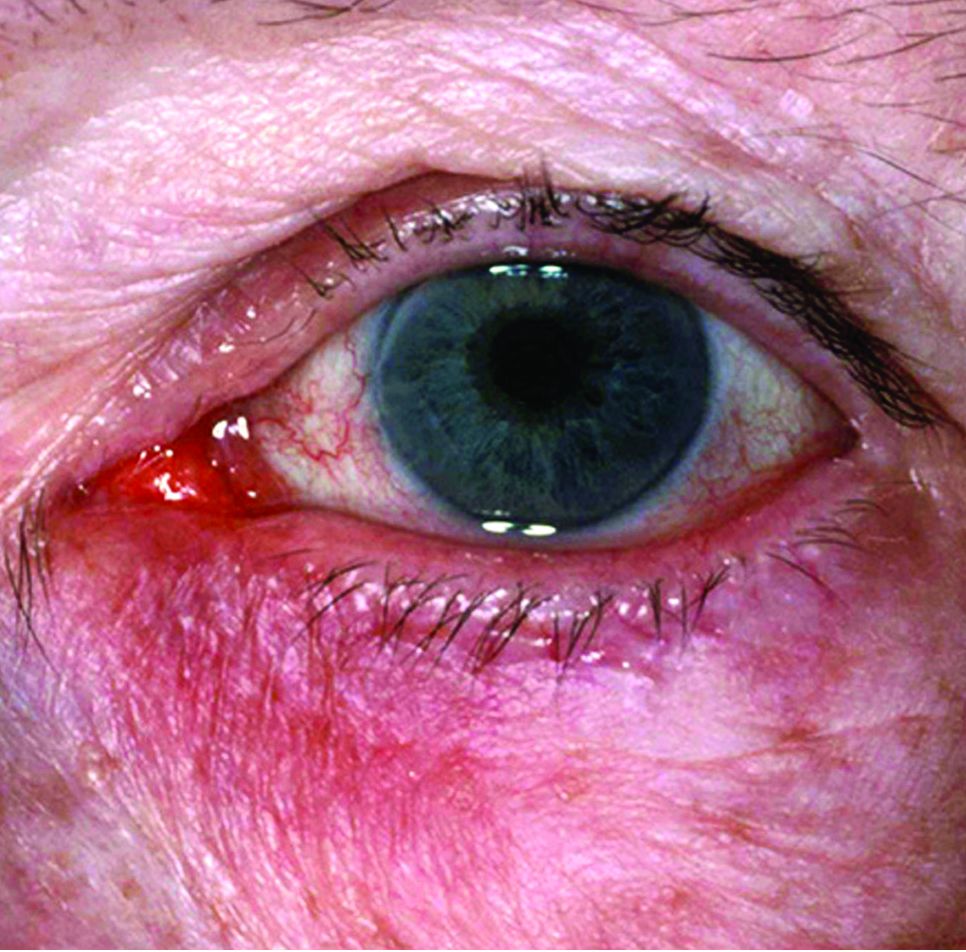
How social media solved a skin outbreak
Several teens who came home from a trip abroad with ugly ulcerated skin lesions in 2014 got vague and unhelpful diagnoses: Physicians thought they had bug bites. True, but that was only part of the story. It took an alert dermatologist and Facebook to identify the true cause, spread the word, and stop the outbreak.
“Social media facilitated communication between patients, crowd sourcing a diagnosis,” said Kanokporn Mongkolrattanothai, MD, who treated three of the teens at Children’s Hospital Los Angeles.
What did the kids have? Read on and see if you can make the diagnosis yourself.

Upon their return, pruritic red papules appeared on a 16-year-old girl’s ankle and thigh. They transformed into ulcers with raised edges and a central crater, according to a report that published online in Pediatric Dermatology (2016 Sep;33[5]:e276-7. doi: 10.1111/pde.12910). At least 12 teens from the trip had similar ulcerated lesions, mostly in exposed areas like arms and legs, said Dr. Mongkolrattanothai, an infectious disease specialist at Children’s Hospital Los Angeles and a coauthor of the report.
Six patients received a diagnosis of insect bites, and one was diagnosed with a bacterial skin infection, noted Dr. Mongkolrattanothai of the University of Southern California, Los Angeles. But these diagnoses were incorrect.
“The light bulb really came on when she mentioned that the lesions were still present several months after the trip to Israel,” said Dr. Krakowski, who was at Rady Children’s Hospital–San Diego at the time. “On physical exam, the lesions were ulcerated and eroded and did not look to be typical bug bite reactions.” The Centers for Disease Control and Prevention confirmed the diagnosis.
On Facebook, the teenager posted a picture of a T-shirt with the words “I went to Israel, and all I got was leishmaniasis.” At the same time, another traveler on the same trip posted pictures of lesions. This set off a wave of awareness that sent affected teens to seek care at Children’s Hospital Los Angeles, Mattel Children’s Hospital UCLA, Cedars-Sinai Medical Center, Kaiser Permanente Woodland Hills Medical Center, and Rady Children’s Hospital–San Diego.
“It is likely that our patients became infected with leishmaniasis while camping in the Negev Desert, sleeping on sand dunes at night without use of mosquito netting or tents,” Dr. Mongkolrattanothai said in an interview. “Most of the affected teens did not take precautions against insect bites, which would have included appropriate clothing to minimize areas of exposed skin and the use of repellent products. This placed them at risk for sand fly bites, as sand flies are most active in twilight, evening, and nighttime hours.”
Cutaneous leishmaniasis can lead to permanent scarring, and another form, visceral leishmaniasis, can be fatal.
What helped Dr. Krakowski crack the case? “Training at the University of California at San Diego, in such close proximity to the Navy’s Balboa Medical Center, we are taught from day 1 to think outside of the box because ‘there are zebras in Africa,’ ” he said. “With so much international travel in and out of the region, including to locations where leishmaniasis is endemic, it is warranted to consider that specific diagnosis on the differential. Normally, I do not have to biopsy ‘bug bites,’ but considering the patient’s entire presentation, you almost have to do a biopsy to make sure the lesions were not leishmaniasis.”
Dr. Krakowski praised the CDC. “They have a tremendous amount of resources dedicated to helping investigators work through diagnostic dilemmas such as this, and they helped us – free of charge – to confirm the diagnosis, type the leishmaniasis, and plot a treatment course to resolution,” he said. “They also were instrumental in helping us identify and educate other potentially exposed patients from the camping trip.”
In November, the Infectious Diseases Society of America and the American Society of Tropical Medicine and Hygiene published new guidelines about leishmaniasis in Clinical Infectious Diseases (doi: 10.1093/cid/ciw670). The societies warn that leishmaniasis is becoming more common in the United States, in part because of ecotourists infected in Central and South America and returning soldiers infected in Afghanistan and Iraq.
Dr. Mongkolrattanothai and Dr. Krakowski reported no relevant financial disclosures.
Several teens who came home from a trip abroad with ugly ulcerated skin lesions in 2014 got vague and unhelpful diagnoses: Physicians thought they had bug bites. True, but that was only part of the story. It took an alert dermatologist and Facebook to identify the true cause, spread the word, and stop the outbreak.
“Social media facilitated communication between patients, crowd sourcing a diagnosis,” said Kanokporn Mongkolrattanothai, MD, who treated three of the teens at Children’s Hospital Los Angeles.
What did the kids have? Read on and see if you can make the diagnosis yourself.
Upon their return, pruritic red papules appeared on a 16-year-old girl’s ankle and thigh. They transformed into ulcers with raised edges and a central crater, according to a report that published online in Pediatric Dermatology (2016 Sep;33[5]:e276-7. doi: 10.1111/pde.12910). At least 12 teens from the trip had similar ulcerated lesions, mostly in exposed areas like arms and legs, said Dr. Mongkolrattanothai, an infectious disease specialist at Children’s Hospital Los Angeles and a coauthor of the report.
Six patients received a diagnosis of insect bites, and one was diagnosed with a bacterial skin infection, noted Dr. Mongkolrattanothai of the University of Southern California, Los Angeles. But these diagnoses were incorrect.
“The light bulb really came on when she mentioned that the lesions were still present several months after the trip to Israel,” said Dr. Krakowski, who was at Rady Children’s Hospital–San Diego at the time. “On physical exam, the lesions were ulcerated and eroded and did not look to be typical bug bite reactions.” The Centers for Disease Control and Prevention confirmed the diagnosis.
On Facebook, the teenager posted a picture of a T-shirt with the words “I went to Israel, and all I got was leishmaniasis.” At the same time, another traveler on the same trip posted pictures of lesions. This set off a wave of awareness that sent affected teens to seek care at Children’s Hospital Los Angeles, Mattel Children’s Hospital UCLA, Cedars-Sinai Medical Center, Kaiser Permanente Woodland Hills Medical Center, and Rady Children’s Hospital–San Diego.
“It is likely that our patients became infected with leishmaniasis while camping in the Negev Desert, sleeping on sand dunes at night without use of mosquito netting or tents,” Dr. Mongkolrattanothai said in an interview. “Most of the affected teens did not take precautions against insect bites, which would have included appropriate clothing to minimize areas of exposed skin and the use of repellent products. This placed them at risk for sand fly bites, as sand flies are most active in twilight, evening, and nighttime hours.”
Cutaneous leishmaniasis can lead to permanent scarring, and another form, visceral leishmaniasis, can be fatal.
What helped Dr. Krakowski crack the case? “Training at the University of California at San Diego, in such close proximity to the Navy’s Balboa Medical Center, we are taught from day 1 to think outside of the box because ‘there are zebras in Africa,’ ” he said. “With so much international travel in and out of the region, including to locations where leishmaniasis is endemic, it is warranted to consider that specific diagnosis on the differential. Normally, I do not have to biopsy ‘bug bites,’ but considering the patient’s entire presentation, you almost have to do a biopsy to make sure the lesions were not leishmaniasis.”
Dr. Krakowski praised the CDC. “They have a tremendous amount of resources dedicated to helping investigators work through diagnostic dilemmas such as this, and they helped us – free of charge – to confirm the diagnosis, type the leishmaniasis, and plot a treatment course to resolution,” he said. “They also were instrumental in helping us identify and educate other potentially exposed patients from the camping trip.”
In November, the Infectious Diseases Society of America and the American Society of Tropical Medicine and Hygiene published new guidelines about leishmaniasis in Clinical Infectious Diseases (doi: 10.1093/cid/ciw670). The societies warn that leishmaniasis is becoming more common in the United States, in part because of ecotourists infected in Central and South America and returning soldiers infected in Afghanistan and Iraq.
Dr. Mongkolrattanothai and Dr. Krakowski reported no relevant financial disclosures.