User login
Larynx Preservation Studies Should Consider Treatment Impact
LONDON – Almost one-quarter of patients who had been given induction chemotherapy before radiotherapy for head and neck cancer experienced long-term swallowing difficulties, with another 15% experiencing voice disabilities that correlated with the mobility of the vocal cords.
Long-term data from the GORTEC (Groupe Oncologie Radiothérapie Tête et Cou) 2000-01 larynx preservation trial also show that approximately two-thirds of long-term head and neck cancer survivors experienced severe problems with sticky saliva and dry mouth, which were in turn linked to nutritional problems.
These findings, reported May 9 at the European Society for Therapeutic Radiation Oncology (ESTRO) Anniversary Conference, further confirm that studies looking at the effects of chemoradiotherapy on the larynx in head and neck cancer need to consider prospective assessment of laryngeal function, rather than just looking at anatomical preservation, according to a French radiation oncologist.
Dr. Gilles Calais of the Centre Hôpitalier Régional et Universitaire de Tours (France) presented data from a prospective analysis of 61 patients who had participated in the original 213-patient GORTEC 2000-01 trial. He also presented updated results from the trial using a recently developed composite end point.
"Larynx preservation can be achieved for most of our [head and neck] patients by using three different strategies: induction chemotherapy, concomitant [chemoradiotherapy], or alternating chemoradiotherapy," Dr. Calais observed. Indeed, larynx preservation is a possibility in approximately 80% of patients, he said.
However, anatomical preservation does not mean that laryngeal function is maintained, especially with respect to the ability to speak or to swallow normally. This realization recently resulted in the development of new end points for clinical trials that included both survival and laryngeal function. The new end points are laryngoesophageal dysfunction-free survival (LED-FS) and freedom from laryngoesophageal dysfunction (FF-LED).
"So the purpose of this study was to go back to our data of the GORTEC 2000-01 study and evaluate the results according to these new composite end points," Dr. Calais explained. "In parallel," he added, "we performed a prospective analysis of voice and swallowing function for long-term surviving patients."
Published in 2009, the GORTEC 2000-01 study showed that induction chemotherapy with TPF (docetaxel, cisplatin, and 5-fluorouracil) was superior to pf (cisplatin and 5-FU) in terms of 3-year larynx preservation rates (J. Natl. Cancer Inst. 2009;101:498-506). In all, 110 patients had been treated with TPF and 103 with PF, and the published larynx preservation rates were 70.3% and 57.5%, respectively. Anatomical preservation of the larynx was a possibly in 66 patients, and these patients were assessed for voice and swallowing function.
Recalculating survival curves according to the two new end points showed much lower overall values, compared with the anatomical-only end points, Dr. Calais noted. Considering all patients, the 5-year LED-FS was just 28% and the 5-year FF-LED was 50%.
Patients who were treated with the taxane-containing regimen fared better than those who received the cisplatin and 5-FU chemotherapy. The 5-year LED-FS rates were 36% in the TPF arm vs. 21% for the PF arm (P = .007). The 5-year FF-LED rates were 60% and 39%, respectively (P = .005).
"These data can be used as a reference for comparison with future larynx preservation studies," Dr. Calais said. He noted that it was important to bear in mind that the best treatment to preserve the larynx is not known, referring to the FF-LED rate of 60% with the TPF regimen.
"Of course, [the] patient’s quality of life measure[s used] should be conducted in every future larynx preservation study."
Dr. Calais declared no financial conflicts of interest.
FROM THE EUROPEAN SOCIETY FOR THERAPEUTIC RADIATION ONCOLOGY ANNIVERSARY CONFERENCE
Major Finding: Considering all patients, the 5-year laryngoesophageal dysfunction-free survival (LED-FS) rate was just 28% and the 5-year freedom from laryngoesophageal dysfunction (FF-LED) rate was 50%.
Data Source: Retrospective analysis of 213 patients with locally advanced head and neck cancer who were treated in the GORTEC 2000-01 study and prospective assessment of voice and swallowing function in 61 long-term surviving patients.
Disclosures: Dr. Calais declared no financial conflicts of interest.
Low EGFR Expression Predicts ARCON Benefit in Laryngeal Cancer
LONDON – Expression of the epidermal growth factor receptor predicts whether patients with laryngeal cancer will benefit from hypoxia modification in addition to accelerated radiotherapy.
Study findings reported May 9 at the European Society for Therapeutic Radiation Oncology (ESTRO) Anniversary Conference showed that low expression was associated with higher disease-specific survival and locoregional control than was high expression in patients who received ARCON (accelerated radiotherapy with carbogen and nicotinamide).

EGFR expression is prognostic for patients who are treated with conventional fractionated radiotherapy, but did not predict response in patients undergoing just AR (accelerated radiotherapy) without hypoxia modification.
"EGFR can activate different downstream signaling pathways [in the tumor cell] that – once activated – can influence tumor cell survival, proliferation, [and] migration," all of which can contribute to tumor progression, said Monique Nijkamp, a Ph.D. student in the radiation oncology department of St. Radboud University Medical Centre in Nijmegen, the Netherlands.
High expression has already been linked to worse outcome in patients who were treated with radiotherapy for head and neck cancer (Cancer Res. 2002;62:7350-6). Ms. Nijkamp said that EGFR could be activated by ionizing radiation, and could account for resistance to radiotherapy in some treated individuals.
Hypothesizing that ARCON may help to overcome this resistance, Ms. Nikamp and coworkers examined 272 biopsy samples taken from 345 patients who had been treated with accelerated radiotherapy, with or without hypoxia modification, in a phase III study.
Preliminary results of the study were reported last September at the ESTRO 29 meeting in Barcelona. They showed that ARCON significantly improved regional – but not locoregional – tumor control, compared with AR alone. All of the patients in the trial had advanced-stage laryngeal cancer.
The latest findings suggest that patients with high expression may not gain any extra benefit from the addition of hypoxia modification. Indeed, although not statistically significant, better 5-year locoregional control (88% vs. 71%) and disease-specific survival (91% vs. 78%) were seen in patients with low EGFR expression than in those with high EGFR expression who were treated with ARCON.
Ms. Nijkamp suggested that perhaps high EGFR–expressing tumors were rapidly activating downstream signaling pathways that negated the effects of hypoxia modification. No difference in outcomes was found according to EGFR expression in the AR alone arm.
"There is concern over the methodologies used to measure EGFR, and this could have a large bearing on what conclusions can be drawn from studies," said Dr. Nick Slevin, who was invited to comment on the findings.
"From the literature and from this study, it appears that high EGFR laryngeal cancers benefit from accelerated radiotherapy, but that low EGFR tumors do not," added Dr. Slevin, a consultant clinical oncologist at the Christie NHS Foundation Trust and the University of Manchester (England).
"I think it’s a fair summary to also say that high-EGFR tumors benefit from AR but not from CON [carbogen and nicotinamide] and that low-EGFR tumors benefit from the CON but not the AR. ... So I’m not sure ARCON is the right approach for all tumors."
Other approaches to hypoxia modification for low EGFR–expressing tumors (such as oral nimorazole) may overcome the logistic difficulties of delivering carbogen and nicotinamide, he added.
The study was supported by the Dutch Cancer Society. Ms. Nijkamp and Dr. Slevin declared they had no financial conflicts of interest.
LONDON – Expression of the epidermal growth factor receptor predicts whether patients with laryngeal cancer will benefit from hypoxia modification in addition to accelerated radiotherapy.
Study findings reported May 9 at the European Society for Therapeutic Radiation Oncology (ESTRO) Anniversary Conference showed that low expression was associated with higher disease-specific survival and locoregional control than was high expression in patients who received ARCON (accelerated radiotherapy with carbogen and nicotinamide).
EGFR expression is prognostic for patients who are treated with conventional fractionated radiotherapy, but did not predict response in patients undergoing just AR (accelerated radiotherapy) without hypoxia modification.
"EGFR can activate different downstream signaling pathways [in the tumor cell] that – once activated – can influence tumor cell survival, proliferation, [and] migration," all of which can contribute to tumor progression, said Monique Nijkamp, a Ph.D. student in the radiation oncology department of St. Radboud University Medical Centre in Nijmegen, the Netherlands.
High expression has already been linked to worse outcome in patients who were treated with radiotherapy for head and neck cancer (Cancer Res. 2002;62:7350-6). Ms. Nijkamp said that EGFR could be activated by ionizing radiation, and could account for resistance to radiotherapy in some treated individuals.
Hypothesizing that ARCON may help to overcome this resistance, Ms. Nikamp and coworkers examined 272 biopsy samples taken from 345 patients who had been treated with accelerated radiotherapy, with or without hypoxia modification, in a phase III study.
Preliminary results of the study were reported last September at the ESTRO 29 meeting in Barcelona. They showed that ARCON significantly improved regional – but not locoregional – tumor control, compared with AR alone. All of the patients in the trial had advanced-stage laryngeal cancer.
The latest findings suggest that patients with high expression may not gain any extra benefit from the addition of hypoxia modification. Indeed, although not statistically significant, better 5-year locoregional control (88% vs. 71%) and disease-specific survival (91% vs. 78%) were seen in patients with low EGFR expression than in those with high EGFR expression who were treated with ARCON.
Ms. Nijkamp suggested that perhaps high EGFR–expressing tumors were rapidly activating downstream signaling pathways that negated the effects of hypoxia modification. No difference in outcomes was found according to EGFR expression in the AR alone arm.
"There is concern over the methodologies used to measure EGFR, and this could have a large bearing on what conclusions can be drawn from studies," said Dr. Nick Slevin, who was invited to comment on the findings.
"From the literature and from this study, it appears that high EGFR laryngeal cancers benefit from accelerated radiotherapy, but that low EGFR tumors do not," added Dr. Slevin, a consultant clinical oncologist at the Christie NHS Foundation Trust and the University of Manchester (England).
"I think it’s a fair summary to also say that high-EGFR tumors benefit from AR but not from CON [carbogen and nicotinamide] and that low-EGFR tumors benefit from the CON but not the AR. ... So I’m not sure ARCON is the right approach for all tumors."
Other approaches to hypoxia modification for low EGFR–expressing tumors (such as oral nimorazole) may overcome the logistic difficulties of delivering carbogen and nicotinamide, he added.
The study was supported by the Dutch Cancer Society. Ms. Nijkamp and Dr. Slevin declared they had no financial conflicts of interest.
FROM THE EUROPEAN SOCIETY FOR THERAPEUTIC RADIATION ONCOLOGY ANNIVERSARY CONFERENCE
Major Finding: The rates of 5-year locoregional control (88% vs. 71%) and disease-specific survival (91% vs. 78%) were better with low vs. high EGFR expression in patients who were treated with ARCON.
Data Source: Immunohistochemical analysis of 272 biopsy samples from 345 patients who were treated with accelerated radiotherapy, with or without hypoxia modification, in a phase III study.
Disclosures: The study was supported by the Dutch Cancer Society. Ms. Nijkamp and Dr. Slevin declared that they had no financial conflicts of interest.
Low-Dose Irradiation Allays 93% of Gastric MALT Lymphomas at 10 Years
LONDON – Low-dose irradiation of the stomach in patients with gastric MALT lymphoma absent or independent of infection by Helicobacter pylori is associated with a 93% disease control rate after 10 years.
As presented by Dr. Joachim Yahalom, a radiation oncologist at Memorial Sloan-Kettering Cancer Center in New York, these long-term study findings highlight the high disease-control rates that can be obtained using low-dose involved field radiotherapy (IFRT).
Low-dose IFRT has now become a standard of care in the United States, although approaches still vary in Europe, he reported May 9 at the European Society for Therapeutic Radiation Oncology Anniversary Conference.
Indeed, the latest guidelines set by the National Comprehensive Cancer Network on non-Hodgkin’s lymphomas – which includes gastric MALT (mucosa-associated lymphoid tissue) lymphoma of the stomach – recommend the use of radiotherapy as a first-line option when patients have early (stage I/II) H. pylori–negative disease.
"We started using this [low-dose irradiation] treatment approach in the early 90s. Until then, the standard treatment for this lymphoma in the stomach was total gastrectomy, and sometimes it was followed by radiation therapy," said Dr. Yahalom, a professor of radiation oncology at Cornell University in New York. He added that chemotherapy and rituximab (Rituxan) were not generally very effective.
"Obviously the treatment of choice is to give antibiotics if H. pylori is present," Dr. Yahalom added, noting that substantial regression of MALT lymphoma can occur via treatment with antibiotics that target the gastric bacterium in positive cases. However, when H. pylori is not present, or if there is a lack of response to or residual lymphoma after complete response to antibiotic therapy, then radiotherapy is the recommended next step.
Dr. Yahalom presented data representing 16 years of experience in treating 103 patients (60 women and 43 men) with gastric MALT lymphoma in 1992-2009. The majority of patients (76%) had stage I disease, with 19% having stage II, and fewer than 5% having stage IV.
The median age of the patients was 62 years (range, 25-91 years). Symptoms were relatively mild at presentation, Dr. Yahalom observed: In all, 52% had epigastric pain, 36% had nausea, 29% had gastric bleeding, 18% had anorexia, 15% had anemia, and 10% had unknown or no obvious symptoms.
Almost two-thirds of patients were H. pylori negative. This included four patients who had relapsed after a complete response to antibiotic therapy. A further 20% of patients had persistent lymphoma after H. pylori eradication therapy. Another 15% had H. pylori and lymphoma remaining after antibiotic treatment, but had progressive symptoms or disease that could not wait for another antibiotics trial.
Prior to being treated with IFRT, six patients had undergone surgery, and seven had received medications that failed to control their disease.
Approximately 90% of patients were given IFRT at a median dose of 30 Gy in 1.5 Gy fractions for 4 weeks. The lowest and highest total radiation doses administered were 22.5 Gy and 43.5 Gy.
Patients received regular follow up via endoscopic biopsies, and after a median of 5.5 years, the disease control rate was 98%. One-fifth of patients had been followed for 10 years, and the 10-year freedom from local failure was 93%; the 10-year overall survival rate was 74%.
There were no significant acute or late adverse events, Dr. Yahalom noted, and just eight patients relapsed (six with gastric MALT lymphoma and two with diffuse large B-cell lymphoma). Subsequent other cancers included lymphoma in extragastric sites (eight patients), adenocarcinoma of the stomach (two), and second tumors close to the radiotherapy field (one in the colon and two in the pancreas). "Other deaths appeared unrelated to disease or treatment," he said.
Based on this experience, Dr. Yahalom concluded that "low-dose irradiation of the stomach provides excellent long-term disease control [and] is safe and simple.
"Radiation therapy, in our opinion, is the treatment of choice for patients with MALT lymphoma that have exhausted their antibiotic options or are unlikely to respond to it."
Dr. Yahalom said he had no financial conflicts of interest.
FROM THE EUROPEAN SOCIETY FOR THERAPEUTIC RADIATION ONCOLOGY ANNIVERSARY CONFERENCE
Major Finding: After a median of 5.5 years, the disease control rate was 98%.
Data Source: Data representing 16 years of experience with the use of low-dose irradiation therapy in 103 patients with H. pylori–independent gastric MALT lymphoma.
Disclosures: Dr. Yahalom said he had no financial conflicts of interest.
Fewer Side Effects From Proton Therapy in NSCLC
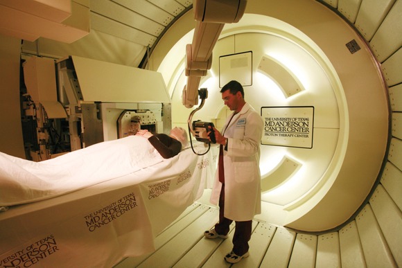
LONDON – Proton beam therapy for non–small cell lung cancer is associated with fewer radiation-induced side effects than are conventional radiotherapy methods when combined with chemotherapy, according to preliminary data from two retrospective studies conducted at the University of Texas M.D. Anderson Cancer Center in Houston.
Significantly less esophagitis, pneumonitis, and bone marrow toxicity were observed with proton beam therapy (PBT) than with intensity-modulated radiotherapy (IMRT), Dr. Ritsuko Komaki reported May 10, at the European Society for Therapeutic Radiation Oncology (ESTRO) Anniversary Conference.
Proton beam therapy also significantly reduced the incidence of esophagitis when compared with IMRT and three-dimensional conformal radiotherapy (3D-CRT). A mean esophageal dose of 40 Gy or higher was identified as the cut-off point for high-grade esophagitis occurring with any method.
"Radiation dose escalation improves local control but increases toxicity, especially when combined with concurrent chemotherapy for non–small cell lung cancer [NSCLC] or even small cell lung cancer," said Dr. Komaki, a professor of radiation oncology at M.D. Anderson, which opened its 94,000-square-foot Proton Therapy Center in 2006. As such, "radiation chemotherapy is a double-edged sword," she observed. "It will kill cancer cells, but it also kills normal tissues, and more targeted treatment is needed."
"One of the most important benefits of PBT is that there is no exit dose," Dr. Komaki added in an interview with Elsevier Global Medical News. "The protons stop after penetrating the tumor, and there is no dose of radiation beyond it."
This has the potential to spare surrounding cells and organs from damage, she observed. Normal tissues that might be affected by radiation therapy for NSCLC include the lungs, esophagus, heart, and bone marrow, which cannot always be avoided by the use of 3D-CRT or even IMRT.
An expensive new technology that delivers highly targeted radiation with electrically charged particles, proton beam therapy is promising but unproven, according to a 2009 review commissioned by the Agency for Healthcare Research and Quality (AHRQ). The authors found few comparative studies to establish effectiveness or safety for the technology, which is housed in a small but growing number of proton beam centers that can cost $100 million to $225 million to build (Ann. Intern. Med. 2009;151:556-65).
Dr. Komaki and her associates are recruiting patients into the first, prospective randomized trial to directly compare proton beam therapy with IMRT in unresectable stage II /III NSCLC. The phase II trial is supported by a grant from the National Cancer Institute, and involves treatment with 74 Gy proton beam therapy or IMRT with concurrent carboplatin and paclitaxel. To date, 107 of the planned 168 patients have been enrolled in the study at the Texas institution and at the Massachusetts General Hospital in Boston, the other participating center, she said.
A recent report from the M.D. Anderson showed that higher doses of proton radiation could be delivered to lung tumors with a lower risk of esophagitis and pneumonitis than either IMRT or 3D-CRT (Cancer 2011;doi.org/10.1002/cncr.25848).
The new data presented by Dr. Komaki showed significantly reduced rates of grade 2 or higher esophagitis (P less than .0001), pneumonitis (P less than .002), hematologic toxicities (P less than .0001 for neutrophil toxicity and P less than .001 for hemoglobin and white blood cell toxicities), and fatigue (P less than.0001) in 60 patients treated with proton beam therapy compared with 75 patients treated with IMRT (Radiother. Oncol. 2011;99:S89-90, abstract 233).
Other research from the M.D. Anderson team focused on esophagitis, and examined dosimetric and clinical factors that could lead to this side effect following proton beam therapy, IMRT, or 3D-CRT for definitive NSCLC treatment (Radiother. Oncol. 2011;99[Suppl.1]:S210, abstract 518).
Dr. Daniel Gomez, a radiation oncologist at M.D. Anderson, presented data on 678 patients treated at the institution between 1999 and 2008. Dr. Gomez explained that the type of radiation therapy received had altered over the years, with 463 patients treated with 3D-CRT between 1999 and 2005, 122 with IMRT between 2005 and 2007, and 94 patients treated with proton beam therapy between 2006 and 2008.
"Esophagitis is a common toxicity in the treatment of NSCLC with definitive radiation," Dr. Gomez observed. Although studies have looked at what factors might predict this life-limiting side effect, conflicting results have been obtained.
Data show, for example, that the presence of acute toxicity is a predictor of late toxicity (Int. J. Radiat. Oncol. Biol. Phys. 2005,61:335-47), but a variety of dosimetric parameters have been noted and there does not appear to be a single threshold at which a toxic effect is or is not likely to be observed (Int. J. Radiat. Oncol. Biol. Phys. 2010;76:S86-93).
Data presented by Dr. Gomez, however, suggest that a mean delivered esophageal dose of above 40 Gy may be predictive of high-grade inflammation regardless of whether proton beams, IMRT or 3D-CRT is used. This research might eventually help develop dosing guides for clinicians to use in routine practice, he suggested.
"Patients receiving IMRT had a higher rate of esophagitis in all grades, including grade 3," Dr. Gomez said. In contrast, "patients receiving proton therapy had lower rates of esophagitis at all grades." The incidence of grade 3 or higher esophagitis was 14% (n = 65) for 3D-CRT, 27% (n = 33) for IMRT, and 6% (n = 6) for proton beam therapy.
Dr. Gomez also reported that grade 3 or higher esophagitis was more likely in patients who received concurrent chemotherapy than in those who did not (18.4% vs. 7.4%, P less than .001). The mean esophageal dose of radiation delivered to patients given concurrent chemotherapy also was, significantly higher (32.2 Gy vs. 15.8 Gy, P less than .001), however.
The M.D. Anderson investigators said they have just finished (May 12) recruiting patients into a phase III trial (Radiation Therapy Oncology Group [RTOG] 0617) that will compare conventional (60 Gy in 6 weeks) vs. high dose (74 Gy in 7.5 weeks) radiation therapy in combination with paclitaxel and carboplatin, with or without the addition of cetuximab (Erbitux) in 500 patients with NSCLC.
Although the trial is not directly comparing the type of radiation treatment used, it should still be possible to retrospectively analyze the results to determine the individual effects of the radiation modalities used at each participating center, Dr. Komaki noted.
"When we started this trial, it was not acquiring patients because some of the radiation and medical oncologists said that it was obvious that patients given 60 Gy would do worse compared to 74 Gy," she added in the interview. "When we included cetuximab based on the results of the RTOG 0324 trial, however, recruitment started to rocket." The RTOG 0324 trial showed the feasibility of combining cetuximab with chemoradiation in NSCLC (J. Clin. Oncol. 2011 May 9 [Epub ahead of print, doi: 10.1200/JCO.2010.31.7875]).
Discussing the downsides of proton beam therapy vs. IMRT, Dr. Komaki conceded that the newer method involved a lot more sophisticated planning and was more expensive. There is also concern that the sharp drop-off of radiation received with proton beam therapy might mean that important areas of the tumor are missed – although this may explain the lower rate of side effects seen with PBT to date. "There is no give," Dr. Komaki said.
As relatively few proton beam facilities are in operation, large cooperative trials are difficult to perform. The prospective phase II trial comparing proton beam therapy and IMRT now being conducted at M.D. Anderson and the Massachusetts General Hospital will be the proof that such trials are possible, and provide valuable information on the comparative safety and efficacy of the two procedures.
Dr. Komaki and Dr. Gomez said they had no financial conflicts of interest.
FROM THE EUROPEAN SOCIETY FOR THERAPEUTIC RADIATION ONCOLOGY ANNIVERSARY CONFERENCE
Major Finding: PBT resulted in significantly lower rates of grade 2 or higher esophagitis (P less than .0001), pneumonitis (P less than .002), hematologic toxicities (P less than .0001 for neutrophil toxicity and P less than .001 for hemoglobin and white blood cell toxicities), and fatigue (P less than .0001) than IMRT.
Data Source: Two retrospective studies: one involving 135 patients with NSCLC treated with concurrent chemoradiation (PBT, IMRT) and one involving 678 patients with NSCLC treated with concurrent chemoradiation between 1999 and 2008.
Disclosures: Dr. Komaki and Dr. Gomez said they had no financial conflicts of interest.
Fewer Side Effects From Proton Therapy in NSCLC
LONDON – Proton beam therapy for non–small cell lung cancer is associated with fewer radiation-induced side effects than are conventional radiotherapy methods when combined with chemotherapy, according to preliminary data from two retrospective studies conducted at the University of Texas M.D. Anderson Cancer Center in Houston.
Significantly less esophagitis, pneumonitis, and bone marrow toxicity were observed with proton beam therapy (PBT) than with intensity-modulated radiotherapy (IMRT), Dr. Ritsuko Komaki reported May 10, at the European Society for Therapeutic Radiation Oncology (ESTRO) Anniversary Conference.
Proton beam therapy also significantly reduced the incidence of esophagitis when compared with IMRT and three-dimensional conformal radiotherapy (3D-CRT). A mean esophageal dose of 40 Gy or higher was identified as the cut-off point for high-grade esophagitis occurring with any method.
"Radiation dose escalation improves local control but increases toxicity, especially when combined with concurrent chemotherapy for non–small cell lung cancer [NSCLC] or even small cell lung cancer," said Dr. Komaki, a professor of radiation oncology at M.D. Anderson, which opened its 94,000-square-foot Proton Therapy Center in 2006. As such, "radiation chemotherapy is a double-edged sword," she observed. "It will kill cancer cells, but it also kills normal tissues, and more targeted treatment is needed."
"One of the most important benefits of PBT is that there is no exit dose," Dr. Komaki added in an interview with Elsevier Global Medical News. "The protons stop after penetrating the tumor, and there is no dose of radiation beyond it."
This has the potential to spare surrounding cells and organs from damage, she observed. Normal tissues that might be affected by radiation therapy for NSCLC include the lungs, esophagus, heart, and bone marrow, which cannot always be avoided by the use of 3D-CRT or even IMRT.
An expensive new technology that delivers highly targeted radiation with electrically charged particles, proton beam therapy is promising but unproven, according to a 2009 review commissioned by the Agency for Healthcare Research and Quality (AHRQ). The authors found few comparative studies to establish effectiveness or safety for the technology, which is housed in a small but growing number of proton beam centers that can cost $100 million to $225 million to build (Ann. Intern. Med. 2009;151:556-65).
Dr. Komaki and her associates are recruiting patients into the first, prospective randomized trial to directly compare proton beam therapy with IMRT in unresectable stage II /III NSCLC. The phase II trial is supported by a grant from the National Cancer Institute, and involves treatment with 74 Gy proton beam therapy or IMRT with concurrent carboplatin and paclitaxel. To date, 107 of the planned 168 patients have been enrolled in the study at the Texas institution and at the Massachusetts General Hospital in Boston, the other participating center, she said.
A recent report from the M.D. Anderson showed that higher doses of proton radiation could be delivered to lung tumors with a lower risk of esophagitis and pneumonitis than either IMRT or 3D-CRT (Cancer 2011;doi.org/10.1002/cncr.25848).
The new data presented by Dr. Komaki showed significantly reduced rates of grade 2 or higher esophagitis (P less than .0001), pneumonitis (P less than .002), hematologic toxicities (P less than .0001 for neutrophil toxicity and P less than .001 for hemoglobin and white blood cell toxicities), and fatigue (P less than.0001) in 60 patients treated with proton beam therapy compared with 75 patients treated with IMRT (Radiother. Oncol. 2011;99:S89-90, abstract 233).
Other research from the M.D. Anderson team focused on esophagitis, and examined dosimetric and clinical factors that could lead to this side effect following proton beam therapy, IMRT, or 3D-CRT for definitive NSCLC treatment (Radiother. Oncol. 2011;99[Suppl.1]:S210, abstract 518).
Dr. Daniel Gomez, a radiation oncologist at M.D. Anderson, presented data on 678 patients treated at the institution between 1999 and 2008. Dr. Gomez explained that the type of radiation therapy received had altered over the years, with 463 patients treated with 3D-CRT between 1999 and 2005, 122 with IMRT between 2005 and 2007, and 94 patients treated with proton beam therapy between 2006 and 2008.
"Esophagitis is a common toxicity in the treatment of NSCLC with definitive radiation," Dr. Gomez observed. Although studies have looked at what factors might predict this life-limiting side effect, conflicting results have been obtained.
Data show, for example, that the presence of acute toxicity is a predictor of late toxicity (Int. J. Radiat. Oncol. Biol. Phys. 2005,61:335-47), but a variety of dosimetric parameters have been noted and there does not appear to be a single threshold at which a toxic effect is or is not likely to be observed (Int. J. Radiat. Oncol. Biol. Phys. 2010;76:S86-93).
Data presented by Dr. Gomez, however, suggest that a mean delivered esophageal dose of above 40 Gy may be predictive of high-grade inflammation regardless of whether proton beams, IMRT or 3D-CRT is used. This research might eventually help develop dosing guides for clinicians to use in routine practice, he suggested.
"Patients receiving IMRT had a higher rate of esophagitis in all grades, including grade 3," Dr. Gomez said. In contrast, "patients receiving proton therapy had lower rates of esophagitis at all grades." The incidence of grade 3 or higher esophagitis was 14% (n = 65) for 3D-CRT, 27% (n = 33) for IMRT, and 6% (n = 6) for proton beam therapy.
Dr. Gomez also reported that grade 3 or higher esophagitis was more likely in patients who received concurrent chemotherapy than in those who did not (18.4% vs. 7.4%, P less than .001). The mean esophageal dose of radiation delivered to patients given concurrent chemotherapy also was, significantly higher (32.2 Gy vs. 15.8 Gy, P less than .001), however.
The M.D. Anderson investigators said they have just finished (May 12) recruiting patients into a phase III trial (Radiation Therapy Oncology Group [RTOG] 0617) that will compare conventional (60 Gy in 6 weeks) vs. high dose (74 Gy in 7.5 weeks) radiation therapy in combination with paclitaxel and carboplatin, with or without the addition of cetuximab (Erbitux) in 500 patients with NSCLC.
Although the trial is not directly comparing the type of radiation treatment used, it should still be possible to retrospectively analyze the results to determine the individual effects of the radiation modalities used at each participating center, Dr. Komaki noted.
"When we started this trial, it was not acquiring patients because some of the radiation and medical oncologists said that it was obvious that patients given 60 Gy would do worse compared to 74 Gy," she added in the interview. "When we included cetuximab based on the results of the RTOG 0324 trial, however, recruitment started to rocket." The RTOG 0324 trial showed the feasibility of combining cetuximab with chemoradiation in NSCLC (J. Clin. Oncol. 2011 May 9 [Epub ahead of print, doi: 10.1200/JCO.2010.31.7875]).
Discussing the downsides of proton beam therapy vs. IMRT, Dr. Komaki conceded that the newer method involved a lot more sophisticated planning and was more expensive. There is also concern that the sharp drop-off of radiation received with proton beam therapy might mean that important areas of the tumor are missed – although this may explain the lower rate of side effects seen with PBT to date. "There is no give," Dr. Komaki said.
As relatively few proton beam facilities are in operation, large cooperative trials are difficult to perform. The prospective phase II trial comparing proton beam therapy and IMRT now being conducted at M.D. Anderson and the Massachusetts General Hospital will be the proof that such trials are possible, and provide valuable information on the comparative safety and efficacy of the two procedures.
Dr. Komaki and Dr. Gomez said they had no financial conflicts of interest.
FROM THE EUROPEAN SOCIETY FOR THERAPEUTIC RADIATION ONCOLOGY ANNIVERSARY CONFERENCE
Major Finding: PBT resulted in significantly lower rates of grade 2 or higher esophagitis (P less than .0001), pneumonitis (P less than .002), hematologic toxicities (P less than .0001 for neutrophil toxicity and P less than .001 for hemoglobin and white blood cell toxicities), and fatigue (P less than .0001) than IMRT.
Data Source: Two retrospective studies: one involving 135 patients with NSCLC treated with concurrent chemoradiation (PBT, IMRT) and one involving 678 patients with NSCLC treated with concurrent chemoradiation between 1999 and 2008.
Disclosures: Dr. Komaki and Dr. Gomez said they had no financial conflicts of interest.
Two Factors Predict Postradiotherapy Swallowing Difficulties
LONDON – Nonglottic cancer and the presence of dysphagia before treatment are highly predictive for severe acute and late swallowing difficulties after radiotherapy for head and neck cancer, according to new data from the DAHANCA 6&7 randomized trial.
Patients with nonglottic cancer were more likely than those with other cancer types to experience severe dysphagia at both 6 and 12 months. Dysphagia before treatment was also associated with both acute and late severe swallowing difficulties.
"The peak incidence of dysphagia is seen during the first 6 months after radiotherapy," Hanna Rahbek Mortensen, Ph.D., reported in an analysis of the DAHANCA (Danish Head and Neck Cancer Group) 6&7 trial findings at the European Society for Therapeutic Radiation Oncology Anniversary Congress.
"After 1 year, however, there is no further increase in severity or prevalence," said Dr. Mortensen of the department of experimental clinical oncology at Åarhus (Denmark) University Hospital.
The trial involved 1,478 patients with squamous cell carcinomas of the glottic larynx, supraglottic larynx, pharynx, or oral cavity who were who were treated with five or six weekly fractions of radiotherapy in 1992-1999. The total dose of radiotherapy delivered was 66-68 Gy in 33-34 fractions.
Efficacy data from the trial have already been published; they showed improved disease-specific but not overall survival of five vs. six fractions of radiotherapy (Lancet 2003;362:933-40).
The aim of the present analysis was to use prospectively collected data from the trial to determine whether any factors could be used to establish which patients may be more likely than others to experience dysphagia following treatment.
"Dysphagia is a common and debilitating side effect of radiotherapy, leading to malnutrition, aspiration, and reduced quality of life," Dr. Mortensen explained. The side effect can be graded using a 5-point scale, with a score of 0 signifying no dysphagia and a score of 3 or 4 indicating considerable or severe dysphagia despite the ingestion of a liquid-only diet.
In all, 1,422 (96%) patients experienced acute dysphagia, including severe grade 3/4 dysphagia in 47% and 38% of those receiving accelerated or conventional radiotherapy, respectively. The two factors most predictive for severe dysphagia at 6 months were nonglottic cancer (odds ratio, 6.73 vs. other sites; P less than .0001) and a baseline dysphagia score of 2-4 vs. 0-1 (OR, 2.11; P =.004).
Odd ratios for other factors predictive for acute – but not late – dysphagia were 1.92 for any (N1-N3) vs. no (N0) nodal involvement (P less than .001), 1.82 for the use of accelerated (six weekly fractions) vs. conventional (five weekly fractions) radiotherapy (P less than .001), and 1.58 for stage T3-T4 vs. stage T1-T2 cancer (P = .02).
After 5-years of regular follow-up, late dysphagia had occurred in 1,205 patients (81%), with grade 3 severity in 24% and grade 4 in 23%. Factors that were predictive for late severe dysphagia that occurred 12 months after radiotherapy were, again, nonglottic cancer (OR, 8.25; P less than .0001) and a baseline dysphagia score greater than 1 (OR, 2.57; P less than .002). Late (T3-T4) tumor stage was also predictive (OR, 1.86; P = .03).
"Predictive factors have been identified to characterize patients at risk of developing acute and late dysphagia," Dr. Mortensen said. These factors may be useful to identify patients who could perhaps benefit from prophylactic measures against swallowing dysfunction.
The DAHANCA 6&7 trial was supported by the Danish Cancer Society, Åarhus University Hospital, Copenhagen University Hospital, Herlev (Denmark) Hospital, Odense (Denmark) University Hospital, and Aalborg (Denmark) Hospital on behalf of DAHANCA. Dr. Mortensen had no personal financial disclosures to declare.
FROM THE EUROPEAN SOCIETY FOR THERAPEUTIC RADIATION ONCOLOGY ANNIVERSARY CONFERENCE
Major Finding: The two factors most predictive for acute severe dysphagia at 6 months were nonglottic cancer (OR, 6.73 vs. other sites; P less than .0001] and a baseline dysphagia score of 0-1 vs. 2-4 (OR, 2.11; P = .004). Both were also highly predictive for late severe dysphagia at 12 months.
Data Source: A phase III, prospective, randomized, multicenter trial of 1,468 patients undergoing accelerated or conventional radiotherapy for squamous cell head and neck carcinoma.
Disclosures: The DAHANCA 6&7 trial was supported by the Danish Cancer Society, Åarhus University Hospital, Copenhagen University Hospital, Herlev University Hospital, Odense University Hospital, Aalborg Hospital. Dr. Mortensen had no personal financial disclosures to declare.
Smoking Cessation Can Reduce Late Side Effects of Radiotherapy
LONDON – One-third of current smokers with head and neck cancer experienced substantial difficulties in swallowing 1 year after radiotherapy, according to the results of a Danish study.
In comparison, 20% of ex-smokers and just 10% of never-smokers experienced dysphagia as a late-occurring side effect. Current smoking also was linked to higher rates of mucosal edema, atrophy, fibrosis, dry mouth, and hoarseness or loss of voice compared with outcomes in ex- and never-smokers.

These findings highlight the importance of smoking cessation before and even during treatment, said Dorthe Wiinholdt, M.P.H., a radiotherapy technician nurse from Copenhagen University Hospital.
"As nurses, we see patients every day with side effects during treatment," she said May 9 at the European Society for Therapeutic Radiation Oncology Anniversary Conference. "We talk to them about quitting smoking before treatment, because we know that smoking has an influence on efficacy and quality of life, and [in this study] we wanted to know if there was anything that patients could do for themselves to decrease the severity of these side effects."
To examine the influence of smoking on late side effects commonly associated with radiotherapy, Ms. Wiinholdt and her colleagues used prospectively collected data from the Danish Head and Neck Cancer Study Group (DAHANCA)’s database.
Between January 2000 and March 2009, they identified 578 men and 217 women with head and neck cancer who had been treated with radiotherapy at Copenhagen University Hospital. The mean age of patients was 60 years.
Treatment consisted of 66-68 Gy for carcinoma of the larynx, pharynx, and oral cavity, with less than 10% receiving concomitant chemotherapy. In the majority (84%) of cases, treatment was intended to cure the cancer.
Before starting treatment, 60 (7.5%) patients were identified as never-smokers, with 290 (36.5%) defined as ex-smokers and the remaining 445 (56%) as current smokers. "Sixty percent of patients quit smoking during treatment, but one-third resumed smoking after treatment," Ms. Wiinholdt reported.
Of the six side effects associated with head and neck radiotherapy included in the analysis, dysphagia and mucosal edema were the most significant in relation to smoking.
Dysphagia occurred in 34% of current smokers, 21% of ex-smokers, and 15% of never-smokers 6 months after stopping radiotherapy, and in 31%, 20%, and 10%, respectively, 1 year after treatment cessation. Corresponding figures for mucosal edema were 25%, 19%, and 10% at 6 months, and 22%, 14%, and 5% at 1 year.
With never-smokers as the reference group, the odds ratios (OR) were 4.3 (P = .02) for the association between smoking and dysphagia and 4.2 (P = .01) for smoking and mucosal edema after 12 months.
"We also saw a significant correlation between daily tobacco use and the number of moderate to severe side effects," Ms. Wiinholdt noted, with respective odds ratios at 6 and 12 months of 2.71 (P = .006) and 2.54 (P = .009).
Importantly, quitting smoking during treatment was found to significantly reduce the severity of voice changes (OR, 1.78; P =.04) and mucosal edema (OR, 1.89; P = .03) after 6 months.
"These findings back up our advice that patients should stop smoking," Ms. Wiinholdt suggested. They not only emphasize the importance of quitting smoking before starting treatment, she added, but also highlight the continued need to support patients with smoking cessation initiatives during treatment.
Ms. Wiinholdt had no financial conflicts of interest.
LONDON – One-third of current smokers with head and neck cancer experienced substantial difficulties in swallowing 1 year after radiotherapy, according to the results of a Danish study.
In comparison, 20% of ex-smokers and just 10% of never-smokers experienced dysphagia as a late-occurring side effect. Current smoking also was linked to higher rates of mucosal edema, atrophy, fibrosis, dry mouth, and hoarseness or loss of voice compared with outcomes in ex- and never-smokers.
These findings highlight the importance of smoking cessation before and even during treatment, said Dorthe Wiinholdt, M.P.H., a radiotherapy technician nurse from Copenhagen University Hospital.
"As nurses, we see patients every day with side effects during treatment," she said May 9 at the European Society for Therapeutic Radiation Oncology Anniversary Conference. "We talk to them about quitting smoking before treatment, because we know that smoking has an influence on efficacy and quality of life, and [in this study] we wanted to know if there was anything that patients could do for themselves to decrease the severity of these side effects."
To examine the influence of smoking on late side effects commonly associated with radiotherapy, Ms. Wiinholdt and her colleagues used prospectively collected data from the Danish Head and Neck Cancer Study Group (DAHANCA)’s database.
Between January 2000 and March 2009, they identified 578 men and 217 women with head and neck cancer who had been treated with radiotherapy at Copenhagen University Hospital. The mean age of patients was 60 years.
Treatment consisted of 66-68 Gy for carcinoma of the larynx, pharynx, and oral cavity, with less than 10% receiving concomitant chemotherapy. In the majority (84%) of cases, treatment was intended to cure the cancer.
Before starting treatment, 60 (7.5%) patients were identified as never-smokers, with 290 (36.5%) defined as ex-smokers and the remaining 445 (56%) as current smokers. "Sixty percent of patients quit smoking during treatment, but one-third resumed smoking after treatment," Ms. Wiinholdt reported.
Of the six side effects associated with head and neck radiotherapy included in the analysis, dysphagia and mucosal edema were the most significant in relation to smoking.
Dysphagia occurred in 34% of current smokers, 21% of ex-smokers, and 15% of never-smokers 6 months after stopping radiotherapy, and in 31%, 20%, and 10%, respectively, 1 year after treatment cessation. Corresponding figures for mucosal edema were 25%, 19%, and 10% at 6 months, and 22%, 14%, and 5% at 1 year.
With never-smokers as the reference group, the odds ratios (OR) were 4.3 (P = .02) for the association between smoking and dysphagia and 4.2 (P = .01) for smoking and mucosal edema after 12 months.
"We also saw a significant correlation between daily tobacco use and the number of moderate to severe side effects," Ms. Wiinholdt noted, with respective odds ratios at 6 and 12 months of 2.71 (P = .006) and 2.54 (P = .009).
Importantly, quitting smoking during treatment was found to significantly reduce the severity of voice changes (OR, 1.78; P =.04) and mucosal edema (OR, 1.89; P = .03) after 6 months.
"These findings back up our advice that patients should stop smoking," Ms. Wiinholdt suggested. They not only emphasize the importance of quitting smoking before starting treatment, she added, but also highlight the continued need to support patients with smoking cessation initiatives during treatment.
Ms. Wiinholdt had no financial conflicts of interest.
FROM THE EUROPEAN SOCIETY FOR THERAPEUTIC RADIATION ONCOLOGY ANNIVERSARY CONFERENCE
Major Finding: Dysphagia occurred in 34% of current smokers, 21% of ex-smokers, and 15% of never-smokers 6 months after stopping radiotherapy, and in 31%, 20%, and 10%, respectively, 1 year after treatment cessation.
Data Source: Epidemiologic study of 795 patients undergoing radiotherapy for head and neck cancer.
Disclosures: Ms. Wiinholdt had no financial conflicts of interest.
Smoking Cessation Can Reduce Late Side Effects of Radiotherapy
LONDON – One-third of current smokers with head and neck cancer experienced substantial difficulties in swallowing 1 year after radiotherapy, according to the results of a Danish study.
In comparison, 20% of ex-smokers and just 10% of never-smokers experienced dysphagia as a late-occurring side effect. Current smoking also was linked to higher rates of mucosal edema, atrophy, fibrosis, dry mouth, and hoarseness or loss of voice compared with outcomes in ex- and never-smokers.
These findings highlight the importance of smoking cessation before and even during treatment, said Dorthe Wiinholdt, M.P.H., a radiotherapy technician nurse from Copenhagen University Hospital.
"As nurses, we see patients every day with side effects during treatment," she said May 9 at the European Society for Therapeutic Radiation Oncology Anniversary Conference. "We talk to them about quitting smoking before treatment, because we know that smoking has an influence on efficacy and quality of life, and [in this study] we wanted to know if there was anything that patients could do for themselves to decrease the severity of these side effects."
To examine the influence of smoking on late side effects commonly associated with radiotherapy, Ms. Wiinholdt and her colleagues used prospectively collected data from the Danish Head and Neck Cancer Study Group (DAHANCA)’s database.
Between January 2000 and March 2009, they identified 578 men and 217 women with head and neck cancer who had been treated with radiotherapy at Copenhagen University Hospital. The mean age of patients was 60 years.
Treatment consisted of 66-68 Gy for carcinoma of the larynx, pharynx, and oral cavity, with less than 10% receiving concomitant chemotherapy. In the majority (84%) of cases, treatment was intended to cure the cancer.
Before starting treatment, 60 (7.5%) patients were identified as never-smokers, with 290 (36.5%) defined as ex-smokers and the remaining 445 (56%) as current smokers. "Sixty percent of patients quit smoking during treatment, but one-third resumed smoking after treatment," Ms. Wiinholdt reported.
Of the six side effects associated with head and neck radiotherapy included in the analysis, dysphagia and mucosal edema were the most significant in relation to smoking.
Dysphagia occurred in 34% of current smokers, 21% of ex-smokers, and 15% of never-smokers 6 months after stopping radiotherapy, and in 31%, 20%, and 10%, respectively, 1 year after treatment cessation. Corresponding figures for mucosal edema were 25%, 19%, and 10% at 6 months, and 22%, 14%, and 5% at 1 year.
With never-smokers as the reference group, the odds ratios (OR) were 4.3 (P = .02) for the association between smoking and dysphagia and 4.2 (P = .01) for smoking and mucosal edema after 12 months.
"We also saw a significant correlation between daily tobacco use and the number of moderate to severe side effects," Ms. Wiinholdt noted, with respective odds ratios at 6 and 12 months of 2.71 (P = .006) and 2.54 (P = .009).
Importantly, quitting smoking during treatment was found to significantly reduce the severity of voice changes (OR, 1.78; P =.04) and mucosal edema (OR, 1.89; P = .03) after 6 months.
"These findings back up our advice that patients should stop smoking," Ms. Wiinholdt suggested. They not only emphasize the importance of quitting smoking before starting treatment, she added, but also highlight the continued need to support patients with smoking cessation initiatives during treatment.
Ms. Wiinholdt had no financial conflicts of interest.
FROM THE EUROPEAN SOCIETY FOR THERAPEUTIC RADIATION ONCOLOY ANNIVERSARY CONFERENCE
GMajor Finding: Dysphagia occurred in 34% of current smokers, 21% of ex-smokers, and 15% of never-smokers 6 months after stopping radiotherapy, and in 31%, 20%, and 10%, respectively, 1 year after treatment cessation.
Data Source: Epidemiologic study of 795 patients undergoing radiotherapy for head and neck cancer.
Disclosures: Ms. Wiinholdt had no financial conflicts of interest.