User login
Imaging techniques will revolutionize cancer detection, expert predicts
PHOENIX –
In a lecture during a multispecialty roundup of cutting-edge energy-based device applications at the annual conference of the American Society for Laser Medicine and Surgery, Dr. Barton, a biomedical engineer who directs the BIO5 Institute at the University of Arizona, Tucson, said that while no current modality exists to enable physicians in dermatology and other specialties to view internal structures throughout the entire body with cellular resolution, refining existing technologies is a good way to start.
In 2011, renowned cancer researchers Douglas Hanahan, PhD, and Robert A. Weinberg, PhD, proposed six hallmarks of cancer, which include sustaining proliferative signaling, evading growth suppressors, resisting cell death, enabling replicative immortality, inducing angiogenesis, and activating invasion and metastasis. Each hallmark poses unique imaging challenges. For example, enabling replicative immortality “means that the cell nuclei change size and shape; they change their position,” said Dr. Barton, who is also professor of biomedical engineering and optical sciences at the university. “If we want to see that, we’re going to need an imaging modality that’s subcellular in resolution.”
Similarly, if clinicians want to view how proliferative signaling is changing, “that means being able to visualize the cell surface receptors; those are even smaller to actually visualize,” she said. “But we have technologies where we can target those receptors with fluorophores. And then we can look at large areas very quickly.” Meanwhile, the ability of cancer cells to resist cell death and evade growth suppressors often results in thickening of epithelium throughout the body. “So, if we can measure the thickness of the epithelium, we can see that there’s something wrong with that tissue,” she said.
As for cancer’s propensity for invasion and metastasis, “here, we’re looking at how the collagen structure [between the cells] has changed and whether there’s layer breakdown or not. Optical imaging can detect cancer. However, high resolution optical techniques can only image about 1 mm deep, so unless you’re looking at the skin or the eye, you’re going to have to develop an endoscope to be able to view these hallmarks.”
OCT images the tissue microstructure, generally in a resolution of 2-20 microns, at a depth of 1-2 mm, and it measures reflected light. When possible, Dr. Barton combines OCT with laser-induced fluorescence for enhanced accuracy of detection of cancer. Induced fluorescence senses molecular information with the natural fluorophores in the body or with targeted exogenous agents. Then there’s multiphoton microscopy, an advanced imaging technique that enables clinicians to view cellular and subcellular events within living tissue. Early models of this technology “took up entire benches” in physics labs, Dr. Barton said, but she and other investigators are designing smaller devices for use in clinics. “This is exciting, because not only do we [view] subcellular structure with this modality, but it can also be highly sensitive to collagen structure,” she said.
Ovarian cancer model
In a model of ovarian cancer, she and colleagues externalized the ovaries of a mouse, imaged the organs, put them back in, and reassessed them at 8 weeks. “This model develops cancer very quickly,” said Dr. Barton, who once worked for McDonnell Douglas on the Space Station program. At 8 weeks, using fluorescence and targeted agents with a tabletop multiphoton microscopy system, they observed that the proliferation signals of cancer had begun. “So, with an agent targeted to the folate receptor or to other receptors that are implicated in cancer development, we can see that ovaries and fallopian tubes are lighting up,” she said.
With proof of concept established with the mouse study, she and other researchers are drawing from technological advances to create tiny laser systems for use in the clinic to image a variety of structures in the human body. Optics advances include bulk optics and all-fiber designs where engineers can create an imaging probe that’s only 125 microns in diameter, “or maybe even as small as 70 microns in diameter,” she said. “We can do fabrications on the tips of endoscopes to redirect the light and focus it. We can also do 3-D printing and spiral scanning to create miniature devices to make new advances. That means that instead of just white light imaging of the colon or the lung like we have had in the past, we can start moving into smaller structures, such as the eustachian tube, the fallopian tube, the bile ducts, or making miniature devices for brain biopsies, lung biopsies, and maybe being able to get into bronchioles and arterioles.”
According to Dr. Barton, prior research has demonstrated that cerebral vasculature can be imaged with a catheter 400 microns in diameter, the spaces in the lungs can be imaged with a needle that is 310 microns in diameter, and the inner structures of the eustachian tube can be viewed with an endoscope 1 mm in diameter.
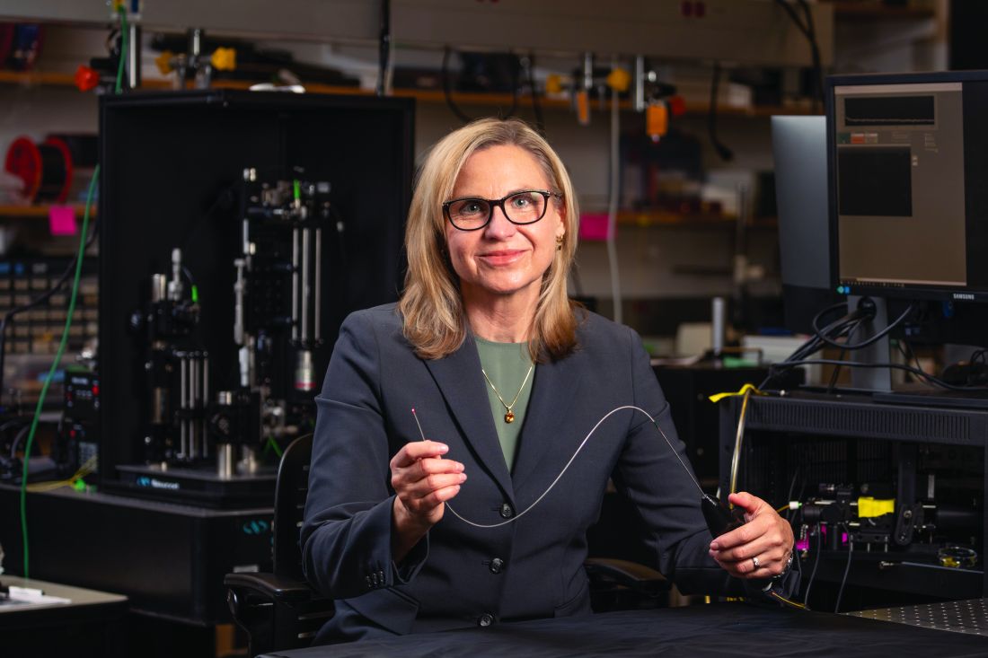
She and her colleagues are developing an OCT/fluorescence imaging falloposcope that is 0.8 mm in diameter, flexible, and steerable, as a tool for early detection of ovarian cancer in humans. “It’s now known that most ovarian cancer starts in the fallopian tubes,” Dr. Barton said. “It’s metastatic disease when those cells break off from the fallopian tubes and go to the ovaries. We wanted to create an imaging system where we created a fiber bundle that we could navigate with white light and with fluorescence so that we can see these early stages of cancer [and] how they fluoresce differently. We also wanted to have an OCT system so that we could image through the wall of the fallopian tube and look for that layer thickening and other precursors to ovarian cancer.”
To date, in vivo testing in healthy women has demonstrated that the miniature endoscope is able to reach the fallopian tubes through the natural orifice of the vagina and uterus. “That is pretty exciting,” she said. “The images may not be of the highest quality, but we are advancing.”
Dr. Barton reported having no relevant financial disclosures.
PHOENIX –
In a lecture during a multispecialty roundup of cutting-edge energy-based device applications at the annual conference of the American Society for Laser Medicine and Surgery, Dr. Barton, a biomedical engineer who directs the BIO5 Institute at the University of Arizona, Tucson, said that while no current modality exists to enable physicians in dermatology and other specialties to view internal structures throughout the entire body with cellular resolution, refining existing technologies is a good way to start.
In 2011, renowned cancer researchers Douglas Hanahan, PhD, and Robert A. Weinberg, PhD, proposed six hallmarks of cancer, which include sustaining proliferative signaling, evading growth suppressors, resisting cell death, enabling replicative immortality, inducing angiogenesis, and activating invasion and metastasis. Each hallmark poses unique imaging challenges. For example, enabling replicative immortality “means that the cell nuclei change size and shape; they change their position,” said Dr. Barton, who is also professor of biomedical engineering and optical sciences at the university. “If we want to see that, we’re going to need an imaging modality that’s subcellular in resolution.”
Similarly, if clinicians want to view how proliferative signaling is changing, “that means being able to visualize the cell surface receptors; those are even smaller to actually visualize,” she said. “But we have technologies where we can target those receptors with fluorophores. And then we can look at large areas very quickly.” Meanwhile, the ability of cancer cells to resist cell death and evade growth suppressors often results in thickening of epithelium throughout the body. “So, if we can measure the thickness of the epithelium, we can see that there’s something wrong with that tissue,” she said.
As for cancer’s propensity for invasion and metastasis, “here, we’re looking at how the collagen structure [between the cells] has changed and whether there’s layer breakdown or not. Optical imaging can detect cancer. However, high resolution optical techniques can only image about 1 mm deep, so unless you’re looking at the skin or the eye, you’re going to have to develop an endoscope to be able to view these hallmarks.”
OCT images the tissue microstructure, generally in a resolution of 2-20 microns, at a depth of 1-2 mm, and it measures reflected light. When possible, Dr. Barton combines OCT with laser-induced fluorescence for enhanced accuracy of detection of cancer. Induced fluorescence senses molecular information with the natural fluorophores in the body or with targeted exogenous agents. Then there’s multiphoton microscopy, an advanced imaging technique that enables clinicians to view cellular and subcellular events within living tissue. Early models of this technology “took up entire benches” in physics labs, Dr. Barton said, but she and other investigators are designing smaller devices for use in clinics. “This is exciting, because not only do we [view] subcellular structure with this modality, but it can also be highly sensitive to collagen structure,” she said.
Ovarian cancer model
In a model of ovarian cancer, she and colleagues externalized the ovaries of a mouse, imaged the organs, put them back in, and reassessed them at 8 weeks. “This model develops cancer very quickly,” said Dr. Barton, who once worked for McDonnell Douglas on the Space Station program. At 8 weeks, using fluorescence and targeted agents with a tabletop multiphoton microscopy system, they observed that the proliferation signals of cancer had begun. “So, with an agent targeted to the folate receptor or to other receptors that are implicated in cancer development, we can see that ovaries and fallopian tubes are lighting up,” she said.
With proof of concept established with the mouse study, she and other researchers are drawing from technological advances to create tiny laser systems for use in the clinic to image a variety of structures in the human body. Optics advances include bulk optics and all-fiber designs where engineers can create an imaging probe that’s only 125 microns in diameter, “or maybe even as small as 70 microns in diameter,” she said. “We can do fabrications on the tips of endoscopes to redirect the light and focus it. We can also do 3-D printing and spiral scanning to create miniature devices to make new advances. That means that instead of just white light imaging of the colon or the lung like we have had in the past, we can start moving into smaller structures, such as the eustachian tube, the fallopian tube, the bile ducts, or making miniature devices for brain biopsies, lung biopsies, and maybe being able to get into bronchioles and arterioles.”
According to Dr. Barton, prior research has demonstrated that cerebral vasculature can be imaged with a catheter 400 microns in diameter, the spaces in the lungs can be imaged with a needle that is 310 microns in diameter, and the inner structures of the eustachian tube can be viewed with an endoscope 1 mm in diameter.
She and her colleagues are developing an OCT/fluorescence imaging falloposcope that is 0.8 mm in diameter, flexible, and steerable, as a tool for early detection of ovarian cancer in humans. “It’s now known that most ovarian cancer starts in the fallopian tubes,” Dr. Barton said. “It’s metastatic disease when those cells break off from the fallopian tubes and go to the ovaries. We wanted to create an imaging system where we created a fiber bundle that we could navigate with white light and with fluorescence so that we can see these early stages of cancer [and] how they fluoresce differently. We also wanted to have an OCT system so that we could image through the wall of the fallopian tube and look for that layer thickening and other precursors to ovarian cancer.”
To date, in vivo testing in healthy women has demonstrated that the miniature endoscope is able to reach the fallopian tubes through the natural orifice of the vagina and uterus. “That is pretty exciting,” she said. “The images may not be of the highest quality, but we are advancing.”
Dr. Barton reported having no relevant financial disclosures.
AT ASLMS 2023
Tackle education and mindset to reduce diabetes distress
, according to two new studies in patients with type 1 diabetes presented at the annual scientific sessions of the American Diabetes Association.
Danielle Hessler Jones, PhD, presented findings from Behavioral Approaches to Reducing Diabetes Distress and Improving Glycemic Control (EMBARK) in adults with type 1 diabetes during an oral session.
The three-arm randomized trial found that patients had the greatest improvements in feelings of powerlessness after a 3-month behavioral intervention that combined type 1 diabetes education plus specific attention to diabetes distress.
And in a late-breaking poster, “Do The Right Thing: Behavioral Intervention for At-Risk T1D Youth,” David V. Wagner, PhD, showed that a behavioral intervention not only improved glycemic management but also reduced cost of care in disadvantaged youth.
“Diabetes distress is the emotional response to living with diabetes, the burden of relentless daily self-management, and the prospect of its long-term complications,” said Dr. Hessler Jones, professor and vice chair for research in the department of family and community medicine at the University of California, San Francisco.
It is common, experienced by 20%-58% of people with type 1 and type 2 diabetes, and is different from depression, as it is associated with glycemic control and disease management. It “is also chronic and does not disappear on its own without intervention,” she stressed.
“It is the expected worries, concerns, and fears that are associated with struggling with a demanding and progressive chronic disease and its management,” she added.
The findings from EMBARK “suggest that distress reductions are greatest when interventions integrate education alongside approaches to address the emotional side of diabetes,” she said.
The group is also analyzing changes in A1c with the three different interventions in EMBARK, with results expected this fall.
Dr. Hessler Jones said they also just received funding for DDASSIST, which will answer the question: “How do I translate this into care in my clinic?” The aim of the clinic training program is to bring the intervention to the diabetes care team.
“Could this program be delivered by somebody else, other than a psychologist?” an audience member asked. They will be looking at this, she replied.
‘Do the right thing’
For the late-breaking poster by Dr. Wagner and colleagues, researchers evaluated direct cost data from three health care systems provided for youth with type 1 diabetes who received an intensive behavioral health intervention, Novel Interventions in Children’s Healthcare (NICH).
Youths were included in the analyses if they had type 1 diabetes and at least 1 year of cost data prior to and following NICH enrollment. Outpatient, emergency department, and inpatient costs were combined. The analysis included 53 youth with the following characteristics: mean age, 14.2 years; 87% Medicaid; 58% female; 32% Black, 29% Non-Hispanic White, 28% Hispanic/Latinx, 7% Pacific Islander, 2% Asian, and 2% other racial and ethnic groups.
Average yearly costs significantly decreased from $20,400 per youth prior to NICH to $9,500 per youth afterward, largely due to inpatient charges.
“These results highlight the benefits of providing access to intensive interventions to pediatric populations experiencing health disparities,” said Dr. Wagner. “Investing early in the lives of youth experiencing health disparities is not only the right thing to do to improve patients’ health but it could also have a positive economic impact down the road.”
Three interventions in 300 adults with type 1 diabetes
Meanwhile, EMBARK recruited 300 patients with type 1 diabetes in the United States from clinics and community organizations who were aged 21 and older and had an elevated type 1 diabetes distress score (> 2.0) and A1c greater than or equal to 7.5%.
Participants were a mean age of 46 years, 79% were female, and 89% were White. They had a type 1 diabetes distress score of 2.8, a mean A1c of 8.3%, and 71% used an insulin pump and 79% used a continuous glucose monitor.
Participants were randomized to one of three interventions:
- Streamline: A traditional diabetes educator-led education and management program.
- Tuned-in: A psychologist-led program focused exclusively on reducing diabetes distress.
- Fixit: An integration of the two programs.
Interventions were given virtually over 3 months to small groups of 8-12 individuals and included initial workshops, one-to-one phone calls, and follow-up group meetings. Participants were then followed for 8 months.
Researchers found statistically significant and substantial reductions in overall diabetes distress in all three interventions, with the greatest reductions in the combined intervention group, which were greater than in the educational approach alone group (P = .005).
The percentage of participants who no longer reported elevated diabetes distress at follow-up was 25% in Streamline, 37% in Tuned-in, and 42% in Fixit.
The percentage of participants who reported a minimal clinically important difference – the smallest change in a treatment outcome that an individual would identify as important – was also greatest in those in the Fixit intervention group (82%) than in the Tuned-in (74%) or Streamline (65%) interventions.
‘Adding the psychologist is where the real magic happens’
“The certified diabetes care specialist intervention is really a very standard thing that most clinicians would have access to; they tend to focus on knowledge and problem solving and some of the psychosocial issues,” Robert Gabbay, MD, PhD, chief scientific and medical officer for the ADA, said in an interview.
The EMBARK trial found a “graded response: CDCS alone, psychologist really focused on diabetes distress, and the two together, which would be the ideal practice model,” he noted.
There are these validated ways of measuring diabetes distress using a diabetes distress survey tool, which is also underutilized.
“Adding the psychologist is really where the real magic happens in terms of diabetes distress,” Dr. Gabbay said.
“As you can imagine, [if] somebody ... feels powerless, it is going to be tough to manage their diabetes and unlikely to be terribly successful,” he observed. Often, these individuals are just not doing well. This study highlighted the importance of identifying this.
“I’m encouraged by the findings from the studies presented during this year’s Scientific Sessions as we continue to seek out innovative, evidence-based solutions that support people living with diabetes when they need it the most,” Dr. Gabbay concluded.
A version of this article originally appeared on Medscape.com.
FROM ADA 2023
‘Striking’ benefit of lipid lowering in primary prevention
SAN DIEGO – two-thirds of whom also had type 2 diabetes, leading to calls for more attention to be paid to this group of patients.
The main results of the CLEAR Outcomes trial of bempedoic acid (Nexletol, Esperion) in a mixed secondary and primary prevention population intolerant to statins, presented in March at the 2023 joint scientific sessions of the American College of Cardiology and the World Heart Federation, showed a 13% relative risk reduction in the main primary endpoint, a composite of cardiovascular death, myocardial infarction, stroke, or coronary revascularization.
This new analysis of the 4,206 high-risk primary prevention patients in the study – 67% of whom also had type 2 diabetes – has shown a 30% relative risk reduction in the same endpoint.
Other key endpoints were reduced to a similar or even greater extent, with the composite of cardiovascular death/stroke/MI showing a 36% relative risk reduction, and a 39% relative risk reduction for cardiovascular death and MI individually.

“These results are frankly striking,” lead investigator Steve Nissen, MD, said in an interview.
“These are really large reductions. These results are telling us that high-risk primary prevention patients, although their absolute event rate is lower than secondary prevention patients, can have very impressive relative risk reductions in major cardiovascular events with lipid-lowering therapy,” he said.
But Dr. Nissen, chief academic officer at the Heart Vascular & Thoracic Institute at the Cleveland Clinic, pointed out that this population of patients is not well treated.
“This is the problem: Less than half of high-risk primary prevention patients in the U.S., and in virtually every other developed country, are receiving cholesterol-lowering medication. These patients tend to get ignored,” he stressed.
Asked what advice he would give to clinicians based on the current findings, Dr. Nissen said: “If a patient is at high risk of developing cardiovascular disease, particularly those with [type 2] diabetes, they need to go on a lipid-lowering drug.”
“If patients can tolerate a statin then that should be the first choice. We know statins work, and they are now inexpensive. They are likely to give the exact same benefit as we have shown in this study with bempedoic acid, as the two drug classes work by very similar mechanisms. But if patients can’t tolerate a statin, then treat them with bempedoic acid. The bottom line is that these patients just need to be treated,” he said.
‘Wake-up call’
He said these new results are a “wake-up call for the medical community that we need to pay far more attention to high-risk primary prevention patients.”
Dr. Nissen does not believe the effect is specific to bempedoic acid; rather, it is more likely an effect of lowering LDL cholesterol (LDL-C) levels.
“This message is not about bempedoic acid, in particular. We have seen similar findings in historical studies with the statins, but that seems to have been forgotten. The message is about lowering LDL in patients who are at high risk of having a first cardiovascular event. We need to identify patients at high risk for a first cardiac event and get them on a cholesterol-lowering drug – and in most cases that will be a statin.”
Dr. Nissen presented the new analysis from the CLEAR OUTCOMES trial at the annual scientific sessions of the American Diabetes Association. It was simultaneously published online in JAMA.
He pointed out that large trials of lipid-lowering therapy in the primary prevention population have not been done for many years.
“All the contemporary trials with lipid-lowering therapy have only included secondary prevention patients and they often enroll patients after an acute coronary syndrome event.
“But for the CLEAR OUTCOMES trial, we included a significant amount of primary prevention patients – those with risk factors such as [type 2] diabetes and hypertension who are considered to be at high risk of developing cardiovascular disease,” he explained.
CLEAR OUTCOMES was a masked, randomized, trial that enrolled 13,970 statin-intolerant patients. The new analysis included 4,206 of those patients with risk factors for heart disease but without a prior cardiovascular event – the primary prevention group. The mean age of these participants was 68 years, 67% had diabetes, and 59% were women.
Treatment with bempedoic acid showed a 22% reduction in LDL-C, compared with placebo, with a reduction of 30.2 mg/dL from a mean baseline of 142.5 mg/dL. High-sensitivity C-reactive protein (CRP) levels were also reduced by 0.56 mg/L (21.5%), from a median baseline of 2.4 mg/L.
Dr. Nissen told a press briefing at the ADA meeting that he believes “it’s the combination of LDL lowering and reduction in CRP that might have been the driver [for the effects we saw in the trial]. Certainly, bempedoic acid lowers both.”
And he noted the recent U.S. approval of a new low dose of colchicine 0.5 mg (Lodoco, Agepha Pharma) with a broad indication for use in atherosclerotic cardiovascular disease (ASCVD), which represents a completely new approach to treatment, specifically targeting inflammation as a driver of atherosclerosis.
Bempedoic acid is a prodrug that works along the same pathways as statins but does not cause muscle pain, which makes many people intolerant to statins. Bempedoic acid was first approved by the Food and Drug Administration in 2020 for the treatment of adults with heterozygous familial hypercholesterolemia or established ASCVD who require additional LDL-C lowering.
Greater benefit in primary prevention?
In this primary prevention group, treatment with bempedoic acid for 40 months was associated with a significant risk reduction for the primary endpoint – a composite of cardiovascular death, nonfatal MI, nonfatal stroke, or coronary revascularization – which occurred in 5.3% of the treatment group versus 7.6% in the placebo group (adjusted hazard ratio, 0.70; P = .002). This represents a 30% relative risk reduction in major cardiovascular events.
Other key secondary endpoints also showed impressive reductions.
The rate of the composite endpoint of cardiovascular death, MI, or stroke was 6.4% in the placebo group and 4.0% with bempedoic acid (HR, 0.64; P < .001); MI occurred in 2.2% versus 1.4% (HR, 0.61), cardiovascular death in 3.1% versus 1.8% (HR, 0.61), and all-cause mortality in 5.2% versus 3.6% (HR, 0.73), respectively.
Adverse effects with bempedoic acid included a higher incidence of gout (2.6% vs 2.0%), cholelithiasis (2.5% vs. 1.1%), and increases in serum creatinine, uric acid, and hepatic enzyme levels.
Dr. Nissen believed these results suggest that there may be a greater benefit of lipid lowering in high-risk primary prevention patients than in the secondary prevention population.
“It may seem paradoxical, but there is actually some history that this may be the case,” he said.
He pointed out that the JUPITER trial of rosuvastatin in 2008 was the last major primary prevention trial of a lipid-lowering agent, which was stopped early with a 44% reduction of the primary endpoint.
He noted that one of the arguments against the use of statins in primary prevention is the belief that absolute risk reductions are quite modest.
“But in this analysis, we found an absolute risk reduction of 2.3% for the primary endpoint. That’s a number needed to treat to prevent 1 event of 43. That’s pretty good,” he said.
Trying to explain why there may be more benefit in the primary prevention population, Dr. Nissen suggested that these patients may have more vulnerable plaques.
“I think high-risk primary prevention patients probably have a lot of lipid-laden plaque – some people call it ‘vulnerable’ plaque. These are softer, cholesterol-laden plaque. We know that treatment with cholesterol-lowering medication causes these plaques to shrink. The lipid core is delipidated and the plaque stabilizes,” he explained. “It may be that in secondary prevention patients to some extent the horse is already out of the barn – they have advanced disease. But primary prevention patients may have plaques that are more amenable to modification by cholesterol lowering.”
He admitted that the idea is only speculation. “But that is a potential explanation for our observations.”
Editorial cautious
In an accompanying editorial, also published in JAMA, Dhruv S. Kazi, MD, Beth Israel Deaconess Medical Center, Boston, said the findings need to be interpreted with caution as they come from one of many subgroup analyses of a larger trial.
Dr. Kazi pointed out that the intervention and control survival curves separate right away, on the first day of follow-up, whereas the true effect of lipid-lowering therapy for primary prevention would be expected to have a somewhat delayed onset, an observation he says supports the argument that this is a chance finding.
Dr. Kazi also reminded clinicians that bempedoic acid should not be regarded as a substitute for statins, which should remain the first-line therapy for primary prevention.
“For now, available evidence suggests that, although bempedoic acid is not a perfect substitute for a statin, it is a reasonable therapeutic choice for primary prevention of ASCVD events in high-risk, statin-intolerant patients,” he concluded.
A version of this article first appeared on Medscape.com.
SAN DIEGO – two-thirds of whom also had type 2 diabetes, leading to calls for more attention to be paid to this group of patients.
The main results of the CLEAR Outcomes trial of bempedoic acid (Nexletol, Esperion) in a mixed secondary and primary prevention population intolerant to statins, presented in March at the 2023 joint scientific sessions of the American College of Cardiology and the World Heart Federation, showed a 13% relative risk reduction in the main primary endpoint, a composite of cardiovascular death, myocardial infarction, stroke, or coronary revascularization.
This new analysis of the 4,206 high-risk primary prevention patients in the study – 67% of whom also had type 2 diabetes – has shown a 30% relative risk reduction in the same endpoint.
Other key endpoints were reduced to a similar or even greater extent, with the composite of cardiovascular death/stroke/MI showing a 36% relative risk reduction, and a 39% relative risk reduction for cardiovascular death and MI individually.
“These results are frankly striking,” lead investigator Steve Nissen, MD, said in an interview.
“These are really large reductions. These results are telling us that high-risk primary prevention patients, although their absolute event rate is lower than secondary prevention patients, can have very impressive relative risk reductions in major cardiovascular events with lipid-lowering therapy,” he said.
But Dr. Nissen, chief academic officer at the Heart Vascular & Thoracic Institute at the Cleveland Clinic, pointed out that this population of patients is not well treated.
“This is the problem: Less than half of high-risk primary prevention patients in the U.S., and in virtually every other developed country, are receiving cholesterol-lowering medication. These patients tend to get ignored,” he stressed.
Asked what advice he would give to clinicians based on the current findings, Dr. Nissen said: “If a patient is at high risk of developing cardiovascular disease, particularly those with [type 2] diabetes, they need to go on a lipid-lowering drug.”
“If patients can tolerate a statin then that should be the first choice. We know statins work, and they are now inexpensive. They are likely to give the exact same benefit as we have shown in this study with bempedoic acid, as the two drug classes work by very similar mechanisms. But if patients can’t tolerate a statin, then treat them with bempedoic acid. The bottom line is that these patients just need to be treated,” he said.
‘Wake-up call’
He said these new results are a “wake-up call for the medical community that we need to pay far more attention to high-risk primary prevention patients.”
Dr. Nissen does not believe the effect is specific to bempedoic acid; rather, it is more likely an effect of lowering LDL cholesterol (LDL-C) levels.
“This message is not about bempedoic acid, in particular. We have seen similar findings in historical studies with the statins, but that seems to have been forgotten. The message is about lowering LDL in patients who are at high risk of having a first cardiovascular event. We need to identify patients at high risk for a first cardiac event and get them on a cholesterol-lowering drug – and in most cases that will be a statin.”
Dr. Nissen presented the new analysis from the CLEAR OUTCOMES trial at the annual scientific sessions of the American Diabetes Association. It was simultaneously published online in JAMA.
He pointed out that large trials of lipid-lowering therapy in the primary prevention population have not been done for many years.
“All the contemporary trials with lipid-lowering therapy have only included secondary prevention patients and they often enroll patients after an acute coronary syndrome event.
“But for the CLEAR OUTCOMES trial, we included a significant amount of primary prevention patients – those with risk factors such as [type 2] diabetes and hypertension who are considered to be at high risk of developing cardiovascular disease,” he explained.
CLEAR OUTCOMES was a masked, randomized, trial that enrolled 13,970 statin-intolerant patients. The new analysis included 4,206 of those patients with risk factors for heart disease but without a prior cardiovascular event – the primary prevention group. The mean age of these participants was 68 years, 67% had diabetes, and 59% were women.
Treatment with bempedoic acid showed a 22% reduction in LDL-C, compared with placebo, with a reduction of 30.2 mg/dL from a mean baseline of 142.5 mg/dL. High-sensitivity C-reactive protein (CRP) levels were also reduced by 0.56 mg/L (21.5%), from a median baseline of 2.4 mg/L.
Dr. Nissen told a press briefing at the ADA meeting that he believes “it’s the combination of LDL lowering and reduction in CRP that might have been the driver [for the effects we saw in the trial]. Certainly, bempedoic acid lowers both.”
And he noted the recent U.S. approval of a new low dose of colchicine 0.5 mg (Lodoco, Agepha Pharma) with a broad indication for use in atherosclerotic cardiovascular disease (ASCVD), which represents a completely new approach to treatment, specifically targeting inflammation as a driver of atherosclerosis.
Bempedoic acid is a prodrug that works along the same pathways as statins but does not cause muscle pain, which makes many people intolerant to statins. Bempedoic acid was first approved by the Food and Drug Administration in 2020 for the treatment of adults with heterozygous familial hypercholesterolemia or established ASCVD who require additional LDL-C lowering.
Greater benefit in primary prevention?
In this primary prevention group, treatment with bempedoic acid for 40 months was associated with a significant risk reduction for the primary endpoint – a composite of cardiovascular death, nonfatal MI, nonfatal stroke, or coronary revascularization – which occurred in 5.3% of the treatment group versus 7.6% in the placebo group (adjusted hazard ratio, 0.70; P = .002). This represents a 30% relative risk reduction in major cardiovascular events.
Other key secondary endpoints also showed impressive reductions.
The rate of the composite endpoint of cardiovascular death, MI, or stroke was 6.4% in the placebo group and 4.0% with bempedoic acid (HR, 0.64; P < .001); MI occurred in 2.2% versus 1.4% (HR, 0.61), cardiovascular death in 3.1% versus 1.8% (HR, 0.61), and all-cause mortality in 5.2% versus 3.6% (HR, 0.73), respectively.
Adverse effects with bempedoic acid included a higher incidence of gout (2.6% vs 2.0%), cholelithiasis (2.5% vs. 1.1%), and increases in serum creatinine, uric acid, and hepatic enzyme levels.
Dr. Nissen believed these results suggest that there may be a greater benefit of lipid lowering in high-risk primary prevention patients than in the secondary prevention population.
“It may seem paradoxical, but there is actually some history that this may be the case,” he said.
He pointed out that the JUPITER trial of rosuvastatin in 2008 was the last major primary prevention trial of a lipid-lowering agent, which was stopped early with a 44% reduction of the primary endpoint.
He noted that one of the arguments against the use of statins in primary prevention is the belief that absolute risk reductions are quite modest.
“But in this analysis, we found an absolute risk reduction of 2.3% for the primary endpoint. That’s a number needed to treat to prevent 1 event of 43. That’s pretty good,” he said.
Trying to explain why there may be more benefit in the primary prevention population, Dr. Nissen suggested that these patients may have more vulnerable plaques.
“I think high-risk primary prevention patients probably have a lot of lipid-laden plaque – some people call it ‘vulnerable’ plaque. These are softer, cholesterol-laden plaque. We know that treatment with cholesterol-lowering medication causes these plaques to shrink. The lipid core is delipidated and the plaque stabilizes,” he explained. “It may be that in secondary prevention patients to some extent the horse is already out of the barn – they have advanced disease. But primary prevention patients may have plaques that are more amenable to modification by cholesterol lowering.”
He admitted that the idea is only speculation. “But that is a potential explanation for our observations.”
Editorial cautious
In an accompanying editorial, also published in JAMA, Dhruv S. Kazi, MD, Beth Israel Deaconess Medical Center, Boston, said the findings need to be interpreted with caution as they come from one of many subgroup analyses of a larger trial.
Dr. Kazi pointed out that the intervention and control survival curves separate right away, on the first day of follow-up, whereas the true effect of lipid-lowering therapy for primary prevention would be expected to have a somewhat delayed onset, an observation he says supports the argument that this is a chance finding.
Dr. Kazi also reminded clinicians that bempedoic acid should not be regarded as a substitute for statins, which should remain the first-line therapy for primary prevention.
“For now, available evidence suggests that, although bempedoic acid is not a perfect substitute for a statin, it is a reasonable therapeutic choice for primary prevention of ASCVD events in high-risk, statin-intolerant patients,” he concluded.
A version of this article first appeared on Medscape.com.
AT ADA 2023
SURMOUNT-2: Tirzepatide rings up major weight loss in type 2 diabetes
SAN DIEGO – in the SURMOUNT-2 pivotal trial, a finding that will likely lead to Food and Drug Administration approval of a new indication for weight loss for tirzepatide.
Tirzepatide received FDA approval as a treatment for type 2 diabetes in adults, marketed as Mounjaro, in 2022. The agent – a “twincretin” that acts as an agonist at both the glucagon-like peptide-1 (GLP-1) receptor and glucose-dependent insulinotropic polypeptide (GIP) receptor – had also previously scored a decisive win for weight loss in adults with overweight or obesity without diabetes in the SURMOUNT-1 pivotal trial.
Taken together, results from SURMOUNT-1 and SURMOUNT-2 appear to make a good case for a weight-loss indication that will not depend on whether a patient also has type 2 diabetes.
“We anticipate that tirzepatide will be [FDA] approved for weight loss later this year,” W. Timothy Garvey, MD, lead researcher for SURMOUNT-2, said during a press briefing at the annual scientific sessions of the American Diabetes Association.
Tirzepatide ‘fills the gap’
Tirzepatide “fills the gap to get [medication-driven] weight loss in the range of 15% of baseline weight or better,” Dr. Garvey noted, which puts it in a favorable position relative to a 2.4-mg weekly subcutaneous injection with the GLP-1 agonist semaglutide (Wegovy), which produced an average weight loss from baseline of about 9.6% in people with type 2 diabetes in the STEP-2 trial.

Although tirzepatide has not been compared head-to-head for weight loss with any of the several available GLP-1 agonists, the reported weight-loss numbers seem to favor tirzepatide, said Dr. Garvey, director of the Diabetes Research Center of the University of Alabama at Birmingham.
“If you look at the degree of weight loss across trials, we see a clinically significant difference in weight loss” compared with semaglutide and other agents that only act on the GLP-1 receptor, he noted. (Although cross-trial comparisons of different medications often have uncertain reliability.)
“The data suggest an incremental effect from tirzepatide” compared with the GLP-1 agonists now approved for weight loss, said Marlon Pragnell, PhD, vice president, research and science, ADA, who was not involved in the tirzepatide studies.
This is a “step forward for treating people with obesity and type 2 diabetes; it’s a very promising treatment option,” Dr. Pragnell said in an interview.
Tirzepatide the ‘most effective agent’
Ildiko Lingvay, MD, the designated discussant for the SURMOUNT-2 presentation at the meeting, fully agreed. The new findings “confirm that tirzepatide is the most effective agent currently on the [U.S.] market to help achieve the two coprimary goals for patients with type 2 diabetes – weight loss and glycemic control – while also having favorable effects on cardiovascular risk factors,” said Dr. Lingvay, an endocrinologist at UT Southwestern Medical Center in Dallas, who was not involved with the SURMOUNT studies.

Dr. Lingvay offered as evidence the performance of tirzepatide’s main rival for weight loss semaglutide (Wegovy), delivered at the 2.4 mg/week subcutaneous injected dosage approved for weight loss. The semaglutide trial that SURMOUNT-2 most resembles is the STEP-2 trial, she said, which showed as its primary outcome a 9.6% average weight loss from baseline after 68 weeks of weekly semaglutide that compares, in a cross-trial way, with the 14.7% average drop from baseline weight with 15 mg tirzepatide weekly for 72 weeks and an average 12.8% weight loss with a weekly 10-mg tirzepatide dose.
“It’s fair to say that tirzepatide has an edge,” despite the limitations of cross-trial comparisons, Dr. Lingvay said in an interview.
But she acknowledged that superior weight loss efficacy takes a back seat in U.S. practice to access and affordability when making a prescribing decision for individual patients as these newer drugs are all expensive.
Affordability and access will remain a ‘big problem’
Dr. Garvey, too, cautioned that access and affordability of tirzepatide as well as other GLP-1 agonists remains a major sticking point.
“These medications are very expensive – more than $1,000 a dose – and this cost limits access ... [which is] a big problem,” Dr. Garvey noted. U.S. health care payers “do not want to open the gates [to expensive treatments] for a disorder that’s as common as obesity.”
“Access and affordability are always an issue for these medications,” agreed Janet Brown-Friday, RN, president, health care and education, ADA, who had no role in the tirzepatide studies.
SURMOUNT-2 randomized 938 adults with type 2 diabetes and overweight or obesity at 77 centers in seven countries including the United States from March 2021 to April 2023. The study had two primary outcomes: Average percent change in body weight from baseline to week 72, and percentage of participants who achieved a weight reduction from baseline of at least 5% after 72 weeks.
In-trial weight loss of 12.8%-14.7%
The in-trial analysis showed that a 10-mg weekly subcutaneous dose of tirzepatide resulted in an average 12.8% weight loss from baseline, and a 15-mg weekly subcutaneous dose led to an average 14.7% drop from baseline weight. People randomized to receive a placebo injection averaged a 3.2% drop from their baseline weight after 72 weeks, a finding that documents significant improvements compared with placebo with both tirzepatide doses.
The percentage of patients who achieved at least a 5% reduction in weight from baseline was 79% with the 10-mg dose of tirzepatide, 83% with the 15-mg dose, and 32% with placebo; these improvements were significant for both tirzepatide doses compared with placebo.
A 15% or greater reduction in weight from baseline occurred in 40%-48% of people who received tirzepatide compared with 3% of those who received placebo. A reduction in weight of this magnitude from baseline “will prevent a broad array of complications,” Dr. Garvey noted.
The results were simulatenously published online in The Lancet.
Glucose control without severe hypoglycemia
The safety profile of tirzepatide in SURMOUNT-2 was consistent with prior studies of the agent, as well as with other medications in the GLP-1 agonist class, with gastrointestinal adverse effects such as nausea and vomiting predominating, especially during the dose-escalation phase at treatment onset.
Dr. Garvey especially highlighted the overall safety of tirzepatide, and particularly its ability to produce clinically important reductions in A1c that averaged more than two percentage points from baseline values without producing a single episode of severe hypoglycemia, and an incidence of milder hypoglycemia of less than a 5%.
The absence of any severe hypoglycemia was “amazing,” Dr. Garvey said, especially given that 46%-49% of people taking tirzepatide in SURMOUNT-2 achieved normalization of their A1c to less than 5.7% on treatment compared with 4% of participants taking placebo.
The results also showed the benefit of a “big reduction in fasting insulin levels,” which averaged a 41% cut from baseline in those who received the 15-mg subcutaneous weekly dose of tirzepatide, coupled with increased insulin sensitivity, Dr. Garvey said.
Dr. Garvey disclosed ties to Eli Lilly, which sponsored SURMOUNT-2 and markets tirzepatide (Mounjaro), as well Boehringer Ingelheim, Novo Nordisk, Pfizer, Fractyl Health, Alnylam Pharmaceuticals, Inogen, and Merck. He has been an investigator for studies sponsored by Novo Nordisk, Epitomee, Neurovalens, and Pfizer. Dr. Pragnell and Dr. Brown-Friday have disclosed no relevant financial relationships.
A version of this article first appeared on Medscape.com.
SAN DIEGO – in the SURMOUNT-2 pivotal trial, a finding that will likely lead to Food and Drug Administration approval of a new indication for weight loss for tirzepatide.
Tirzepatide received FDA approval as a treatment for type 2 diabetes in adults, marketed as Mounjaro, in 2022. The agent – a “twincretin” that acts as an agonist at both the glucagon-like peptide-1 (GLP-1) receptor and glucose-dependent insulinotropic polypeptide (GIP) receptor – had also previously scored a decisive win for weight loss in adults with overweight or obesity without diabetes in the SURMOUNT-1 pivotal trial.
Taken together, results from SURMOUNT-1 and SURMOUNT-2 appear to make a good case for a weight-loss indication that will not depend on whether a patient also has type 2 diabetes.
“We anticipate that tirzepatide will be [FDA] approved for weight loss later this year,” W. Timothy Garvey, MD, lead researcher for SURMOUNT-2, said during a press briefing at the annual scientific sessions of the American Diabetes Association.
Tirzepatide ‘fills the gap’
Tirzepatide “fills the gap to get [medication-driven] weight loss in the range of 15% of baseline weight or better,” Dr. Garvey noted, which puts it in a favorable position relative to a 2.4-mg weekly subcutaneous injection with the GLP-1 agonist semaglutide (Wegovy), which produced an average weight loss from baseline of about 9.6% in people with type 2 diabetes in the STEP-2 trial.
Although tirzepatide has not been compared head-to-head for weight loss with any of the several available GLP-1 agonists, the reported weight-loss numbers seem to favor tirzepatide, said Dr. Garvey, director of the Diabetes Research Center of the University of Alabama at Birmingham.
“If you look at the degree of weight loss across trials, we see a clinically significant difference in weight loss” compared with semaglutide and other agents that only act on the GLP-1 receptor, he noted. (Although cross-trial comparisons of different medications often have uncertain reliability.)
“The data suggest an incremental effect from tirzepatide” compared with the GLP-1 agonists now approved for weight loss, said Marlon Pragnell, PhD, vice president, research and science, ADA, who was not involved in the tirzepatide studies.
This is a “step forward for treating people with obesity and type 2 diabetes; it’s a very promising treatment option,” Dr. Pragnell said in an interview.
Tirzepatide the ‘most effective agent’
Ildiko Lingvay, MD, the designated discussant for the SURMOUNT-2 presentation at the meeting, fully agreed. The new findings “confirm that tirzepatide is the most effective agent currently on the [U.S.] market to help achieve the two coprimary goals for patients with type 2 diabetes – weight loss and glycemic control – while also having favorable effects on cardiovascular risk factors,” said Dr. Lingvay, an endocrinologist at UT Southwestern Medical Center in Dallas, who was not involved with the SURMOUNT studies.
Dr. Lingvay offered as evidence the performance of tirzepatide’s main rival for weight loss semaglutide (Wegovy), delivered at the 2.4 mg/week subcutaneous injected dosage approved for weight loss. The semaglutide trial that SURMOUNT-2 most resembles is the STEP-2 trial, she said, which showed as its primary outcome a 9.6% average weight loss from baseline after 68 weeks of weekly semaglutide that compares, in a cross-trial way, with the 14.7% average drop from baseline weight with 15 mg tirzepatide weekly for 72 weeks and an average 12.8% weight loss with a weekly 10-mg tirzepatide dose.
“It’s fair to say that tirzepatide has an edge,” despite the limitations of cross-trial comparisons, Dr. Lingvay said in an interview.
But she acknowledged that superior weight loss efficacy takes a back seat in U.S. practice to access and affordability when making a prescribing decision for individual patients as these newer drugs are all expensive.
Affordability and access will remain a ‘big problem’
Dr. Garvey, too, cautioned that access and affordability of tirzepatide as well as other GLP-1 agonists remains a major sticking point.
“These medications are very expensive – more than $1,000 a dose – and this cost limits access ... [which is] a big problem,” Dr. Garvey noted. U.S. health care payers “do not want to open the gates [to expensive treatments] for a disorder that’s as common as obesity.”
“Access and affordability are always an issue for these medications,” agreed Janet Brown-Friday, RN, president, health care and education, ADA, who had no role in the tirzepatide studies.
SURMOUNT-2 randomized 938 adults with type 2 diabetes and overweight or obesity at 77 centers in seven countries including the United States from March 2021 to April 2023. The study had two primary outcomes: Average percent change in body weight from baseline to week 72, and percentage of participants who achieved a weight reduction from baseline of at least 5% after 72 weeks.
In-trial weight loss of 12.8%-14.7%
The in-trial analysis showed that a 10-mg weekly subcutaneous dose of tirzepatide resulted in an average 12.8% weight loss from baseline, and a 15-mg weekly subcutaneous dose led to an average 14.7% drop from baseline weight. People randomized to receive a placebo injection averaged a 3.2% drop from their baseline weight after 72 weeks, a finding that documents significant improvements compared with placebo with both tirzepatide doses.
The percentage of patients who achieved at least a 5% reduction in weight from baseline was 79% with the 10-mg dose of tirzepatide, 83% with the 15-mg dose, and 32% with placebo; these improvements were significant for both tirzepatide doses compared with placebo.
A 15% or greater reduction in weight from baseline occurred in 40%-48% of people who received tirzepatide compared with 3% of those who received placebo. A reduction in weight of this magnitude from baseline “will prevent a broad array of complications,” Dr. Garvey noted.
The results were simulatenously published online in The Lancet.
Glucose control without severe hypoglycemia
The safety profile of tirzepatide in SURMOUNT-2 was consistent with prior studies of the agent, as well as with other medications in the GLP-1 agonist class, with gastrointestinal adverse effects such as nausea and vomiting predominating, especially during the dose-escalation phase at treatment onset.
Dr. Garvey especially highlighted the overall safety of tirzepatide, and particularly its ability to produce clinically important reductions in A1c that averaged more than two percentage points from baseline values without producing a single episode of severe hypoglycemia, and an incidence of milder hypoglycemia of less than a 5%.
The absence of any severe hypoglycemia was “amazing,” Dr. Garvey said, especially given that 46%-49% of people taking tirzepatide in SURMOUNT-2 achieved normalization of their A1c to less than 5.7% on treatment compared with 4% of participants taking placebo.
The results also showed the benefit of a “big reduction in fasting insulin levels,” which averaged a 41% cut from baseline in those who received the 15-mg subcutaneous weekly dose of tirzepatide, coupled with increased insulin sensitivity, Dr. Garvey said.
Dr. Garvey disclosed ties to Eli Lilly, which sponsored SURMOUNT-2 and markets tirzepatide (Mounjaro), as well Boehringer Ingelheim, Novo Nordisk, Pfizer, Fractyl Health, Alnylam Pharmaceuticals, Inogen, and Merck. He has been an investigator for studies sponsored by Novo Nordisk, Epitomee, Neurovalens, and Pfizer. Dr. Pragnell and Dr. Brown-Friday have disclosed no relevant financial relationships.
A version of this article first appeared on Medscape.com.
AT ADA 2023
The metaverse is the dermatologist’s ally
MADRID – There are endless possibilities within the dermoverse (a term coined by joining “dermatology” and “metaverse”), from a robot office assistant to the brand new world it offers for virtual training and simulation.
A group of dermatologists expert in new technologies came together at the 50th National Congress of the Spanish Academy for Dermatology and Venereology to discuss the metaverse: that sum of all virtual spaces that bridges physical and digital reality, where users interact through their avatars and where these experts are discovering new opportunities for treating their patients. The metaverse and AI offer a massive opportunity for improving telehealth visits, immersive surgical planning, or virtual training using 3-D skin models. These are just a few examples of what this technology may eventually provide.
“The possibilities offered by the metaverse in the field of dermatology could be endless,” explained Miriam Fernández-Parrado, MD, dermatologist at Navarre Hospital, Pamplona, Spain. To her, “the metaverse could mean a step forward in teledermatology, which has come of age as a result of the pandemic.” These past few years have shown that it’s possible to perform some screenings online. This, in turn, has produced significant time and cost savings, along with greater efficacy in initial screening and early detection of serious diseases.
The overall percentage of cases that are potentially treatable in absentia is estimated to exceed 70%. “This isn’t a matter of replacing in-person visits but of finding a quality alternative that, far from dehumanizing the doctor-patient relationship, helps to satisfy the growing need for this relationship,” said Dr. Fernández-Parrado.
Always on duty
Julián Conejo-Mir, MD, PhD, professor and head of dermatology at the Virgen del Rocío Hospital in Seville, Spain, told this news organization that AI will help with day-to-day interactions with patients. It’s already a reality. “But to say that with a simple photo, we can address 70% of dermatology cases without being physically present with our patients – I don’t think that will become a reality in the next 20 years.”
Currently, algorithms can identify tumors with high success rates (80%-90%) using photographs and dermoscopic images; rates increase significantly when both kinds of images are available. These high success rates are possible because tumor morphology is stationary. “However, for inflammatory conditions, accurate diagnosis generally doesn’t exceed 60%, since these are conditions in which morphology can change a lot from one day to the next and can vary significantly, depending on their anatomic location or the patient’s age.”
Maybe once metaclinics, with 3-D virtual reality, have been established and clinicians can see the patient in real time from their offices, the rate of accurate diagnosis will reach 70%, especially with patients who have limited mobility or who live at a distance from the hospital. “But that’s still 10-15 years away, since more powerful computers are needed, most likely quantum computers,” cautioned Dr. Conejo-Mir.
The patient’s ally
In clinical practice, facilitating access to the dermoverse may help reduce pain and divert the patient’s attention, especially during in-person visits that require bothersome or uncomfortable interventions. “This is especially effective in pediatric dermatology, since settings of immersive virtual reality may contribute to relaxation among children,” explained Dr. Fernández-Parrado. She also sees potential applications among patients who need surgery. The metaverse would allow them to preview a simulation of their operation before undergoing it, thus reducing their anxiety and allaying their fears about these procedures.
Two lines are being pursued: automated diagnosis for telehealth consultations, which are primarily for tumors, and robotic office assistants.
“We have been using the first one in clinical practice, and we can achieve a success rate of 85%-90%.” The second one is much more complex, “and we’re having a hard time moving it forward within our research team, since it doesn’t involve only one algorithm. Instead, it requires five algorithms working together simultaneously (chatbot, automatic writing, image analysis, selecting the most appropriate treatment, ability to make recommendations, and even an additional one involving feelings),” explained Dr. Conejo-Mir.
A wise consultant
Dr. Conejo-Mir offered examples of how this might work in the near future. “In under 5 years, you’ll be able to sit in front of a computer or your smartphone, talk to an avatar that we’re able to select (sex, appearance, age, kind/serious), show the avatar your lesions, and it will tell us a basic diagnostic impression and even the treatment.”
With virtual learning, physicians can also gather knowledge or take refresher courses, using skin models in augmented reality with tumors and other skin lesions, or using immersive simulation courses that aid learning. Digital models that replicate the anatomy and elasticity of the skin or other characteristics unique to the patient can be used to reach decisions regarding surgeries and to practice interventions before entering the operating room, explained Dr. Fernández-Parrado.
Optimal virtual training
Virtual reality and simulation will doubtless play a major role in this promising field of using these devices for training purposes. “There will be virtual dermatology clinics or metaclinics, where you can do everything with virtual simulated patients, from gaining experience in interviews or health histories (even with patients who are difficult to deal with), to taking biopsies and performing interventions,” said Dr. Conejo-Mir.
A recent study titled “How the World Sees the Metaverse and Extended Reality” gathered data from 29 countries regarding the next 10 years. One of the greatest benefits of this technology is expected in health resources (59%), even more than in the trading of digital assets. While it is difficult to predict when the dermoverse will be in operation, Dr. Fernández-Parrado says she’s a techno-optimist. Together with Dr. Héctor Perandones, MD, a dermatologist at the University Healthcare Complex in León, Spain, and coauthor with Dr. Fernández-Parrado of the article, “A New Universe in Dermatology: From Metaverse to Dermoverse,” she’s convinced that “if we can imagine it, we can create it.”
A differential diagnostician
Over the past 10 years, AI has become a major ally of dermatology, providing new techniques that simplify the diagnosis and treatment of patients. There are many applications for which it adds tremendous value in dermatology: establishing precise differential diagnoses for common diseases, such as psoriasis, atopic dermatitis, or acne; eveloping personalized therapeutic protocols; and predicting medium- and long-term outcomes.
Furthermore, in onco-dermatology, AI has helped to automate the diagnosis of skin tumors by making it possible to differentiate between melanocytic and nonmelanocytic lesions. This distinction promotes early diagnosis and helps produce screening systems that are capable of prioritizing cases on the basis of their seriousness.
When asked whether any group has published any promising tools with good preliminary results, Dr. Conejo-Mir stated that his group has produced three articles that have been published in top-ranking journals. In these articles, “we explain our experience with artificial intelligence in Mohs surgery, in automated diagnosis, and for calculating the thickness of melanomas.” The eight-person research team, which comprises dermatologists and software engineers, has been working together in this area for the past 4 years.
Aesthetic dermatology
Unlike other specialists, dermatologists have 4-D vision when it comes to aesthetics, since they are also skin experts. AI plays a major role in aesthetic dermatology. It supports this specialty by providing a greater analytic capacity and by evaluating the procedure and technique to be used. “It’s going to help us think and make decisions. It has taken great strides in aesthetic dermatology, especially when it comes to techniques and products. There have been products like collagen, hyaluronic acid, then thread lifts ... Also, different techniques have been developed, like Botox, for example. Before, Botox was given following one method. Now, there are other methods,” explained Dr. Conejo-Mir.
He explained, “We have analyzed the facial image to detect wrinkles, spots, enlarged pores, et cetera, to see whether there are any lesions, and, depending on what the machine says you have, it provides you with a personalized treatment. It tells you the pattern of care that the patient should follow. It also tells you what you’re going to do, whether or not there is any problem, depending on the location and on what the person is like, et cetera. Then, for follow-up, you’re given an AI program that tells you if you’re doing well or not. Lastly, it gives you product recommendations.
“We are among the specialties that are going through the most change,” said Dr. Conejo-Mir.
An intrusive technology?
AI will be a tremendous help in decision-making, to the point where “in 4 or 5 years, it will become indispensable, just like the loupe in years past, and then the dermatoscope.” However, the machine will have to depend on human beings. “They won’t replace us, but they will become unavoidable assistants in our day-to-day medical practice.”
Questions have arisen regarding the potential dangers of these new technologies, like that of reducing the number of dermatologists within the population, and whether they might encourage intrusiveness. Dr. Conejo-Mir made no bones about it. “AI will never cut back the number of specialists. That is false. When AI supports us in teledermatology, even currently on our team, it spits out information, but the one making the decision is the practitioner, not the machine.”
AI is a tool but is not in itself something that treats patients. It is akin to the dermatoscope. Dermatologists use these tools every day, and they help arrive at diagnoses in difficult cases, but they are not a replacement for humans. “At least for the next 50 years, then we’ll see. In 2050 is when they say AI will surpass humans in its intelligence and reasoning capacity,” said Dr. Conejo-Mir.
Dr. Conejo-Mir has disclosed no relevant financial relationships.
This article was translated from the Medscape Spanish Edition. A version of this article appeared on Medscape.com.
FDA approves ritlecitinib for ages 12 and up for alopecia areata
Taken as a once-daily pill, ritlecitinib is a dual inhibitor of the TEC family of tyrosine kinases and of Janus kinase 3 (JAK3). The recommended dose of ritlecitinib, which will be marketed as Litfulo, is 50 mg once a day, according to the statement announcing the approval from Pfizer.
It is the second JAK inhibitor approved for treating alopecia areata, following approval of baricitinib (Olumiant) in June 2022 for AA in adults. Ritlecitinib is the first JAK inhibitor approved for children ages 12 and older with AA.
The European Medicines Agency has also accepted the Marketing Authorization Application for ritlecitinib in the same population and a decision is expected in the fourth quarter of this year.
Approval based on ALLEGRO trials
Approval was based on previously announced results from trials, including the phase 2b/3 ALLEGRO study of ritlecitinib in 718 patients aged 12 years and older with alopecia areata, with 50% of more scalp hair loss, as measured by the Severity of Alopecia Tool (SALT), including patients with alopecia totalis (complete scalp hair loss) and alopecia universalis (complete scalp, face, and body hair loss).
Patients in the trial were experiencing a current episode of alopecia areata that had lasted between 6 months and 10 years. They were randomized to receive once-daily ritlecitinib at doses of 30 mg or 50 mg (with or without 1 month of initial treatment with once-daily ritlecitinib 200 mg), ritlecitinib 10 mg, or placebo.
Statistically significantly higher proportions of patients treated with ritlecitinib 30 mg and 50 mg (with or without the loading dose) had 80% or more scalp hair coverage, as measured by a SALT score of 20 or less after 6 months of treatment versus placebo. After 6 months of treatment, among those on the 50-mg dose, 23% had achieved a SALT score of 20 or less, compared with 2% of those on placebo. The results were published in The Lancet.
According to the company release, efficacy and safety of ritlecitinib was consistent between those ages 12-17 and adults, and the most common adverse events reported in the study, in at least 4% of patients treated with ritlecitinib, were headache (10.8%), diarrhea (10%), acne (6.2%), rash (5.4%), and urticaria (4.6%).
Ritlecitinib labeling includes the boxed warning about the risk for serious infections, mortality, malignancy, major adverse cardiovascular events, and thrombosis, which is included in the labels for other JAK inhibitors.
Ritlecitinib evaluated for other diseases
In addition to alopecia areata, ritlecitinib has shown efficacy and acceptable safety in treating ulcerative colitis and is being evaluated for treating vitiligo, Crohn’s disease, and rheumatoid arthritis.
In the statement, the company says that ritlecitinib will be available “in the coming weeks.” The manufacturer says it also has completed regulatory submissions for ritlecitinib in the United Kingdom, China, and Japan, and expects decisions this year.
Alopecia areata affects about 6.8 million people in the United States and 147 million globally.
In a statement, Nicole Friedland, president and CEO of the National Alopecia Areata Foundation, said that NAAF “is thrilled to have a second FDA-approved treatment for alopecia areata, which is the first approved for adolescents.”
A version of this article first appeared on Medscape.com.
AAP issues guidance on inguinal hernias
Faraz A. Khan, MD, an adjunct associate professor in the division of pediatric surgery at Loma Linda (Calif.) University Children’s Hospital, led the AAP’s Committee on Fetus and Newborn, sections on surgery and urology, in writing the guidance, published in Pediatrics.

An inguinal hernia, a common pediatric surgical condition (90% are in boys, the authors wrote), appears as a bulge in the groin or scrotum and requires surgical repair to prevent a more severe incarcerated hernia, which occurs when organs from the abdomen become trapped in the hernia.
The risk of that incarceration drives the preference and timing of surgical repair, the authors wrote.
The incidence of inguinal hernias is about 8-50 per 1,000 live births in term infants and is much higher in extremely low-birth-weight infants.
Ankush Gosain, MD, PhD, chief of pediatric surgery at Children’s Hospital Colorado, Aurora, who was not involved in the AAP clinical report, said in an interview that the best timing for the surgery on a premature infant has been an unanswered question and this guidance is helpful.
Inguinal hernias in preterm infants are especially common. The incidence is reported to be as high as 20%.
Repair can wait until babies have left NICU
The authors concluded that there was moderate-quality evidence supporting deferring hernia repair until after discharge from the neonatal intensive care unit because this may reduce the risk of respiratory problems without increasing risk of incarceration or another operation.
But Dr. Gosain noted that the authors left the door open for data from a study that recently finished enrolling patients. That trial (Dr. Gosain is a site investigator) is expected to help determine whether an early- or late-term approach is best in preterm infants.
“There are pluses and minuses that we and the neonatologists and the anesthesiologists recognize,” he said.
Laparoscopic approach as good, sometimes better
Dr. Gosain also said he was glad to see the authors addressed the merits of the laparoscopic approach and when it is preferred.
The authors noted that a laparoscopic approach is increasingly popular – rates have grown fivefold between 2009 and 2018 – and they found it is “at least as effective as, if not better than,” the current preferred method, traditional open high ligation of the hernia sac.
Laparoscopy also appears to be a feasible option in managing recurrent hernias.
Dr. Gosain said that, when the laparoscopic approach was developed, there was concern that it would lead to higher recurrence of the hernias. “That concern has diminished over time,” he added. The paper helps give surgeons and pediatricians peace of mind that this is a safe approach.
Who should perform the surgeries?
The authors concluded that, ideally, pediatric surgical specialists, pediatric urologists, or general surgeons with a significant yearly case volume should perform the surgeries.
They found a significant inverse relationship between recurrence rates and general surgeon case volume: general surgeons who completed fewer than 10 pediatric inguinal hernias per year had the highest recurrence rates and the highest-volume general surgeons had recurrence rates similar to pediatric surgical specialists.
Pediatric surgical specialists trained in fellowships had the lowest rate of hernia recurrences.
Dr. Gosain said he was glad the authors pointed out that both the surgeon and the anesthesiologist ideally should have that specialty training.
No evidence that anesthetic exposure affects neurodevelopment
The researchers found no conclusive evidence that otherwise-healthy children’s exposure to a single relatively short duration of anesthetic adds any significant risk to neurodevelopment or academic performance, or increases risk of ADHD or autism spectrum disorder.
Contralateral exploration with unilateral hernia
Providers continue to debate contralateral exploration among patients with unilateral inguinal hernia. Proponents of exploration cite a 10%-15% rate of developing of a hernia at a later time. Therefore, routine exploration and, if identified, ligation of a patent processus vaginalis (PPV) may avoid a subsequent anesthetic.
Opponents counter that not all PPVs will become clinically significant inguinal hernias, and doing routine exploration exposes the patient to potentially unnecessary complications.
The authors wrote: “In the absence of strong data for or against repair of incidentally discovered contralateral PPV, family values related to the risks and benefits of each approach from a nuanced preoperative discussion should be considered.”
Dr. Gosain said that, with all of the guidance points, “you need to have a true conversation between the surgeon and the parents with pluses and minuses of the different approaches because one is not necessarily absolutely better than the other.”
The authors and Dr. Gosain declare no relevant financial relationships.
Faraz A. Khan, MD, an adjunct associate professor in the division of pediatric surgery at Loma Linda (Calif.) University Children’s Hospital, led the AAP’s Committee on Fetus and Newborn, sections on surgery and urology, in writing the guidance, published in Pediatrics.
An inguinal hernia, a common pediatric surgical condition (90% are in boys, the authors wrote), appears as a bulge in the groin or scrotum and requires surgical repair to prevent a more severe incarcerated hernia, which occurs when organs from the abdomen become trapped in the hernia.
The risk of that incarceration drives the preference and timing of surgical repair, the authors wrote.
The incidence of inguinal hernias is about 8-50 per 1,000 live births in term infants and is much higher in extremely low-birth-weight infants.
Ankush Gosain, MD, PhD, chief of pediatric surgery at Children’s Hospital Colorado, Aurora, who was not involved in the AAP clinical report, said in an interview that the best timing for the surgery on a premature infant has been an unanswered question and this guidance is helpful.
Inguinal hernias in preterm infants are especially common. The incidence is reported to be as high as 20%.
Repair can wait until babies have left NICU
The authors concluded that there was moderate-quality evidence supporting deferring hernia repair until after discharge from the neonatal intensive care unit because this may reduce the risk of respiratory problems without increasing risk of incarceration or another operation.
But Dr. Gosain noted that the authors left the door open for data from a study that recently finished enrolling patients. That trial (Dr. Gosain is a site investigator) is expected to help determine whether an early- or late-term approach is best in preterm infants.
“There are pluses and minuses that we and the neonatologists and the anesthesiologists recognize,” he said.
Laparoscopic approach as good, sometimes better
Dr. Gosain also said he was glad to see the authors addressed the merits of the laparoscopic approach and when it is preferred.
The authors noted that a laparoscopic approach is increasingly popular – rates have grown fivefold between 2009 and 2018 – and they found it is “at least as effective as, if not better than,” the current preferred method, traditional open high ligation of the hernia sac.
Laparoscopy also appears to be a feasible option in managing recurrent hernias.
Dr. Gosain said that, when the laparoscopic approach was developed, there was concern that it would lead to higher recurrence of the hernias. “That concern has diminished over time,” he added. The paper helps give surgeons and pediatricians peace of mind that this is a safe approach.
Who should perform the surgeries?
The authors concluded that, ideally, pediatric surgical specialists, pediatric urologists, or general surgeons with a significant yearly case volume should perform the surgeries.
They found a significant inverse relationship between recurrence rates and general surgeon case volume: general surgeons who completed fewer than 10 pediatric inguinal hernias per year had the highest recurrence rates and the highest-volume general surgeons had recurrence rates similar to pediatric surgical specialists.
Pediatric surgical specialists trained in fellowships had the lowest rate of hernia recurrences.
Dr. Gosain said he was glad the authors pointed out that both the surgeon and the anesthesiologist ideally should have that specialty training.
No evidence that anesthetic exposure affects neurodevelopment
The researchers found no conclusive evidence that otherwise-healthy children’s exposure to a single relatively short duration of anesthetic adds any significant risk to neurodevelopment or academic performance, or increases risk of ADHD or autism spectrum disorder.
Contralateral exploration with unilateral hernia
Providers continue to debate contralateral exploration among patients with unilateral inguinal hernia. Proponents of exploration cite a 10%-15% rate of developing of a hernia at a later time. Therefore, routine exploration and, if identified, ligation of a patent processus vaginalis (PPV) may avoid a subsequent anesthetic.
Opponents counter that not all PPVs will become clinically significant inguinal hernias, and doing routine exploration exposes the patient to potentially unnecessary complications.
The authors wrote: “In the absence of strong data for or against repair of incidentally discovered contralateral PPV, family values related to the risks and benefits of each approach from a nuanced preoperative discussion should be considered.”
Dr. Gosain said that, with all of the guidance points, “you need to have a true conversation between the surgeon and the parents with pluses and minuses of the different approaches because one is not necessarily absolutely better than the other.”
The authors and Dr. Gosain declare no relevant financial relationships.
FROM PEDIATRICS
Study supports new NCCN classification for cutaneous squamous cell carcinoma
, according to new findings.
In addition, regardless of the NCCN risk group, the study found that Mohs surgery or peripheral and deep en face margin assessment (PDEMA) conferred a lower risk of developing LR, DM, and disease-related death.
“Although the NCCN included this new high-risk group in the last iteration of the guidelines, there were no studies that identified whether the high-risk group achieved the goal of identifying riskier tumors,” said senior author Emily Ruiz, MD, MPH, associate physician at the Mohs and Dermatologic Surgery Center at Brigham and Women’s Faulkner Hospital, Boston. “Based on the data in our study, the risk groups did risk stratify tumors and so clinicians can utilize the high-risk group risk factors to identify which tumors may require additional surveillance or treatment.”
The study was published online in JAMA Dermatology.
Most patients with CSCC are successfully treated with Mohs micrographic surgery or wide local excision (WLE) alone, but a subset will experience more severe and aggressive disease. While useful for prognostication, current staging systems do not incorporate patient factors or other high-risk tumor features that influence outcomes, which led to the NCCN reclassifying CSCC into low-, high-, and very high-risk groups. The NCCN guidelines also made a new recommendation that Mohs or PDEMA be the preferred method for tissue processing for high- and very-high-risk tumors, based on this new stratification.
However, these changes to the NCCN guidelines have not been validated. The goal of this study was to compare outcomes in very-high-, high-, and low-risk NCCN groups as well as comparing outcomes of CSCCs stratified by Mohs and WLE.
Dr. Ruiz and colleagues conducted a retrospective cohort study using patient data from two tertiary care academic medical centers. Their analysis included 10,196 tumors from 8,727 patients that were then stratified into low-risk (3,054 tumors [30.0%]), high-risk (6,269 tumors [61.5%]), and very-high-risk (873 tumors [8.6%]) groups.
Tumors in the very-high-risk group were more likely to have high-risk tumor and histologic features, such as large-caliber perineural invasion, large diameter, invasion beyond the subcutaneous fat or bone, poor differentiation, and lymphovascular invasion.
The authors found that, compared with the low-risk group, the high- and very-high-risk groups demonstrated a greater risk of LR (high-risk subhazard ratio, 1.99; P = .007; very-high-risk SHR, 12.66; P < .001); NM (high-risk SHR, 4.26; P = .02; very-high-risk SHR, 62.98; P < .001); DM (high-risk SHR, 2.2 × 10⁷; P < .001; very-high-risk SHR, 6.3 × 10⁸; P < .001); and DSD (high-risk SHR, 4.02; P = .03; very-high-risk SHR, 93.87; P < .001).
Adjusted 5-year cumulative incidence was also significantly higher in very-high- vs. high- and low-risk groups for all endpoints.
They next compared the procedures used to treat the tumors. Compared with WLE, patients treated with Mohs or PDEMA had a lower risk of LR (SHR, 0.65; P = .009), DM (SHR, 0.38; P = .02), and DSD (SHR, 0.55; P = .006).
Mohs and PDEMA have already became preferred surgical modalities for high- and very-high-risk tumors, and Dr. Ruiz pointed out that their analysis was for the entire cohort.
“We did not stratify this by risk group,” she said. “So our results do not change anything clinically at this time, but support prior studies that have found Mohs/PDEMA to have improved outcomes, compared to WLE. Further studies are needed evaluating surgical approach by risk-group.”
However, she emphasized, “our studies further validate prior evidence showing Mohs/PDEMA to have the lowest rates of recurrence and in this study, even disease-related death.”
Approached for an independent comment, Jeffrey M. Farma, MD, codirector of the melanoma and skin cancer program, and interim chair, department of surgical oncology, Fox Chase Cancer Center, Philadelphia, noted that this study supports the new reclassification of CSCC tumors by the NCCN, and confirms that the high-risk and very-high-risk tumors surely have a higher propensity for worse outcomes overall.
“That being said, the notion for type of resection and margin assessment is still an area of controversy in the dermatology, surgical oncology, and pathology community,” said Dr. Farma, who is also on the NCCN panel. “I believe we need further studies to truly understand the role of the type of resection and the pathologic evaluation play in this disease process.”
He also pointed out that it is unclear in this dataset if patients initially had any imaging to evaluate for local or regional metastatic disease. “It would be helpful to have a further understanding of which type of provider was performing the excisions, the type of excision decided upon, and if there was a standardized approach to [decide] which patients had MOHS or PDEMA and what was the surveillance for these patients both with imaging and physical examinations,” said Dr. Farma. “This data also evaluated patients over a long time period where practice patterns have evolved.”
Finally, he noted that the number of local and metastatic events subjectively seems low in this cohort. “We also do not know any information about the initial workup of the patients, patterns of recurrence, and adjuvant or palliative treatment after recurrence,” he added. “It is unclear from this manuscript how the type of resection or pathologic evaluation of margins leads to improved outcomes and further prospective studies are warranted.”
Dr. Ruiz reports reported serving as a coinvestigator and principal investigator for Regeneron Pharmaceuticals and as a coinvestigator for Merck and consulting for Checkpoint Therapeutics, BDO, and Genentech outside the submitted work. Dr. Farma has no disclosures other than the NCCN panel. The study was supported by Harvard Catalyst and the Harvard University Clinical and Translational Science Center and by Harvard University and its affiliated academic health care centers and partially supported by the Melvin Markey Discovery Fund at Cleveland Clinic Foundation.
FROM JAMA DERMATOLOGY
Multiprong strategy makes clinical trials less White
CHICAGO – Clinical trials are so White. Only a small percentage of eligible patients participate in clinical trials in the first place, and very few come from racial and ethnic minority groups.
For example, according to the Food and Drug Administration, in trials that resulted in drug approvals from 2017 to 2020, only 2%-5% of participants were Black patients.
When clinical trials lack diverse patient populations, those who are left out have fewer opportunities to get new therapies. Moreover, the scope of the research is limited by smaller phenotypic and genotypic samples, and the trial results are applicable only to more homogeneous patient groups.
There has been a push to include more underrepresented patients in clinical trials. One group reported its success in doing so here at the annual meeting of the American Society of Clinical Oncology.
a period that included a pandemic-induced hiatus in clinical trials in general.
Alliance member Electra D. Paskett, PhD, from the College of Public Health at the Ohio State University in Columbus, presented accrual data from 117 trials led by the Alliance from 2014 to 2022.
During this period, accrual of racial and ethnic minority patients increased from 13.6% to 25.3% for cancer treatment trials and from 13% to 21.5% for cancer control trials.
Overall, the recruitment program resulted in an absolute increase from 13.5 % to 23.6% of underrepresented populations, which translated into a relative 74.8% improvement.
“We’re focusing now on monitoring accrual of women, rural populations, younger AYAs [adolescents and young adults] and older patients, and we’ll see what strategies we need to implement,” Dr. Packett told this news organization.
The Alliance has implemented a real-time accrual dashboard on its website that allows individual sites to review accrual by trial and overall for all of the identified underrepresented populations, she noted.
Program to increase underrepresented patient accrual
The impetus for the program to increase enrollment of underrepresented patients came from the goal set by Monica M. Bertagnolli, MD, group chair of the Alliance from 2011 to 2022 and currently the director of the U.S. National Cancer Institute.
“Our leader, Dr. Bertagnolli, set out a group-wide goal for accrual of underrepresented minorities to our trials of 20%, and that gave us permission to implement a whole host of new strategies,” Dr. Paskett said in an interview.
“These strategies follow the Accrual of Clinical Trials framework, which essentially says that the interaction between the patient and the provider for going on a clinical trial is not just an interaction between the patient and provider but recognizes, for example, that the provider has coworkers and they have norms and beliefs and attitudes, and the patient comes from a family with their own values. And then there are system-level barriers, and there are community barriers that all relate to this interaction about going on a trial,” Dr. Packett said.
What works?
The study was presented as a poster at the meeting. During the poster discussion session, comoderator Victoria S. Blinder, MD, from Memorial Sloan Kettering Cancer Center in New York, asked Dr. Paskett, “If you had a certain amount of money and you really wanted to use that resource to focus on one area, where would you put that resource?”
“I’m going to violate the rules of your question,” Dr. Paskett replied.
“You cannot change this problem by focusing on one thing, and that’s what we showed in our Alliance poster, and what I’ve said is based on over 30 years of work in this area,” she said.
She cited what she considered as the two most important components for improving accrual of underrepresented populations: a commitment by leadership to a recruitment goal, and the development of protocols with specific accrual goals for minority populations.
Still, those are only two components of a comprehensive program that includes the aforementioned accrual goal set by Dr. Bertagnolli, as well as the following:
- Funding of minority junior investigators and research that focuses on issues of concern to underrepresented populations.
- Establishment of work groups that focus on specific populations with the Alliance health disparities committee.
- Translation of informational materials for patients.
- Opening studies at National Cancer Institute Community. Oncology Research Program–designated minority underserved sites.
- Real-time monitoring of accrual demographics by the Alliance and at the trial site.
- Closing protocol enrollment to majority populations.
- Increasing the study sample sizes to enroll additional minority participants and to allow for subgroup analyses.
The study was funded by the National Institutes of Health. Dr. Packett and Dr. Blinder reported no relevant financial relationships.
A version of this article originally appeared on Medscape.com.
AT ASCO 2023
CBSM phone app eases anxiety, depression in cancer patients
CHICAGO – One-third of patients with cancer also experience anxiety or depression, and an estimated 70% of the 18 million patients with cancer and cancer survivors in the US experience emotional symptoms, including fear of recurrence.
Despite many having these symptoms, few patients with cancer have access to psycho-oncologic support.
A digital cognitive-behavioral stress management (CBSM) application may help to ease some of the burden, reported Allison Ramiller, MPH, of Blue Note Therapeutics in San Francisco, which developed the app version of the program.
In addition, patients assigned to the CBSM app were twice as likely as control persons to report that their symptoms were “much” or “very much” improved after using the app for 12 weeks, Ms. Ramiller reported at an oral abstract session at the annual meeting of the American Society of Clinical Oncology (ASCO).
However, the investigators did not report baseline characteristics of patients in each of the study arms, which might have helped to clarify the depth of the effects they saw.
The CBSM program was developed by Michael H. Antoni, PhD, and colleagues in the University of Miami Health System. It is based on cognitive-behavioral therapy but also includes stress management and relaxation techniques to help patients cope with cancer-specific stress.
“”It has been clinically validated and shown to benefit patients with cancer,” Ms. Ramiller said. “However, access is a problem,” she said.
“There aren’t enough qualified, trained providers for the need, and patients with cancer encounter barriers to in-person participation, including things like transportation or financial barriers. So to overcome this, we developed a digitized version of CBSM,” she explained.
Impressive and elegant
“Everything about [the study] I thought was very impressive, very elegant, very nicely done,” said invited discussant Raymond U. Osarogiagbon, MBBS, FACP, chief scientist at Baptist Memorial Health Care Corp in Memphis, Tenn.
“They showed efficacy, they showed safety – very nice – user friendliness – very good. Certainly they look like they’re trying to address a highly important, unmet need in a very elegant way. Certainly, they pointed out it needs longer follow-up to see sustainability. We need to see will this work in other settings. Will this be cost-effective? You’ve gotta believe it probably will be,” he said.
CBSM has previously been shown to help patients with cancer reduce stress, improve general and cancer-specific quality of life at various stages of treatment, reduce symptom burden, and improve coping skills, Ms. Ramiller said.
To see whether these benefits could be conveyed digitally rather than in face-to-face encounters, Ms. Ramiller and colleagues worked with Dr. Antoni to develop the CBSM app.
Patients using the app received therapeutic content over 10 sessions with audio, video, and interactive tools that mimicked the sessions they would have received during in-person interventions.
They then compared the app against the control educational app in the randomized, decentralized RESTORE study.
High-quality control
Ms. Ramiller said that the control app set “a high bar.”
“The control also offered 10 interactive self-guided sessions. Both treatment apps were professionally designed and visually similar in styling, and they were presented as digital therapeutic-specific for cancer patients. And they were also in a match condition, meaning they received the same attention from study staff and cadence of reminders, but importantly, only the intervention app was based on CBSM,” she explained.
A total of 449 patients with cancers of stage I–III who were undergoing active systemic treatment or were planning to undergo such treatment within 6 months were randomly assigned to the CBSM app or the control app.
The CBSM app was superior to the control app for the primary outcome of anxiety reduction over baseline, as measured at 4, 8 and 12 weeks by the Patient-Reported Outcomes Measurement Information System Anxiety Scale (PROMIS-A) (beta = -.03; P = .019).
CBSM was also significantly better than the control app for the secondary endpoints of reducing symptoms of depression, as measured by the PROMIS-D scale (beta = -.02, P = .042), and also at increasing the percentage of patients who reported improvement in anxiety and depression symptoms on the Patient Global Impression of Change instrument (P < .001)
An extension study of the durability of the effects at 3 and 6 months is underway.
The investigators noted that the incremental cost of management of anxiety or depression is greater than $17,000 per patient per year.
“One of the big promises of a digital therapeutic like this is that it could potentially reduce costs,” Ms. Ramiller told the audience, but she acknowledged, “More work is really needed, however, to directly test the potential savings.”
The RESTORE study is funded by Blue Note Therapeutics. Dr. Osarogiagbon owns stock in Gilead, Lilly, and Pfizer, has received honoraria from Biodesix and Medscape, and has a consulting or advisory role for the American Cancer Society AstraZeneca, Genentech/Roche, LUNGevity, National Cancer Institute, and Triptych Health Partners.
A version of this article originally appeared on Medscape.com.
AT ASCO 2023