User login
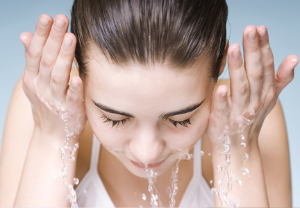
Cosmetic Corner: Dermatologists Weigh in on Face Washes
To improve patient care and outcomes, leading dermatologists offered their recommendations on the top face washes. Consideration must be given to:
• Clean & Clear Morning Burst Facial Cleanser
Johnson & Johnson Consumer Companies, Inc
“Clean & Clear Morning Burst is a facial cleanser which I love to use after working out. The combination of caffeine, lemon, and papaya wakes my skin up and keeps it looking fresh.”—Elizabeth K. Hale, MD, New York, New York
Neutrogena Corporation
Recommended by Marian Northington, MD, Birmingham, Alabama
PCA Skin
“It is an aloe-based cleanser with low level α-hydroxy acids in it. It is very well tolerated and leaves the skin with a squeaky clean feeling.”—Mark G. Rubin, MD, Beverly Hills, California
Valeant Consumer Products, a division of Valeant Pharmaceuticals North America
Recommended by Marian Northington, MD, Birmingham, Alabama
Allergan, Inc
"It is great for patients with oily or acne-prone skin. It is recommended to use several times per week, but I find that it can be used daily to decrease oil. It also helps with seborrheic dermatitis.—Amy J. Derick, MD, Barrington, Illinois
Cutis invites readers to send us their recommendations. Antiwrinkle treatments, men’s cosmetics, and facial moisturizers will be featured in upcoming editions of Cosmetic Corner Please e-mail your recommendation(s) to msteiger@frontlinemedcom.com.
Disclaimer: Opinions expressed herein do not necessarily reflect those of Cutis or Frontline Medical Communications Inc and shall not be used for product endorsement purposes. Any reference made to a specific commercial product does not indicate or imply that Cutis or Frontline Medical Communications Inc endorses, recommends, or favors the product mentioned. No guarantee is given to the effects of recommended products.
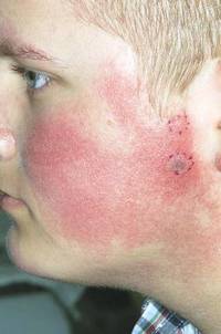
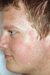
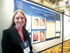
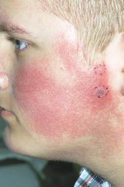
Pulsed dye laser targets keratosis pilaris rubra
MILWAUKEE – Pulsed dye laser therapy reduced the redness associated with chronic keratosis pilaris rubra in as little as one session in a case series of seven adolescents.
The problem, however, is convincing insurers to pay for the procedure.
"That’s part of why we brought this to a forum like this meeting," Dr. Jennifer J. Schoch said at the annual meeting of the Society for Pediatric Dermatology. "These kids are so significantly affected by this and embarrassed [by the condition], that if we can do something in just one treatment and have a good response, it makes sense. But these kids required a lot of letters to the insurance companies, and a lot of them paid out of pocket."
Several attendees at the meeting echoed these comments, and some observed that pulse dye laser (PDL) therapy is not as effective in patients with higher Fitzpatrick skin types or that it was out of reach for their patients at a price tag of $200 or more per session.
"It seems to make a lot of sense, but I don’t think it would be worth the cost for my patients," Dr. Aimee Smidt, University of New Mexico, Albuquerque, said in an interview.
Keratosis pilaris rubra is often viewed as a benign condition, but one patient found it so socially disturbing that he flew from Alabama to Minnesota for treatment, said Dr. Schoch, a dermatologist at the Mayo Clinic in Rochester, Minn.
Dr. Schoch noted that there are few data in the literature on PDL treatment of keratosis pilaris rubra, and the current series is occurring over a 13-year-period at the clinic. In this series, adolescents aged 14-17 years received one to four sessions of PDL at a wavelength of 585 nm or 595 nm, for erythema and hyperkeratotic follicular papules on both cheeks. Two patients also received treatment to the chin, forehead and/or neck.
All patients had Fitzpatrick skin type I or II, five were male, and two also had ulerythema ophryogenes. Some patients had been misdiagnosed with severe acne, and all had failed a range of treatments including emollients, lactic acid, topical retinoids, urea, sulfacetamide lotion, and weak topical corticosteroids, as well as laser therapy with intense pulsed light, Fraxel, and Nd:YAG lasers, she said.
PDL treatment was physician-dependent. Starting fluences ranged from 4-9 J/cm2, with the goal of achieving a mild, bruiselike response. The spot size was predominantly 7 mm, and pulse duration was 1.5, 3, or 10 msec.
"I hypothesize that people are probably undertreating because it’s a benign condition, and you don’t want to cause problems, but to be effective, it seems you have to go for a little more aggressive response," Dr. Schoch said in an interview.
All patients experienced significant improvement after one to four treatments, based on patient report or provider assessment. Bruising resolved in 1-2 weeks. Resolution of erythema was observed, but not specifically measured. Patients also experienced some transient purpura, which was not well documented, said Dr. Schoch.
After 1-19 months follow-up, most patients remained satisfied with the results, although some flushing returned in two patients, she said. Blanching has not been a significant problem, and overlapping the treated areas reduced the risk of a honeycomb pattern developing on the skin.
The investigators are considering a prospective study to more objectively monitor responses. Treatment parameters will depend on patient’s response to test spots, Dr. Schoch said.
Dr. Schoch and her coauthors reported having no financial disclosures.
AT THE SPD ANNUAL MEETING
Cosmetic Corner: Dermatologists Weigh in on Sunscreens (2013)
To improve patient care and outcomes, leading dermatologists offered their recommendations on the top sunscreen products. Consideration must be given to:
• Anthelios 40 Sunscreen
La Roche-Posay Laboratoire Dermatologique
“Anthelios SPF 40 with Mexoryl has the best UVA protection available.”—Jeffrey M. Weinberg, MD, New York, New York
• Anthelios 50 Mineral Ultra Light Sunscreen Fluid
La Roche-Posay Laboratoire Dermatologique
“The fact that this product is a total blocker with active ingredients blocking UVB and Mexoryl SX for additional UVA photoprotection makes it one of the ideal sunscreens. In addition, the formulation feels great on the skin.”—Marta I. Rendon, MD, Miami, Florida
“I like this product because it provides both UVA and UVB protection utilizing the physical blocker titanium dioxide that also is stabilized with the company’s Cell-Ox Shield technology. It is fragrance free and paraben free as well.”—Anthony M. Rossi, MD, New York, New York
• EltaMD UV Sport Broad-Spectrum SPF 50
EltaMD
“This product is noncomedogenic, paraben free, sensitivity free, and fragrance free. It contains 9% zinc oxide, the broadest spectrum UVA/UVB physical blocker.”—Basil M. Hantash, MD, PhD, Turlock, California
• Physical Fusion UV Defense SPF 50
SkinCeuticals
“This chemical-free sunscreen is a cosmetically elegant choice for people who want to have a mineral sunscreen but can’t tolerate the white film.”—Amy J. Derick, MD, Barrington, Illinois
• Ultra Sheer Dry-Touch Sunscreen Broad Spectrum SPF 85
Neutrogena Corporation
“It has excellent UVA and UVB protection in a nongreasy base that most patients like a lot. It’s particularly popular with patients who have oily skin and with male patients who don’t like a creamy-feeling product.” —Mark G. Rubin, MD, Beverly Hills, California
• Wet Skin Sunscreen Spray
Neutrogena Corporation
“This is a spray that is easy to apply to children and is great to quickly spray extremities before sports and can be reapplied on wet skin but not to the face.”—Marian Northington, MD, Birmingham, Alabama
Cutis invites readers to send us their recommendations. Men’s cosmetics, facial moisturizers, and shampoos will be featured in upcoming editions of Cosmetic Corner. Please e-mail your recommendation(s) to msteiger@frontlinemedcom.com.
Disclaimer: Opinions expressed herein do not necessarily reflect those of Cutis or Frontline Medical Communications Inc and shall not be used for product endorsement purposes. Any reference made to a specific commercial product does not indicate or imply that Cutis or Frontline Medical Communications Inc endorses, recommends, or favors the product mentioned. No guarantee is given to the effects of recommended products.
Difficult to treat hyperpigmentation – eyelids, axillae, and neck
Persons with Fitzpatrick skin types III-VI and those of certain ethnic groups tend to have a higher risk of darker pigmentation on certain parts of the body. Melasma, postinflammatory hyperpigmentation, and lentigines often respond to treatment with topical antipigment agents, chemical peels, and lasers. Darker pigment on the elbows and knees, if bothersome also can be treated with topical antipigment creams, plus or minus topicals that promote exfoliation (such as urea-based topicals). If the skin is acanthotic on elbows or knees, a topical steroid can be used first to thin the area before applying a lightening agent.
But what about pigmentation of the eyelids, axillae, and neck? At these thinner, more sensitive areas of skin, the cause of darker pigment could be multifactorial. Treatment can be difficult because the same methods we use to treat pigmentation in other areas can be too aggressive for these locations. A recent study by Saedi and Ganesan (J. Drugs Dermatol. 2013;12:563-7) surveyed practicing dermatologists’ methods of treating hyperpigmentation of the eyelids, axillae, and neck. Fifty dermatologists completed the survey, and 46 (92%) reported treating patients with darker skin. The ethnic groups treated included Hispanic (97.8%), black (97.8%), Middle Eastern (77.6%), and Asian (88.9%). Thirty-six survey respondents reported treating patients with hyperpigmentation under the eyes, and 22 (61.1%) thought the hyperpigmentation was a result of idiopathic increase in melanin deposition. Forty-two responded to treating hyperpigmentation in the axilla, most of whom thought it was related to acanthosis nigricans (69.0%) or contact dermatitis (59.5%). Forty responded to treating hyperpigmentation on the neck, most of whom treated the condition with hydroquinone (66%). Treatments for these three areas were not found to be effective.
Pigment in these areas could be normal and purely genetic, such as variations in skin pigment because of embryonic pigment demarcation areas, versus an underlying pathology.
For the eyelids, that pathology could include increased pigment from inflammatory conditions (eczema, allergies, allergic or irritant contact dermatitis, photodermatitis), autoimmune conditions (dermatomyositis, lupus), medications (bimatoprost, among others), heavy metal poisoning (colloidal silver, lead, mercury), or increased vascularity. Treating these underlying conditions may help improve the appearance of darker eyelids. Hyperpigmentation treatment options include a series of light chemical peels, topical lightening agents such as kojic acid, and resurfacing lasers, but caution must be taken to avoid additional postinflammatory pigmentation from these procedures. Long-term sun protection and sunscreen use is imperative in any area after treatment.
Tear trough deformity because of volume loss under the eyes also can cause the appearance of darker lower eyelids. However, hyperpigmentation of the skin is not the primary issue in these cases, and the appearance can often improve with placement of dermal fillers.
For the axillae and neck, conditions that could promote hyperpigmentation include postinflammatory pigmentation (caused by irritant or allergic contact dermatitis, infection, waxing, or friction), UV exposure, acanthosis nigricans, and photodermatitis, especially from photosensitizing medications. All of these conditions may also respond to topical antipigment ingredients and attention to the underlying condition, but unfortunately, not always to the patient’s greatest satisfaction.
What are your strategies for hyperpigmentation on the tough-to-treat areas of the eyelids, neck, or axillae?
Dr. Wesley practices dermatology in Beverly Hills, Calif.
Do you have questions about treating patients with dark skin? If so, send them to sknews@frontlinemedcom.com.

Blue light
Photodynamic light therapies (PDLs) have emerged as significant adjuvant approaches for treating acne. In particular, such therapies have been used for acne refractory to standard retinoid or combined retinoid and antimicrobial regimens. Why write about PDL in a column devoted to topical cosmeceutical products and ingredients? Blue light warrants inclusion because it has been studied in comparison to topical cosmeceutical treatments, and it is used in conjunction with other topical approaches.
Blue light exerts a phototoxic effect on the heme metabolism of Propionibacterium acnes, and it is considered effective by targeting part of the etiologic pathway of acne. It has become a widely used option for inflammatory acne (J. Drugs. Dermatol. 2006;5:605-10).
Early work
In 1990, Meffert et al. were the first to show that a blue light–type, high-pressure lamp could improve acne and seborrhea (10 10-minute treatments, cumulative light dose 325 J/cm2). Given the copious amounts of porphyrins stored in lipophilic P. acnes, the technology could be targeted to destroy propionibacteria, and the researchers observed a decline in the porphyrin content inside acne follicles. They concluded that short-range visible light (400-420 nm) was a viable option for acne treatment during the light-poor season of the year (Dermatol. Monatsschr. 1990;176:597-603). It was subsequently established that treatment with UV-free blue light in the range of 405-420 nm leads to the elimination of acne bacteria by virtue of the effects on the porphyrins generated naturally by P. acnes (J. Cosmet. Laser. Ther. 2003;5:111-7). Notably, blue light appears to photoinactivate P. acnes, but it does not penetrate deeply into the skin (Dermatol. Online J. 2011;17:2).
In an open study of the then-novel high-intensity, enhanced, narrow-band, blue-light phototherapy, Kawada et al. treated 30 acne patients (27 female, 3 male) twice a week for up to 5 weeks. A reduction of 64% was seen in acne lesions, and in vitro data showed a significant decline in P. acnes, but not in Staphylococcus epidermidis (J. Dermatol. Sci. 2002;30:129-35).
Mechanism of action
In 2006, Shnitkind et al. studied the effect of narrow-band blue light on the inflammatory process in the presence and absence of cytokines and ultraviolet B using interleukin-1 alpha (IL-1alpha) and intercellular adhesion molecule 1 (ICAM-1) as markers for inflammation. They found that blue light treatment of HaCaT and hTERT cells decreased levels of IL-1alpha by 82% in HaCaT and by 75% in hTERT. When blue light was combined with ultraviolet B, the respective reductions were 95% and 91%. Similar reductions in ICAM-1 expression were seen in HaCaT, but not in hTERT. The researchers concluded that narrow-band blue light exerts anti-inflammatory effects on keratinocytes by reducing cytokine-induced synthesis of IL-1alpha and ICAM-1. They suggested that these findings imply a broader range of effects is exerted on the inflammatory process by narrow-band blue light than previously understood (J. Drugs. Dermatol. 2006;5:605-10).
Comparison studies
In 2000, Papageorgiou et al. randomized 107 patients with mild to moderate acne to four treatment groups: blue light (peak at 415 nm), mixed blue and red light (peaks at 415 and 660 nm), cool white light, and 5% benzoyl peroxide cream, for 12 weeks of active treatment. Phototherapy using portable light sources was conducted daily for 15 minutes; comparative assessments among the three phototherapy groups were done with observers blinded. The investigators found that the greatest improvement in acne lesions occurred in the blue and red light combined group. The blue/red treatment was significantly superior to blue light alone at 4 and 8 weeks but not 12 weeks, benzoyl peroxide at weeks 8 and 12, and white light at all assessments (Br. J. Dermatol. 2000;142:973-8).
Gold et al. performed a multicenter clinical evaluation comparing blue light to topical 1% clindamycin solution with respect to safety and efficacy. They found that clindamycin was associated with a 14% reduction of inflammatory lesions, but blue light reduced such lesions by an average of 34% (J. Drugs. Dermatol. 2005;4:64-70).
In another study, 20 patients with moderate to severe facial acne were treated in four weekly sessions with topical aminolevulinic acid (ALA)-photodynamic therapy with blue light (415 nm) on the right side of the face, or blue light alone on the left side of the face. At 4, 8, 12, and 16 weeks after the start of treatment, the mean percent reductions in inflamed lesions were higher in the ALA-PDT areas (32%, 50.9%, 65.9%, and 71.1%, respectively) than in the blue light–only treatment areas (20.7%, 27%, 57.7%, and 56.7%), but the differences were not statistically significant. Side effects, which included pain, stinging, erythema, itching, peeling, oozing, and pustules, were more pronounced in the areas treated with ALA-PDT (Photodermatol. Photoimmunol. Photomed. 2007;23:186-90).
However, in a 2007 study of 22 patients with moderate to severe acne randomized to one of three ALA-PDT treatments – intense pulsed light (IPL; 600-850 nm), a combination of IPL (580-980 nm) and bipolar radiofrequency (RF) energies, or blue light (417 nm) – in three sessions at 2-week intervals, ALA-PDT with activation by IPL yielded the most consistent, lasting improvement in moderate to severe acne (J. Drugs Dermatol. 2007;6:1010-6).
In a study of 60 volunteers with facial acne (grades II and III), de Arruda et al. compared the safety and efficacy of blue light to topical benzoyl peroxide 5% over 4 weeks. Thirty patients received blue light treatment twice a week, and the other 30 self-applied the topical formulation twice daily. The improvements were similar in both groups, but side effects were milder in the blue light group (An. Bras. Dermatol. 2009;84:463-8).
Choi et al. compared the bactericidal effects of 5-aminolevulinic acid (ALA) with blue and red light on P. acnes and found that blue light was more effective than red light phototherapy in eliminating the bacteria unless ALA was added, which substantially augmented red light phototherapy efficacy (J. Dermatol. 2011;38:661-6).
In summarizing the off-label uses of light-based treatments and PDT using topical precursors of porphyrins, Sakamoto et al. observed that blue light alone lessens acne severity due to anti-inflammatory effects; PDT using 5-ALA or ALA derivatives provides antimicrobial and anti-inflammatory activity; and, at high doses, red-light PDT may suppress or eradicate sebaceous glands, yielding clinical improvement (J. Am. Acad. Dermatol. 2010;63:183-93).
Combination therapy
In 2011, Wheeland and Dhawan assessed the efficacy and tolerability of treating mild to moderate facial acne using a novel, handheld, light-emitting diode (LED) blue-light device, along with a foam cleanser containing 5% glycolic acid and 2% salicylic acid, plus a serum containing 1.25% salicylic acid, 0.5% niacinamide, 0.08% liposomal-based azelaic acid, and superoxide dismutase. In all, 28 of 35 adults aged 25-45 years completed the 8-week study, in which they used the device twice daily (in addition to the cleanser before treatments and the serum after nighttime treatments). Significant reductions in inflammatory lesion counts were seen from week 1 onward, and significant reductions in noninflammatory lesion counts were seen from week 4 onward compared with baseline counts. The number and severity of flares were significantly reduced from baseline as well.
Therapy was well received by patients, with more than 90% claiming improvement in overall skin appearance and other parameters. In addition, 86% described the treatment as gentler than other therapies. (J. Drugs Dermatol. 2011;10:596-602).
Products
The Skin Clarifying Blue Light device has a power density of 400 mW/cm2, which is 10 times the power of other LED acne devices. This high power density allows the Skin Clarifying Blue Light device to deliver high levels of efficacy with short treatment times. When used as stand-alone treatment, it delivered a 70% reduction in inflammatory lesions in 8 weeks. The success of products designed for self-administration has coincided with the rampant interest in the creation of smart phone applications (J. Cosmet. Laser. Ther. 2011;13:308-14; J. Clin. Aesthet. Dermatol. 2009;2:40-4; J. Clin. Aesthet. Dermatol. 2009;2:44-50). That is, blue-light therapy apps have been developed for the iPhone and the iPod touch. None, however, has been approved by the Food and Drug Administration, although at least one is said to be under investigation in a clinical trial. I am skeptical about the efficacy of these LED-backlit apps and concerned about potentially prolonged magnetic radiation exposure that would be required. The self-administered devices appear to be a more reliable alternative.
Conclusion
Blue light can effectively be used to treat mild to moderate acne by causing photodynamic destruction of P. acnes. It can be used alone or in combination with topical or other light regimens. Additional studies are needed before a consensus protocol can be established, and future research goals should include establishing the optimal incubation time, activating light source, and frequency of treatment.
Dr. Baumann is in private practice in Miami Beach. She did not disclose any conflicts of interest. To respond to this column, or to suggest topics for future columns, write to her at sknews@frontlinemedcom.com.

Make your scalp surgery seamless
WASHINGTON – To make scalp surgery seamless, remember what makes the scalp unique: hair and tension, Dr. Mark Welch said.
The scalp is "a bloodless plain," said Dr. Welch of the Skin Cancer Surgery Center in Bethesda, Md.
Also, the scalp is painless, so it’s possible to go beyond the field of anesthesia, he noted at the Atlantic Dermatological Conference.
To keep hair out of the surgical field, Dr. Welch uses a razor to shave the immediate area, and then tapes down the surrounding hair. "The surrounding hair will find its way into your surgery site and wound," he said. Alternatively, moistening the hair with saline or water can keep it away from the surgical field. Tubular bandaging also can be used to hold the hair away from the surgery site.
The tension on the scalp presents a unique surgical challenge, said Dr. Welch. "The scalp skin is holding the weight of the body; there’s lots of tension up there."
Dr. Welch said he starts with a temporary pulley stitch to decrease the distance across the wound, which allows easier placement of subcutaneous stitches. "Then the pulley stitch can come out," he said.
"One technique I use a lot is preplaced subcutaneous sutures," Dr. Welch said. "You leave yourself a tail long enough to tie, then go posterior to the first subcutaneous stitch, and then go back and tie the first stitch, then the second, then go to the external stitches."
Dr. Welch said he uses a running stitch for external stitches. "On the last external running stitch, go out and come back in on the same side, and angle it back slightly." This technique allows for a more perpendicular closing to the wound edge, he explained.
Dr. Welch cited one case of a large defect in a patient with skin cancer on the scalp. He opted for a pulley stitch with gel foam in the center, and some silver nitrate. The wound was essentially healed in 8 weeks, even without the defect being completely closed.
For scalp dressings, Dr. Welch said he often prefers a Xeroform gauze bolster, which he sews in place, "so we don’t have to use any tape." When the stitches come out after a week, flexible collodion can be used. "It hardens, and over the next 3 or 4 weeks of shampooing, it flakes off."
When using wraps, Dr. Welch recommends combining vertical and horizontal wraps to create tension and promote healing.
He said he had no financial conflicts to disclose.
EXPERT ANALYSIS FROM THE ATLANTIC DERMATOLOGICAL CONFERENCE
Practice Question Answers: Anesthetics in Dermatology
1. A 17-year-old adolescent girl presents to the emergency department with breathing trouble. She has no history of respiratory disease and no medical history of consequence. After leaving an uncomplicated laser hair removal appointment, she developed shortness of breath. On examination she is tachypneic and tachycardic with a pulse oximetry of 88% on 90% nonrebreather mask. What is the appropriate course of action?
a. epinephrine and intravenous diphenhydramine
b. intravenous methylene blue (1 mg/kg)
c. intravenous methylprednisolone sodium succinate (Solu-Medrol, Pharmacia & Upjohn Co)
d. oxygen and nebulizer treatments
e. spiral chest computed tomography
2. Which is the most likely order of symptoms in a patient with increasing lidocaine toxicity?
a. coma, anxiety, disorientation, focal seizures
b. nausea, bradypnea, metallic taste, dizziness
c. perioral numbness, diplopia, bradycardia, cardiac arrest
d. seizure, somnolence, vomiting, coma
e. slurred speech, dizziness, bradycardia, tinnitus, seizure
3. What is the maximum amount of 1% lidocaine (with epinephrine) that can be safely administered to a 50-kg healthy adult?
a. 150 mg
b. 175 mg
c. 200 mg
d. 250 mg
e. 350 mg
4. Which of the following will not decrease pain from local administration of lidocaine for most patients?
a. addition of sodium bicarbonate to the preparation
b. background music or conversation
c. quick administration
d. tapping the skin
e. warming the anesthetic
5. Which of the following can be safely used in a patient with a p-aminobenzoic acid allergy?
a. chloroprocaine
b. lidocaine (preservative free)
c. procaine
d. proparacaine
e. tetracaine
Anesthetics in Dermatology

Nail surgery made simple
WASHINGTON – There’s a lot of anxiety about nail surgery, particularly nail biopsies, for both physicians and patients, according to Dr. Maral K. Skelsey.
The goals of successful nail surgery are threefold: avoid complications, reduce patient pain and anxiety, and optimize pathologic diagnosis, said Dr. Skelsey of Georgetown University Medical Center in Washington.
Because nail surgery is often performed to obtain a clinical diagnosis, a good specimen is needed to allow the dermatopathologist to make a diagnosis, she noted at the Atlantic Dermatological Conference.
Approach preoperative assessment for nail surgery as any other surgery, said Dr. Skelsey. Take a full history, including information about vascular impairment, arterial disease, latex allergies, and a history of anticoagulant use. "We don’t stop anticoagulants, usually," Dr. Skelsey noted, but she does assess the prothrombin time (PT/INR) within 1 week.
Also, don’t underestimate the value of an x-ray. "One thing I have found physicians don’t do often" is to x-ray to check for bony changes and any anatomic abnormalities, she said.
To help optimize nail surgery outcomes, Dr. Skelsey recommended the following preoperative instructions for patients: Remove nail polish, scrub the area with povidone-iodine twice daily for 3 days prior to surgery, bring open-toed shoes (for toenail surgeries), arrange for a ride home, and plan to elevate the hand or foot as much as possible for the first 48 hours following the procedure.
Also, it "will help reduce morbidity if you tell your patients ahead of time to reduce their exercise and activity" immediately after the procedure, she said.
The right tools "will make your nail surgery much more successful," Dr. Skelsey said. Her essential tools: a nail splitter, nail nipper, and nail elevator.
Allow the patient to recline with goggles and ear phones to reduce anxiety during the procedure, she said.
For anesthesia, "I always use a 30-gauge needle, injecting very slowly," she said. She prefers a wing block, injecting slowly at a 45-degree angle towards the bone. This injection also acts as a volumetric tourniquet.
When obtaining the specimen during nail surgery, "visualize the location of the pathology by reflecting the proximal nail fold with a suture of skin hook and full or partial nail avulsion," said Dr. Skelsey.
"You can use a punch biopsy for longitudinal melanonychia less than 3 mm," she noted, but for anything more than 3 mm, a transverse excision or shave biopsy with a tangential excision is needed.
After the biopsy, Dr. Skelsey said that she applies an absorbable gelatin sponge saturated in aluminum chloride.
"What’s very important is giving your dermatopathologist a good specimen," she said. Don’t forget to ink the margins and orient the specimen. "You don’t want to go through all this trouble and have someone tell you there is nothing there," she added. She also recommended using separate, labelled formalin jars for the nail plate, bed, and matrix.
Dr. Skelsey said that she had no financial conflicts to disclose.
EXPERT ANALYSIS FROM THE ATLANTIC DERMATOLOGICAL CONFERENCE
Cosmetic Corner: Dermatologists Weigh in on Redness-Reducing Products
To improve patient care and outcomes, leading dermatologists offered their recommendations on the top redness-reducing products. Consideration must be given to:
• CeraVe Facial Moisturizing Lotion AM
Valeant Consumer Products, a division of Valeant Pharmaceuticals North America LLC
“Sunscreen is a must. I recommend CeraVe.”—Gary Goldenberg, MD, New York, New York
• Oxymetazoline such as Afrin Nasal Spray
MSD Consumer Care, Inc
“The mechanism is sympathomimetic/vasoconstrictive. I tell patients to take the nozzle off and apply to red areas of the face with a cotton ball if they want to get a few hours of decreased redness. It works on only a short-term basis for things like an event/wedding/photo to decrease erythema/flushing for many patients. Rebound redness can occur with overuse, similar to rebound congestion when it is used intranasally.”—Lorraine L. Rosamilia, MD, State College, Pennsylvania
• PÊCHE Redness Control
Neocutis Inc
“Patients have found [this product] to be soothing. Patients with rosacea have tolerated it well and found a decrease in redness.”—Mary P. Lupo, MD, New Orleans, Louisiana
“ROSAPLEX [in this product] is an exclusive redness-balancing complex with dextran sulfate, zinc, caffeine, and glycerin, which helps to eliminate the appearance of blotchiness and blemishes, and improve overall skin tone. PÊCHE is a very versatile cream and can be used alone or in conjunction with prescription products for rosacea.”—Marta I. Rendon, MD, Boca Raton, Florida
• Redness Solutions Daily Relief Cream
Clinique Laboratories, LLC
“This cream is safe for patients with rosacea to use as a general moisturizer and it has an underlying green base, which cuts the appearance of red.”—Anthony M. Rossi, MD, New York, New York
• Redness Solutions Soothing Cleanser
Clinique Laboratories, LLC
“[I recommend that patients] use the Redness Solutions Soothing Cleanser, then apply the Redness Solutions Daily Relief Cream.”—Gary Goldenberg, MD, New York, New York
Cutis invites readers to send us their recommendations. Sunscreens, body moisturizers, and face washes will be featured in upcoming editions of Cosmetic Corner. Please e-mail your recommendation(s) to msteiger@frontlinemedcom.com.
Disclaimer: Opinions expressed herein do not necessarily reflect those of Cutis or Frontline Medical Communications Inc and shall not be used for product endorsement purposes. Any reference made to a specific commercial product does not indicate or imply that Cutis or Frontline Medical Communications Inc endorses, recommends, or favors the product mentioned. No guarantee is given to the effects of recommended products.