User login
Consider Marking Facial Lines Before Challenging Repairs
SAN DIEGO – A chief goal during cutaneous surgery in the facial region should be to avoid disturbing or crossing contour lines such as the alar fold, the junction of the eyebrow and forehead, and the eyelid margin, according to Dr. Jenny Kim.
"Alterations in these aesthetic lines will have a profound impact on an individual’s appearance," Dr. Kim said at the meeting sponsored by the University of California, San Diego School of Medicine and the Scripps Clinic. "Functional complications may also result from crossing these lines."
If the defect involves one or more cosmetic units, such as the cheek and nose, "repair each unit separately," advised Dr. Kim, associate professor of medicine and dermatology at the University of California, Los Angeles. "This allows for incisions to be hidden along contour lines and will provide you with a better aesthetic appearance."
She also recommends marking contour lines prior to challenging repairs, such as those in the vermilion border of the lip or in the nasolabial fold.
Skin tension lines, she said, follow the direction of the greatest intrinsic tension in the skin. Also known as maximal skin tension lines, relaxed skin tension lines, and Langer’s lines, skin tension lines "are less well defined compared with contour lines, and incisions placed parallel to these lines heal with a finer, more cosmetically pleasing scar," Dr. Kim said.
Unlike contour lines, skin tension lines vary widely among individuals due to various intrinsic and extrinsic factors "One of the most important intrinsic factors is the collagen in our dermis," she said. "Type I and type III collagen provide 70%-80% of wound volume in the dermis as well as tensile properties of the skin. It resists extensibility under tension and elongates with prolonged tension."
On the other hand, she continued, elastic tissue "will easily be extensible under minimal tension. It has the capacity to resume its original shape after tension is released. Elastic fibers maintain static tension of the skin and provide tensile strength."
Elastic tissue has an increased tension vector parallel to skin tension lines and a decreased tension vector perpendicular to skin tension lines. "These are some of the reasons why you want your wound to be parallel to your skin tension lines," said Dr. Kim, who is also a codirector of the medical school’s Procedural Dermatology Fellowship.
Extrinsic factors that play a role in skin tension lines include the muscles of facial expression, sun damage, aging, and smoking. Aging, for example, leads to fragmentation and loss of elastic fiber, "which is why there’s more variability in skin tension lines as you age," she said.
In order to best determine skin tension lines prior to surgery, have the patient sit upright. "This is because the lines can appear different when a person is lying down versus sitting due to gravity," Dr. Kim said. "Creases and folds secondary to muscle movement can be determined by having patients move facial muscles."
She makes it a point to mark skin tension lines before using anesthesia, "as the swelling from anesthesia makes these lines visible and a patient may not be able to contract muscles later. Draw in the skin tension lines before beginning the surgery. You want to make sure that you maintain function [of the lines], especially around the eyes and the mouth. Evaluate each patient individually, because there are always exceptions to our rules."
Dr. Kim said that she had no relevant financial conflicts to disclose.
EXPERT ANALYSIS FROM A MEETING ON SUPERFICIAL ANATOMY AND CUTANEOUS SURGERY
Don't Penny-Pinch on Dermatologic Surgery Instruments
SAN DIEGO – If you perform excisional surgery in your dermatology practice, don’t skimp on instrumentation, advised Dr. David E. Kent.
"Spend your money on instrumentation that’s going to get you out of trouble," Dr. Kent said at the meeting, which was sponsored by the University of California, San Diego School of Medicine and the Scripps Clinic.
His list of recommended instrumentation includes hemostats to clamp and tie off blood vessels, skin hooks to improve visualization, suction to remove excess blood, cotton tip applicators, and 4-by-4-inch gauze. "Be aware that the least expensive gauze may not have the best quality, so you want to evaluate different vendors," said Dr. Kent, a clinical instructor in the division of dermatology at the Medical College of Georgia, Augusta.
He also recommends having electrosurgical devices on hand, liquid thrombin, Gelfoam, and oxidized cellulose to place in wounds that are going to heal by second intention. Xenografts, "which can be helpful for temporary hemostasis over a wound with exposed muscle, may serve as a very nice scaffold to seal the wound and are easy to apply," he said.
Applying pressure to the wound after surgery is key, he added. "In all of our patients who are on any aspirin products, after any closure, my nurse holds pressure for 10 minutes. We’ve found that to be very helpful."
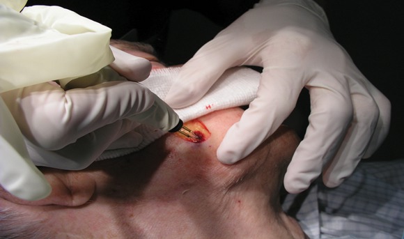
He finds the Geiger Thermal Cautery Unit useful for patients who have implantable cardiac defibrillators. "We did a study of this unit years ago and found that a setting between 6 and 7.5 is fairly ideal," Dr. Kent said. "It holds its temperature reasonably well in a wet field, compared with handheld units."
For handheld cautery, he recommends the LMA Perfect Temp device for isolated small pinpoint areas of bleeding. For solid state electrosurgical generators, "there are many manufacturers including Valleylab, Bard Medical, and Aaron Medical, to name a few," he said. "When using electrosurgical devices, it is important to avoid skin edges. This can be done by approaching the bleeding site at 90 degrees to the skin edge to avoid epidermal thermal injury. Use the lowest possible setting to control bleeding."
Another worthwhile instrument to have is a hemostatic scalpel, which provides heat energy to seal vessels and tissue. "It's excellent for skeletal muscle and large defects into muscle," Dr. Kent said. "If you're doing a lot of larger cases, it can really help you avoid excessive bleeding. But they are costly," he said. Used hemostatic scalpels can cost as much as $5,000. Blades cost $10 apiece and are not reusable.
If postoperative bleeding occurs after the patient has gone home, see the patient as soon as possible. "The next day is not soon enough," Dr. Kent said. "Have someone there to help you; make sure you have a nurse on call if you need one." On return, make sure the patient's vital signs are stable. Is the bandage soiled? Is there active bleeding? "Consider removing one or two sutures to see if there is brisk bleeding," Dr. Kent said. "Try to establish if it is a single skin edge or something more. If uncertain, you may need to take the entire closure down, inspect, and control what is bleeding."
Dr. Kent said that he had no relevant financial conflicts to disclose.
SAN DIEGO – If you perform excisional surgery in your dermatology practice, don’t skimp on instrumentation, advised Dr. David E. Kent.
"Spend your money on instrumentation that’s going to get you out of trouble," Dr. Kent said at the meeting, which was sponsored by the University of California, San Diego School of Medicine and the Scripps Clinic.
His list of recommended instrumentation includes hemostats to clamp and tie off blood vessels, skin hooks to improve visualization, suction to remove excess blood, cotton tip applicators, and 4-by-4-inch gauze. "Be aware that the least expensive gauze may not have the best quality, so you want to evaluate different vendors," said Dr. Kent, a clinical instructor in the division of dermatology at the Medical College of Georgia, Augusta.
He also recommends having electrosurgical devices on hand, liquid thrombin, Gelfoam, and oxidized cellulose to place in wounds that are going to heal by second intention. Xenografts, "which can be helpful for temporary hemostasis over a wound with exposed muscle, may serve as a very nice scaffold to seal the wound and are easy to apply," he said.
Applying pressure to the wound after surgery is key, he added. "In all of our patients who are on any aspirin products, after any closure, my nurse holds pressure for 10 minutes. We’ve found that to be very helpful."
He finds the Geiger Thermal Cautery Unit useful for patients who have implantable cardiac defibrillators. "We did a study of this unit years ago and found that a setting between 6 and 7.5 is fairly ideal," Dr. Kent said. "It holds its temperature reasonably well in a wet field, compared with handheld units."
For handheld cautery, he recommends the LMA Perfect Temp device for isolated small pinpoint areas of bleeding. For solid state electrosurgical generators, "there are many manufacturers including Valleylab, Bard Medical, and Aaron Medical, to name a few," he said. "When using electrosurgical devices, it is important to avoid skin edges. This can be done by approaching the bleeding site at 90 degrees to the skin edge to avoid epidermal thermal injury. Use the lowest possible setting to control bleeding."
Another worthwhile instrument to have is a hemostatic scalpel, which provides heat energy to seal vessels and tissue. "It's excellent for skeletal muscle and large defects into muscle," Dr. Kent said. "If you're doing a lot of larger cases, it can really help you avoid excessive bleeding. But they are costly," he said. Used hemostatic scalpels can cost as much as $5,000. Blades cost $10 apiece and are not reusable.
If postoperative bleeding occurs after the patient has gone home, see the patient as soon as possible. "The next day is not soon enough," Dr. Kent said. "Have someone there to help you; make sure you have a nurse on call if you need one." On return, make sure the patient's vital signs are stable. Is the bandage soiled? Is there active bleeding? "Consider removing one or two sutures to see if there is brisk bleeding," Dr. Kent said. "Try to establish if it is a single skin edge or something more. If uncertain, you may need to take the entire closure down, inspect, and control what is bleeding."
Dr. Kent said that he had no relevant financial conflicts to disclose.
EXPERT ANALYSIS FROM A MEETING ON SUPERFICIAL ANATOMY AND CUTANEOUS SURGERY
Innovations in Photoprotection
Photoaging in Skin of Color
Three-Dimensional Rejuvenation of the Photoaged Body
What's New in Photodynamic Therapy for Photorejuvenation?
Does That Really Work?
Successful Treatment of Chickenpox Scars With Microdermabrasion and a Nonablative, Submillisecond, 1064-nm Nd:YAG Laser
Thulium Laser Yields 'Dramatic' Resolution of AKs
SEOUL, SOUTH KOREA – The nonablative fractionated thulium laser at 1,927-nm wavelength is a promising new noninvasive therapy for actinic keratoses.
The laser was actually developed for superficial skin resurfacing, an application for which it is particularly well suited because the 1,927-nm wavelength minimizes patient discomfort. But while investigating the device for improvement of pigmentation, Dr. Roy G. Geronemus noted incidentally that patients were also achieving "a rather dramatic resolution" of multiple facial actinic keratoses (AKs). So he decided to conduct a formal examination of the laser’s performance for this purpose, he said at the World Congress of Dermatology
To date, in 15 patients followed for 1-6 months after the last of several thulium laser treatment sessions for multiple facial AKs, the mean clearance of the lesions was 84%-91%.
"This compares very favorably to other modalities that are out there, including the topical chemotherapies, immunomodulatory agents, and photodynamic therapy. The advantage of this is not only do you improve the AKs, but you’re also getting the cosmetic benefit simultaneously," said Dr. Geronemus, medical director of the Laser and Skin Surgery Center of New York.
Patients received up to four treatments at 2- to 6-week intervals. The laser setting was 5-20 mJ, with 30%-70% coverage per session. Topical anesthetic was utilized for 1 hour, supplemented as needed by intramuscular ketorolac.
After a single treatment a mean of 63% of AKs were cleared. After two, 84%, and after three, 85%.
The laser therapy was well tolerated. The average pain score during treatment was 2.7 on a 0-9 scale. No scarring or infections have occurred. Mild redness and peeling typically lasted 4-5 days.
Dr. Geronemus said he and his colleagues have also found that the 1,927-nm fractionated thulium laser brings about "dramatic improvement" in actinic cheilitis, and is also highly effective for the thorny problem of enlarged facial pore size.
Dr. Geronemus is a shareholder in Solta Medical, which markets the 1,927-nm fractionated thulium laser. He also is on the advisory boards of numerous dermatologic laser manufacturers.
FROM THE WORLD CONGRESS OF DERMATOLOGY
Ablative Fractional Resurfacing Smooths Atrophic Scars
SEOUL, SOUTH KOREA – Ablative fractional resurfacing with a 30-W carbon dioxide laser is a safe and effective therapy for atrophic scarring due to surgery or trauma, according to a prospective study featuring quantitative assessment of improvement in scar depth and volume.
"We’re now using this routinely on the surgical side of our practice," Dr. Roy G. Geronemus said at the World Congress of Dermatology.
He presented a study of 12 women with 19 nonacne atrophic scars, each of whom underwent three ablative fractional resurfacing (AFR) sessions at 1- to 4-month intervals. Follow-up continued for another 6 months after the final treatment, with grading of adverse events and efficacy provided both by patients and by nonblinded investigators.
The study also featured quantitative measurements of volumetric scar improvement using the Primos Imaging three-dimensional optical profiling system, which generates high-resolution topographic images of cutaneous scars. That’s a particularly noteworthy aspect of the study. The field of cosmetic dermatology is often heavily criticized for a glaring absence of this sort of objective before-and-after data.
The laser used to achieve AFR in this study was Solta Medical’s Fraxel re:pair Laser. The device delivers a pixelated pattern of microscopic ablative wounds surrounded by healthy tissue. This pattern results in zones of thermal coagulation and tissue ablation that run much deeper than those obtainable with traditional CO2 laser ablative resurfacing. This leads to more impressive neocollagenesis and dermal remodeling accompanied by shorter healing times and better safety than can be obtained with traditional broad-stroke ablative resurfacing, said Dr. Geronemus, medical director of the Laser and Skin Surgery Center of New York.
At the 6-month follow-up visit, Primos Imaging analysis demonstrated a mean 38% reduction in scar volume and a 36% decrease in maximum scar depth, compared with baseline.
Mean patient and investigator scores indicated a moderate 26%-50% improvement in scar texture, atrophy, and pigmentation. Patients and investigators respectively characterized 63% and 89% of scars as showing a 51% or greater overall improvement. A 76% or greater overall improvement was achieved in 42% and 16% of treated scars, according to patients and investigators, respectively.
Treatment-related erythema was typically rated moderate to severe, and it peaked at 72 hours after each treatment session, dropping to mild to moderate by 1 week. Edema peaked at a mild to moderate level within an hour post treatment. No incidents of delayed-onset hypopigmentation occurred during 6 months of post-AFR follow-up; in contrast, this complication has been noted in 20% or more of patients following traditional CO2 laser resurfacing, the dermatologist noted.
As to the AFR treatment settings, facial scars were typically addressed at 70 mJ per pulse, an energy density of 200 microscopic treatment zones/cm2 per pass, and 2-3 passes per session, resulting in 27%-38% coverage. Scars on the body were best addressed at 40 mJ per pulse, 200 microscopic treatment zones/cm2 per pass, and 2-3 passes, for 20%-30% coverage. Higher fluences off-face appeared to be more effective but brought prolonged erythema, which many patients find objectionable.
In an earlier study, Dr. Geronemus and his coworkers showed that the same CO2 AFR laser was effective for treatment of acne scars, achieving a mean 67% improvement in scar depth as measured using the Primos Imaging system (Lasers Surg. Med. 2008;40:381-6).
Dr. Geronemus is a shareholder in Solta Medical, and is on the advisory boards of numerous dermatologic laser manufacturers.
FROM THE WORLD CONGRESS OF DERMATOLOGY