User login
Hyaluronic Acid Filler Shows Long-Term Durability for Midface Deficits
SEOUL, SOUTH KOREA — The hyaluronic acid filler Voluma demonstrated excellent efficacy, durability, and patient satisfaction in correcting midface volume deficits at the interim 78-week analysis in a large prospective multicenter study.
At 78 weeks, 84% of patients maintained a clinically meaningful improvement after a single baseline injection and a touch-up injection, if needed, at week 4, Dr. Greg J. Goodman said at the World Congress of Dermatology.
Moreover, 95% of participants pronounced themselves satisfied or very satisfied with the product at the 78-week mark. An equal percentage indicated they would recommend the treatment to others, added Dr. Goodman of Monash University, Melbourne.
Based upon these interim results of what will be a 104-week study – the first-ever formal study of the filler’s long-term effects in daily clinical practice – correction of mild to severe midface volume deficits with Voluma is a safe, effective, long-lasting, and cost-effective alternative to surgical correction, he continued.
The study involved 103 middle-aged subjects with a baseline score of 2-5 on the 6-point Mid-Face Volume-Deficit Scale (MVDS). They were corrected to a 0 or 1, meaning no or mild deficit, by means of a baseline injection of up to 2 cc of Voluma per side. This was followed by an additional injection of up to 2 cc per side if needed at week 4. Patients received a mean total volume of 3.1 mL per side. No further retreatment was permitted until week 78.
A greater than 1-point improvement over baseline MVDS was present in 96% of patients at week 8, 90% at week 52, and 84% at week 78. Similarly, 100% of patients had a greater than 1-point improvement on the 5-point Global Aesthetic Improvement Scale (GAIS)at week 8, 92% at week 52, and 82% at week 78. In all, 78% of patients rated themselves as having more than a 1-point improvement on the GAIS at week 78.
At week 78, 32% of patients received a supplemental injection of Voluma, based upon protocol-defined criteria indicating they had returned to within less than 1 MVDS point of their pretreatment midface volume.
The chief side effect of treatment was mild to moderate bruising at the injection site. This was much more common early in the study, when Voluma was new to Australian physicians.
"We were finding our way back then," Dr. Goodman recalled.
The study was sponsored by Allergan. Dr. Goodman is a consultant to the company.
FROM THE WORLD CONGRESS OF DERMATOLOGY
Interim Study Results Find Artefill Safe
DANA POINT, CALIF. – The rate of granuloma formation following use of Artefill for the correction of nasolabial folds stands at 0.59%, interim results from a 5-year study have shown.
Dr. Christopher B. Zachary presented 36-month results from the prospective study at the SDEF Summit in Aesthetic Medicine. The study was required by the Food and Drug Administration and is the largest and longest prospective clinical study to date for dermal fillers in the United States and in the European Union, according to Dr. Zachary.
The purpose of the study is to assess the safety of Artefill (Suneva Medical) in 1,008 patients, based on the incidence of anticipated and unanticipated adverse events and serious adverse events, the incidence of granuloma formation, and subjects' assessment of satisfaction.
Patients were treated for the correction of nasolabial folds at baseline. They received two touch-up treatments if needed, said Dr. Zachary, professor and chair of the department of dermatology at the University of California, Irvine. The second clinic visit consisted of a clinical evaluation and photos of the treated site 3 months after the last treatment.
Patients filled out questionnaires at 6, 12, and 18 months and at 2 and 3 years, and will again do so at years 4 and 5. Adverse events were reported to the site for investigation. Final visits are scheduled for 5 years after the last Artefill treatment.
The mean age of the 1,008 patients was 54 years, 89% were female, and 88% were white. Most subjects (975) are still in the trial, while 29 have been lost to follow-up or withdrew consent for personal reasons, and four non–treatment-related deaths have occurred.
A total of 114 adverse events deemed device related have occurred to date, for a rate of 11%. These include local complications such as lumpiness, swelling, and redness. In addition, 11 lesions have been identified and biopsied: 4 were viewed as unremarkable at biopsy, 1 was categorized as a foreign body reaction consistent with implant material, and 6 were granulomas, for a rate of 0.59%. Five granulomas resolved completely and one is responding well to treatment.
"You might say that the 0.59% incidence of granulomas is a bit high when you consider that the worldwide reported incidence of granulomas after using Artefill is 0.04%," said Dr. Zachary. "If the FDA required an intense 5-year study of all the commonly used filler products, where every adverse event [AE] was reported, then you would expect the overall AE incidence to be significantly higher. So what is the real incidence of AEs in fillers, and does reliance on voluntary reporting give us misleading results?"
Patient satisfaction scores have remained high over time. For example, at 6 months, 81% reported being "very satisfied" or "satisfied" with the cosmetic results, compared with 79% at 18 months, and 78% at 24 months.
"I'm not up here promoting the product, but I do think Artefill is very safe," Dr. Zachary said. "In practice, this study demonstrates that the product is probably just as safe as hyaluronic acids. Some of my colleagues hate to hear this, and I am not really a fan of permanent fillers, but if we determine that a company needs to perform a comprehensive 5-year safety study, then we need to sit up and take notice of the results. To do otherwise would be ignorant."
Dr. Zachary disclosed that he has received research support, discounts on devices, and honoraria from numerous laser and device companies, including Suneva Medical. He is a member of Suneva’s scientific advisory board.
SDEF and this news organization are owned by Elsevier.
FROM THE SDEF SUMMIT IN AESTHETIC MEDICINE
Major Finding: A total of 114 adverse events deemed device related have occurred to date, for a rate of 11%. The rate of granuloma formation to date is 0.59%
Data Source: Interim results from a 5-year study of 1,008 patients who have received Artefill for the correction of nasolabial folds.
Disclosures: Dr. Zachary disclosed that he has received research support, discounts on devices, and honoraria from numerous laser and device companies, including Suneva Medical. He is a member of Suneva’s scientific advisory board.
Expert Offers Facial Fat Augmentation Pearls
DANA POINT, CALIF. – Before undergoing facial fat augmentation, patients routinely ask Dr. Jonathan M. Sykes how long their results will last.
"I tell them that the longevity of fat is variable in different parts of the face," Dr. Sykes said at the. Summit in Aesthetic Medicine sponsored by Skin Disease Education Foundation (SDEF). He described long-term results in the infraorbital region, the malar region, and the tear trough as "great"; in the melolabial fold and pre-jowl sulcus as "good"; and in the lips as "not so good."
"I think lips are the hardest area to treat," said Dr. Sykes, director of facial plastic and reconstructive surgery at the University of California Davis Health System, Sacramento.
One advantage of facial fat augmentation is that most patients have an abundant supply of autologous fat, with the exception of bodybuilders and patients who have been on long-term antiretroviral medicines for HIV, he said. "It’s also easy to perform simultaneously with other surgical rejuvenative procedures, and it does not add significantly to time or cost. I harvest the fat at the beginning, and put it in at the end. Other physicians put in the fat right away and then do the surgical procedure."
Disadvantages of facial fat augmentation, he said, are that the procedure is time and technique sensitive, donor site contour irregularities are possible, donor site pain/ecchymosis is possible, and it is difficult to modulate the results.
Instruments he uses for most procedures include four cannulas made by San Diego–based Tulip Medical: a 0.9-mm spoon tip cannula that is 4 cm long for periorbital injections, a 1.2-mm spoon tip cannula that’s 6 cm long for all-purpose injections, a 3.0-mm bullet tip cannula that is 15 cm long for all-purpose fat harvesting, and a 2.1-mm multiport cannula that is 12 cm long and used as an optional secondary cannula for thin patients.
Dr. Sykes, who is also the current president of the American Academy of Facial Plastic and Reconstructive Surgery, said he prefers the upper/outer hip as a donor source, but makes it a point to ask patients where they retain the most fat. "In women, usually it’s the abdomen, hips, and inner/outer thighs, while in men it’s usually the abdomen or the hips," he said. "There is some evidence that outer thigh fat persists a bit better because it’s less vascular."
To harvest the fat he uses four to eight 10-cc Luer lock syringes with low negative pressure. He then stands the syringes upright for about 15 minutes, "typically while other surgical procedures are being performed," he said.
Next, he places the fat into a centrifuge at 3,000 rpm for 1-3 minutes and transfers the fat into one 20- or 35-cc syringe. He uses a Leur lock transfer hub to transfer the fat into several 1-cc Leur lock syringes.
Dr. Sykes said he routinely uses local anesthesia with epinephrine 20 minutes prior to injection. "That gives us less bleeding and less ecchymosis," he said.
He then injects small parcels of fat as the cannula is withdrawn, aiming for 30-50 passes per 1 cc of fat.
In most patients, adding fat volume to the face "creates a rejuvenated, youthful appearance," Dr. Sykes concluded, noting that he performs fat augmentation in about 70% of face-lift procedures.
Dr. Sykes disclosed that he has served as a paid trainer and speaker for Sanofi Aventis and Medicis.
SDEF and this news organization are owned by Elsevier.
EXPERT ANALYSIS FROM THE SDEF SUMMIT IN AESTHETIC MEDICINE
Steaming Veins: The Skinny Podcast
In this month's podcast, Skin & Allergy News' reporters discuss cancer, wrinkles, and veins.
Positive clinical trial results for new melanoma therapies were presented at the American Society of Clinical Oncology. Jeff Evans interviews Dr. Lynn Schuchter about the findings, and what they mean for patients.
Also in this episode, Heidi Splete offers a report on the link between wrinkles and bone health, which may have you sending your wrinkled patients to the bone doctor.
Then, Sherry Boschert talks with Dr. Rene Milleret about a new endovenous ablation technique for varicose veins that uses steam; the procedure is catching on in Europe.
In this month's Cosmetic Counter segment, Dr. Lily Talakoub tackles nanotechnology.
And finally, Dr. Alan Rockoff assures everyone that, contrary to any rumors, he has not kicked the bucket.
Purse String Stitch Handy for Lip Defects
LAS VEGAS – A simple purse string stitch can provide an elegant closure of challenging lip defects after Mohs surgery.
"It's quite easy to perform. It's useful for numerous areas of the lip. It's low-risk, with low morbidity, and requires little down time," yet provides an excellent aesthetic outcome, Dr. Kenny J. Omlin said at the annual meeting of the American College of Mohs Surgery.
The keys to reconstruction using the purse string stitch start with undermining the entire surgical wound in the subdermal plane to decrease sheering forces.
Next, uniformly place an absorbable suture in the deep dermis using a small needle, with circumferential tissue advancement to distribute the tension uniformly, explained Dr. Omlin, chief of Mohs surgery for Kaiser Permanente Napa-Solano County, Vacaville, Calif.
"There is a uniform stitch all the way around the perimeter" of the wound with particular attention to precisely aligning the vermilion/cutaneous lip junction, he said.
The purse string stitch creates a trestle-like framework that supports normal wound healing.
As with any reconstructions on the lower cosmetic subunits of the face, he tells patients to practice a "ventriloquist act" while healing and not talk much or move their mouths much.
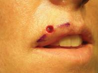
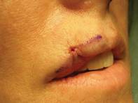
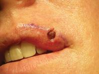
One of his patients provided an excellent case-control comparison of wound closures. She initially presented with a basal cell carcinoma that intersected both the cutaneous and vermilion margins of her left upper lip.
After Mohs surgery, Dr. Omlin did a standard, complex linear closure, followed later by two pulsed-dye laser treatments. At 1-year follow-up, the patient was satisfied with an acceptable cosmetic outcome.
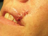
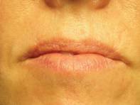
Six months later, she presented with a nearly identical basal cell carcinoma on the right upper lip. This time, Dr. Omlin used a purse string stitch after Mohs surgery. "It takes all of 5-10 minutes," he noted. The aesthetic result was "nearly perfect" a month later, said Dr. Omlin, also of the University of California, Davis.
On the central upper lip, "a lot of our older patients tend to have absent philtral columns or an absent Cupid's bow. Again, this is an excellent place for a purse string stitch," he said.
For patients on warfarin, the purse string stitch is great for hemostasis when repairing Mohs defects of the lip, Dr. Omlin added.
He also likes to use it for defects at the oral commissure. "Sure, you can use an elaborate cross-lip commissuroplasty or other elaborate techniques," but a simple purse string stitch reproduces the accordion-like structure of the oral commissure.
After wound granulation and healing, at 3 months it's hard to tell that a defect was ever there, he said.
In an interview after his presentation, he said he was pleased by the enthusiasm expressed by other attendees at the meeting for his simple surgical pearl.
Dr. Omlin said he has no relevant financial disclosures.
LAS VEGAS – A simple purse string stitch can provide an elegant closure of challenging lip defects after Mohs surgery.
"It's quite easy to perform. It's useful for numerous areas of the lip. It's low-risk, with low morbidity, and requires little down time," yet provides an excellent aesthetic outcome, Dr. Kenny J. Omlin said at the annual meeting of the American College of Mohs Surgery.
The keys to reconstruction using the purse string stitch start with undermining the entire surgical wound in the subdermal plane to decrease sheering forces.
Next, uniformly place an absorbable suture in the deep dermis using a small needle, with circumferential tissue advancement to distribute the tension uniformly, explained Dr. Omlin, chief of Mohs surgery for Kaiser Permanente Napa-Solano County, Vacaville, Calif.
"There is a uniform stitch all the way around the perimeter" of the wound with particular attention to precisely aligning the vermilion/cutaneous lip junction, he said.
The purse string stitch creates a trestle-like framework that supports normal wound healing.
As with any reconstructions on the lower cosmetic subunits of the face, he tells patients to practice a "ventriloquist act" while healing and not talk much or move their mouths much.
One of his patients provided an excellent case-control comparison of wound closures. She initially presented with a basal cell carcinoma that intersected both the cutaneous and vermilion margins of her left upper lip.
After Mohs surgery, Dr. Omlin did a standard, complex linear closure, followed later by two pulsed-dye laser treatments. At 1-year follow-up, the patient was satisfied with an acceptable cosmetic outcome.
Six months later, she presented with a nearly identical basal cell carcinoma on the right upper lip. This time, Dr. Omlin used a purse string stitch after Mohs surgery. "It takes all of 5-10 minutes," he noted. The aesthetic result was "nearly perfect" a month later, said Dr. Omlin, also of the University of California, Davis.
On the central upper lip, "a lot of our older patients tend to have absent philtral columns or an absent Cupid's bow. Again, this is an excellent place for a purse string stitch," he said.
For patients on warfarin, the purse string stitch is great for hemostasis when repairing Mohs defects of the lip, Dr. Omlin added.
He also likes to use it for defects at the oral commissure. "Sure, you can use an elaborate cross-lip commissuroplasty or other elaborate techniques," but a simple purse string stitch reproduces the accordion-like structure of the oral commissure.
After wound granulation and healing, at 3 months it's hard to tell that a defect was ever there, he said.
In an interview after his presentation, he said he was pleased by the enthusiasm expressed by other attendees at the meeting for his simple surgical pearl.
Dr. Omlin said he has no relevant financial disclosures.
EXPERT ANALYSIS FROM THE ANNUAL MEETING OF THE AMERICAN COLLEGE OF MOHS SURGERY
Anchoring Cartilage Grafts to Alar Rim Is Simple, Effective
LAS VEGAS – Traditional techniques for anchoring cartilage grafts after Mohs surgery on the nose might be more complicated than necessary.
Well-known textbook descriptions of cartilage grafting for alar rim reconstruction involve harvesting a large piece of cartilage and securing the grafted cartilage with multiple sutures medially to the lower lateral cartilage and laterally to the periosteum of the piriform aperture of the maxilla.
This technique is appropriate for large defects that require reconstruction of the entire alar subunit, but is unnecessary for many of the smaller alar defects that commonly are encountered in Mohs surgery, Dr. Ravi S. Krishnan said.
"While this does produce nice results, I don't like it for two reasons," he said at the annual meeting of the American College of Mohs Surgery. "It requires a very large cartilage graft, and it often requires more effort than you sometimes need."
In his approach to performing a cartilage graft with a skin graft on top of it, he starts the conventional way by making some stab incisions on either side of the wound to create a pocket for the cartilage graft. What most surgeons would do next is to place either a figure-of-eight suture or some interrupted sutures to hold the graft in place.
"While these techniques are fine, I sometimes still have problems with them getting the cartilage graft flush against the alar remnant," noted Dr. Krishnan of Virginia Mason Medical Center, Seattle.
Instead, he starts suturing from inside the nose, pushing the suture through the nose behind the cartilage graft, then pulling it back through the cartilage graft and the nose, finally pulling inferiorly as the knot is tied. He repeats this process, so that there are two sutures anchoring the cartilage graft along the alar rim. These sutures are left in place for 2 weeks to allow some fibrosis to occur.
"The reason I like this technique is because it pulls the graft inferiorly so that it's perfectly flush against the alar rim remnant," he said. It also leaves more of the wound base exposed so that any overlying skin graft will be well perfused.
This is not necessarily a new technique, Dr. Krishnan said, but he could find no published description of it. It doesn't necessarily give better results, but it's easier to execute than are traditional methods, he added.
His techniques for anchoring cartilage grafts along the alar rim also work well with flaps including nasolabial transposition flaps, bilobed flaps, and interpolated paranasal flaps, resulting in good contour and symmetry and very acceptable results, he believes.
He typically follows these patients for 6 months after surgery, and while it's possible that the cartilage graft could shift after 6 months, "I doubt this would be the case."
One small drawback is that epithelium can start growing over the anchoring sutures during the 2 weeks that they are left in place, making them difficult to remove.
"Some people worry about infection, but I've never seen an infection with this technique," he added. All patients in his practice that receive cartilage grafts get perioperative antibiotics.
The advantages outweigh any potential drawbacks of the technique, in his opinion. It's easy to perform, and results are at least as good as those with more difficult techniques. His method precisely places the cartilage "exactly where you want it" along the alar rim, and apposes the cartilage graft to the mucosal lining, he said. When used in conjunction with a full-thickness skin graft, it allows the skin graft to come into contact with as much of the base of the wound as possible.
"It is important to remember that this technique is applicable only to smaller alar defects," he said. "For larger alar defects, using a large cartilage graft secured in the traditional manner is the preferred approach."
Dr. Krishnan said he has no relevant financial disclosures.
EXPERT ANALYSIS FROM THE ANNUAL MEETING OF THE AMERICAN COLLEGE OF MOHS SURGERY
Laser-Assisted Liposuction 'Still Finding Its Place'
DANA POINT, CALIF. – In the opinion of Dr. Gordon Sasaki, laser-assisted liposuction is still "finding its place" as a treatment option for invasive body shaping.
"We have to keep in mind that gold standard still is traditional liposuction," Dr. Sasaki said at the Summit in Aesthetic Medicine, sponsored by Skin Disease Education Foundation (SDEF). "Any other types of devices that come on the market have to be measured against that."
While there are currently six devices cleared by the Food and Drug Administration for laser-assisted liposuction, Dr. Sasaki discussed the one he has the most experience with: Cynosure's Smartlipo, which contains a laser that fires at three wavelengths: 1,064 nm, 1,320 nm, and 1,440 nm.
"I believe that the primary effect of laser lipolysis is collagen for tissue contraction, more than skin accommodation, redistribution, or retraction," said Dr. Sasaki, clinical professor of plastic surgery at Loma Linda (Calif.) University. "I think the secondary effect is lipolysis."
Since June 2008, Dr. Sasaki has treated 252 patients with Smartlipo. Their average age was 48 years, 91% were female, and their average body mass index was 24.9 kg/m2. Per case, the average total infiltrate was about 2,500 cc, the average total aspirate was about 2,400 cc, and the average total amount of fat removed was about 2,000 cc.
"That means the average fat/aspirate ratio is 87%, which is comparable to other liposuction methods," said Dr. Sasaki, who has a private aesthetic plastic surgery practice in Pasadena, Calif.
Preoperative medications include 5-10 mg of diazepam or Norco 10/325 as needed. After making 5-by-5-cm preoperative markings in the skin, he delivers 50-100 cc per 5-by-5-cm2 of tumescent anesthesia to the treatment area. Next, he delivers deep laser energy to the skin. For example, his protocol for the 1440-nm laser is to deliver 1,000-1,500 J per 5-by-5-cm2 to the body area and 200-500 J per 5-by-5-cm2 to the face.
The next step involves liposuction, which enables "a better clinical assessment of the contouring," he explained. "You remove all of that debris, so when you bring in the heating for the subdermis of the skin the process goes much faster because you don’t have to heat up the materials that you have already destroyed."
This is followed by shallow laser-assisted liposuction "where the skin is heated in a controlled fashion to 38-42 C," Dr. Sasaki said. "At this time, I use either the 1,440-nm laser or a combination of the 1,064-nm and 1,320-nm, depending upon its usages either for the skin of the facial area or to other parts of the body."
For postoperative management, he uses quarter-inch Penrose drains, which are removed the day after the procedure.
"I use compression garments as long as the patients can tolerate them, usually for 1 or 2 weeks," he said. He also uses external ultrasound treatment to smooth out the areas of lymphatic drainage and light-emitting diode skin rejuvenation sessions for inflammation.
Dr. Sasaki said that off-label uses of laser-assisted liposuction "are beginning to be investigated to expand its potential therapies in other areas, especially cellulite."
Dr. Sasaki disclosed that he has been a paid consultant for many laser and device companies, including Cynosure.
SDEF and this news organization are owned by Elsevier.
EXPERT ANALYSIS FROM THE SDEF SUMMIT IN AESTHETIC MEDICINE
Steam Offers Novel Approach to Treating Varicose Veins
Steam Technology Appears Promising
SEOUL, SOUTH KOREA – Steam ablation of varicose veins appears to be a safe, effective, and relatively simple new endovascular thermal therapy with excellent patient acceptance, according to Dr. Martino Neumann.
"Maybe water will be the future for your practice," said Dr. Neumann. He presented the results of a pilot study of steam ablation at the World Congress of Dermatology.
Steam may offer a safer alternative to endovascular laser ablation of saphenous varicose veins. "If you look at your laser probe after treating a vessel, you can see strong carbonization and slight damage to the tip of the probe. This foreign material may stay within the body," said Dr. Neumann of Erasmus University Medical Center, Rotterdam, the Netherlands.
Endovascular laser ablation of varicose veins has become a popular procedure in recent years. But it results in temperatures of 600°-1,000° degrees C, causing blood to literally boil and carbonize. In contrast, steam ablation is performed at a temperature of 120° C. The pulsed steam is released under pressure into the blood vessel through two holes near the tip.
Steam ablation utilizes a 1.2-mm highly flexible catheter which is introduced directly through the puncturing needle without need for a sheath or guidewire. This makes for a simpler and safer procedure than with the stiff glass fibers used in laser ablation, said Dr. Neumann.
The pilot study entailed steam ablation of 17 great saphenous veins and 3 small saphenous veins in 19 patients. The mean treated vessel length was 25 cm, with an average of 50 steam pulses or puffs administered per treated vein. Each treated vein utilized roughly 2 mL of sterile water. The procedure was conducted on an outpatient basis under local tumescent anesthesia.
Nine patients had ecchymoses at the puncture site, and one had a transient superficial phlebitis. There were no cases of deep vein thrombosis, infection, or any other serious side effects.
All treated veins were occluded upon ultrasound examination 1 week post treatment. At 6 months follow-up, ultrasound examination showed 13 of 20 veins were completely occluded; the other 7 showed a small segment of recanalization that was not clinically relevant.
The investigators continue to search for the optimal dose of steam, expressed as puffs per treated centimeter of vein, to eliminate any recanalization.
Median patient satisfaction with the treatment was 9.25 on a 0-10 scale. Median maximal pain after the procedure was 1 on a 10-point scale.
Based upon the favorable pilot study results, a definitive head-to-head comparative study is underway. Approximately 250 patients at three Dutch medical centers were randomized to steam ablation or laser ablation; participants are now in the follow-up phase of the trial.
Studies in sheep demonstrated that the mechanism of steam ablation involves endothelial destruction, thickening of the vessel wall with fibrosis and inflammation, and alteration of collagen and elastic fibers in the media. The diameter of treated vessels decreased over time, with a mean 56% reduction 3 months post treatment.
If steam ablation is to make substantial inroads on endovascular laser ablation, it will have to be on the basis of safety, cost, and patient and operator satisfaction. Laser ablation is tough to beat on the basis of efficacy.
In a meta-analysis carried out by Dr. Neumann and coinvestigators, the 5-year success rate with endovascular laser ablation of saphenous varicose veins was 95%, compared with 80% for nonsegmental radiofrequency ablation, 74% with ultrasound-guided foam sclerotherapy, and 76% with traditional surgery involving ligation and stripping of the veins.
The steam ablation studies were conducted using the Steam Vein Sclerosis, or SVS, system manufactured by CERMA, a French company. Dr. Neumann declared having no relevant financial relationships.
Depending on how the studies pan out, I
think steam endovascular ablation is going to be cheaper for physicians
because it utilizes steam and not laser or expensive fiber. Secondly, I
think it's going to be just as effective, and it's going to be a lot
quicker. Less time in the OR translates into a procedure that's going to
be cheaper for patients. Because of all those reasons, it's going to
give the current technology a run for its money.
By Dr. Margaret W. Mann |

Currently, with our laser fibers, one of the difficulties is that the
fiber is fairly rigid. Tortuous veins can be difficult to get to. I
haven't personally seen the steam device yet, but from videos that I
have seen, it does appear to be much more flexible. You can easily feed
it into a vein that is tortuous.
The first-generation radiofrequency device was somewhat flexible,
but the second-generation device, which is faster, is somewhat rigid.
You can only really treat the great saphenous vein, but for tributaries
and for really tortuous veins, you are not able to treat. I think you
really can do it with steam.
In my experience, I'd say 80%-90% of the time, it's fairly easy to
feed the laser or radiofrequency device into these veins. For that
5%-10% of the time that it’s difficult, however, I think this will be
advantageous.
In the pilot study of 20 patients, the success rate was about
60%-70%. That certainly is much lower than what we are accustomed to
with laser and radiofrequency. Success rates are about 95% with laser
and about 89% with radiofrequency devices. However, investigators are
still trying to figure out the best energy delivery, how many pulses of
steam are necessary. I think if they do some more tinkering with it, and
graph the optimal energy, they're going to get better results
Based on the pilot study, would I go out and
purchase this and use it on my patients? No, but I think if they get
more effective results, then, it's going to be a great technology.
There are several advantages I see with this compared with the other
devices: With laser, you create carbonization on the laser fiber tip,
which leads to less uniform heating. You also have to wear safety
glasses, and there's the potential that this rigid fiber can perforate
vein walls. With the radiofrequency device, it's a slower process. It
takes at least 20-30 minutes or longer to close the vein. And the
catheter is quite expensive. With sclerotherapy, there's always the
possibility of an allergic reaction and potential for the foam bubbles
to go through the patent foraminal valley and cause some sort of
neurologic deficit.
You eliminate all those disadvantages with the steam system.
The technology is a novel but quite simple idea, and I think patients
will understand that and may prefer it to the other modalities.
Dr. Mann is co-director of dermatologic surgery and chief
of clinical services in dermatology at the University of California,
Irvine. She has been a consultant to Merz Aesthetics, which markets a sclerotherapy product.
Depending on how the studies pan out, I
think steam endovascular ablation is going to be cheaper for physicians
because it utilizes steam and not laser or expensive fiber. Secondly, I
think it's going to be just as effective, and it's going to be a lot
quicker. Less time in the OR translates into a procedure that's going to
be cheaper for patients. Because of all those reasons, it's going to
give the current technology a run for its money.
By Dr. Margaret W. Mann |
Currently, with our laser fibers, one of the difficulties is that the
fiber is fairly rigid. Tortuous veins can be difficult to get to. I
haven't personally seen the steam device yet, but from videos that I
have seen, it does appear to be much more flexible. You can easily feed
it into a vein that is tortuous.
The first-generation radiofrequency device was somewhat flexible,
but the second-generation device, which is faster, is somewhat rigid.
You can only really treat the great saphenous vein, but for tributaries
and for really tortuous veins, you are not able to treat. I think you
really can do it with steam.
In my experience, I'd say 80%-90% of the time, it's fairly easy to
feed the laser or radiofrequency device into these veins. For that
5%-10% of the time that it’s difficult, however, I think this will be
advantageous.
In the pilot study of 20 patients, the success rate was about
60%-70%. That certainly is much lower than what we are accustomed to
with laser and radiofrequency. Success rates are about 95% with laser
and about 89% with radiofrequency devices. However, investigators are
still trying to figure out the best energy delivery, how many pulses of
steam are necessary. I think if they do some more tinkering with it, and
graph the optimal energy, they're going to get better results
Based on the pilot study, would I go out and
purchase this and use it on my patients? No, but I think if they get
more effective results, then, it's going to be a great technology.
There are several advantages I see with this compared with the other
devices: With laser, you create carbonization on the laser fiber tip,
which leads to less uniform heating. You also have to wear safety
glasses, and there's the potential that this rigid fiber can perforate
vein walls. With the radiofrequency device, it's a slower process. It
takes at least 20-30 minutes or longer to close the vein. And the
catheter is quite expensive. With sclerotherapy, there's always the
possibility of an allergic reaction and potential for the foam bubbles
to go through the patent foraminal valley and cause some sort of
neurologic deficit.
You eliminate all those disadvantages with the steam system.
The technology is a novel but quite simple idea, and I think patients
will understand that and may prefer it to the other modalities.
Dr. Mann is co-director of dermatologic surgery and chief
of clinical services in dermatology at the University of California,
Irvine. She has been a consultant to Merz Aesthetics, which markets a sclerotherapy product.
Steam Technology Appears Promising
Steam Technology Appears Promising
SEOUL, SOUTH KOREA – Steam ablation of varicose veins appears to be a safe, effective, and relatively simple new endovascular thermal therapy with excellent patient acceptance, according to Dr. Martino Neumann.
"Maybe water will be the future for your practice," said Dr. Neumann. He presented the results of a pilot study of steam ablation at the World Congress of Dermatology.
Steam may offer a safer alternative to endovascular laser ablation of saphenous varicose veins. "If you look at your laser probe after treating a vessel, you can see strong carbonization and slight damage to the tip of the probe. This foreign material may stay within the body," said Dr. Neumann of Erasmus University Medical Center, Rotterdam, the Netherlands.
Endovascular laser ablation of varicose veins has become a popular procedure in recent years. But it results in temperatures of 600°-1,000° degrees C, causing blood to literally boil and carbonize. In contrast, steam ablation is performed at a temperature of 120° C. The pulsed steam is released under pressure into the blood vessel through two holes near the tip.
Steam ablation utilizes a 1.2-mm highly flexible catheter which is introduced directly through the puncturing needle without need for a sheath or guidewire. This makes for a simpler and safer procedure than with the stiff glass fibers used in laser ablation, said Dr. Neumann.
The pilot study entailed steam ablation of 17 great saphenous veins and 3 small saphenous veins in 19 patients. The mean treated vessel length was 25 cm, with an average of 50 steam pulses or puffs administered per treated vein. Each treated vein utilized roughly 2 mL of sterile water. The procedure was conducted on an outpatient basis under local tumescent anesthesia.
Nine patients had ecchymoses at the puncture site, and one had a transient superficial phlebitis. There were no cases of deep vein thrombosis, infection, or any other serious side effects.
All treated veins were occluded upon ultrasound examination 1 week post treatment. At 6 months follow-up, ultrasound examination showed 13 of 20 veins were completely occluded; the other 7 showed a small segment of recanalization that was not clinically relevant.
The investigators continue to search for the optimal dose of steam, expressed as puffs per treated centimeter of vein, to eliminate any recanalization.
Median patient satisfaction with the treatment was 9.25 on a 0-10 scale. Median maximal pain after the procedure was 1 on a 10-point scale.
Based upon the favorable pilot study results, a definitive head-to-head comparative study is underway. Approximately 250 patients at three Dutch medical centers were randomized to steam ablation or laser ablation; participants are now in the follow-up phase of the trial.
Studies in sheep demonstrated that the mechanism of steam ablation involves endothelial destruction, thickening of the vessel wall with fibrosis and inflammation, and alteration of collagen and elastic fibers in the media. The diameter of treated vessels decreased over time, with a mean 56% reduction 3 months post treatment.
If steam ablation is to make substantial inroads on endovascular laser ablation, it will have to be on the basis of safety, cost, and patient and operator satisfaction. Laser ablation is tough to beat on the basis of efficacy.
In a meta-analysis carried out by Dr. Neumann and coinvestigators, the 5-year success rate with endovascular laser ablation of saphenous varicose veins was 95%, compared with 80% for nonsegmental radiofrequency ablation, 74% with ultrasound-guided foam sclerotherapy, and 76% with traditional surgery involving ligation and stripping of the veins.
The steam ablation studies were conducted using the Steam Vein Sclerosis, or SVS, system manufactured by CERMA, a French company. Dr. Neumann declared having no relevant financial relationships.
FROM THE WORLD CONGRESS OF DERMATOLOGY
Major Finding: At 6 months' follow-up, ultrasound examination showed 13 of 20 veins were completely occluded; the other 7 showed a small segment of recanalization that was not clinically relevant.
Data Source: The pilot study entailed steam ablation of 17 great saphenous veins and 3 small saphenous veins in 19 patients.
Disclosures: Dr. Neumann declared having no relevant financial relationships. Dr. Mann has been a consultant to Merz Aesthetics, which markets a sclerotherapy product.
High-Intensity Focused Ultrasound Slims Waists
GRAPEVINE, TEX. – High-intensity focused ultrasound body contouring was both objectively and subjectively superior to sham treatment for reducing waist circumference in a randomized controlled trial of 180 adults who sought removal of excess abdominal fat.
"High-intensity focused ultrasound is a noninvasive, effective, and well-tolerated option for body contouring," said Dr. Jeremy B. Green of Skin Care Physicians, Chestnut Hill, Mass.
The patients were enrolled at nine clinical sites. They were aged 18-65 years, with subcutaneous abdominal adipose tissue depth of 2.5 cm or greater but with body mass indexes no greater than 30 kg/m2.
The majority (80%) were white women, with an average weight of 70 kg and BMI of approximately 25 kg/m2. They were randomized to one of three groups: a "low-energy" HIFU dose of 141 J/cm2 (three passes of 47 J/cm2), a "high-energy" dose of 177 J/cm2 (three passes of 59 J/cm2), or a sham control using three passes with 0 J/cm2.
The primary outcome was mean change from baseline waist circumference at 12 weeks post treatment.
In the per protocol population of 168 patients, there were significantly greater least squares mean reductions of 2.5 cm with the high-energy treatment and 2.1 cm with the low-energy treatment, compared with a drop of 1.2 cm in the sham group. (Subjects had agreed not to change their diet or exercise.)
There were also significantly greater waist circumference reductions for the high-energy HIFU at 4 weeks (1.8 cm vs. 0.2 cm with sham) and for both energy levels at week 8 (2.6 cm with high-energy and 2.6 cm with low-energy vs. 0.9 cm with sham).
Significance was achieved for the primary outcome with the high-energy treatment and some of the secondary outcomes in the intent-to-treat population of all 180 enrolled patients, he noted.
Subjective aesthetic assessments by both investigators and patients were consistent with the primary outcome measure. Investigator Global Aesthetic Improvement Scale (GAIS) ratings were significantly improved compared with sham treatments for both energy levels at weeks 4-8.
At 8 weeks, investigators deemed two-thirds of the patients as "much improved" or "improved" with both high- and low-energy HIFU, compared with just 19% for sham treatment, Dr. Green reported.
On patient satisfaction questionnaires, 58% treated with the low-energy and 67% with high-energy HIFU reported being "satisfied" or "very satisfied" at week 12, compared with 48% of those who received sham treatment. The difference was statistically significant for the high-energy HIFU.
Adverse events included pain during and after the procedure, ecchymosis, and edema. Six of the 180 enrolled patients did not complete the treatment because of pain. However, more than two-thirds of each group reported just mild or no pain, and all resolved by 16 days.
Half of the active treatment patients had mild bruising, all of which resolved in 10-14 days. There were no unanticipated adverse events. Clinical laboratory tests did not reveal any abnormalities or fluctuations with regard to lipid profiles, markers of inflammation, coagulation, hepatic or renal function, hematologic assessments, or blood chemistry, he reported.
Patients were compliant in maintaining their pretreatment diet and exercise habits, and there were minimal weight fluctuations during the study.
FROM THE ANNUAL MEETING OF THE AMERICAN SOCIETY FOR LASER MEDICINE AND SURGERY
Major Finding: In the per protocol population of 168 patients, there was a significantly greater least squares mean drop of 2.5 cm with the high-energy treatment, compared with a drop of just 1.2 cm in the sham group.
Data Source: A randomized controlled trial of 180 adults with excess abdominal fat.
Disclosures: The study was funded by Medicis Technologies, which manufactures the HIFU device called Liposonix.
Wrinkles May Help Predict Bone Mineral Density
BOSTON – Skin wrinkling and rigidity could give physicians a clue to bone mineral density, at least among early postmenopausal women, based on a study presented as a poster at the annual meeting of the Endocrine Society.
"In the women that we're talking about, skin wrinkling and skin rigidity – features that are easily appreciable across the table when you are looking at the patient – tie in with bone mineral density as assessed by clinical gold standards, such a as dual x-ray absorptiometry," Dr. Lubna Pal said during a press conference at the meeting.
The researchers explored possible relationships between skin wrinkling/rigidity and bone mineral density (BMD) in a cohort of early menopausal women who were enrolled in the Kronos Early Estrogen Prevention Study, a longitudinal trial of menopausal hormone therapy.
The skin ancillary study to the ongoing Kronos clinical trial included 114 women who had their last menstrual period within the past 3 years. Most of the participants were white, although 30% were not. Cross-sectional baseline data were used, said Dr. Pal, who is a reproductive endocrinologist at Yale University, New Haven, Conn.
Skin wrinkles were assessed at 11 sites on the face and neck using the validated Lemperle wrinkle scale. Skin rigidity was assessed at the forehead and cheek using a durometer. Participants also underwent BMD assessment by dual energy x-ray absorptiometry at the lumbar spine, left hip, and total body. The patients also underwent quantitative heel ultrasound.
Stepwise multivariable linear regression analyses explored the relationship between skin parameters and BMD. Covariates included age, body mass, race/ethnicity, age at menopause, history of smoking, multivitamins intake, and enrollment site.
The researchers found that skin wrinkle severity correlates with BMD. In particular, when wrinkles are severe, BMD is low.
"Our hypothesis, I'm very pleased to say, was substantiated by these findings," said Dr. Pal. "But we are really seeing the tip of the iceberg here. This is a tantalizing association.
"The quest for all of us really is, can we pick out markers in a cost-effective manner that may translate into overall risk detection that would prevent [fractures]?" she added.
Why look at skin wrinkles and bone density? "Well, when you look at the architecture of the skeleton and the architecture of the skin, about 90% of shared properties within tissues exist, which are the protein building blocks," Dr. Pal said.
In the skeleton, bone mineral must be deposited on some struts, and proteins provide that infrastructure, she explained. So, loss of protein in the skeleton translates into increased skeletal fragility. That structural deterioration is also seen in the skin. "As we age, the protein texture in our dermis and the deeper layers of our skin also deteriorate," Dr. Pal noted.
The women in the study are being followed longitudinally. Thus, the researchers may be able to answer questions such as whether more wrinkles are associated with a faster rate of bone loss and whether estrogen therapy has an effect on such a relationship, Dr. Pal said.
Dr. Pal and her coinvestigators reported that they have no relevant financial relationships.
FROM THE ANNUAL MEETING OF THE ENDOCRINE SOCIETY
Major Finding: The worse the skin wrinkles were in terms of depth and number, the lower BMD was in that individual.
Data Source: A skin ancillary study of 114 women also enrolled in the Kronos Early Estrogen Prevention Study, a longitudinal trial of menopausal hormone therapy.
Disclosures: Dr. Pal and her coinvestigators reported that they have no relevant financial relationships.