User login
Identifying and targeting malignant aging in sAML

Photo courtesy of the University
of California San Diego
Researchers say they have identified RNA-based biomarkers that distinguish normal, aged hematopoietic stem and progenitor cells (HSPCs) from leukemia stem cells (LSCs) associated with secondary acute myeloid leukemia (sAML).
The team believes their discovery may provide a new way to predict leukemic relapse and identify targets for drug development.
Catriona Jamieson, MD, PhD, of the University of California San Diego in La Jolla, and her colleagues described the discovery in Cell Stem Cell.
The researchers noted that age-related hematopoietic stem cell exhaustion and myeloid-lineage skewing promote the transformation of hematopoietic progenitors into therapy-resistant LSCs in sAML. However, the contribution of RNA processing alterations to HSPC aging and LSC generation remains unclear.
With this study, Dr Jamieson and her colleagues discovered RNA splice isoform expression patterns that distinguished normal, aged HSPCs from sAML LSCs.
The team said the aged HSPCs displayed pro-apoptotic BCL2 splice isoform switching, while sAML LSCs favored pro-survival expression of BCL2L1 (BCL-XL).
“These splicing signatures could potentially be used as clinical biomarkers to detect blood stem cells that show signs of early aging or leukemia and to monitor patient responses to treatment,” said study author Leslie Crews, PhD, of the University of California San Diego.
“By being able to distinguish benign from malignant aging based on distinctive RNA splicing patterns, we can develop therapeutic strategies that selectively target leukemia stem cells while sparing normal hematopoietic stem cells,” Dr Jamieson added.
In fact, she and her colleagues were able to show that treatment with a pharmacologic splicing modulator known as 17S-FD-895 could eradicate sAML LSCs while sparing normal HSPCs. The compound reversed pro-survival splice isoform switching and impaired LSC maintenance.
“Our findings show that RNA splicing is a unique therapeutic vulnerability for secondary AML,” Dr Jamieson said. “RNA-splicing-targeted therapies may be a potent and selective way to clear leukemia stem cells and prevent relapse.” ![]()
Treatment may allow HSCT without radiation, chemotherapy
A new therapy combining an anti-c-Kit monoclonal antibody with a CD47 blocker allowed hematopoietic stem cell engraftment in immunocompetent mice without the need for toxic preconditioning using radiation or chemotherapy, according to a report published in Science Translational Medicine.
Until now, hematopoietic stem cell transplantation has required rigorous conditioning regimens to clear out the host’s bone marrow, which can cause lifelong complications. So the procedure has been reserved for patients whose life-threatening disorders justified such toxicity. “Safer and more targeted conditioning protocols could both improve the safety of transplantation and extend the existing clinical utility of this powerful form of cell therapy,” said Akanksha Chhabra, PhD, of the department of blood and marrow transplantation, Stanford (Calif.) University, and her associates.
They assessed the new combined treatment in a series of laboratory and mouse studies. The opsonizing anti-c-Kit monoclonal antibodies induced robust depletion of functional hematopoietic stem cells in immunocompetent mice, which allowed donor stem cells to engraft in these hosts. Adding the T-cell–depleting CD47-antagonists further facilitated immune ablation of host stem cells and progenitor cells. Combined, the two agents eliminated more than 99% of host hematopoietic stem cells in the bone marrow and enabled strong engraftment of the donor stem cells, while avoiding radiation- and chemotherapy-related adverse effects.
The main toxicities that occurred in treated mice were, as expected, reductions in hematologic parameters, especially red blood cell indices. This may be related to a factor in mouse physiology that is not present in humans. But if such toxicities do develop in human subjects, they can be mitigated by careful monitoring and occasional supportive transfusions, Dr. Chhabra and her associates said (Sci Transl Med. 2016;8:351ra105).
These two types of antibodies are already being investigated separately in early-phase clinical trials. If the combined treatment proves effective and safe in humans – a question that awaits further clinical studies – hematopoietic stem cell transplantation might be extended to nonmalignant conditions such as inherited immunodeficiency, inborn errors of metabolism, and hemoglobinopathies. It might also be adapted for use in solid-organ transplants, the researchers added.
This work was supported by the Virginia and D.K. Ludwig Fund for Cancer Research and several other nonprofit organizations, the California Institute for Regenerative Medicine, and the National Institutes of Health. Dr. Chhabra is a coinventor on a patent described in this article, and her associates are cofounders of Forty Seven, the company that licensed the technology for radiation- and chemotherapy-free stem-cell transplantation. Two associates also serve as advisors for Alexo Therapeutics, which develops CD47-based treatments.
FROM SCIENCE TRANSLATIONAL MEDICINE
Key clinical point: A new treatment allowed hematopoietic stem cell engraftment in immunocompetent mice without the need for toxic preconditioning using radiation or chemotherapy.
Major finding: The combined therapy eliminated more than 99% of host hematopoietic stem cells.
Data source: A series of laboratory and mouse studies of combined treatment with anti-c-Kit monoclonal antibodies plus CD47 blockers.
Disclosures: This work was supported by the Virginia and D.K. Ludwig Fund for Cancer Research and several other nonprofit organizations, the California Institute for Regenerative Medicine, and the National Institutes of Health. Dr. Chhabra is a coinventor on a patent described in this article, and her associates are cofounders of Forty Seven, the company that licensed the technology for radiation- and chemotherapy-free stem-cell transplantation. Two associates also serve as advisors for Alexo Therapeutics, which develops CD47-based treatments.
Venetoclax can produce short-term responses in AML

Results of a phase 2 trial suggest the BCL2 inhibitor venetoclax can produce responses in patients with acute myelogenous leukemia (AML) who do not respond to or cannot tolerate chemotherapy.
However, the overall response rate in this trial was low, and responses were not durable.
All of the patients studied discontinued venetoclax, most due to disease progression.
The study was published in Cancer Discovery. It was funded by AbbVie in collaboration with Genentech/Roche.
The trial included 32 patients with AML and a median age of 71 (range, 19–84). Thirteen patients had an antecedent hematologic disorder or myeloproliferative neoplasm, and 4 had therapy-related AML with complex cytogenetics.
Twelve patients had mutations in IDH genes, and 6 had a high BCL2-sensitive protein index.
Thirty patients had received at least 1 prior therapy, and 13 had received at least 3 prior treatment regimens. Two patients were considered unfit for intensive chemotherapy and were treatment-naive at study entry.
The patients received venetoclax at 800 mg daily. All 32 patients received at least 1 dose, and 26 patients received at least 4 weeks of therapy.
Efficacy
The overall response rate was 19%. Two patients had a complete response (CR), and 4 had a CR with incomplete blood count recovery. Three of the 6 responders had an antecedent hematologic disorder.
“[E]ven among pretreated patients whose AML was refractory to intensive chemotherapy, there was evidence of exceptional sensitivity to selective BCL2 inhibition, even to the point of complete remissions,” said study author Anthony Letai, MD, PhD, of the Dana-Farber Cancer Institute in Boston, Massachusetts.
The median duration of therapy in responders was 144.5 days, and the median duration of CR was 48 days.
The 4 patients who had CRs with incomplete count recovery had IDH mutations. Response to the drug correlated with biomarker results, including indices of BCL2 protein expression and BH3 profiling.
“This is significant as it supports the mechanism of action of venetoclax as an on-target inhibitor of BCL2,” Dr Letai said. “Moreover, it offers the possibility of using BH3 profiling as a potential predictive biomarker for clinical use of BH3 mimetics.”
Safety and discontinuation
All of the patients discontinued therapy—29 due to progressive disease and 1 due to an adverse event (terminal ileitis). One patient withdrew consent, and 1 proceeded to allogeneic transplant after achieving stable disease.
All of the patients experienced treatment-emergent adverse events. The most common were nausea (59%), diarrhea (56%), hypokalemia (41%), vomiting (41%), fatigue (34%), headache (34%), hypomagnesemia (34%), febrile neutropenia (31%), and hypophosphatemia (31%).
Serious adverse events occurred in 84% of patients. These included febrile neutropenia (28%), pneumonia (16%), abdominal pain (6%), acute renal failure (6%), failure to thrive (6%), hypotension (6%), sepsis (6%), and urinary tract infection (6%).
Based on the results of this trial, the researchers concluded that venetoclax may be a viable treatment option for AML patients when used in combination with other therapies.
“We believe that venetoclax will soon become an equal partner to standard-of-care chemotherapy in elderly patients with AML when used in combinations with hypomethylating agents and other approaches,” said study author Marina Konopleva, MD, PhD, of MD Anderson Cancer Center in Houston, Texas.
“Planned studies will test the hypothesis that venetoclax may likewise improve outcomes in younger AML patients when combined with high-dose chemotherapy.” ![]()
HDAC inhibitor granted breakthrough designation

Image by Eric Smith
The US Food and Drug Administration (FDA) has granted breakthrough therapy designation for the histone deacetylase (HDAC) inhibitor pracinostat to be used in combination with azacitidine to treat newly diagnosed acute myeloid leukemia (AML) patients who are 75 and older or unfit for intensive chemotherapy.
The FDA’s breakthrough designation is intended to expedite the development and review of new therapies for serious or life-threatening conditions.
To earn the designation, a treatment must show encouraging early clinical results demonstrating substantial improvement over available therapies with regard to a clinically significant endpoint, or it must fulfill an unmet need.
The breakthrough therapy designation for pracinostat is supported by data from a phase 2 study of the HDAC inhibitor in combination with azacitidine in elderly patients with newly diagnosed AML who were not candidates for induction chemotherapy.
Detailed results from this trial were presented at the 20th Congress of the European Hematology Association last year. The research was sponsored by MEI Pharma, the company developing pracinostat.
The study included 50 AML patients who had a median age of 75 (range, 66-84).
The patients received pracinostat at 60 mg orally on days 1, 3, and 5 of each week for 21 days of each 28-day cycle. They received azacitidine subcutaneously or intravenously on days 1-7 or days 1-5 and 8-9 (per site preference) of each 28-day cycle.
According to updated data from MEI Pharma, the complete response rate was 42% (n=21), and the median overall survival was 19.1 months.
The company said these data compare favorably to a phase 3 study of azacitidine (AZA-AML-0011), which showed a median overall survival of 10.4 months with azacitidine alone and a complete response rate of 19.5% in a similar patient population.
The combination of pracinostat and azacitidine was thought to be well tolerated overall, with no unexpected toxicities. The most common grade 3-4 treatment-emergent adverse events included febrile neutropenia, thrombocytopenia, anemia, and fatigue. ![]()
Radiologists no longer have higher risk of cancer-related death

Photo by Rhoda Baer
Radiologists who graduated from medical school after 1940 do not have an increased risk of dying from radiation-related causes such as cancers, according to a study published in Radiology.
However, the study suggested that male radiologists who graduated before 1940 had a higher risk of death from certain cancers, including acute myeloid leukemia and non-Hodgkin lymphoma.
Researchers said these findings point to the success of efforts to reduce occupational radiation doses over the past several decades.
The team noted that female radiologists did not have an increased risk of all-cause mortality or cancer-related mortality, regardless of when they graduated from medical school.
However, the small number of women in this study prevented the researchers from studying the subjects’ mortality rates in detail. And very few female radiologists worked during the early period of the study, when radiation exposures were likely highest.
To conduct this study, the researchers analyzed records from the American Medical Association Physician Masterfile, a database established in 1906 that has grown to include current and historical data for more than 1.4 million physicians, residents, and medical students in the US.
The team compared cancer incidence and mortality rates between 43,763 radiologists and 64,990 psychiatrists who graduated from medical school between 1916 and 2006. Psychiatrists were chosen as a comparison group because they are unlikely to have had occupational radiation exposure.
“Our most important finding is that radiologists have lower death rates from all causes of death combined, compared to psychiatrists, and had similar risks of cancer deaths overall,” said study author Martha Linet, MD, of the National Cancer Institute in Bethesda, Maryland.
Results in males
The researchers found that, among male subjects who graduated after 1940, the risk of all-cause mortality was lower for the radiologists than the psychiatrists (relative risk [RR]=0.94; 95% CI: 0.90, 0.97), and the risk of death from cancer was similar (RR=1.00; 95% CI: 0.93, 1.07).
In contrast, male radiologists who graduated before 1940 had higher mortality rates from certain cancers.
They had a higher risk of skin cancer mortality (RR=6.38; 95% CI: 1.75, 23.20) that was driven by an excess of melanoma (RR=8.75; 95% CI: 1.89, 40.53).
They had an increased risk of death from all myeloid leukemias (RR=1.43; 95% CI: 1.00, 2.05) that was driven by acute myeloid leukemia and/or myelodysplastic syndromes (RR=4.68; 95% CI: 0.91, 24.18).
And they had an increased risk of death from lymphomas (RR=2.24; 95% CI: 1.31, 3.86) that was driven by non-Hodgkin lymphoma (RR=2.69; 95% CI: 1.33, 5.45).
The researchers also found an increased risk of cerebrovascular deaths in the male radiologists who graduated before 1940 (RR=1.49; 95% CI: 1.11, 2.01).
The team said the reduced health risks for more recent radiology graduates are likely due to developments and improvements in radiation protection and monitoring, along with improvements in equipment safety.
“Most of the findings of increased risk were in the earlier radiologists,” Dr Linet noted. “We do feel there is evidence that decreases in dose in the United States and other countries seem to have paid off, reducing risks in recent graduates.”
Results in females
The researchers said there were no clear increases in mortality in the female radiologists compared with the female psychiatrists.
The risk of all-cause mortality was lower in the radiologists, as was the risk of death from circulatory diseases, but the risk of cancer-related mortality was similar between the radiologists and the psychiatrists.
However, the researchers said the relatively small number of female deaths in this study prevented detailed investigation. Only 2% of female radiologists (208/8851) and 3% of female psychiatrists (524/17,493) died, compared to 12% of male radiologists (4260/43,763) and 16% of male psychiatrists (7815/47,443). ![]()
Antimalarials, antifungals may fight leukemia
A new study has shed light on the process leukemia cells use to evade apoptosis and revealed drugs that can fight this process.
Investigators found that leukemia cells expel a molecule that starts apoptosis, but antimalarial drugs and antifungal drugs can halt this process and force the leukemia cells to self-destruct.
Alexandre Chigaev, PhD, of the University of New Mexico in Albuquerque, and his colleagues described these discoveries in Oncotarget.
The investigators theorized that leukemia cells might evade death by regulating 3’-5’-cyclic adenosine monophosphate (cAMP), which is associated with pro-apoptotic signaling.
The team thought leukemic cells might possess mechanisms that efflux cAMP from the cytoplasm, thereby protecting them from apoptosis.
In studying VLA-4—an adhesion molecule that keeps each cell in its niche—the investigators discovered that cAMP reduces VLA-4’s adhesive properties, allowing cells to detach.
“That’s how we stumbled upon it,” Dr Chigaev said. “Cyclic AMP reduces cell adhesion, and maybe that’s one of the mechanisms by which [leukemic] cells leave bone marrow niches.”
The investigators then confirmed that cAMP starts apoptosis in leukemia cells. And they showed that leukemia cells could efflux cAMP, but normal blood cells could not.
The team substantiated their findings by testing several drugs that block cAMP efflux.
The drugs, which are all approved by the US Food and Drug Administration, are used to fight malaria or fungal infections. They include artesunate, dihydroartemisinin, clioquinol, cryptotanshinone, parthenolide, and patulin.
The investigators found these drugs successfully decreased cAMP efflux and induced apoptosis in a model of acute myeloid leukemia, in B-lineage acute lymphoblastic leukemia cell lines, and in samples from patients with B-lineage acute lymphoblastic leukemia.
On the other hand, the drugs did not affect peripheral blood mononuclear cells.
“This particular mechanism of action has not been reported for these drugs,” Dr Chigaev said. “And the idea that cells can have this apoptotic escape, or apoptotic evasion, through cyclic AMP pumping, that’s new. It’s never been reported previously.”
Dr Chigaev and his colleagues noted that repurposing already-approved drugs could greatly shorten the approval process to use them for leukemia.
Since the drugs were tested on cells from leukemia patients, the team hopes to continue seeing promising results. They have begun animal studies in preparation for clinical trials. ![]()
Predicting outcomes in AML patients

Photo by Darren Baker
An international competition has produced models that can help predict outcomes in patients with acute myeloid leukemia (AML), according to researchers.
For the competition, known as the DREAM 9 challenge, 31 teams of computational researchers attempted to predict outcomes using data from hundreds of patients with AML.
DREAM, which stands for Dialogue for Reverse Engineering Assessment and Methods, is a platform for crowd-sourced studies that focus on developing computational tools to solve biomedical problems.
Essentially, it’s a competition that serves as a large, long-standing, international scientific collaboration.
“We used DREAM as a way to get general insight into making more accurate predictive models of clinical outcomes,” said Amina Qutub, PhD, of Rice University in Houston, Texas.
She and her colleagues described this effort in PLOS Computational Biology.
For the DREAM 9 challenge, each team was presented with training data from 191 AML patients, which included demographic information, such as age and gender, and more complex proteomic and phosphoprotein data that describes signaling protein pathways believed to play a role in AML.
The teams were also presented with a test set of 100 AML patients and were asked to predict response to therapy, remission duration, or overall survival for these patients.
The top-performing models—by Team EvoMed of Arizona State University and Team Chipmunks of the Ontario Institute for Cancer Research—were able to predict patient response to therapy with an accuracy of close to 80%.
Both of these models were impacted by the perturbation of PIK3CA and NPM1, which singles out these proteins as candidates for further study, according to researchers.
Another discovery resulting from this competition was that, overall, the 31 models were not as effective for predicting outcomes in patients classified as “resistant to therapy” than for responsive patients.
The median model prediction accuracy was 42% for resistant patients and 73% for responsive patients. ![]()
Cell of origin impacts aggressiveness of AML

New research suggests the cell of origin controls the aggressiveness and outcome of acute myeloid leukemia (AML) driven by MLL-AF9.
Using a mouse model, investigators found that MLL-AF9 expression in long-term hematopoietic stem cells (HSCs) causes invasive, chemoresistant AML that expresses genes related to epithelial-mesenchymal transition (EMT).
The researchers also found these genes were associated with poor survival in humans with AML.
“The prognosis thus depends on the particular hematopoietic stem or precursor cells in which the genetic alteration occurs and what genes are expressed,” said Antoine Peters, PhD, of Friedrich Miescher Institute for Biomedical Research in Basel, Switzerland.
Dr Peters and his colleagues related these findings in Cancer Cell.
The investigators compared the results of MLL-AF9 expression in long-term HSCs and granulocyte-macrophage progenitors (GMPs) in vitro. They found that colonies derived from long-term HSCs exhibited a greater capacity to migrate and expressed genes known to be involved in cell migration and tissue invasion.
With in vivo experiments, the researchers found that long-term HSCs were more potent in inducing AML than GMPs. Twenty percent of long-term HSC-derived AMLs proved especially aggressive, exhibiting extensive tissue infiltration, resistance to chemotherapy, and the expression of EMT genes such as EVI1, ERG, and ZEB1.
The investigators then tested the function of EMT-related transcription factors in leukemic cell migration and invasion. And they found that knocking down ZEB1 significantly reduced leukemic blast invasion.
Next, the researchers classified mouse and human leukemias according to EVI1/Evi1 and ERG/Erg expression and compared transcriptional profiles between species.
This revealed 111 genes that were more highly expressed in EVI1highERGhigh AML and Evi1highErghigh iMLL-AF9 long-term-HSC-early AML. These genes were dubbed ‘‘cluster I.’’
The investigators also identified 40 genes that were more highly expressed in EVI1lowERGlow AML samples and in iMLL-AF9 GMP-derived AML. These were called ‘‘cluster II.’’
Finally, the team evaluated how the 2 gene clusters related to overall survival in patients with 11q23+ AML.
Patients with high expression of cluster I genes tended to have poor survival rates, while patients with high levels of cluster II genes had variable survival rates and a propensity for poor outcomes if they had co-expression of a “substantial fraction” of cluster I genes.
The researchers noted that many cluster I genes are implicated in cell migration, invasion, EMT, and inflammation.
“The expression of genes such as EVI1, ERG, or ZEB1 now allows us to classify patients into different groups according to prognosis and, if necessary, to adapt treatment,” said Jürg Schwaller, MD, of University Children’s Hospital and University of Basel in Switzerland.
“Our findings should also enable us to develop new, more personalized therapies for these patients.” ![]()
New research suggests the cell of origin controls the aggressiveness and outcome of acute myeloid leukemia (AML) driven by MLL-AF9.
Using a mouse model, investigators found that MLL-AF9 expression in long-term hematopoietic stem cells (HSCs) causes invasive, chemoresistant AML that expresses genes related to epithelial-mesenchymal transition (EMT).
The researchers also found these genes were associated with poor survival in humans with AML.
“The prognosis thus depends on the particular hematopoietic stem or precursor cells in which the genetic alteration occurs and what genes are expressed,” said Antoine Peters, PhD, of Friedrich Miescher Institute for Biomedical Research in Basel, Switzerland.
Dr Peters and his colleagues related these findings in Cancer Cell.
The investigators compared the results of MLL-AF9 expression in long-term HSCs and granulocyte-macrophage progenitors (GMPs) in vitro. They found that colonies derived from long-term HSCs exhibited a greater capacity to migrate and expressed genes known to be involved in cell migration and tissue invasion.
With in vivo experiments, the researchers found that long-term HSCs were more potent in inducing AML than GMPs. Twenty percent of long-term HSC-derived AMLs proved especially aggressive, exhibiting extensive tissue infiltration, resistance to chemotherapy, and the expression of EMT genes such as EVI1, ERG, and ZEB1.
The investigators then tested the function of EMT-related transcription factors in leukemic cell migration and invasion. And they found that knocking down ZEB1 significantly reduced leukemic blast invasion.
Next, the researchers classified mouse and human leukemias according to EVI1/Evi1 and ERG/Erg expression and compared transcriptional profiles between species.
This revealed 111 genes that were more highly expressed in EVI1highERGhigh AML and Evi1highErghigh iMLL-AF9 long-term-HSC-early AML. These genes were dubbed ‘‘cluster I.’’
The investigators also identified 40 genes that were more highly expressed in EVI1lowERGlow AML samples and in iMLL-AF9 GMP-derived AML. These were called ‘‘cluster II.’’
Finally, the team evaluated how the 2 gene clusters related to overall survival in patients with 11q23+ AML.
Patients with high expression of cluster I genes tended to have poor survival rates, while patients with high levels of cluster II genes had variable survival rates and a propensity for poor outcomes if they had co-expression of a “substantial fraction” of cluster I genes.
The researchers noted that many cluster I genes are implicated in cell migration, invasion, EMT, and inflammation.
“The expression of genes such as EVI1, ERG, or ZEB1 now allows us to classify patients into different groups according to prognosis and, if necessary, to adapt treatment,” said Jürg Schwaller, MD, of University Children’s Hospital and University of Basel in Switzerland.
“Our findings should also enable us to develop new, more personalized therapies for these patients.”
Immunotherapy may benefit relapsed HSCT recipients
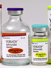
Photo from Business Wire
Results of a phase 1 study suggest that repeated doses of the immunotherapy drug ipilimumab is a feasible treatment option for patients with hematologic diseases who relapse after allogeneic hematopoietic stem cell transplant (HSCT).
Seven of the 28 patients studied responded to the treatment, but immune-mediated toxic effects and graft-vs-host disease (GVHD) occurred as well.
These results were published in NEJM.
Ipilimumab, which is already approved to treat unresectable or metastatic melanoma, works by blocking the immune checkpoint CTLA-4. Blockade of CTLA-4 has been shown to augment T-cell activation and proliferation.
“We believe [,in the case of relapse after HSCT,] the donor immune cells are present but can’t recognize the tumor cells because of inhibitory signals that disguise them,” said study author Matthew Davids, MD, of the Dana-Farber Cancer Institute in Boston, Massachusetts.
“By blocking the checkpoint, you allow the donor cells to see the cancer cells.”
Dr Davids and his colleagues tested this theory in 28 patients who had relapsed after allogeneic HSCT. The patients had acute myeloid leukemia (AML, n=12), Hodgkin lymphoma (n=7), non-Hodgkin lymphoma (n=4), myelodysplastic syndromes (MDS, n=2), multiple myeloma (n=1), myeloproliferative neoplasm (n=1), or acute lymphoblastic leukemia (n=1).
Patients had received a median of 3 prior treatment regimens, excluding HSCT (range, 1 to 14), and 20 patients (71%) had received treatment for relapse after transplant. Eight patients (29%) previously had grade 1/2 acute GVHD, and 16 (57%) previously had chronic GVHD.
The median time from transplant to initial treatment with ipilimumab was 675 days (range, 198 to 1830), and the median time from relapse to initial treatment with ipilimumab was 97 days (range, 0 to
1415).
Patients received induction therapy with ipilimumab at a dose of 3 mg/kg or 10 mg/kg every 3 weeks for a total of 4 doses. Those who had a clinical benefit received additional doses every 12 weeks for up to 60 weeks.
Safety
Five patients discontinued ipilimumab due to dose-limiting toxic effects. Four of these patients had GVHD, and 1 had severe immune-related adverse events.
Dose-limiting GVHD presented as chronic GVHD of the liver in 3 patients and acute GVHD of the gut in 1 patient.
Immune-related adverse events included death (n=1), pneumonitis (2 grade 2 events, 1 grade 4 event), colitis (1 grade 3 event), immune thrombocytopenia (1 grade 2 event), and diarrhea (1 grade 2 event).
Efficacy
There were no responses in patients who received ipilimumab at 3 mg/kg. Among the 22 patients who received ipilimumab at 10 mg/kg, 5 had a complete response, and 2 had a partial response.
Six other patients did not qualify as having responses but had a decrease in their tumor burden. Altogether, ipilimumab reduced tumor burden in 59% of patients.
The complete responses occurred in 4 patients with extramedullary AML and 1 patient with MDS developing into AML. Two of the AML patients remained in complete response at 12 and 15 months, and the patient with MDS remained in complete response at 16 months.
At a median follow-up of 15 months (range, 8 to 27), the median duration of response had not been reached. Responses were associated with in situ infiltration of cytotoxic CD8+ T cells, decreased activation of regulatory T cells, and expansion of subpopulations of effector T cells.
The 1-year overall survival rate was 49%.
The investigators said these encouraging results have set the stage for larger trials of checkpoint blockade in this patient population. Further research is planned to determine whether immunotherapy drugs could be given to high-risk patients to prevent relapse.
Ipilimumab may restore antitumor immunity after relapse from HSCT
Early data hint that immune checkpoint inhibitors may be able to restore antitumor activity in patients with hematologic malignancies that have relapsed after allogeneic transplant.
Among 22 patients with relapsed hematologic cancers following allogeneic hematopoietic stem cell transplantation (HSCT) in a phase I/Ib study, treatment with the anti-CTLA-4 antibody ipilimumab (Yervoy) at a dose of 10 mg/kg was associated with complete responses in five patients, partial responses in two, and decreased tumor burden in six, reported Matthew S. Davids, MD, of the Dana-Farber Cancer Institute in Boston, and his colleagues.
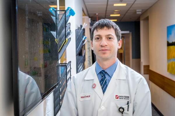
“CTLA-4 blockade was a feasible approach for the treatment of patients with relapsed hematologic cancer after transplantation. Complete remissions with some durability were observed, even in patients with refractory myeloid cancers,” they wrote (N Engl J Med. 2016 Jul 14. doi: 10.1056/NEJMoa1601202).
More than one-third of patients who undergo HSCT for hematologic malignancies such as lymphoma, multiple myeloma, or leukemia will experience a relapse, and most will die within a year of relapse despite salvage therapies or retransplantation, the authors noted.
“Immune escape (i.e., tumor evasion of the donor immune system) contributes to relapse after allogeneic HSCT, and immune checkpoint inhibitory pathways probably play an important role,” they wrote.
Selective CTLA-4 blockade has been shown in mouse models to treat late relapse after transplantation by augmenting graft-versus-tumor response without apparent exacerbation of graft-versus-host disease (GVHD). To see whether the use of a CTLA-4 inhibitor could have the same effect in humans, the investigators instituted a single-group, open-label, dose-finding, safety and efficacy study of ipilimumab in 28 patients from six treatment sites.
The patients had all undergone allogeneic HSCT more than 3 months before the start of the study. The diagnoses included acute myeloid leukemia (AML) in 12 patients (including 3 with leukemia cutis and 1 with a myeloid sarcoma), Hodgkin lymphoma in 7, non-Hodgkin lymphoma in 4, myelodysplastic syndrome (MDS) in 2, and multiple myeloma, myeloproliferative neoplasm, and acute lymphoblastic leukemia in 1 patient each. Eight of the patients had previously had either grade I or II acute GVHD; 16 had had chronic GVHD.
Patients received induction therapy with ipilimumab at a dose of either 3 mg/kg (6 patients), or 10 mg/kg (22 patients) every 3 weeks for a total of 4 doses. Patients who experienced a clinical benefit from the drug could receive additional doses every 12 weeks for up to 60 weeks.
There were no clinical responses meeting study criteria in any of the patients who received the 3-mg/kg dose. Among the 22 who received the 10-mg/kg dose, however, the rate of complete responses was 23% (5 of 22), partial responses 9% (2 of 22), and decreased tumor burden 27% (6 of 22). The remaining nine patients experienced disease progression.
Four of the complete responses occurred in patients with extramedullary AML, and one occurred in a patient with MDS transforming into AML.
The safety analysis, which included all 28 patients evaluable for adverse events, showed four discontinuations due to dose-limiting chronic GVHD of the liver in the 3 patients, and acute GVHD of the gut in 1, and to severe immune-related events in one additional patient, leading to the patient’s death.
Other grade 3 or greater adverse events possibly related to ipilimumab included acute kidney injury (one patient) , corneal ulcer (one), thrombocytopenia (nine), neutropenia (three), anemia and pleural effusion (two).
The investigators point out that therapy to stimulate a graft-versus-tumor effect has the potential to promote or exacerbate GVHD, as occurred in four patients in the study. The GVHD in these patients was effectively managed with glucocorticoids, however.
The National Institutes of Health, Leukemia and Lymphoma Society, Pasquarello Tissue Bank, and Dana-Farber Cancer Institute supported the study. Dr. Davids disclosed grants from ASCO, the Pasquarello Tissue Bank, NIH, NCI, and Leukemia and Lymphoma society, and personal fees from several companies outside the study. Several coauthors disclosed relationships with various pharmaceutical companies, including Bristol-Myers Squibb, maker of ipilimumab.
Early data hint that immune checkpoint inhibitors may be able to restore antitumor activity in patients with hematologic malignancies that have relapsed after allogeneic transplant.
Among 22 patients with relapsed hematologic cancers following allogeneic hematopoietic stem cell transplantation (HSCT) in a phase I/Ib study, treatment with the anti-CTLA-4 antibody ipilimumab (Yervoy) at a dose of 10 mg/kg was associated with complete responses in five patients, partial responses in two, and decreased tumor burden in six, reported Matthew S. Davids, MD, of the Dana-Farber Cancer Institute in Boston, and his colleagues.
“CTLA-4 blockade was a feasible approach for the treatment of patients with relapsed hematologic cancer after transplantation. Complete remissions with some durability were observed, even in patients with refractory myeloid cancers,” they wrote (N Engl J Med. 2016 Jul 14. doi: 10.1056/NEJMoa1601202).
More than one-third of patients who undergo HSCT for hematologic malignancies such as lymphoma, multiple myeloma, or leukemia will experience a relapse, and most will die within a year of relapse despite salvage therapies or retransplantation, the authors noted.
“Immune escape (i.e., tumor evasion of the donor immune system) contributes to relapse after allogeneic HSCT, and immune checkpoint inhibitory pathways probably play an important role,” they wrote.
Selective CTLA-4 blockade has been shown in mouse models to treat late relapse after transplantation by augmenting graft-versus-tumor response without apparent exacerbation of graft-versus-host disease (GVHD). To see whether the use of a CTLA-4 inhibitor could have the same effect in humans, the investigators instituted a single-group, open-label, dose-finding, safety and efficacy study of ipilimumab in 28 patients from six treatment sites.
The patients had all undergone allogeneic HSCT more than 3 months before the start of the study. The diagnoses included acute myeloid leukemia (AML) in 12 patients (including 3 with leukemia cutis and 1 with a myeloid sarcoma), Hodgkin lymphoma in 7, non-Hodgkin lymphoma in 4, myelodysplastic syndrome (MDS) in 2, and multiple myeloma, myeloproliferative neoplasm, and acute lymphoblastic leukemia in 1 patient each. Eight of the patients had previously had either grade I or II acute GVHD; 16 had had chronic GVHD.
Patients received induction therapy with ipilimumab at a dose of either 3 mg/kg (6 patients), or 10 mg/kg (22 patients) every 3 weeks for a total of 4 doses. Patients who experienced a clinical benefit from the drug could receive additional doses every 12 weeks for up to 60 weeks.
There were no clinical responses meeting study criteria in any of the patients who received the 3-mg/kg dose. Among the 22 who received the 10-mg/kg dose, however, the rate of complete responses was 23% (5 of 22), partial responses 9% (2 of 22), and decreased tumor burden 27% (6 of 22). The remaining nine patients experienced disease progression.
Four of the complete responses occurred in patients with extramedullary AML, and one occurred in a patient with MDS transforming into AML.
The safety analysis, which included all 28 patients evaluable for adverse events, showed four discontinuations due to dose-limiting chronic GVHD of the liver in the 3 patients, and acute GVHD of the gut in 1, and to severe immune-related events in one additional patient, leading to the patient’s death.
Other grade 3 or greater adverse events possibly related to ipilimumab included acute kidney injury (one patient) , corneal ulcer (one), thrombocytopenia (nine), neutropenia (three), anemia and pleural effusion (two).
The investigators point out that therapy to stimulate a graft-versus-tumor effect has the potential to promote or exacerbate GVHD, as occurred in four patients in the study. The GVHD in these patients was effectively managed with glucocorticoids, however.
The National Institutes of Health, Leukemia and Lymphoma Society, Pasquarello Tissue Bank, and Dana-Farber Cancer Institute supported the study. Dr. Davids disclosed grants from ASCO, the Pasquarello Tissue Bank, NIH, NCI, and Leukemia and Lymphoma society, and personal fees from several companies outside the study. Several coauthors disclosed relationships with various pharmaceutical companies, including Bristol-Myers Squibb, maker of ipilimumab.
FROM THE NEW ENGLAND JOURNAL OF MEDICINE
Key clinical point: Anti-CTLA-4 therapy may restore graft-versus-tumor effect in patients with hematologic malignancies relapsed after allogeneic transplantation.
Major finding: Five of 22 patients on a 10-mg/kg dose of ipilimumab had a complete response.
Data source: Phase I/Ib investigator-initiated study of 28 patients with hematologic malignancies relapsed after allogeneic hematopoietic stem cell transplantation.
Disclosures: The National Institutes of Health, Leukemia and Lymphoma Society, Pasquarello Tissue Bank, and Dana-Farber Cancer Institute supported the study. Dr. Davids disclosed grants from ASCO, the Pasquarello Tissue Bank, NIH, NCI, and Leukemia and Lymphoma society, and personal fees from several companies outside the study. Several coauthors disclosed relationships with various pharmaceutical companies, including Bristol-Myers Squibb, maker of ipilimumab.
