User login
Drug can improve upon chelation therapy in thalassemia major

An anti-hypertensive drug can improve the effects of chelation therapy in patients with thalassemia major and cardiac iron overload, according to research published in Blood.
Researchers found that administering the drug, amlodipine, in conjunction with conventional chelation therapy significantly reduced myocardial iron concentration (MIC) in patients with cardiac iron overload at baseline, when compared to chelation therapy alone.
In addition, amlodipine did not cause any serious adverse events.
“The drug has been used clinically for decades and is considered safe for adults and children,” said study author Juliano de Lara Fernandes, MD, PhD, of José Michel Kalaf Research Institute in Campinas, São Paulo State, Brazil.
“As an adjunct to standard treatment, it can be greatly beneficial to patients and has few side effects.”
Dr Fernandes said he and his colleagues decided to test amlodipine in patients with thalassemia major because although chelation therapy works well in peripheral organs, it’s difficult to remove iron from the heart.
“Myocardial dysfunctions are currently the main cause of death among patients with thalassemia and can emerge in children from the age of 10,” Dr Fernandes said.
The most serious problem of all, he added, is caused by an accumulation of non-transferrin bound iron (NTBI) in myocardial cells. NTBI enters and leaves the liver without causing much damage to the organ, but it enters the heart via a channel whose main role is to carry calcium into cells.
“It occurred to us that drugs capable of blocking the calcium channel could also prevent NTBI from entering the heart and therefore increase the efficacy of chelation therapy,” Dr Fernandes said.
He and his colleagues tested this hypothesis in 62 patients with thalassemia major. The patients were divided into 2 treatment groups. One group received conventional chelation therapy along with amlodipine (5 mg/day), and the other received chelation therapy plus oral placebo.
Patients were further divided according to cardiac iron levels at baseline. Cardiac iron overload was defined as T2* < 35 ms. Fifty percent of patients in the amlodipine arm and 52% of those in the placebo arm had cardiac iron overload at baseline.
Results
The study’s main outcome was change in MIC, as determined by magnetic resonance imaging at 12 months.
At that point, among patients with cardiac iron overload at baseline, there was a significant decrease in MIC in the amlodipine arm but not the placebo arm. The median change in MIC was -0.26 mg/g and 0.01 mg/g, respectively (P=0.02).
The median MIC in the amlodipine arm went from 1.31 mg/g at baseline to 1.05 mg/g at 12 months (P=0.02). The median MIC in the placebo arm went from 0.77 mg/g at baseline to 0.75 at 12 months (P=0.76).
“Myocardial iron concentration fell 21% in patients with initial iron overload who were treated with chelation plus amlodipine, whereas it increased by 2% in those with initial overload who were treated with chelation plus placebo,” Dr Fernandes said.
On the other hand, there were no significant differences in MIC from baseline to 12 months among patients who received amlodipine but did not have cardiac iron overload at baseline.
For patients without cardiac iron overload at baseline, the median change in MIC was +0.04 mg/g in the amlodipine arm and -0.01 in the placebo arm (P=0.07).
The median MIC in the amlodipine arm went from 0.54 mg/g at baseline to 0.55 mg/g at 12 months. The median MIC in the placebo arm went from 0.55 mg/g at baseline to 0.52 mg/g at 12 months.
“Perhaps we would have needed to monitor these patients for a longer period to see the benefits of preventive therapy with amlodipine for people who don’t have excess iron in their organs,” Dr Fernandes said.
“For those who do, however, the results show it’s worth using amlodipine. There’s no need to change the existing therapy. It’s enough to administer the anti-hypertensive orally every day.”
There were 4 mild adverse events in the amlodipine arm but none in the placebo arm (13% vs 0%, P=0.11).
Three patients (10%) in the amlodipine arm had their initial dose of 5 mg/day reduced to 2.5 mg/day because of mild ankle edema (n=2) and dizziness (n=1). One patient had a mild cutaneous allergic reaction and stopped receiving amlodipine after 15 days but continued on study.
There were no deaths or hospital admissions due to cardiovascular complications during the trial. ![]()
HU trial to prevent stroke in SCA feasible in Nigeria

© Todd Buchanan 2016
SAN DIEGO—High rates of recruitment (90%), enrollment (92%), and adherence to study drug and follow-up visits have confirmed the feasibility of conducting a trial of hydroxyurea (HU) for stroke prevention in Nigeria (the SPIN trial), researchers say.
The 235 children with sickle cell anemia (SCA)enrolled on the SPIN trial did not miss any of the scheduled monthly visits, and drug adherence was 84% based on the increase in their mean corpuscular volume (MCV) by 10 fL.
These data provide strong evidence, researchers believe, for patient and family acceptability of the trial and potential safety of a moderate dose of HU to prevent stroke in children with SCA in Nigeria.
Nigeria has the largest burden of sickle cell disease (SCD) in the world, Najibah Galadanci, MBBS, of Bayero University/Aminu Kano Teaching Hospital in Nigeria, said at the 2016 ASH Annual Meeting.
Every year, about 150,000 children in Nigeria are born with SCD. This compares with 2400 children in the United States and 300 children in the United Kingdom.
“And it is estimated that 15,000 children with SCA per year in Nigeria will have strokes,” Dr Galadanci added.
She presented data from the SPIN trial (NCT01801423) at ASH as abstract 122.
At present, she explained, primary stroke prevention consists of regular blood transfusions for patients with transcranial Doppler (TCD) measurements higher than 200 cm/second.
However, distinct challenges with this prevention method exist in sub-Saharan Africa, such as inadequate blood supply, cost, unsafe transfusion practice, and the high probability of blood-borne infections.
HU is the only drug approved by the US Food and Drug administration to treat SCD. It increases total hemoglobin level, which is associated with a decreased risk of strokes.
In addition, HU significantly decreases TCD ultrasound velocity in children with SCD and abnormal TCD and is cost-effective and practical in sub-Saharan Africa.
So investigators at Aminu Kano teaching hospital in Nigeria undertook to study the feasibility of using HU to prevent stroke in children with SCD.
The team based their decision on 3 main components: recruitment rate, retention rate, and adherence to study medication.
SPIN trial
Children ages 5 to 12 were eligible if they had a diagnosis of SCA, either HbSS or HbSb0. They had to have 2 independent readings of elevated TCD velocity of 200 to 219 cm/second or 1 reading of 220 cm/second or higher.
Investigators enrolled 25 children on the treatment arm. The children received a moderate dose (20 mg/kg/day) of HU for 3 years.
Investigators also enrolled a comparison group of 210 children with SCA who had a TCD velocity of less than 200 cm/second.
The median follow-up was 2.1 years. The median age was 6.8 years and 8 years in the treatment and comparison groups, respectively.
The treatment group had a total of 603 follow-up visits.
The recruitment rate was 90% (335 of 370 families), the enrollment rate for the treatment arm was 92% (25 of 27 patients), and the adherence rate to monthly visits was 100%. Eighty-four percent of patients (21/25) adhered to the medication regimen, based on their increased MCV.
HU therapy
Investigators observed no laboratory evidence of severe myelosuppression or toxicity.
Of 712 complete blood counts performed on 25 study participants, 2 patients had repeated hemoglobin counts of less than 6 g/dL, and no participant had a repeat platelet count below 80 x 109/L nor a repeat absolute neutrophil count of less than 1.2 x 10⁹/L.
Investigators found no significant difference overall (P=0.37) in the rate of hospitalization between the treatment and comparison groups based on hospitalizations for acute chest syndrome, pain, stroke, transfusion, malaria, and infection.
Investigators also found no significant difference (P=0.67) in rates of severe adverse events between the study and comparison groups.
Twelve deaths occurred during the study period, 2 in the treatment group (2.69/100 patient years) and 10 in the comparison group (1.81/100 patient years).
Deaths in the treatment arm were due to sepsis and progressive renal disease. Deaths in the comparison group were due to severe anemia, infection, and malaria.
“The most interesting finding of our study,” Dr Galadanci indicated, “was the 85% reduction in TCD velocity after starting hydroxyurea therapy.”
Baseline TCD measurements went from 211 cm/second to 165 cm/second at 24 months.
Dr Galadanci said next steps include conducting a phase 3, multicenter, randomized controlled trial (NCT 02560935) comparing low-dose (10 mg/kg/day) and moderate-dose (20 mg/kg/day) HU therapy for preventing primary strokes in children with SCA living in Nigeria (SPRING Trial).
Investigators hypothesize there will be a 66% reduction over 3 years in relative risk of primary strokes in children with SCA and elevated TCD velocity in the moderate-dose group compared to the low-dose group. ![]()
Drug granted fast track designation for PNH

The US Food and Drug Administration (FDA) has granted fast track designation for the complement C3 inhibitor APL-2.
The designation applies to APL-2 in the treatment of patients with paroxysmal nocturnal hemoglobinuria (PNH) who continue to experience hemolysis and require red blood cell transfusions despite receiving therapy with eculizumab.
APL-2 is also being developed as a treatment for PNH patients not previously treated with eculizumab.
The company developing APL-2 is Apellis Pharmaceuticals, Inc.
APL-2 is a synthetic cyclic peptide conjugated to a polyethylene glycol polymer that binds specifically to C3 and C3b, blocking all 3 pathways of complement activation (classical, lectin, and alternative).
According to Apellis, this comprehensive inhibition of complement-mediated pathology may have the potential to control symptoms and modify underlying disease in patients with PNH.
Results from a pair of phase 1 studies of APL-2 in healthy volunteers were recently presented at the 2016 ASH Annual Meeting (abstract 1251).
Now, Apellis is evaluating APL-2 in a pair of phase 1b clinical trials of patients with PNH.
In PADDOCK (NCT02588833), researchers are assessing the safety, tolerability, pharmacokinetics, pharmacodynamics, and preliminary efficacy of multiple doses of APL-2 administered by daily subcutaneous injection in patients with PNH who have not received the standard of care in the past.
In PHAROAH (NCT02264639), researchers are assessing the safety, tolerability, pharmacokinetics, and pharmacodynamics of single and multiple doses of APL-2 administered by subcutaneous injection as an add-on to the standard of care in patients with PNH.
About fast track designation
The FDA’s fast track program is designed to facilitate the development and expedite the review of products intended to treat or prevent serious or life-threatening conditions and address unmet medical need.
Through the FDA’s fast track program, a product may be eligible for priority review. In addition, the company developing the product may be allowed to submit sections of the biologic license application or new drug application on a rolling basis as data become available.
Fast track designation also provides the company with opportunities for more frequent meetings and written communications with the FDA. ![]()
Agios stops developing drug for PK deficiency

Agios Pharmaceuticals, Inc. is no longer developing one of its pyruvate kinase-R (PKR) activators, AG-519, for the treatment of pyruvate kinase (PK) deficiency.
The company withdrew its investigational new drug application for AG-519 following a verbal notification of a clinical hold from the US Food and Drug Administration (FDA).
The hold resulted from an adverse event—cholestatic hepatitis—observed in a phase 1 trial of healthy volunteers.
“[W]e received feedback from the FDA that AG-519 no longer has an appropriate risk-benefit ratio to move forward in clinical development and was placed on clinical hold due to that case of cholestatic hepatitis,” said David Schenkein, MD, chief executive officer at Agios.
“We made the decision to withdraw the IND [investigational new drug application] and discontinue development of AG-519 and advance AG-348, our first-in-class and lead pyruvate kinase activator into pivotal development. We share the FDA’s commitment to patient safety and believe this is the right decision to ultimately help people with PK deficiency.”
About AG-519
Agios has described AG-519 as a potent, highly selective, and orally bioavailable PKR activator devoid of the aromatase inhibitory effects that were observed with the company’s other PKR activator, AG-348.
AG-519 was evaluated in a phase 1 study of healthy volunteers in the UK. The goal of this study was to assess the drug’s safety, tolerability, pharmacokinetics, pharmacodynamics, and bioavailability.
A case of drug-induced cholestatic hepatitis occurred in the bioavailability portion of the study. This volunteer continues to be monitored and is showing improvement, according to Dr Schenkein.
Agios said other adverse events observed in this trial were largely mild or moderate (grade 1/2). The most common of these was headache.
The company did note a case of grade 2 thrombocytopenia that resolved spontaneously within 7 days after the last dose of AG-519.
Results from this trial were presented at the 2015 ASH Annual Meeting (abstract 1264).
AG-519 was also under investigation in a palatability study of volunteers in the US. The goal of this study was to develop a formulation of the drug for potential future development.
In total, 98 volunteers have received AG-519. No volunteers or patients are currently receiving the drug.
About AG-348
Agios’s decision to stop developing AG-519 does not affect the company’s ongoing phase 2 study (DRIVE PK) of AG-348, an activator of both wild-type and mutated PKR enzymes.
“AG-348 and AG-519 are different molecules with different structures,” Dr Schenkein noted.
Agios is advancing AG-348 into development as the first potential disease-modifying treatment for PK deficiency.
Results from a pair of phase 1 studies of AG-348 were presented at the 2014 ASH Annual Meeting (abstract 4007).
About PK deficiency
PK deficiency is a rare inherited disease that presents as hemolytic anemia. The inherited mutations in PKR enzymes cause a deficit in cellular energy within the red blood cell, as evidenced by lower PK enzyme activity, a decline in adenosine triphosphate levels, and a build-up of upstream metabolites, including 2,3-DPG.
The current standard of care for PK deficiency is supportive care, including blood transfusions, splenectomy, chelation therapy to address iron overload, and/or interventions for other treatment- and disease-related morbidities.
There is, at present, no approved therapy to treat the underlying cause of PK deficiency. ![]()
MDS patients with mutated IDH2 benefit from enasidenib

Photo courtesy of ASH
SAN DIEGO—Daily treatment with enasidenib monotherapy in patients with mutated IDH2-positive myelodysplastic syndromes (MDS) induced responses in the majority of patients treated, according to a presentation at the 2016 ASH Annual Meeting.
The study was a portion of a larger phase 1/2 trial of the agent in patients with acute myeloid leukemia (AML) and other hematologic malignancies, so the subset was relatively small, numbering 17 patients.
Nevertheless, enasidenib was well tolerated and induced responses in these predominantly higher-risk patients.
Enasidenib (AG-221/CC-9007) is a selective, oral, potent inhibitor of mutant IDH2 (mIDH2), which produces 2-HG and thus alters DNA methylation and blocks cellular differentiation of hematopoietic progenitor cells.
Approximately 15% of AML patients and 5% of MDS patients have mIDH2. So investigators undertook the study to evaluate the safety and efficacy of enasidenib monotherapy in these diseases.
Eytan Stein, MD, of Memorial Sloan Kettering Cancer Center in New York, New York, presented the analysis of enasidenib in mIDH2-positive MDS patients as abstract 343.*
Study design
MDS patients were allowed to enroll during the dose-escalation and expansion phase of the study, Dr Stein explained.
Patients had to be 18 or older and have an advanced hematologic malignancy with mutated IDH2—relapsed or refractory AML, relapsed or refractory MDS, untreated AML, or other hematologic malignancy with mIDH2.
MDS patients could not be candidates for other therapies, had to be IPSS-R high risk, and had to have relapsed or refractory RAEB-1/RAEB-2 disease.
Investigators also performed co-molecular profiling using next-generation sequencing with a FoundationOne® Heme Panel.
All patients received daily oral enasidenib at 100 mg daily in 28-day cycles.
Patient characteristics
The study accrued a total of 239 patients—176 with relapsed or refractory AML, 37 with untreated AML, 9 with another hematologic malignancy, and 17 with MDS.
The median age of the MDS patients was 67 (range, 45-78), and 71% were male. All had the IDH2 mutation, 88% had R140 mutations, and 12% had R172.
Thirteen patients (76%) had an ECOG performance status of 0-1, and 4 (24%) had a performance status of 2.
A little more than a third (35%) of patients had 2 or more prior anti-cancer regimens.
Two patients (12%) received prior lenalidomide therapy, 8 (47%) received other treatments, including sorafenib (n=2) and vosaroxin, epoetin alfa, pracinostat, cytarabine plus clofarabine, ruxolitinib, and rigosertib (n=1 each). Four patients (24%) were untreated.
“I want to make the point,” Dr Stein said, “that, of those patients, three quarters of them, 76% [n=13], had received a prior hypomethylating agent, really understanding that this is a very poor-risk group of patients that we are studying here.”
About half of patients (47%) had intermediate-2/high IPSS risk status, good MDS cytogenetic risk, and high/very high IPSS-R risk status.
Adverse events
Grade 3-4 treatment-emergent adverse events (AEs) occurring in 2 or more patients were hyperbilirubinemia (n=5), pneumonia (n=4), thrombocytopenia (n=4), anemia (n=3), hypokalemia (n=3), dyspnea (n=2), and tumor lysis syndrome (n=2).
“As I’ve said in a number of meetings where I’ve talked about IDH2 inhibitors, and specifically enasidenib, the hyperbilirubinemia that is seen with this drug is an indirect hyperbilirubinemia,” Dr Stein said.
“A known off-target effect of this drug is inhibition of the UGT1a1 enzyme, which conjugates bilirubin, and this indirect hyperbilirubinemia, which is typically relatively mild [and] does not appear to have any clinical sequelae.”
Investigators considered 9 of the AEs reported for 6 patients to be drug-related.
Four serious enasidenib-related AEs included tumor lysis syndrome (n=2), increased blood bilirubin (n=1), and transaminitis (n=1).
There were no treatment-related deaths.
Response and survival
Ten of 17 patients (59%) achieved an overall response, defined as complete response (CR) plus partial response, plus marrow CR, plus hematologic improvement (HI).
One patient achieved CR, 1 had a partial response, 3 had marrow CR, and 5 had HI.
Dr Stein noted that, of the 13 patients who had received prior hypomethylating agent therapy, 7 (54%) had a response with enasidenib.
Of the patients who attained HI, 2 had trilineage and 2 had bilineage improvement.
The median time to response was 21 days (range, 10-87), and the median number of treatment cycles was 3.0.
Patients remained on study for up to 24 months. Four patients in hematologic remission are still on study, and 3 patients proceeded to stem cell or bone marrow transplant, Dr Stein said.
Two patients discontinued because of investigator decision, and 8 discontinued therapy due to death or disease progression.
The limitation of the study regarding overall survival, Dr Stein said, is that the number is very small.
At a median follow-up of 7.5 months, the median overall survival was not reached.
“So I’m not arguing that this is the end word of this,” he said. “This is going to be dynamic as more patients come on these studies. But I think it’s a nice indication that this treatment appears to be well-tolerated and doing good for a large subset of patients.”
Co-occurring mutations
The investigators also analyzed co-occurring mutations in 13 MDS patients.
The small patient population prevented the investigators from making definitive conclusions regarding potential correlations between response and co-mutations.
Nevertheless, Dr Stein said the analysis revealed something “very, very intriguing.”
He noted that 7 patients had ASXL1 mutations, and “those are typically patients who are bad actors.”
“Five of those 7 patients had a response,” Dr Stein said. “And of those 5, 3 of them had received a prior hypomethylating agent. I think it’s at least food for thought that you can salvage a patient who has failed a hypomethylating agent, with poor risk disease. I think this is very, very exciting.”
Celgene Corporation and its collaborator, Agios Pharmaceuticals, sponsored the trial. ![]()
*Information in the abstract differs from the presentation.
Predicting the risk of CKD in sickle cell anemia
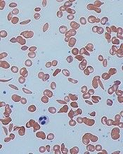
and normal red blood cells
Image by Graham Beards
Researchers say they have identified a genetic risk profile that can be used to predict which patients with sickle cell anemia (SCA) are likely to develop chronic kidney disease (CKD).
The team found evidence to suggest that a profile incorporating APOL1 G1/G2, α-thalassemia, and BCL11A can help physicians categorize SCA patients as having a high or low risk of CKD.
The researchers reported these findings in Haematologica.
Identifying SCA patients at the greatest risk of CKD can help physicians develop proactive, individualized strategies to reduce the high rate of early mortality due to CKD, said study author Santosh Saraf, MD, of the University of Illinois at Chicago.
“We looked at the genetic factors already known to be associated with kidney disease or the degree of red blood cell hemolysis and examined the relationship they had with the condition in sickle cell patients,” Dr Saraf said.
“Our hypothesis was that a genetic risk profile that integrated APOL1, α-thalassemia, and BCL11A would improve our ability to predict a patient’s risk for developing chronic kidney disease.”
To test this theory, Dr Saraf and his colleagues recruited 262 adult patients with SCA treated at a single center between 2010 and 2016. The team collected patient data, drew blood, analyzed genetic markers, and prospectively followed the patients to see if they developed CKD.
Stratification
The researchers used their genetic profile to stratify patients according to risk for CKD.
Patients were considered high-risk if they had APOL1 G1/G2 and wild-type BCL11A but did not have α-thalassemia.
Patients were considered low-risk if they were negative for APOL1 G1/G2 but had α-thalassemia (either α-/αα or α-/α-) and the BCL11A rs1427407 T allele (either G/T or T/T).
The researchers defined all other combinations as intermediate-risk.
The team found the genetic profile identified SCA patients at high and low risk for albuminuria or an estimated glomerular filtration rate less than 60 mL/min/1.73m2.
The researchers also said application of the genetic profile revealed progressively higher rates of CKD progression.
Mechanisms
Dr Saraf and his colleagues noted that homozygosity or compound heterozygosity for APOL1 G1/G2 is the strongest genetic association for CKD in African Americans, and APOL1 G1/G2 is associated with proteinuria and albuminuria in SCA patients.
The researchers pointed out that APOL1 G1/G2 variants associate with CKD in African
Americans by unknown mechanisms, but the team found an association with
hemolysis in SCA, as reflected by hemoglobinuria.
As for α-thalassemia, it’s associated with reduced hemolysis and protection from albuminuria. The researchers said α-thalassemia reduces hemolysis in SCA by decreasing the
intra-erythrocyte concentration of sickle hemoglobin (HbS) and reducing
HbS polymerization.
Finally, Dr Saraf and his colleagues noted that BCL11A rs1427407 leads to higher fetal hemoglobin (HbF) levels, reduced hemolysis, and amelioration of SCA-related complications. The team said the BCL11A rs1427407 T variant leads to decreased function of BCL11A at the HbF promoter and therefore increases HbF, leading to decreased HbS polymerization.
“The results of this study are encouraging,” Dr Saraf said. “By understanding more about the genetic risk factors of kidney disease in sickle cell patients, we are one step closer to improving the length and quality of life for the millions of people worldwide living with sickle cell disease.”
“Using combinations of genes to better predict complications in sickle cell anemia is a new approach,” added study author Victor Gordeuk, MD, of the University of Illinois at Chicago.
“The results of this study indicate that it is effective and probably can be improved on in the future to be an important part of our evaluation of patients.”
The researchers said the small sample size and observational nature of this study are limitations, and they hope to validate the results with larger studies. ![]()
Another treatment on the horizon for SCD

Photo courtesy of ASH
SAN DIEGO—The first-in-class humanized anti-P-selectin antibody SelG1, also known as crizanlizumab, significantly reduced sickle cell pain crises (SCPC) when compared to placebo in the phase 2 SUSTAIN trial.
The higher dose of SelG1 tested reduced the annual rate of SCPC by 45% (P=0.01) and the annual rate of uncomplicated SCPC by 63% (P=0.015).
Acute painful crises are the primary cause for patients with sickle cell disease (SCD) to seek medical attention.
Kenneth I. Ataga, MD, of the University of North Carolina at Chapel Hill, explained that upregulation of P-selectin on endothelial cells and platelets contributes to the cell-cell interaction involved in the pathogenesis of SCPC. SelG1 binds to P-selectin and inhibits its interaction with P-selectin glycoprotein ligand 1.
Dr Ataga presented results from the SUSTAIN study during the plenary session of the 2016 ASH Annual Meeting (abstract 1).
The study was also published in NEJM. The research was sponsored by Selexys Pharmaceuticals Corporation, which was recently acquired by Novartis.
Patient population
A total of 198 patients were randomized, 67 to high-dose SelG1 (5.0 mg/kg), 66 to low-dose SelG1 (2.5 mg/kg), and 65 to placebo.
They received a loading dose in the first 2 weeks of treatment, followed by monthly dosing for a year.
Patients had to have a diagnosis of SCD, including genotypes HbSS, HbSC, HbSb0-thalassemia, or HbSB+-thalassemia.
They had to have at least 2 but not more than 10 acute sickle-related pain events within 12 months of study entry.
Patients ranged in age from 16 to 57 years, about 70% had HbSS, and 60% were on concomitant hydroxyurea therapy.
Patients not already receiving hydroxyurea were not permitted to start it during the study. And patients could not be on chronic transfusion therapy.
Study endpoints
The primary endpoint was the annual rate of adjudicated SCPC.
“Painful crisis was defined as an active episode of pain, and it was felt to be related to sickle cell disease-specific events and no other medically defined causes,” Dr Ataga explained.
The pain episodes also had to result in a visit to a medical facility and require treatment with parenteral or oral narcotics or parenteral nonsteroidal anti-inflammatory drugs.
The definition of SCPC included not only typical painful episodes but also acute chest syndrome (ACS), hepatic or splenic sequestration, and priapism.
Secondary endpoints included the annual rate of days hospitalized, time to first and second SCPC, the annual rate of uncomplicated pain crises, and the annual rate of ACS, among others.
Efficacy
The median annual rate of SCPC was 1.63 in the high-dose arm, 2.01 in the low-dose arm, and 2.98 in the placebo arm, amounting to a significant 45.3% reduction with the higher dose of SelG1 compared to placebo. The low dose resulted in a reduction of 32.6% compared to placebo, but this was not significant.
Twenty-four patients in the high-dose arm had an SCPC rate of 0, compared with 12 in the low-dose arm and 11 on placebo.
SCD genotype or concomitant hydroxyurea use did not impact these results.
Patients in the high-dose arm had a median annual rate of 4 hospitalization days, compared with 6.87 in both the low-dose and placebo groups. The difference was not significant, although the reduction in the high-dose arm was 41.8%.
The median annual rate of uncomplicated SCPC was 1.08 in the high-dose arm, 2.00 in the low-dose arm, and 2.91 in the placebo arm. Uncomplicated SCPC excluded ACS, splenic or hepatic sequestration, and priapism.
The reduction in the high-dose arm compared to placebo was significant, at 62.9% (P=0.015).
“The rate of ACS was pretty rare,” Dr Ataga said, “so the median rate across the various groups was 0.”
And time to the first SCPC was 4.1 months (P=0.001) in the high-dose group, 2.2 months (P=0.136) in the low-dose group, and 1.4 months in the placebo group.
“The curves separated pretty early,” Dr Ataga noted, “and were maintained throughout the course of the treatment phase, suggesting that the beneficial effect of SelG1 manifested pretty early following initiation of treatment.”
The time to second event was also significant in the high-dose arm compared to placebo, at 10.3 months and 5.1 months, respectively (P=0.022).
Safety
One or more adverse events occurred in over 85% of patients in each group.
Adverse events that occurred in at least 10% of SelG1-treated patients and amounted to at least double the number in the placebo group were arthralgia, diarrhea, pruritus, vomiting, and chest pain.
Five patients died while on study, but none of these deaths were related to the study drug.
Despite the adverse events, Dr Ataga said the drug was, overall, well-tolerated among the patients who received treatment. ![]()
Photo courtesy of ASH
SAN DIEGO—The first-in-class humanized anti-P-selectin antibody SelG1, also known as crizanlizumab, significantly reduced sickle cell pain crises (SCPC) when compared to placebo in the phase 2 SUSTAIN trial.
The higher dose of SelG1 tested reduced the annual rate of SCPC by 45% (P=0.01) and the annual rate of uncomplicated SCPC by 63% (P=0.015).
Acute painful crises are the primary cause for patients with sickle cell disease (SCD) to seek medical attention.
Kenneth I. Ataga, MD, of the University of North Carolina at Chapel Hill, explained that upregulation of P-selectin on endothelial cells and platelets contributes to the cell-cell interaction involved in the pathogenesis of SCPC. SelG1 binds to P-selectin and inhibits its interaction with P-selectin glycoprotein ligand 1.
Dr Ataga presented results from the SUSTAIN study during the plenary session of the 2016 ASH Annual Meeting (abstract 1).
The study was also published in NEJM. The research was sponsored by Selexys Pharmaceuticals Corporation, which was recently acquired by Novartis.
Patient population
A total of 198 patients were randomized, 67 to high-dose SelG1 (5.0 mg/kg), 66 to low-dose SelG1 (2.5 mg/kg), and 65 to placebo.
They received a loading dose in the first 2 weeks of treatment, followed by monthly dosing for a year.
Patients had to have a diagnosis of SCD, including genotypes HbSS, HbSC, HbSb0-thalassemia, or HbSB+-thalassemia.
They had to have at least 2 but not more than 10 acute sickle-related pain events within 12 months of study entry.
Patients ranged in age from 16 to 57 years, about 70% had HbSS, and 60% were on concomitant hydroxyurea therapy.
Patients not already receiving hydroxyurea were not permitted to start it during the study. And patients could not be on chronic transfusion therapy.
Study endpoints
The primary endpoint was the annual rate of adjudicated SCPC.
“Painful crisis was defined as an active episode of pain, and it was felt to be related to sickle cell disease-specific events and no other medically defined causes,” Dr Ataga explained.
The pain episodes also had to result in a visit to a medical facility and require treatment with parenteral or oral narcotics or parenteral nonsteroidal anti-inflammatory drugs.
The definition of SCPC included not only typical painful episodes but also acute chest syndrome (ACS), hepatic or splenic sequestration, and priapism.
Secondary endpoints included the annual rate of days hospitalized, time to first and second SCPC, the annual rate of uncomplicated pain crises, and the annual rate of ACS, among others.
Efficacy
The median annual rate of SCPC was 1.63 in the high-dose arm, 2.01 in the low-dose arm, and 2.98 in the placebo arm, amounting to a significant 45.3% reduction with the higher dose of SelG1 compared to placebo. The low dose resulted in a reduction of 32.6% compared to placebo, but this was not significant.
Twenty-four patients in the high-dose arm had an SCPC rate of 0, compared with 12 in the low-dose arm and 11 on placebo.
SCD genotype or concomitant hydroxyurea use did not impact these results.
Patients in the high-dose arm had a median annual rate of 4 hospitalization days, compared with 6.87 in both the low-dose and placebo groups. The difference was not significant, although the reduction in the high-dose arm was 41.8%.
The median annual rate of uncomplicated SCPC was 1.08 in the high-dose arm, 2.00 in the low-dose arm, and 2.91 in the placebo arm. Uncomplicated SCPC excluded ACS, splenic or hepatic sequestration, and priapism.
The reduction in the high-dose arm compared to placebo was significant, at 62.9% (P=0.015).
“The rate of ACS was pretty rare,” Dr Ataga said, “so the median rate across the various groups was 0.”
And time to the first SCPC was 4.1 months (P=0.001) in the high-dose group, 2.2 months (P=0.136) in the low-dose group, and 1.4 months in the placebo group.
“The curves separated pretty early,” Dr Ataga noted, “and were maintained throughout the course of the treatment phase, suggesting that the beneficial effect of SelG1 manifested pretty early following initiation of treatment.”
The time to second event was also significant in the high-dose arm compared to placebo, at 10.3 months and 5.1 months, respectively (P=0.022).
Safety
One or more adverse events occurred in over 85% of patients in each group.
Adverse events that occurred in at least 10% of SelG1-treated patients and amounted to at least double the number in the placebo group were arthralgia, diarrhea, pruritus, vomiting, and chest pain.
Five patients died while on study, but none of these deaths were related to the study drug.
Despite the adverse events, Dr Ataga said the drug was, overall, well-tolerated among the patients who received treatment.
Predicting therapy-related myeloid neoplasms

Photo courtesy of
MD Anderson Cancer Center
SAN DIEGO―Clonal hematopoiesis could be used as a predictive marker to identify cancer patients at risk of developing therapy-related myeloid neoplasms (t-MNs), according to researchers.
The team conducted a case-control study, which showed that patients who developed t-MNs—acute myeloid leukemia and myelodysplastic syndromes—were significantly more likely than patients without t-MNs to have clonal hematopoiesis at the time of primary cancer diagnosis.
“Based on these findings, we believe pre-leukemic mutations may function as a new biomarker that would predict t-MN development,” said Andy Futreal, PhD, of The University of Texas MD Anderson Cancer Center in Houston.
Dr Futreal and his colleagues reported these findings in The Lancet Oncology.
Co-author Koichi Takashi, MD, also of MD Anderson, presented the findings at the 2016 ASH Annual Meeting (abstract 38).
Initial cohort
The researchers analyzed data on patients treated at MD Anderson from 1997 to 2015.
The 14 cases the team identified had been treated for a primary cancer and later developed t-MNs. The 54 age-matched control subjects had been treated for lymphoma, received combination chemotherapy, and did not develop t-MNs after at least 5 years of follow-up.
For both cases and controls, the researchers performed gene sequencing on pre-treatment peripheral blood samples. For cases, the researchers also performed targeted gene sequencing on bone marrow samples taken at t-MN diagnosis.
“We found that prevalence of pre-leukemic mutations was significantly higher in patients who developed t-MNs versus those who did not,” Dr Futreal said.
Clonal hematopoiesis was present in 71% of cases (n=10) and 31% of controls (n=17).
“We found genetic mutations that are present in t-MNs leukemia samples actually could be found in blood samples obtained at the time of their original cancer diagnosis,” Dr Takashi noted.
Overall, the cumulative incidence of t-MNs at 5 years was significantly higher in patients with clonal hematopoiesis than in those without it—30% and 7%, respectively (P=0.016).
Validation cohort
The researchers also assessed clonal hematopoiesis in an external cohort of 74 patients with lymphoma who were treated in a trial of front-line chemotherapy with cyclophosphamide, doxorubicin, vincristine, and prednisone, with or without melatonin.
In this cohort, 7% (n=5) of patients developed t-MNs. Eighty percent of these patients (n=4) had clonal hematopoiesis.
Of the 69 patients who did not develop t-MNs, 16% (n=11) had clonal hematopoiesis.
The cumulative incidence of t-MNs at 10 years was significantly higher in patients with clonal hematopoiesis than in those without it—29% and 0%, respectively (P=0.0009).
Multivariate analysis suggested clonal hematopoiesis significantly increased the risk of t-MNs, with a hazard ratio of 13.7 (P=0.013).
“[W]e believe the data suggest potential approaches of screening for clonal hematopoiesis in cancer patients that may identify patients at risk of developing t-MNs, although further studies are needed,” Dr Takashi concluded.
Fanconi anemia linked to cancer gene
Researchers say they have discovered an important molecular link between Fanconi anemia (FA) and PTEN, a gene associated with uterine, prostate, and brain cancer.
They say this discovery enhances our understanding of the molecular basis of Fanconi anemia and could lead to improved treatment outcomes for both Fanconi anemia and cancer patients.
The researchers detailed their discovery in Scientific Reports.
They explained that Fanconi anemia proteins function primarily in DNA interstrand crosslink (ICL) repair, and they wanted to determine the role of the PTEN phosphatase in this process.
“The PTEN gene codes for a phosphatase—an enzyme that removes phosphate groups from proteins,” said study author Niall Howlett, PhD, of the University of Rhode Island in Kingston, Rhode Island.
“Many Fanconi anemia proteins have phosphate groups attached to them when they become activated. However, how these phosphate groups are removed is poorly understood.”
With this in mind, the researchers performed an experiment to determine if Fanconi anemia and PTEN are biochemically linked.
The team knew that cells from Fanconi anemia patients are sensitive to ICL-inducing agents, so they set out to determine if PTEN-deficient cells are sensitive to these agents as well.
“By testing if cells with mutations in the PTEN gene were also sensitive to [ICL-inducing] agents, we discovered that Fanconi anemia patient cells and PTEN-deficient cells were practically indistinguishable in terms of sensitivity to these drugs,” Dr Howlett said.
“This strongly suggested that the Fanconi anemia proteins and PTEN might work together to repair the DNA damage caused by [ICL-inducing] agents.”
Using epistasis analysis, Dr Howlett and his colleagues found that Fanconi anemia proteins and PTEN do indeed function together in ICL repair.
“Before this work, Fanconi anemia and PTEN weren’t even on the same radar,” Dr Howlett said. “This is really important to understanding how this disease arises and what its molecular underpinnings are. The more we can find out about its molecular basis, the more likely we are to come up with strategies to treat the disease.”
Dr Howlett and his colleagues believe their research is equally important to cancer patients. Since this study showed that cells missing PTEN are highly sensitive to ICL-inducing agents, the team believes it should be possible to predict whether a particular cancer patient will respond to this class of drugs by conducting a simple DNA test.
“We can now predict that if a patient has cancer associated with mutations in PTEN, then it is likely that the cancer will be sensitive to [ICL-inducing] agents,” Dr Howlett said. “This could lead to improved outcomes for patients with certain types of PTEN mutations.”
VIDEO: SPIN tests hydroxyurea to prevent pediatric stroke in sickle cell disease
SAN DIEGO – Whether hydroxyurea can avert a first stroke in children with sickle cell disease is a question that may be answered by a soon-to-be-started trial in Nigeria.
An estimated 15,000 children with sickle cell anemia have strokes each year in Nigeria, which is considered to have the largest burden of sickle cell disease in the world. Without treatment, about half of those children will have a second stroke within 2 years.
With completion of its feasibility trial results, SPIN (Primary Stroke Prevention in Children With Sickle Cell Anemia in Nigeria) will begin to examine two doses of hydroxyurea therapy, 20 mg/kg per day and 10 mg/kg per day, to determine whether the drug can prevent a first stroke in high-risk children.
In a video interview, Najibah A. Galadanci, MBBS, MPH, of Aminu Kano (Nigeria) Teaching Hospital, discusses how SPIN is addressing the unresolved clinical issue of hydroxyurea’s risks and benefits in children, and how research in Nigeria may provide adequate patient numbers to address a wide range of clinical questions about sickle cell disease.
The video associated with this article is no longer available on this site. Please view all of our videos on the MDedge YouTube channel
mdales@frontlinemedcom.com
On Twitter @maryjodales
AT ASH 2016