User login
Team characterizes therapy-resistant ALL cells

Image by Vashi Donsk
Researchers say they have characterized a subpopulation of leukemia cells that are responsible for relapse in acute lymphoblastic leukemia (ALL).
The team identified these dormant, therapy-resistant cells in mouse models of ALL and found that removing the cells from their environment makes them sensitive to treatment.
The researchers believe these findings could pave the way to better relapse prevention in patients with ALL.
“Previously, the biological principles responsible for a relapse in leukemia were not fully understood,” said study author Irmela Jeremias, MD, PhD, of Helmholtz Zentrum München in Munich, Germany.
“Our new approach is to isolate dormant cells, which gives us the first possibility of developing therapies that switch off these cells.”
Dr Jeremias and colleagues described this approach in Cancer Cell.
First, the researchers created mouse models recapitulating minimal residual disease (MRD) and relapse in ALL patients.
The team then used genetic engineering and proliferation-sensitive dyes to isolate and characterize relapse-inducing cells.
This revealed a subpopulation of leukemia cells that exhibited long-term dormancy, treatment resistance, and stemness. These cells were similar to primary ALL cells isolated from pediatric and adult patients with MRD.
However, the dormant leukemia cells found in the mice changed once they were removed from the in vivo environment. They began to proliferate and became sensitive to ex vivo treatment with chemotherapy drugs.
“[T]hese cells, once they have been dissolved out of their surroundings, are indeed susceptible to therapy and react well to therapeutics,” said study author Erbey Özdemir, a doctoral candidate at Helmholtz Zentrum München.
The researchers therefore believe that therapeutic strategies aimed at dissociating dormant leukemia cells from their protective niche might prevent relapse in ALL patients.
“This has brought us a small step closer to the global goal of preventing disease relapse in patients suffering from leukemia,” Dr Jeremias said. “It might serve as basis for new therapies that destroy resistant leukemia cells before they induce relapse.” ![]()
Drugs may be effective against hematologic, other cancers

Image courtesy of PNAS
A diabetes medication and an antihypertensive drug may prove effective in the treatment of hematologic malignancies and other cancers, according to preclinical research published in Science Advances.
Past research has shown that metformin, a drug used to treat type 2 diabetes, has anticancer properties.
However, the usual therapeutic dose is too low to effectively fight cancer, and higher doses of metformin could be too toxic.
With the current study, researchers found that the antihypertensive drug syrosingopine enhances the anticancer efficacy of metformin without harming normal blood cells.
The team screened over a thousand drugs to find one that could boost metformin’s efficacy against cancers.
They identified syrosingopine and tested it in combination with metformin—at concentrations substantially below the drugs’ therapeutic thresholds—on a range of cancer cell lines and in mouse models of liver cancer.
Thirty-five of the 43 cell lines tested were susceptible to both syrosingopine and metformin. This included leukemia, lymphoma, and multiple myeloma cell lines.
In addition, the mice given a short course of syrosingopine and metformin experienced a reduction in the number of visible liver tumors.
The researchers also tested syrosingopine and metformin in peripheral blasts from 12 patients with acute myeloid leukemia and a patient with blast crisis chronic myeloid leukemia. All 13 samples responded to the treatment.
On the other hand, syrosingopine and metformin did not affect peripheral blood cells from healthy subjects.
“[A]lmost all tumor cells were killed by this cocktail and at doses that are actually not toxic to normal cells,” said study author Don Benjamin, of the University of Basel in Switzerland.
“And the effect was exclusively confined to cancer cells, as the blood cells from healthy donors were insensitive to the treatment.”
The researchers believe metformin functions by lowering blood glucose levels for cancer cells, starving them of essential nutrients needed for their survival. However, it is not clear how syrosingopine works in conjunction with metformin.
The team emphasized the need for more research evaluating the drugs in combination.
“We have been able to show that the 2 known drugs lead to more profound effects on cancer cell proliferation than each drug alone,” Dr Benjamin said. “The data from this study support the development of combination approaches for the treatment of cancer patients.” ![]()
Combos prove no better than 7+3 for AML
Photo courtesy of
MD Anderson Cancer Center
SAN DIEGO—Neither a 2-drug combination nor a 3-drug combination is superior to 7+3 chemotherapy in younger patients with previously untreated acute myeloid leukemia (AML), according to a phase 3 trial.
Treatment
with idarubicin and high-dose cytarabine (IA), with or without
vorinostat (V), was no more effective than standard cytarabine plus
daunorubicin (7+3) in this trial.
In fact, among patients with favorable cytogenetics, outcomes with IA or IA+V were inferior to outcomes with 7+3.
Guillermo Garcia-Manero, MD, of The University of Texas MD Anderson Cancer Center in Houston, presented these results at the 2016 ASH Annual Meeting (abstract 901*).
In a phase 2 trial, Dr Garcia-Manero and his colleagues found that IA+V produced a high response rate (85%) in patients with previously untreated AML or high-risk myelodysplastic syndromes.
So the researchers conducted a phase 3 study (SWOG S1203) to determine if IA or IA+V could improve outcomes for younger AML patients when compared to 7+3.
Treatment
Induction therapy was as follows:
- 7+3 arm—daunorubicin** at 90 mg/m2 once daily on days 1 to 3 with cytarabine at 100 mg/m2 once daily on days 1 to 7.
- IA arm—idarubicin at 12 mg/m2 once daily on days 1 to 3 with cytarabine at 1.5 gm/m2 once daily on days 1 to 4.
- IA+V arm—vorinostat at 500 mg orally 3 times a day on days 1 to 3, idarubicin at 12 mg/m2 once daily on days 4 to 6, and cytarabine at 1.5 gm/m2 once daily on days 4 to 7.
Consolidation was as follows:
- 7+3 arm—standard high-dose cytarabine at 3 gm/m2 over 3 hours every 12 hours x 6 doses for 1 to 4 cycles, depending on transplant availability.
- IA arm—idarubicin at 8 mg/m2 once daily on days 1 to 2 with cytarabine at 0.75 mg/m2 for 3 days on days 1 to 3 for 4 cycles.
- IA+V arm—vorinostat at 500 mg orally 3 times a day on days 1 to 3, idarubicin at 8 mg/m2 once daily on days 4 to 5, and cytarabine at 0.75 gm/m2 once daily on days 4 to 6.
The number of consolidation cycles varied depending on transplant indication. In all, 43% of patients (n=317) proceeded to allogeneic transplant. (Details on these patients were presented at ASH as abstract 1166.)
Patients in the IA+V arm also received maintenance with vorinostat at 300 mg 3 times a day for 14 days every 28 days.
**There was a shortage of daunorubicin during this trial. So if daunorubicin was not available, patients received idarubicin at 12 mg/m2 once daily on days 1 to 3. Dr Garcia-Manero could not provide data on how many patients assigned to daunorubicin actually received idarubicin.
Patients
There were a total of 738 eligible patients—261 in the 7+3 arm, 261 in the IA arm, and 216 in the IA+V arm. Dr Garcia-Manero said baseline characteristics were well balanced among the arms.
Overall, the median age was 49 (range, 18-60), 49% of patients were female, and 13% had a performance status of 2-3.
Thirteen percent of patients had favorable cytogenetics, 22% had high-risk cytogenetics, 16% had FLT3-ITD, and 21% had mutated NPM1.
Results
The complete response rates were 62% overall, 63% for 7+3, 64% for IA, and 60% for IA+V (P=0.58).
The rates of complete response with incomplete count recovery were 15%, 13%, 16%, and 17%, respectively. The failure rates were 23%, 25%, 21%, and 23%, respectively.
The rate of mortality within 30 days was 4% overall, 3% for 7+3, 6% for IA, and 4% for IA+V (P=0.013). The rate of mortality within 60 days was 7%, 5%, 9%, and 9%, respectively (P=0.097).
The rate of event-free survival was 42% overall, 43% for 7+3, 43% for IA, and 40% for IA+V.
There was no significant difference in event-free survival between IA+V and IA (P=0.66), IA+V and 7+3 (P=0.91), or IA and 7+3 (P=0.76).
The rate of overall survival (OS) was 62% overall, 62% for 7+3, 63% for IA, and 59% for IA+V.
There was no significant difference in OS between IA+V and IA (P=0.6), IA+V and 7+3 (P=0.67), or IA and 7+3 (P=0.92).
Among patients with favorable cytogenetics, there was no significant difference in OS between IA and IA+V (P=0.8). However, patients who received IA (P=0.011) or IA+V (P=0.012) had significantly better OS than patients who received 7+3.
There were more grade 5 adverse events (AEs) in the IA (n=19) and IA+V arms (n=16) than in the 7+3 arm (n=6).
Grade 5 AEs in the 7+3 arm were classified as follows: cardiac disorder (n=1), gastrointestinal disorder (n=1), general disorders (n=2), hepatobiliary disorder (n=1), and respiratory/thoracic/mediastinal disorder (n=1).
Grade 5 AEs in the IA arm included cardiac disorders (n=3), gastrointestinal disorder (n=1), general disorders (n=2), infections and infestations (n=7), nervous system disorder (n=1), respiratory/thoracic/mediastinal disorders (n=4), and vascular disorder (n=1).
Grade 5 AEs in the IA+V arm included cardiac disorder (n=1), general disorders (n=2), infections and infestations (n=7), nervous system disorder (n=1), and respiratory/thoracic/mediastinal disorders (n=5).
“In newly diagnosed adults with AML ages 18 to 60, neither IA [plus] vorinostat nor IA were superior to standard 7+3,” Dr Garcia-Manero said in closing.
“Indeed, 7+3 was superior to IA and IA [plus] vorinostat for those patients with favorable cytogenetics, reinforcing the need for high-dose ara-C during the consolidation phase. Newer studies with other combinations, including, perhaps, nucleoside analogues, monoclonal antibodies, or targeted agents are needed.” ![]()
*Some data in the abstract differ from the presentation.
All cases of CRS are not created equal
2016 ASH Annual Meeting
SAN DIEGO—Investigators have found that life-threatening cytokine release syndrome (CRS) and its symptoms are due to the release of macrophage activation syndrome (MAS) cytokines, such as IL-6, IL-8, and IL2RA.
MAS cytokines, at least in vitro, are not made by chimeric antigen receptor (CAR) T cells and are not necessary for CAR T-cell efficacy, the team says.
The cytokines are produced by antigen-presenting cells (APCs) in response to CAR-mediated killing of leukemia.
What’s more, they say, is that this is likely to be different for each CAR structure and possibly even tumor type.
“Understanding these mechanisms, as it relates to our treatment, will be critical to understanding how best to take care of patients and maintain efficacy without toxicity,” said David Barrett, MD, PhD, of the University of Pennsylvania in Philadelphia.
Dr Barrett discussed the relationship between IL-6, CRS, and CAR T-cell therapy at the 2016 ASH Annual Meeting (abstract 654).
“Every CAR system is slightly different,” he explained, “and it’s very important to understand that when we’re talking about efficacy and toxicity.”
Dr Barrett focused on CTL019 (also known as CART19), the CD19-directed 4-1BB CD3ζ CAR used at the Children’s Hospital of Philadelphia (CHOP).
In pediatric acute lymphoblastic leukemia (ALL), CTL019 produced a 93% response rate at 1 month and an overall survival rate of 79% at 12 months in 59 patients.
“Some relapses take place,” Dr Barrett noted. “This is not a perfect therapy, although it has been transformative in the care of patients.”
Eighty-eight percent of the patients experienced CRS of any grade, and 2 died from it. CRS causes high fever and myalgias, and severe CRS causes unstable hypotension that can require mechanical ventilation.
Tocilizumab, the IL-6R blocking antibody, was used in 27% of the patients, generally for grade 4 CRS.
CRS with CTL019
Dr Barrett described CRS in the first patient treated with CTL019 at CHOP in April 2012. The CRS was quite severe, with high fevers and unstable hypotension requiring multiple vasopressors and the need for mechanical ventilation.
“[W]e had no idea what was happening,” he said. “We didn’t understand what the source of the illness was.”
The patient did not respond to steroids or to etanercept, which Dr Barrett indicated is known to help in acute respiratory distress in transplant patients.
“And it was only through some incredible clinical acumen of the treating physicians as well as incredible critical care that was delivered by our ICU that kept this patient alive long enough for us to try tocilizumab,” Dr Barrett continued, “which, thankfully, worked by blocking the most severe side effects in this patient and allowed her to survive.”
Dr Barrett described the course of another patient who developed grade 4 CRS that continued to get worse even after he received tocilizumab, siltuximab, and steroids.
The patient required vasoactive drugs, had seizures, required milrinone, and was placed on a ventilator. One year after receiving CAR T-cell therapy, he recovered.
“This is an incredibly terrifying syndrome to take care of when we don’t understand what’s triggering it or how to stop it,” Dr Barrett emphasized.
Studying CRS
IL-6 is clearly a critical cytokine in the toxicity of CAR T-cell therapy, Dr Barrett said, but IFNγ and other cytokines are also important.
He and his colleagues performed a comprehensive cytokine analysis of pediatric patients treated with CTL019—specifically, engineered T cells composed of an anti-CD19 single-chain variable fragment, CD3ζ activation domain, a 4-1BB costimulatory domain, and transduced with a lentivirus grown on CD3/CD28 beads with a little bit of IL-2.
With that specific CAR, Dr Barrett said they observed a MAS pattern—IFNγ, IL-10, IL-6, and IL-8, which are most elevated in grades 4 and 5 CRS.
“[S]o this pattern, and this clinical syndrome [CRS] was what we believe was driving toxicity in this model,” he said.
To figure out why this was happening, the investigators created 4-1BB CAR-mediated CRS in a mouse model.
The team took leukemia cells from the first patient treated and clinical T cells from her CAR product and put them in an NSG mouse model that they had used for preclinical development.
The investigators then measured cytokine production in the serum of animals 3 and 7 days post-treatment with CTL019.
“And nothing happened,” Dr Barrett said. “The mice didn’t get sick, they cleared their leukemia, and when you looked for cytokines, you found IFNγ, IL-2, and GM-CSF, but you did not find IL-6.”
The team had also included etanercept and tocilizumab in this model, but since the mice didn’t make the toxic cytokines, the antibodies didn’t do anything.
“So why did she get so sick but yet her cancer and her CAR T cells did not make these mice sick and not generate these cytokines?” Dr Barrett asked.
The investigators hypothesized that APCs—not the CAR T cells—were responsible for the toxic cytokines secreted.
“[I]t would be the CAR T-cell-mediated killing of leukemia which would induce this cytokine release from the antigen-presenting cell lineages,” Dr Barrett explained.
To test this theory, the investigators co-cultured CTL019 and Nalm-6 leukemia, with or without cells derived from peripheral blood monocytes.
The team found that IL-6 levels were elevated several logs when CAR T cells killed leukemia in the presence of the APCs.
On the other hand, co-culture of only CTL019 and Nalm-6 produced high levels of GM-CSF, IFNγ, IL-2, and IL-10 but no detectable IL-6 or IL-8.
Transwell in vitro experiments separating CTL019 and Nalm-6 from the APCs showed the same pattern.
The investigators thus confirmed that IL-6 is made by APCs in response to CAR-mediated killing of leukemia.
Nanostring profiling
The team then performed nanostring RNA analysis of separated cell populations recovered from that experiment.
They found that IL-6 and IL-8 are produced by APCs but not by CTL019. IL-2 and IFNγ are produced by CTL019 and not by APCs, and GM-CSF was produced from CTL019.
“There was a clear separation in cytokine production in this model,” Dr Barrett said.
The investigators also observed that the CTL019 nanostring profile was unaffected by proximity to the APCs and all the IL-6 they make.
“CART19 T cells did not seem to care, on a transcriptional level, that all this IL-6 was floating around,” Dr Barrett said.
In contrast, the APCs do change, he said, when CAR T cells are killing leukemia nearby.
“There are dozens and dozens of changes,” he said, “including many in chemokines and IL-6 and IL-8.”
The investigators performed multiple in vitro killing assays and found no difference in CAR T-cell killing potential in the presence or absence of the MAS cytokines.
They also performed peripheral blood analysis of patients experiencing CRS of grades 2 to 5. The team observed that clinical CRS may be divided into MAS and not-MAS patterns. In addition, they detected no IL-6 transcript in any of the CAR T cells isolated from these patients.
“I think we’re going to discover that cytokine release syndrome is a clinical entity that has multiple mechanisms,” Dr Barrett said. “And so it’s very important, when we are talking about our models and talking about our results, to be sure that we’re all speaking the same language.” ![]()
Company withdraws MAA for biosimilar pegfilgrastim

Image by Volker Brinkmann
Gedeon Richter Plc. has withdrawn its marketing authorization application (MAA) for the biosimilar pegfilgrastim product Cavoley from the European Medicines Agency (EMA).
Richter was seeking approval of Cavoley for the same indications as the reference product, Neulasta, a pegylated recombinant granulocyte-colony stimulating factor used to reduce the duration of neutropenia and the occurrence of febrile neutropenia in adults receiving cytotoxic chemotherapy to treat malignancies (except chronic myeloid leukemia and myelodysplastic syndromes).
The MAA filing for Cavoley was based on data from Richter’s completed biosimilar development program.
The company presented to the EMA results of studies in healthy volunteers designed to show that Cavoley is highly similar to Neulasta in terms of chemical structure, purity, the way it works, and how the body handles the drug.
Richter also presented results of a study comparing the safety and effectiveness of Cavoley and Neulasta in breast cancer patients receiving cytotoxic chemotherapy (EudraCT 2013-003166-14).
Richter withdrew the MAA for Cavoley after the EMA’s Committee for Medicinal Products for Human Use (CHMP) evaluated the documentation provided by the company and formulated lists of questions.
After the CHMP had assessed the company’s responses to the last round of questions, there were still some unresolved issues.
Based on the review of the data and Richter’s response to the CHMP’s list of questions, at the time of the withdrawal, the CHMP had some concerns and was of the provisional opinion that Cavoley could not have been approved.
The CHMP said the company had not demonstrated that Cavoley is highly similar to Neulasta.
In its letter notifying the EMA of the MAA withdrawal, Richter said it would continue developing Cavoley and follow the CHMP’s advice to eliminate the remaining uncertainty that Cavoley is highly similar to Neulasta. ![]()
FDA places AML trials on full, partial clinical hold

The US Food and Drug Administration (FDA) has placed holds on 3 early stage trials of vadastuximab talirine (SGN-CD33A) in acute myeloid leukemia (AML).
A phase 1/2 trial of vadastuximab talirine monotherapy in pre- and post-allogeneic transplant patients has been placed on full clinical hold.
This means no new subjects can be enrolled on the trial, and there can be no further dosing of subjects who are already enrolled.
Two phase 1 trials of vadastuximab talirine have been placed on partial clinical hold. This means no new subjects can be enrolled, but existing patients may continue treatment with re-consent.
In one of the trials on partial hold, researchers are investigating vadastuximab talirine alone and in combination with hypomethylating agents in AML patients who either relapsed after induction/consolidation or declined treatment with high-dose induction/consolidation.
In the other trial on partial hold, researchers are testing vadastuximab talirine in combination with 7+3 chemotherapy in newly diagnosed AML patients. Results from this trial were presented at the 2016 ASH Annual Meeting.
All 3 clinical holds were initiated to evaluate the potential risk of hepatotoxicity in patients who were treated with vadastuximab talirine and received allogeneic stem cell transplant either before or after treatment.
There have been 6 patients with hepatotoxicity, including several cases of veno-occlusive disease, with 4 fatal events.
Seattle Genetics, Inc., the company developing vadastuximab talirine, said it is working with the FDA to determine whether there is any association between hepatotoxicity and treatment with vadastuximab talirine to identify appropriate protocol amendments for patient safety and to enable continuation of these trials.
No new studies of vadastuximab talirine will be initiated until the clinical holds are lifted.
Seattle Genetics’ other ongoing trials of vadastuximab talirine, including the phase 3 CASCADE trial in older AML patients and phase 1/2 trial in patients with myelodysplastic syndrome (MDS), are proceeding with enrollment.
Overall, more than 300 patients have been treated with vadastuximab talirine in clinical trials across multiple treatment settings.
Vadastuximab talirine is an investigational antibody-drug conjugate (ADC) targeted to CD33, which is expressed on most AML and MDS blast cells. The CD33 engineered cysteine antibody is stably linked to a DNA binding agent called a pyrrolobenzodiazepine (PBD) dimer via site-specific conjugation technology (EC-mAb).
PBD dimers are said to be significantly more potent than systemic chemotherapeutic drugs, and the EC-mAb technology allows uniform drug-loading onto an ADC. The ADC is designed to be stable in the bloodstream and to release its PBD agent upon internalization into CD33-expressing cells. ![]()
Combo produces high response rate in CLL trial

Results of a phase 2 trial suggest a 2-drug combination may be effective in patients with chronic lymphocytic leukemia (CLL), particularly those with high-risk disease.
The combination consists of ublituximab (TG-1101), a glycoengineered anti-CD20 monoclonal antibody, and the oral BTK inhibitor ibrutinib.
Six months after starting treatment, the overall response rate was 88% among all evaluable patients and 95% among those with high-risk CLL.
Researchers said the long-term clinical benefit of the combination will be defined by an ongoing phase 3 trial.
The team reported results from the phase 2 trial in the British Journal of Haematology. The study was sponsored by TG Therapeutics, Inc., the company developing ublituximab.
The trial included 45 patients. Their median age was 71 (range, 39-86), about half were female, and the median ECOG performance score was 1.
Nearly half of patients (47%, n=21) had high-risk CLL. Twelve patients had del 17p, 12 had del 11q, 5 patients had both, and 2 had a TP53 mutation.
The patients had a median of 2 (range, 1-7) prior treatments, including purine analogues (n=22), bendamustine (n=21), idelalisib (n=2), a spleen-tyrosine kinase inhibitor (n=2), and the BTK inhibitor CC-292 (n=1).
Treatment
For this study, patients received ibrutinib at 420 mg once daily and 2 different doses of ublituximab. The study had a dose-confirmation safety run-in period that was followed by an open enrollment into phase 2.
The dose-confirmation safety assessment enrolled 6 patients in each of 2 cohorts. Patients in cohort 1 received ublituximab at 600 mg on days 1, 8, and 15 of cycle 1. If there was ≤1 dose-limiting toxicity (DLT) in this cohort, the dose escalation would proceed to cohort 2.
In cohort 2, patients’ ublituximab dose increased to 900 mg on days 1, 8, and 15 of cycle 1. If ≤ 1 DLT was reported in this cohort, the dose was considered safe for phase 2.
There were no DLTs observed in either cohort. So subsequent patients were enrolled into the open phase 2 part of the study, in which they received ublituximab at 900 mg on days 1, 8, and 15 of cycle 1, as well as on day 1 of cycles 2 to 6.
Patients had response assessments at cycles 3 and 6. After that, they continued on ibrutinib monotherapy off study.
Safety
All 45 patients were evaluable for safety. The most common adverse events (AEs) were infusion-related reactions (IRRs, 53%), diarrhea (40%), fatigue (33%), cough (27%), rash (27%), and nausea (24%).
Grade 3/4 AEs included anemia (11%), neutropenia (11%), IRRs (7%), thrombocytopenia (7%), diarrhea (4%), and arthralgia (2%).
All rash and grade 3/4 diarrhea events were attributed to ibrutinib, and all IRRs were related to ublituximab. Twenty-one patients (47%) had dose interruptions due to IRRs, and 1 patient had a dose reduction to 600 mg.
Four patients had ublituximab-related dose interruptions—2 due to neutropenia and 2 because of elevated aspartate aminotransferase.
Two patients had ibrutinib-related dose reductions (for diarrhea and dizziness). Ten patients had ibrutinib-related dose interruptions—3 due to rash, 2 due to neutropenia, and 1 each because of anemia, thrombocytopenia, nausea, hypercalcemia, and dehydration.
Efficacy
Forty-one patients were evaluable for efficacy. Two patients were lost to follow-up, and 2 discontinued due to AEs. One of the AEs, diarrhea, was considered related to ibrutinib. The other patient discontinued due to pneumonia and pleural effusion, which were not attributed to study treatment.
At 6 months, the overall response rate was 88% among evaluable patients and 95% among high-risk patients. The median time to response was 8 weeks.
Two patients had a complete response, 34 had a partial response, and 3 had stable disease.
Both complete responders and 1 of the partial responders achieved minimal residual disease negativity. All 3 of these patients had high-risk disease.
“[T]he addition of ublituximab to ibrutinib not only produced high response rates but also allowed patients to achieve deeper responses, with complete responses and minimal residual disease negativity seen, which is rare with ibrutinib alone,” said study author Jeff Sharman, MD, of Willamette Valley Cancer Institute in Eugene, Oregon.
“We look forward to exploring how the increased depth of response may affect the sequence of treatments given to patients.” ![]()
Results of a phase 2 trial suggest a 2-drug combination may be effective in patients with chronic lymphocytic leukemia (CLL), particularly those with high-risk disease.
The combination consists of ublituximab (TG-1101), a glycoengineered anti-CD20 monoclonal antibody, and the oral BTK inhibitor ibrutinib.
Six months after starting treatment, the overall response rate was 88% among all evaluable patients and 95% among those with high-risk CLL.
Researchers said the long-term clinical benefit of the combination will be defined by an ongoing phase 3 trial.
The team reported results from the phase 2 trial in the British Journal of Haematology. The study was sponsored by TG Therapeutics, Inc., the company developing ublituximab.
The trial included 45 patients. Their median age was 71 (range, 39-86), about half were female, and the median ECOG performance score was 1.
Nearly half of patients (47%, n=21) had high-risk CLL. Twelve patients had del 17p, 12 had del 11q, 5 patients had both, and 2 had a TP53 mutation.
The patients had a median of 2 (range, 1-7) prior treatments, including purine analogues (n=22), bendamustine (n=21), idelalisib (n=2), a spleen-tyrosine kinase inhibitor (n=2), and the BTK inhibitor CC-292 (n=1).
Treatment
For this study, patients received ibrutinib at 420 mg once daily and 2 different doses of ublituximab. The study had a dose-confirmation safety run-in period that was followed by an open enrollment into phase 2.
The dose-confirmation safety assessment enrolled 6 patients in each of 2 cohorts. Patients in cohort 1 received ublituximab at 600 mg on days 1, 8, and 15 of cycle 1. If there was ≤1 dose-limiting toxicity (DLT) in this cohort, the dose escalation would proceed to cohort 2.
In cohort 2, patients’ ublituximab dose increased to 900 mg on days 1, 8, and 15 of cycle 1. If ≤ 1 DLT was reported in this cohort, the dose was considered safe for phase 2.
There were no DLTs observed in either cohort. So subsequent patients were enrolled into the open phase 2 part of the study, in which they received ublituximab at 900 mg on days 1, 8, and 15 of cycle 1, as well as on day 1 of cycles 2 to 6.
Patients had response assessments at cycles 3 and 6. After that, they continued on ibrutinib monotherapy off study.
Safety
All 45 patients were evaluable for safety. The most common adverse events (AEs) were infusion-related reactions (IRRs, 53%), diarrhea (40%), fatigue (33%), cough (27%), rash (27%), and nausea (24%).
Grade 3/4 AEs included anemia (11%), neutropenia (11%), IRRs (7%), thrombocytopenia (7%), diarrhea (4%), and arthralgia (2%).
All rash and grade 3/4 diarrhea events were attributed to ibrutinib, and all IRRs were related to ublituximab. Twenty-one patients (47%) had dose interruptions due to IRRs, and 1 patient had a dose reduction to 600 mg.
Four patients had ublituximab-related dose interruptions—2 due to neutropenia and 2 because of elevated aspartate aminotransferase.
Two patients had ibrutinib-related dose reductions (for diarrhea and dizziness). Ten patients had ibrutinib-related dose interruptions—3 due to rash, 2 due to neutropenia, and 1 each because of anemia, thrombocytopenia, nausea, hypercalcemia, and dehydration.
Efficacy
Forty-one patients were evaluable for efficacy. Two patients were lost to follow-up, and 2 discontinued due to AEs. One of the AEs, diarrhea, was considered related to ibrutinib. The other patient discontinued due to pneumonia and pleural effusion, which were not attributed to study treatment.
At 6 months, the overall response rate was 88% among evaluable patients and 95% among high-risk patients. The median time to response was 8 weeks.
Two patients had a complete response, 34 had a partial response, and 3 had stable disease.
Both complete responders and 1 of the partial responders achieved minimal residual disease negativity. All 3 of these patients had high-risk disease.
“[T]he addition of ublituximab to ibrutinib not only produced high response rates but also allowed patients to achieve deeper responses, with complete responses and minimal residual disease negativity seen, which is rare with ibrutinib alone,” said study author Jeff Sharman, MD, of Willamette Valley Cancer Institute in Eugene, Oregon.
“We look forward to exploring how the increased depth of response may affect the sequence of treatments given to patients.”
KTE-C19 feasible in most young, high-risk ALL patients, study suggests

Photo by Bill Branson
SAN DIEGO—Trial results suggest treatment with the chimeric antigen receptor (CAR) T-cell therapy KTE-C19 is feasible for most young patients with high-risk B-cell acute lymphoblastic leukemia (ALL).
Nearly all ALL patients in this trial were able to receive their assigned dose of KTE-C19 after a preparative chemotherapy regimen.
The complete response (CR) rate in these patients was 62%, and the rate of severe cytokine release syndrome (CRS) was low.
Daniel W. Lee III, MD, of the University of Virginia in Charlottesville, presented these results at the 2016 ASH Annual Meeting (abstract 218).
Dr Lee noted that CAR T cells have shown promise in early studies, but morbidity related to high-grade CRS and/or neurotoxicity could limit wide applicability of this treatment in patients with high disease burden. Among those who achieve CR to CD19 CAR T-cell therapy, nearly half of patients relapse in the first year.
At ASH, Dr Lee reported results of a non-randomized clinical trial of KTE-C19, a CD19 CAR T-cell therapy under development by Kite Pharmaceuticals. The company did not sponsor this study, although investigators reported relationships with Kite and other companies. The trial was sponsored by the National Cancer Institute.
The trial included 53 children and young adults with relapsed/refractory ALL (n=51) or diffuse large B-cell lymphoma (n=2). The patients’ median age was 13 (range, 4-30), and most were male (n=41).
Of the ALL patients, 11 had primary refractory disease, 5 had Ph-positive ALL, 3 had Down syndrome, 6 had central nervous system (CNS) disease (2 with CNS3, 4 with CNS2), and 2 had MLL-rearranged ALL. The median ALL disease burden was 27%.
The first 21 patients received a low-dose fludarabine/cyclophosphamide preparative regimen, and the subsequent 32 patients received an alternative intensified preparative regimen in an attempt to mitigate severe CRS risk and improve response.
Possible intensive preparative regimens included higher-dose fludarabine/cyclophosphamide, fludarabine/high-dose cytarabine/G-CSF, and ifosfamide/etoposide.
All 53 patients had peripheral blood cells collected, and 52 were infused with CAR T cells. One patient did not receive an infusion due to progressive fungal pneumonia, and 2 patients received less than their assigned dose.
Therefore, Dr Lee said KTE-C19 was feasible in 94% of patients.
Efficacy
The median follow-up was 18.7 months.
Dr Lee said KTE-C19 “produced robust responses in very high-risk ALL patients.” He noted, however, that the CR rate was lower among patients with high disease burden.
The CR rate among the ALL patients was 62%. Of the 31 patients who achieved a CR, 28 had a minimal residual disease (MRD)-negative remission.
The rate of MRD-negative CR was 100% among the 11 patients with primary refractory ALL, 100% among the 6 patients with CNS disease, 60% among the 5 patients with Ph+ ALL, and 67% among the 3 with Down syndrome. Neither of the 2 patients with MLL-rearranged ALL responded.
“Attempts to increase response rate by modifying the preparative regimen have not yet been successful,” Dr Lee pointed out.
However, he noted superior response and overall survival rates among patients who received a fludarabine/cyclophosphamide preparative regimen.
“Median overall survival in all enrolled patients is 13.3 months with fludarabine/cyclophosphamide prep versus 5.5 months with other regimens,” he said.
The overall survival rate for the ALL patients was 28%, and the median overall survival was 11.2 months.
For patients who achieved an MRD-negative remission, the leukemia-free survival (LFS) rate was 56%. The median LFS was not reached.
Dr Lee noted that hematopoietic stem cell transplant (HSCT) after KTE-C19 correlated with decreased relapse rates and led to superior LFS.
Of the 28 patients who achieved MRD-negative CR, 21 went on to HSCT after KTE-C19. The median time to HSCT after CAR T-cell dose was 54 days. (Ten of the 28 patients had HSCT before receiving KTE-C19.)
Nineteen (91%) of the patients who proceeded to HSCT after KTE-C19 did not relapse, compared to 1 (14%) of the patients who did not have a post-CAR T transplant.
The median LFS was 4.9 months among the MRD responders who did not proceed to HSCT and undefined among MRD responders with a transplant after KTE-C19.
The probability of survival was 65% beginning at 18 months among patients who underwent HSCT and 14% beginning at 9.8 months among patients without a post-KTE-C19 transplant.
CD19 escape remains a challenge, Dr Lee said. The risk may be diminished, but not eradicated, with HSCT.
Toxicity
“There was a low rate of CRS, which was successfully managed with a grade-driven algorithm,” Dr Lee noted.
Five patients (10%) had grade 3 CRS, and 2 (4%) had grade 4 CRS.
Other grade 3/4 adverse events that were considered at least possibly related to therapy included fever (38% grade 3), febrile neutropenia (23% grade
3), hypotension (9% grade 3, 4% grade 4), LV systolic dysfunction (9% grade 3), prolonged QTc (2% grade 3), dysphasia (2% grade 3), cardiac arrest (2% grade 4), multi-organ failure (2% grade 3), hypoxia (2% grade 3, 2% grade 4), and pulmonary embolism (2% grade 3).
“There were no severe or permanent neurologic toxicities,” Dr Lee said. “Intensive neuropsychological testing in 13 patients revealed no consistent treatment-related neurocognitive decline, and several patients improved following therapy.”
In all, there were 46 cases of neurotoxicity, including visual hallucination (8 grade 1, 17%), headache (1 grade 3 [2%], 3 grade 2 [6%]), confusion (2 grade 1, 4%),
dysphasia (1 grade 3, 2%), delirium (1 grade 3, 2%), seizure (1 grade 2, 1 grade 1 [2% each]), ataxia (1 grade 2, 2%), tremor (1 grade 2, 2%), dysesthesia (1 grade 2, 2%), and dysarthria (1 grade 1, 2%).
Intermittent fasting fights ALL, not AML, in mice
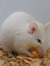
Photo by Steve Berger
Intermittent fasting inhibits the development and progression of acute lymphoblastic leukemia (ALL), according to preclinical research published in Nature Medicine.
Fasting had an inhibitory effect in mouse models of T-cell and B-cell ALL but not acute myeloid leukemia (AML).
“This study using mouse models indicates that the effects of fasting on blood cancers are type-dependent and provides a platform for identifying new targets for leukemia treatments,” said study author Chengcheng “Alec” Zhang, PhD, of UT Southwestern Medical Center in Dallas, Texas.
“We also identified a mechanism responsible for the differing response to the fasting treatment.”
For this study, Dr Zhang and his colleagues created mouse models of acute leukemia—N-Myc B-ALL, activated Notch1 T-ALL, MLL-AF9 AML, and AML driven by the AML1-Eto9a oncogene—and tested the effects of various dietary restriction plans.
The team used green or yellow florescent proteins to mark and trace the leukemia cells so they could determine if the cells’ levels rose or fell in response to the fasting treatment.
“Strikingly, we found that, in models of ALL, a regimen consisting of 6 cycles of 1 day of fasting followed by 1 day of feeding completely inhibited cancer development,” Dr Zhang said.
At the end of 7 weeks, fasted mice with B-ALL had virtually no detectible cancerous cells—an average of 0.48%—compared to an average of 67.68% of cells found to be cancerous in the test areas of the non-fasted B-ALL mice.
Dr Zhang noted that, compared to B-ALL mice that ate normally, the mice on alternate-day fasting had dramatic reductions in the percentage of ALL cells in the bone marrow and spleen, as well as reduced numbers of white blood cells.
In addition, the spleens and lymph nodes in the fasted mice with B-ALL were similar in size to those of normal mice.
“Although initially cancerous, the few fluorescent cells that remained in the fasted mice after 7 weeks appeared to behave like normal cells,” Dr Zhang said. “Mice in the [B-ALL] model group that ate normally died within 59 days, while 75% of the fasted mice survived more than 120 days without signs of leukemia.”
Dr Zhang and his colleagues said they observed similar results in the T-ALL model but not the AML models. There was no decrease in leukemia cells among fasted mice with AML. And fasting actually shortened survival time in these mice.
Identifying the mechanism
Fasting is known to reduce the level of leptin, a cell signaling molecule created by fat tissue. In addition, previous studies have shown weakened activity by leptin receptors in humans with ALL. For those reasons, the researchers studied both leptin levels and leptin receptors in the mouse models.
The team found that mice with ALL showed reduced leptin receptor activity that increased with intermittent fasting.
“We found that fasting decreased the levels of leptin circulating in the bloodstream as well as decreased the leptin levels in the bone marrow,” Dr Zhang said. “These effects became more pronounced with repeated cycles of fasting. After fasting, the rate at which the leptin levels recovered seemed to correspond to the rate at which the cancerous ALL cells were cleared from the blood.”
The researchers also found that AML was associated with higher levels of leptin receptors that were unaffected by fasting, which could help explain why the fasting treatment was ineffective against this type of leukemia.
It also suggests a mechanism—the leptin receptor pathway—by which fasting exerts its effects in ALL, Dr Zhang said.
“It will be important to determine whether ALL cells can become resistant to the effects of fasting,” he noted. “It also will be interesting to investigate whether we can find alternative ways that mimic fasting to block ALL development.”
Given that this study did not involve drug treatment, researchers are discussing with clinicians whether the tested regimen might be able to move forward quickly to clinical trials.
Ruxolitinib may prevent CRS after CAR T-cell therapy
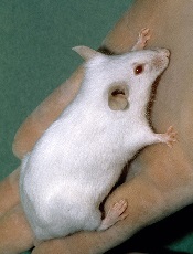
Photo courtesy of NCI
SAN DIEGO—A novel xenograft model of acute myeloid leukemia (AML) demonstrated that the JAK/STAT inhibitor ruxolitinib can prevent severe cytokine release syndrome (CRS) without impairing the anti-tumor effect of chimeric antigen receptor (CAR) T cells, according to research presented at the 2016 ASH Annual Meeting.
Almost all patients responding to CART-cell therapy develop CRS, and up to 60% develop severe CRS.
The research team believes the mouse model and findings with ruxolitinib will provide an important platform for studying CRS prevention and treatment.
At ASH, Saad Kenderian, MD, of the Mayo Clinic in Rochester, Minnesota, explained that CRS produces very high levels of the inflammatory protein IL-6.
Treatment with ruxolitinib in clinical studies has reduced human inflammatory cytokines. Therefore, it made sense to the investigators to study ruxolitinib as a means to prevent CRS after CAR T-cell therapy.
Tocilizumab has been used to treat grade 3 and 4 CRS, but physicians are concerned that earlier introduction during the course of CRS may impair CAR T-cell function.
At present, no relevant preclinical model for CRS after CAR T-cell therapy exists, “which is limiting the development of CRS preventative modalities that could, in turn, enhance the feasibility of CAR T-cell therapy,” Dr Kenderian said.
And so the investigators decided to create an animal model.
Dr Kenderian described the work at the meeting as abstract 652.
Mouse model for human CRS
Using NSG-S mice (non-obese diabetic, SCID ɣ -/- mice additionally transgenic for human stem cell factor, IL-3, and GM-CSF), investigators injected them with blasts from AML patients. After 3 to 4 weeks, investigators treated the mice with 1 x 10⁶ CD123-directed CAR T cells.
Dr Kenderian noted this dose of CART123 was 10 times higher than doses previously used in primary AML xenograft models.
The mice became weak, emaciated, developed hunched bodies, became withdrawn, had poor motor responses, and died in 7 to 10 days. The illness started within 1 week of CAR T-cell injection and correlated with significant expansion of T cells in the peripheral blood of these mice.
The team studied the serum from these mice 7 days after CART123 injection. They found extreme elevation of human IL-6, interferon-γ, tumor necrosis factor-α, and other inflammatory cytokines. This response resembled human CRS after CAR T-cell therapy.
Ruxolitinib treatment
The investigators first studied ruxolitinib activity in vitro with CART123 cells and found that ruxolitinib did not impair CAR T-cell effector functions.
“And also, ruxolitinib was not directly toxic to CAR T cells,” Dr Kenderian added.
But ruxolitinib did slow CAR T-cell proliferation in vitro.
They next tested ruxolitinib and CART123 in the mouse model.
Once the mice experienced high-burden disease, investigators treated them with CART123. That same day, investigators began treating the mice with ruxolitinib for 1 week. The mice were randomized to 30, 60, 90 mg/kg, or vehicle twice a day.
Twenty-nine days after AML injection, the mice treated with CART123 plus 90 mg or 60 mg of ruxolitinib experienced less weight loss than those treated with CART123 plus 30 mg of ruxolitinib or CART123-only.
“And more importantly, all mice had eradication of their disease,” Dr Kenderian said.
Mice treated with CART123 plus 90 mg, 60 mg, or 30 mg of ruxolitinib or CART123 alone had fewer AML blasts at day 28 than mice treated with 60 mg of ruxolitinib alone.
The investigators then analyzed the effect of ruxolitinib on the anti-tumor effect of CART123 and found that ruxolitinib did not impair it.
The attenuation of inflammatory cytokines translated to a survival advantage for mice treated with CART123 and ruxolitinib.
The investigators believe the addition of ruxolitinib to CAR T-cell therapy is a modality that should be investigated in patients at high-risk of developing CRS.
Dr Kenderian disclosed patents, royalties, and research funding from Novartis.