User login
Team uncovers novel function of p53

Andrei Thomas Tikhonenko
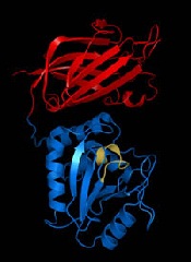
Investigators have uncovered a novel role for the tumor suppressor p53, according to a paper published in Nature Cell Biology.
The research showed that loss of p53 function caused overproduction of the Aurora A kinase, an enzyme involved in cell division.
That overproduction led to mitotic spindle malformation and aberrant separation of duplicated chromosomes over daughter cells, a phenomenon that predicts tumor metastasis and poor patient outcomes.
“Attempts to identify which genetic defects drive chromosome reshuffling in human cancer led us to focus on cyclin B1 and B2, two key regulators of the stage in the cell cycle where duplicated chromosomes normally separate,” said principal investigator Jan van Deursen, PhD, of the Mayo Clinic in Rochester, Minnesota.
Dr van Deursen and his colleague, Hyun-Ja Nam, PhD, used mouse models to mimic the cyclin B1 and B2 gene defects observed in treatment-resistant human cancers. And the pair discovered that both cyclin B1 and B2 induce chromosome reshuffling and tumor formation.
Subsequent experiments investigating cyclin B2’s mechanism of action pinpointed Aurora A kinase hyperactivity as the main culprit and showed that damage or loss of p53 is a mimetic of cyclin B2 gene defects.
The investigators said the next step for this research will be testing whether anticancer drugs that inhibit Aurora A kinase can be effective in treating cancer patients whose tumors have defects in p53. ![]()
Andrei Thomas Tikhonenko
Investigators have uncovered a novel role for the tumor suppressor p53, according to a paper published in Nature Cell Biology.
The research showed that loss of p53 function caused overproduction of the Aurora A kinase, an enzyme involved in cell division.
That overproduction led to mitotic spindle malformation and aberrant separation of duplicated chromosomes over daughter cells, a phenomenon that predicts tumor metastasis and poor patient outcomes.
“Attempts to identify which genetic defects drive chromosome reshuffling in human cancer led us to focus on cyclin B1 and B2, two key regulators of the stage in the cell cycle where duplicated chromosomes normally separate,” said principal investigator Jan van Deursen, PhD, of the Mayo Clinic in Rochester, Minnesota.
Dr van Deursen and his colleague, Hyun-Ja Nam, PhD, used mouse models to mimic the cyclin B1 and B2 gene defects observed in treatment-resistant human cancers. And the pair discovered that both cyclin B1 and B2 induce chromosome reshuffling and tumor formation.
Subsequent experiments investigating cyclin B2’s mechanism of action pinpointed Aurora A kinase hyperactivity as the main culprit and showed that damage or loss of p53 is a mimetic of cyclin B2 gene defects.
The investigators said the next step for this research will be testing whether anticancer drugs that inhibit Aurora A kinase can be effective in treating cancer patients whose tumors have defects in p53. ![]()
New insight into PTEN’s role in cancers
Researchers say they’ve uncovered new details that help explain how the PTEN gene exerts its anticancer effects and how PTEN loss or alteration can set cells on a cancerous course.
The team’s study, published in Cell, reveals that PTEN loss and PTEN mutations are not synonymous.
This discovery provides additional insight into basic tumor biology and offers a potential new direction in the pursuit of cancer therapies, according to the researchers.
“By characterizing the ways that 2 specific PTEN mutations regulate the tumor suppressor function of the normal PTEN protein, our findings suggest that different PTEN mutations contribute to tumorigenesis by regulating different aspects of PTEN biology,” said study author Pier Paolo Pandolfi, MD, PhD, of Beth Israel Deaconess Medical Center in Boston.
“It has been suggested that cancer patients harboring mutations in PTEN had poorer outcomes than cancer patients with PTEN loss. Now, using mouse modeling, we are able to demonstrate that this is indeed the case. Because PTEN mutations are extremely frequent in various types of tumors, this discovery could help pave the way for a new level of personalized cancer treatment.”
Several of the proteins that PTEN acts upon, both lipids and proteins, are known to promote cancer when bound to a phosphate. Consequently, when PTEN removes their phosphates, it is acting as a tumor suppressor to prevent cancer. When PTEN is mutated, it loses this suppressive ability, and the cancer-promoting proteins are left intact and uninhibited.
This new study showed that the PTEN mutant protein is not only functionally impaired, it also acquires the ability to affect the function of normal PTEN proteins, thereby gaining a pro-tumorigenic function. But the researchers also wanted to determine the difference between PTEN mutation and PTEN loss.
“We wanted to know, would outcomes differ in cases when PTEN was not expressed, compared with cases when PTEN was expressed but encoded a mutation within its sequence?” said study author Antonella Papa, PhD, an investigator in the Pandolfi lab.
To find out, the team created several genetically modified strains of mice to mimic the PTEN mutations found in human cancer patients.
“All mice [and humans] have 2 copies of the PTEN gene,” Dr Papa explained. “The genetically modified mice in our study had 1 copy of the PTEN gene that contained a cancer-associated mutation [either PTENC124S or PTENG129E] and 1 normal copy of PTEN. Other mice in the study had only 1 copy of the normal PTEN gene, and the second copy was removed.”
The researchers found that the mice with a single mutated copy of PTEN were more tumor-prone than the mice with a deleted copy of PTEN. They also discovered that the mutated protein that was produced by PTENC124S or PTENG129E was binding to and inhibiting the PTEN protein made from the normal copy of the PTEN gene.
“This was very surprising, as we were expecting a reduction in tumorigenesis,” Dr Papa said. “Instead, mechanistically, we found that PTEN exists as a dimer, and, in this new conformation, the mutated protein prevents the normal protein from functioning.”
At the molecular level, this generates an increased activation of a PTEN target—the protein Akt—which is what leads to the augmented tumorigenesis in the mice. Akt is part of a signaling pathway that regulates cell growth, division and metabolism.
When PTEN is prevented from inhibiting Akt, the pathway becomes overactive. As a result, targeting Akt and its pathway may be an effective treatment strategy for patients with PTEN mutations, the researchers said, adding that inhibitors to affect this pathway are currently being tested and developed.
“This defines a new working model for the function and regulation of PTEN and tells us that PTEN mutational status can be used to determine which cancer patients might benefit from earlier and more radical therapeutic interventions and, ultimately, better prognosis,” Dr Pandolfi said.
“Our findings may help to better identify and stratify patients and their response to treatment based on the different genetic alterations found in the PTEN gene. Importantly, our study shows that cancer therapy should be tailored on the basis of the very specific type of mutations that the tumor harbors.”
“This adds a new layer of complexity but also a new opportunity for precision medicine. I would say that, based on these thorough genetic analyses, this story represents the ultimate example of why personalized cancer medicine is so urgently needed.” ![]()
Bortezomib available for newly diagnosed MM patients

Credit: CDC
Patients with newly diagnosed multiple myeloma (MM) will now be guaranteed access to bortezomib (Velcade) through the National Health Service (NHS), according to the UK’s National Institute for Health and Care Excellence (NICE).
NICE has issued a guidance recommending bortezomib (given with dexamethasone or dexamethasone and thalidomide) as induction treatment for adults with newly diagnosed MM who are eligible for high-dose chemotherapy and stem cell transplant.
Bortezomib should be available on the NHS within 3 months.
The current standard induction therapy for MM patients in the UK is the combination of cyclophosphamide, thalidomide, and dexamethasone.
“We are pleased to recommend bortezomib as a first treatment for people with multiple myeloma before bone marrow transplant,” said Carole Longson, director of the NICE center for health technology evaluation.
“The evidence presented to our independent committee showed that having bortezomib at this stage will help more patients go on to a bone marrow transplant and, consequently, prevent the disease from progressing for longer.”
The committee concluded that, although there was uncertainty in the magnitude of overall survival gain associated with bortezomib, it was plausible that bortezomib’s impact on induction response could be associated with improved overall survival.
The cost of bortezomib is £762.38 per 3.5-mg vial. The average cost of a course of bortezomib given with dexamethasone is estimated to be £12,260.91. And the average cost of a course of bortezomib given with dexamethasone and thalidomide is estimated to be £24,840.10.
The cost of bortezomib, thalidomide, and dexamethasone compared with thalidomide and dexamethasone was likely to be below £30,000 per quality-adjusted life-year gained.
The same was true for bortezomib and dexamethasone compared with cyclophosphamide, thalidomide, and dexamethasone or with vincristine, doxorubicin, and dexamethasone. Therefore, bortezomib was considered an acceptable use of NHS resources.
NICE already recommends bortezomib monotherapy to treat progressive MM in patients at first relapse who have undergone, or are unsuitable for, stem cell transplant. ![]()
Protein helps HSP90 inhibitors fight cancers

Credit: PNAS
Researchers have discovered how a molecule called CUL5 helps HSP90 inhibitors kill cancer cells, according to a study published in Proceedings of the National Academy of Sciences.
The team found that CUL5 is required for the degradation of proteins that promote cancer cell proliferation, and CUL5 works in opposition to HSP90.
When cancer cells are treated with HSP90 inhibitors, CUL5 immediately steps in to help dispose of the proliferation-promoting proteins.
Based on these findings, the researchers speculate that some patients may be resistant to HSP90 inhibitors if their cancer cells have lower amounts of CUL5. And conversely, the drugs may work better in patients with higher CUL5 levels.
Paul Workman, PhD, of The Institute of Cancer Research in London, UK, and his colleagues conducted this research in cell lines of melanoma, as well as colon, breast, and lung cancers.
They first tested the HSP90 inhibitor 17-AAG in HT29 cells and found that CUL5 is involved in the drug-induced degradation of several protein kinase clients of HSP90.
Then, the researchers assessed the effects of silencing CUL5 and discovered that it delays the abrogation of protein signaling caused by an HSP90 inhibitor.
Furthermore, silencing CUL5 reduced cellular sensitivity to 3 different HSP90 inhibitors across the 4 different cancer types studied, which, as the researchers pointed out, are driven by different protein kinases.
So the team believes this research could apply to a number of different cancers. HSP90 inhibitors have proven effective against a range of malignancies, including leukemias, lymphomas, and multiple myeloma.
“We’ve known for some time that drugs that block HSP90 have great potential as treatments for cancers . . . , and we had an initial clue that the protein CUL5 may be involved in some way in how these drugs work,” Dr Workman said.
“Our new research shows that CUL5 is not only vital in the response of cancer cells to HSP90 inhibitors but also reveals surprising insights into precisely how it works by acting at several different levels. What also surprised us was that CUL5 gets rid of many more of the cancer-causing proteins than we’d previously imagined and that it’s effective across several types of tumor.”
“This suggests that a test for CUL5 in patients could help us tell whether they might respond to HSP90-blocking drugs, as well as pointing to new targets to develop more effective drugs.” ![]()
Reovirus shows synergy with agents used to treat MM
SAN DIEGO—Reovirus can induce cell death in a range of multiple myeloma (MM) cell lines, and it is synergistic with drugs used to treat MM, according to research presented at the AACR Annual Meeting 2014.
Six of the 7 MM cell lines tested were at least moderately sensitive to reovirus, and introducing the virus in combination with the proteasome inhibitor carfilzomib or the Akt inhibitor perifosine increased antimyeloma activity.
Chandini M. Thirukkumaran, PhD, of The University of Calgary in Canada, and her colleagues presented these results at the meeting as abstract 1709. The study was funded by the Cancer Research Society of Canada.
“Our university, about 15 years ago, found that reovirus can infect all cells, but it will specifically kill cancer cells and not harm normal cells,” Dr Thirukkumaran said. “This is because cancer cells have aberrant Ras signaling pathways, and the virus utilizes that aberrant signaling for its replication.”
“Reovirus kills a lot of myeloma cell lines and cells from patients too, but you always find that therapy-resistant population. So we wondered if we could combine reovirus with common myeloma drugs, like carfilzomib and perifosine, and see whether we could get synergy.”
Dr Thirukkumaran and her colleagues first wanted to quantify the effect of reovirus alone on MM cell lines. So they incubated the cell lines RPMI 8226, MM1.S, NCIH929, U266, INA-6, KMS11, and OPM2 with live reovirus or UV-inactivated reovirus and assessed cell death.
They found that RPMI8226 was highly sensitive to reovirus, and OPM2 was resistant to it. The remaining cell lines were somewhat sensitive to the virus.
The researchers chose RPMI8226, KMS-11, and OPM2 to test reovirus in combination with either carfilzomib or perifosine. They tested the drugs at various concentrations to determine effective dose 50% (ED50) values. They combined ED50 values for each drug and reovirus at various concentrations but with consistent ratios and determined toxicity.
The team used software to generate combination index (CI) values and determine synergism per the Chou-Talalay method. A CI equal to 1 suggested an additive effect, a CI greater than 1 suggested an antagonistic effect, and a CI less than 1 suggested a synergistic effect.
Results showed that reovirus synergized with both carfilzomib and perifosine. Furthermore, the greater a cell line’s resistance to reovirus, the greater the synergy.
For instance, with reovirus and carfilzomib in combination, the ED50 was 1.06 + 0.15 in the reovirus-sensitive RPMI8226 cell line, 0.78 + 0.14 in the moderately sensitive KMS11 cell line, and 0.57 + 0.05 in the resistant OPM2 cell line.
When reovirus and perifosine were combined, the ED50 was 0.97 + 0.19 in the RMI8226 cell line, 0.26 + 0.11 in the KMS11 cell line, and 0.88 + 0.22 in the OPM2 cell line.
The researchers said these results highlight the significance of preclinical studies in evaluating reovirus-drug combinations that could be extrapolated to a clinical setting.
Two phase 2 trials evaluating reovirus in combination therapy for MM are now underway. Meanwhile, Dr Thirukkumaran and her colleagues are hoping to gain more insight into how reovirus-drug combinations work by testing them in mouse models of MM. ![]()
ASCO releases guidelines for managing cancer survivors

with a cancer patient
NCI/Mathews Media Group
The American Society of Clinical Oncology (ASCO) has issued 3 practice guidelines for preventing and managing symptoms that can affect adult cancer survivors—neuropathy, fatigue, and depression/anxiety.
The guideline on chemotherapy-induced peripheral neuropathy (CIPN) lists a few options for treating the condition but discourages interventions to prevent CIPN, as there is insufficient evidence that these interventions benefit patients.
The guideline on fatigue recommends that healthcare providers start screening cancer patients for the condition at diagnosis and emphasizes the importance of educating patients about fatigue.
The guideline on depression and anxiety recommends periodic evaluations for symptoms of depression and anxiety in all cancer patients. It also suggests that all patients be offered supportive care services.
All 3 of these guidelines are published in the Journal of Clinical Oncology.
Treating and preventing CIPN
ASCO’s guideline on CIPN lists a handful of drugs that may be helpful in diminishing the symptoms of CIPN, but it does not recommend any agents for preventing the condition.
In fact, the guideline provides a list of agents that should not be offered for the prevention of CIPN, including acetyl-L-carnitine, amifostine, amitriptyline, CaMg, diethyldithio-carbamate, glutathione, nimodipine, Org 2766, all-trans retinoic acid, rhuLIF, and vitamin E.
“There is no clear panacea for neuropathy,” said Gary Lyman, MD, MPH, co-chair of the ASCO Survivorship Guidelines Advisory Group.
“Some of the drugs used for prevention or treatment of neuropathy may cause side effects or interfere with other drugs. We want to be clear that if there is no evidence of benefit from those drugs, it’s probably best not to take them.”
As for treatment, the guideline states that data support a “moderate” recommendation for duloxetine.
It also notes that there is no strong evidence of benefit for the use of tricyclic antidepressants, gabapentin, and a topical gel containing baclofen, amitriptyline, and ketamine. However, it may be reasonable to try those agents in select patients.
To develop this guideline, an ASCO panel conducted a systematic review of relevant medical literature. They analyzed data from 48 randomized, clinical trials focused on managing CIPN.
Screening and managing fatigue
ASCO’s guideline on fatigue recommends that all patients be screened for fatigue from the point of diagnosis onward. Healthcare providers should assess fatigue history, disease status, and treatable contributing factors.
All patients should be educated about the differences between normal and cancer-related fatigue, causes of fatigue, and contributing factors.
Healthcare providers should discuss with patients strategies to manage fatigue, including physical activity, psychosocial interventions (such as cognitive and behavioral therapies or psycho-educational therapies), and mind-body interventions (such as yoga or acupuncture).
To develop this guideline, an ASCO panel conducted a systematic review of clinical practice guideline databases and relevant medical literature. The adaptation is based on a Pan-Canadian guideline on fatigue and 2 National Comprehensive Cancer Network guidelines on cancer-related fatigue and survivorship.
Handling anxiety and depression
ASCO’s guideline on anxiety and depression recommends that healthcare providers periodically evaluate all cancer patients for symptoms of depression and anxiety. The assessments should be performed using validated, published measures and procedures.
All patients should have the option of receiving supportive care services, such as education about the normalcy of stress in the context of cancer, signs and symptoms of distress, and stress reduction strategies.
Patients who display moderate or severe symptoms of anxiety and depression should be referred for the appropriate psychological, psychosocial, or psychiatric interventions.
“Doctors sometimes don’t give these symptoms much attention because they think it’s normal that their patients are a little anxious or depressed about their disease,” Dr Lyman said. “But it’s important to keep an eye on the symptoms and step in when they start to interfere with the patients’ quality of life.”
To develop this guideline, an ASCO panel conducted a systematic review of clinical practice guideline databases and relevant medical literature. The adaptation is based on a Pan-Canadian practice guideline on psychological distress in adults with cancer. ![]()
Heparanase regulates response to chemo in MM, team says
SAN DIEGO—Experiments conducted in the lab and the clinic suggest the enzyme heparanase enhances resistance to chemotherapy in multiple myeloma (MM).
Researchers first found that expression of heparanase, an endoglycosidase that cleaves heparan sulfate, is highly elevated in MM patients after chemotherapy.
The team then used MM cell lines to investigate the mechanism behind this phenomenon.
Their results indicate that, by inhibiting heparanase, we might be able to prevent or delay relapse in MM.
Vishnu Ramani, PhD, of the University of Alabama Birmingham, and his colleagues conducted this research and presented the results at the AACR Annual Meeting 2014 (abstract 1708).
Several years ago, Dr Ramani’s colleagues (in the lab of Ralph Sanderson, PhD) identified heparanase as a master regulator of aggressive MM. Since then, research has suggested that heparanase fuels aggressive MM by upregulating the expression of pro-angiogenic genes, driving osteolysis, upregulating prometastatic molecules, and controlling the tumor microenvironment.
“We have done a lot of work on the biology of how this molecule works in myeloma, but the one thing I was really interested in was its role in drug resistance,” Dr Ramani said.
So he and his colleagues decided to study heparanase levels in 9 MM patients undergoing chemotherapy. The team isolated tumor cells from patients before and after 2 rounds of chemotherapy and compared heparanase levels at the different time points.
“What we find—and this is really remarkable—is that the expression of heparanase over rounds of therapy goes up several thousand-fold, and this is in the majority of patients,” Dr Ramani said. “In 8 out of 9 patients that we studied, at the end of chemotherapy, the cells that survive have extremely high levels of heparanase.”
To gain more insight into this phenomenon, the researchers studied it in MM cell lines. The team introduced bortezomib to RPMI-8226 and CAG cells and found that heparanase levels increased “dramatically” after treatment.
“The treatment is not only increasing the heparanase expression inside the cell,” Dr Ramani explained. “What the cells do is that, if you continue the treatment, they die, but they don’t take the heparanase with them. They leave it out in the media, and this can be taken in by other cells. So this can activate other cells to promote aggressive tumor growth too.”
Additional investigation revealed that the NF-κB pathway plays a role—namely, chemotherapy activates the pathway to upregulate heparanase. But inhibiting NF-κB activity can prevent that increase in heparanase.
The researchers tested the NF-κB inhibitors BAY 11-7085 and BMS345541 in combination with bortezomib. And they found that both agents prevented bortezomib from elevating heparanase expression in CAG cells.
Dr Ramani and his colleagues also evaluated heparanase levels in chemoresistant MM cell lines. Heparanase levels were 4-fold higher in a doxorubicin-resistant MM cell line and 10-fold higher in a melphalan-resistant cell line, when compared to a wild-type MM cell line.
Next, the researchers compared MM cells with high heparanase expression to those with low heparanase expression. And they discovered that high heparanase levels protect cells from chemotherapy.
After treatment with bortezomib, cells with high heparanase expression were significantly more viable than those with low expression (P<0.05). And there was a significantly higher percentage of apoptotic cells among the low-heparanase population compared to the high-heparanase population (P<0.05).
“If you take cells that have high heparanase and another group of cells that have low heparanase and expose both of them to therapy, the cells with high heparanase always survive better because the heparanase upregulates certain pathways, like the MAP kinase pathway, which helps the cells to survive the onslaught of chemotherapy,” Dr Ramani said. “So myeloma cells are actually hijacking the heparanase pathway to survive better after therapy.”
Building upon that finding, the researchers decided to assess whether inhibiting ERK activity might help cells overcome heparanase-mediated chemoresistance. And experiments showed that the ERK inhibitor U0126 can sensitize cells with high heparanase levels to treatment with bortezomib.
To take this research to the next level, Dr Ramani and his colleagues are collaborating with a company called Sigma Tau, which is developing a heparanase inhibitor called SST-0001. A phase 1 study of the drug in MM patients has been completed, and phase 2 studies are currently recruiting patients in Europe.
Dr Ramani is now conducting experiments in mice to determine how the inhibitor might work in combination with chemotherapy and when it should be administered in order to overcome treatment resistance. He is also looking for other molecular pathways that could be involved in heparanase-related treatment resistance. ![]()
SAN DIEGO—Experiments conducted in the lab and the clinic suggest the enzyme heparanase enhances resistance to chemotherapy in multiple myeloma (MM).
Researchers first found that expression of heparanase, an endoglycosidase that cleaves heparan sulfate, is highly elevated in MM patients after chemotherapy.
The team then used MM cell lines to investigate the mechanism behind this phenomenon.
Their results indicate that, by inhibiting heparanase, we might be able to prevent or delay relapse in MM.
Vishnu Ramani, PhD, of the University of Alabama Birmingham, and his colleagues conducted this research and presented the results at the AACR Annual Meeting 2014 (abstract 1708).
Several years ago, Dr Ramani’s colleagues (in the lab of Ralph Sanderson, PhD) identified heparanase as a master regulator of aggressive MM. Since then, research has suggested that heparanase fuels aggressive MM by upregulating the expression of pro-angiogenic genes, driving osteolysis, upregulating prometastatic molecules, and controlling the tumor microenvironment.
“We have done a lot of work on the biology of how this molecule works in myeloma, but the one thing I was really interested in was its role in drug resistance,” Dr Ramani said.
So he and his colleagues decided to study heparanase levels in 9 MM patients undergoing chemotherapy. The team isolated tumor cells from patients before and after 2 rounds of chemotherapy and compared heparanase levels at the different time points.
“What we find—and this is really remarkable—is that the expression of heparanase over rounds of therapy goes up several thousand-fold, and this is in the majority of patients,” Dr Ramani said. “In 8 out of 9 patients that we studied, at the end of chemotherapy, the cells that survive have extremely high levels of heparanase.”
To gain more insight into this phenomenon, the researchers studied it in MM cell lines. The team introduced bortezomib to RPMI-8226 and CAG cells and found that heparanase levels increased “dramatically” after treatment.
“The treatment is not only increasing the heparanase expression inside the cell,” Dr Ramani explained. “What the cells do is that, if you continue the treatment, they die, but they don’t take the heparanase with them. They leave it out in the media, and this can be taken in by other cells. So this can activate other cells to promote aggressive tumor growth too.”
Additional investigation revealed that the NF-κB pathway plays a role—namely, chemotherapy activates the pathway to upregulate heparanase. But inhibiting NF-κB activity can prevent that increase in heparanase.
The researchers tested the NF-κB inhibitors BAY 11-7085 and BMS345541 in combination with bortezomib. And they found that both agents prevented bortezomib from elevating heparanase expression in CAG cells.
Dr Ramani and his colleagues also evaluated heparanase levels in chemoresistant MM cell lines. Heparanase levels were 4-fold higher in a doxorubicin-resistant MM cell line and 10-fold higher in a melphalan-resistant cell line, when compared to a wild-type MM cell line.
Next, the researchers compared MM cells with high heparanase expression to those with low heparanase expression. And they discovered that high heparanase levels protect cells from chemotherapy.
After treatment with bortezomib, cells with high heparanase expression were significantly more viable than those with low expression (P<0.05). And there was a significantly higher percentage of apoptotic cells among the low-heparanase population compared to the high-heparanase population (P<0.05).
“If you take cells that have high heparanase and another group of cells that have low heparanase and expose both of them to therapy, the cells with high heparanase always survive better because the heparanase upregulates certain pathways, like the MAP kinase pathway, which helps the cells to survive the onslaught of chemotherapy,” Dr Ramani said. “So myeloma cells are actually hijacking the heparanase pathway to survive better after therapy.”
Building upon that finding, the researchers decided to assess whether inhibiting ERK activity might help cells overcome heparanase-mediated chemoresistance. And experiments showed that the ERK inhibitor U0126 can sensitize cells with high heparanase levels to treatment with bortezomib.
To take this research to the next level, Dr Ramani and his colleagues are collaborating with a company called Sigma Tau, which is developing a heparanase inhibitor called SST-0001. A phase 1 study of the drug in MM patients has been completed, and phase 2 studies are currently recruiting patients in Europe.
Dr Ramani is now conducting experiments in mice to determine how the inhibitor might work in combination with chemotherapy and when it should be administered in order to overcome treatment resistance. He is also looking for other molecular pathways that could be involved in heparanase-related treatment resistance.
How autophagy helps cancer cells evade death

Credit: Sarah Pfau
SAN DIEGO—New research suggests that autophagy may allow cancer cells to recover and divide, rather than die, when faced with chemotherapy.
“What we showed is that if this mechanism doesn’t work right—for example, if autophagy is too high or if the target regulated by autophagy isn’t around—cancer cells may be able to rescue themselves from death caused by chemotherapies,” said study author Andrew Thorburn, PhD, of the University of Colorado Denver.
He and his colleagues believe this finding has important implications. It demonstrates a mechanism whereby autophagy controls cell death, and it further reinforces the clinical potential of inhibiting autophagy to sensitize cancer cells to chemotherapy.
Dr Thorburn and his colleagues recounted their research in Cell Reports and presented it in an education session at the AACR Annual Meeting 2014.
The researchers had set out to examine how autophagy affects canonical death receptor-induced mitochondrial outer membrane permeabilization (MOMP) and apoptosis. They found that MOMP occurs at variable times in cells, and it’s delayed by autophagy.
Furthermore, autophagy leads to inefficient MOMP. This causes some cells to die via a slower process than typical apoptosis, which allows them to eventually recover and divide.
Specifically, the researchers found that, as a cancer cell begins to die, mitochondrial cell walls break down. And the cell’s mitochondria release proteins via MOMP.
But then, high autophagy allows the cell to encapsulate and “digest” these released proteins before MOMP can keep the cell well and truly dead. The cell recovers and goes on to divide.
“The implication here is that if you inhibit autophagy, you’d make this less likely to happen; ie, when you kill cancer cells, they would stay dead,” Dr Thorburn said.
He and his colleagues also found that autophagy depends on the target PUMA to regulate cell death. When PUMA is absent, it doesn’t matter if autophagy is inhibited. Without the communicating action of PUMA, cancer cells evade apoptosis and continue to survive.
The researchers said this suggests autophagy can control apoptosis via a regulator that makes MOMP faster and more efficient, thus ensuring the rapid completion of apoptosis.
“Autophagy is complex and, as yet, not fully understood,” Dr Thorburn said. “But now that we see a molecular mechanism whereby cell fate can be determined by autophagy, we hope to discover patient populations that could benefit from drugs that inhibit this action.”
Nanocapsules prevent release of nontargeted radiation

Credit: Rhoda Baer
A novel type of nanocapsule can safely and effectively store isotopes that emit ionizing radiation, according to a paper published in Biochimica et Biophysica Acta.
In experiments, the nanocapsules were taken up by cells, accumulated near the perinuclear region, and persisted without degrading.
They also prevented nontargeted, radioactive daughter ions from escaping. These ions could cause significant damage if released, such as prompting the development of leukemia.
This research suggests the nanocapsules have the potential to advance radiation therapy, according to study author John M. Tomich, PhD, of Kansas State University’s Johnson Cancer Research Center.
Dr Tomich and his colleagues created the nanocapsules, called branched amphiphilic peptide capsules (BAPCs), by combining 2 related sequences of amino acids—bis(FLIVI)-K-KKKK and bis(FLIVIGSII)-K-KKKK.
“We found that the 2 sequences come together to form a thin membrane that assembled into little spheres, which we call capsules,” Dr Tomich said. “While other vesicles have been created from lipids, most are much less stable and break down. Ours are like stones, though. They’re incredibly stable and are not destroyed by cells in the body.”
The capsules’ ability to stay intact with the isotope inside and remain undetected by the body’s clearance systems prompted Dr Tomich to investigate using BAPCs for radiation therapies.
“The problem with current alpha-particle radiation therapies used to treat cancer is that they lead to the release of nontargeted, radioactive daughter ions into the body,” Dr Tomich said. “Radioactive atoms break down to form new atoms, called daughter ions, with the release of some form of energy or energetic particles. Alpha emitters give off an energetic particle that comes off at nearly the speed of light.”
The alpha particle destroys DNA and whatever vital cellular components are in its path. Similarly, the daughter ions recoil with high energy on ejection of the alpha particle. The daughter ions have enough energy to escape the targeting and containment molecules that are currently in use.
“Once freed, the daughter isotopes can end up in places you don’t want them, like bone marrow, which can then lead to leukemia and new challenges,” Dr Tomich said.
To see if the BAPCs could prevent the release of daughter isotopes, the researchers loaded the nanoparticles with 225Actinium. Upon decay, this compound releases 4 alpha particles and numerous daughter ions.
The team found that BAPCs loaded with the compound readily entered cells and migrated to a position alongside the nucleus.
As the alpha-particle-emitting isotopes decayed, the recoiled daughter ions collided with the capsule walls, essentially bouncing off them, and remained trapped inside the BAPCs. This completely blocked the release of the daughter ions, which prevented uptake in nontarget tissues.
Dr Tomich said more studies are needed to add target molecules to the surface of the BAPCs. But he believes the particles could provide a safer option for treating tumors with radiation therapy by reducing the amount of radioisotope required for killing cancer cells and reducing the side effects caused by off-target accumulation of radioisotopes.
“These capsules are easy to make and easy to work with,” Dr Tomich said. “I think we’re just scratching the surface of what we can do with them to improve human health and nanomaterials.”
NHL among top 10 most common cancers in US

Credit: Rhoda Baer
A new report shows the rate of invasive cancer among US men and women dropped slightly from 2009 to 2010, and the most common cancers were solid tumor malignancies.
However, non-Hodgkin lymphoma (NHL) consistently rated among the top 10 most common cancers, regardless of patient gender or race.
The report was prepared by the Centers for Disease Control and Prevention (CDC) and appears in the current Morbidity and Mortality Weekly Report.
Researchers analyzed new cases of invasive cancers diagnosed in 2010 and reported to the CDC’s National Program of Cancer Registries and the National Cancer Institute’s Surveillance, Epidemiology and Results Program.
Data from all states (except Arkansas and Minnesota) and the District of Columbia were included in the analysis, which covered 97% of the US population.
The researchers found the rates of invasive cancer cases dropped from 459 per 100,000 people in 2009 to 446 per 100,000 in 2010.
Cancer rates were higher among men (503 per 100,000) than women (405 per 100,000). In all, there were 745,383 cases reported among men and 711,113 among women in 2010.
The highest rates were for cancers of the prostate (126 per 100,000), female breast (119 per 100,000), lung and bronchus (62 per 100,000), and colon and rectum (40 per 100,000). Together, these accounted for half of all cancer cases in the US.
However, hematologic malignancies were fairly common as well. NHL was the 6th most common cancer for men of all races/ethnicities except Hispanic. For this group, NHL was the 4th most common cancer.
NHL was the 7th most common cancer for black and white women and 6th for the remaining groups, which included American Indian/Alaskan native, Asian/Pacific Islander, and Hispanic women.
Leukemia and myeloma were also among the top 10 most common invasive cancers for certain patients.
Leukemia was the 9th most common cancer for Hispanic and white men and the 10th for American Indian/Alaskan Native women. Myeloma was the 8th most common cancer for black women.
Overall, cancer rates were highest among black patients (455 per 100,000), followed by whites (445 per 100,000), Hispanics (344 per 100,000), Asian/Pacific Islanders (289 per 100,000), and American Indians/Alaskan Natives (270 per 100,000).