User login
Why fewer blood cancer patients receive hospice care
New research provides an explanation for the fact that US patients with hematologic malignancies are less likely to enroll in hospice care than patients with solid tumor malignancies.
Results of a national survey suggest that concerns about the adequacy of hospice may prevent hematologic oncologists from referring their patients.
Researchers say this finding, published in Cancer, points to potential means of improving end-of-life care for patients with hematologic malignancies.
Oreofe Odejide, MD, of the Dana-Farber Cancer Institute in Boston, Massachusetts, and her colleagues carried out this study.
The team conducted a survey of a national sample of hematologic oncologists listed in the publicly available clinical directory of the American Society of Hematology.
More than 57% of physicians who were contacted provided responses, for a total of 349 respondents.
The survey included questions about views regarding the helpfulness and adequacy of home hospice services for patients with hematologic malignancies, as well as factors that would impact oncologists’ likelihood of referring patients to hospice.
More than 68% of hematologic oncologists strongly agreed that hospice care is “helpful” for patients with hematologic malignancies.
However, 46% of the oncologists felt that home hospice is “inadequate” for the needs of patients with hematologic malignancies, when compared to inpatient hospice.
Still, most of the respondents who believed home hospice is inadequate said they would be more likely to refer patients if platelet and red blood cell transfusions were readily available.
“Our findings are important as they shed light on factors that are potential barriers to hospice referrals,” Dr Odejide said. “These findings can be employed to develop targeted interventions to address hospice underuse for patients with blood cancers.” ![]()
Smoldering myeloma progressed more rapidly in patients with elevated BMIs
An elevated body mass index appears to be a risk factor for progression of smoldering multiple myeloma, according to Wilson I. Gonsalves, MD, and his colleagues at Mayo Clinic, Rochester, Minn.
The findings, based on median follow up data of 106 months from 306 patients diagnosed with smoldering multiple myeloma from 2000-2010 at the Mayo Clinic, need to be confirmed in larger studies. Nevertheless, the results imply that patient weight is a potentially modifiable risk factor for progression from smoldering disease to multiple myeloma, Dr. Gonsalves and his colleagues wrote.
At initial evaluation, 28% of patients had myeloma defining events, such as a serum free light chain ratio greater than 100 or over 60% clonal bone marrow plasma cells. Myeloma defining events were present in 17% of patients with normal BMIs and 33% of patients with elevated BMIs, a statistically significant difference (P = .011).
When the analysis was limited to the 187 patients without myeloma-defining events at initial evaluation, the 2-year rate of progression to symptomatic multiple myeloma was 15% in those with a normal BMI and 33% in those with an elevated BMI (P = .013).
In a multivariable model, only elevated BMI (P = .004) and increasing clonal bone marrow plasma cells (P = .001) were statistically significant in predicting 2-year progression to multiple myeloma.
At last follow-up, 66% of patients had progressed to symptomatic multiple myeloma.
Dr. Gonsalves had no relationships to disclose.
The impact of body mass index on the risk of early progression of smoldering multiple myeloma to symptomatic myeloma. 2017 ASCO annual meeting. Abstract No: 8032.
mdales@frontlinemedcom.com
On Twitter @maryjodales
FROM ASCO 2017
Key clinical point:
Major finding: The 2-year rate of progression from smoldering disease to symptomatic multiple myeloma was 16% in patients with a normal BMI and 42% in patients with an elevated BMI (P less than 0.0001).
Data source: Median follow up data of 106 months from 306 patients diagnosed with smoldering multiple myeloma during 2000-2010 at the Mayo Clinic.
Disclosures: Dr. Gonsalves had no relationships to disclose.
Citation: The impact of body mass index on the risk of early progression of smoldering multiple myeloma to symptomatic myeloma. 2017 ASCO annual meeting. Abstract No: 8032.
Myeloma patients who get solid tumor cancers do as well as other cancer patients
With improved treatment, patients with multiple myeloma are surviving long enough to develop other cancers, Jorge J. Castillo, MD, and Adam J. Olszewski, MD, reported in a poster to be presented at the annual meeting of the American Society of Clinical Oncology.
The good news is that myeloma patients, when diagnosed with a subsequent solid tumor, are just as likely to respond to treatment and do just as well as patients without myeloma, according to Dr. Castillo of Dana-Farber Cancer Institute, Boston, and Dr. Olszewski of Alpert Medical School of Brown University, Providence, R.I.
They based their conclusion on Surveillance, Epidemiology, and End Results (SEER) data for patients diagnosed with six common cancers from 2004-2013.
“Among them, we identified [nearly 1,300] myeloma survivors, and we matched each to 50 randomly sampled controls with the same cancer by age, sex, race, and year of diagnosis. We then compared [cancer specific survival], cumulative incidence function (CIF) for death from the non-myeloma index cancer, and whether patients had surgery for non-metastastic, stage-matched tumors only,” the researchers wrote in their abstract.
They did analyses for breast, lung, prostate, colorectal, melanoma, and bladder cancers. The median time from diagnosis of myeloma to diagnosis of the second ranged from 35 months (bladder [133 myeloma patients] and lung [286 myeloma patients] cancers) to 50 months (melanoma [140 myeloma patients]). The median time after myeloma diagnosis was 40 months for those patients who developed breast, prostate, or colorectal cancers.
In the comparisons, myeloma survivors were significantly older (P less than .001) than patients initially diagnosed with the same respective cancers. In the case-control analysis, breast (P = .002) and lung cancers (P = .003) were more often diagnosed at an early stage among myeloma survivors.
The hazard ratio (HR) for cancer-specific survival for 189 myeloma patients diagnosed with breast cancer as compared to other breast cancer patients, for example, was 0.99, 95% confidence interval (CI) 0.61-1.61. The HR for the cumulative incidence function of cancer death was 0.82, 95% CI 0.50-1.35.
Myeloma patients were no less likely than were case-control subjects to have surgery for their cancers, with the exception of the 330 myeloma patients who developed prostate cancer (odds ratio, 0.59, 95% CI, 0.44-0.81).
Cancer-specific survival significantly differed (P less than .05) only for lung cancer, and was better among the 286 myeloma patients with lung cancer even when stratified by stage (HR 0.64, 95% CI 0.54-0.75). For cumulative incidence function of cancer death for lung cancer, the hazard ratio was 0.52 (95% CI 0.44-0.61). Better outcomes in lung cancer are not fully explained by earlier detection, suggesting a biological difference, the researchers reported.
Cumulative incidence function of cancer death was significantly lower for myeloma patients with lung and colorectal cancers.
Dr. Castillo disclosed honoraria from Celgene and Janssen; a consulting or advisory role with Biogen, Otsuka, and Pharmacyclics; and institutional research funding from Abbvie, Gilead Sciences, Millennium, and Pharmacyclics. Dr. Olszewski disclosed institutional research funding from Genentech, Incyte, and TG Therapeutics.
Citation: Outcomes of secondary cancers among myeloma survivors. 2017 ASCO annual meeting. Abstract No. 8043.
mdales@frontlinemedcom.com
On Twitter @maryjodales
FROM 2017 ASCO ANNUAL MEETING
Key clinical point:
Major finding: Cancer-specific survival significantly differed (P less than .05) only for lung cancer, and was better among the 286 myeloma patients with lung cancer even when stratified by stage (HR, 0.64, 95% CI 0.54-0.75).
Data source: Surveillance, Epidemiology, and End Results (SEER) data for patients diagnosed with six common cancers from 2004-2013.
Disclosures: Dr. Castillo disclosed honoraria from Celgene and Janssen; a consulting or advisory role with Biogen, Otsuka, and Pharmacyclics; and institutional research funding from Abbvie, Gilead Sciences, Millennium, and Pharmacyclics. Dr. Olszewski disclosed institutional research funding from Genentech, Incyte, and TG Therapeutics.
Citation: Outcomes of secondary cancers among myeloma survivors. 2017 ASCO annual meeting. Abstract No. 8043.
Lenalidomide maintenance boosted depth of response in myeloma patients
Lenalidomide maintenance therapy further improved depth of response in newly diagnosed, transplant-eligible patients with multiple myeloma in the EMN02/HO95 trial, based on the abstract of a poster to be presented at the annual meeting of the American Society of Clinical Oncology.
The study results also show that using multiparameter flow cytometry to monitor minimal residual disease (MRD) was predictive of patient outcome. A high-risk cytogenetic profile – defined as having del17, translocation (4;14), or translocation (14;16) – was the most important prognostic factor in MRD-positive patients, according to Stefania Oliva, MD, of the University of Torino [Italy] and her colleagues.
At 3 years, progression-free survival was 50% in MRD-positive patients and 77% in MRD-negative patients (hazard ratio, 2.87; P less than .001). High-risk cytogenetics was the most important risk factor (HR, 9.87; interaction-P = .001). Further, 48% of patients who had MRD before maintenance therapy and had a second evaluation for minimal residual disease after at least 1 year of lenalidomide therapy became MRD negative.
The trial (NCT01208766) participants were no older than age 65 years and received received bortezomib-cyclophosphamide-dexamethasone (VCD) induction therapy, then bortezomib-melphalan-prednisone (VMP) or high-dose melphalan intensification therapy followed by stem cell transplant, and subsequently bortezomib-lenalidomide-dexamethasone (VRD) consolidation therapy or no consolidation therapy, followed by lenalidomide maintenance therapy.
Of 316 patients who were evaluable before maintenance therapy, 18% had International Staging System stage III disease (beta-2 microglobulin of 5.5 mg/L or greater) and 22% had a high-risk cytogenetic profile.
For intensification therapy, 63% had received high-dose melphalan and 37% got VMP; thereafter 51% had received VRD. Nearly two-thirds of the 76% of patients who were MRD negative got high-dose melphalan, with a median follow-up of 30 months from MRD enrollment.
Patients who had at least a very good partial response underwent minimal residual disease evaluation before starting maintenance therapy and then every 6-12 months during maintenance therapy. Multiparameter flow cytometry was performed on bone marrow according to Euroflow-based methods (eight colors, two tubes) with a sensitivity of 10-5, and quality checks were performed to compare sensitivity and to show correlation between protocols.
Dr. Oliva disclosed receiving honoraria from Celgene and Takeda.
Citation: Minimal residual disease (MRD) monitoring by multiparameter flow cytometry (MFC) in newly diagnosed transplant eligible multiple myeloma (MM) patients: Results from the EMN02/HO95 phase 3 trial. 2017 ASCO annual meeting. Abstract No: 8011
mdales@frontlinemedcom.com
On Twitter @maryjodales
FROM 2017 ASCO ANNUAL MEETING
Key clinical point:
Major finding: 48% of patients who had minimal residual disease before maintenance therapy and had a second evaluation for MRD after at least 1 year of lenalidomide therapy became MRD negative.
Data source: A 3-year study of 316 patients who were evaluable before maintenance therapy in the EMN02/HO95 trial.
Disclosures: Dr. Oliva disclosed receiving honoraria from Celgene and Takeda.
Citation: Minimal residual disease (MRD) monitoring by multiparameter flow cytometry (MFC) in newly diagnosed transplant eligible multiple myeloma (MM) patients: Results from the EMN02/HO95 phase 3 trial. 2017 ASCO annual meeting. Abstract No: 8011
Compound could treat lymphoma, myeloma
A nucleoside analog has shown potential for treating primary effusion lymphoma (PEL) and multiple myeloma (MM), according to researchers.
The compound, 6-ethylthioinosine (6-ETI), killed both PEL and MM cells in vitro.
6-ETI also reduced tumor burden and prolonged survival in mouse models of MM and PEL.
Ethel Cesarman, MD, PhD, of Weill Cornell Medicine in New York, New York, and her colleagues conducted this research and disclosed their results in the Journal of Clinical Investigation.
The researchers identified 6-ETI via high-throughput screening, and they initially tested the compound in PEL cell lines. 6-ETI induced necrosis, apoptosis, and autophagy in these cells.
In a xenograft model of PEL, mice treated with 6-ETI experienced “striking and immediate regression of the implanted xenograft within 3 days of treatment,” according to the researchers.
In addition, mice that received 6-ETI had significantly longer progression-free and overall survival than control mice (P<0.0001 for both), and the researchers said there were no obvious toxicities from the treatment.
Looking into the mechanism of 6-ETI, the researchers found that adenosine kinase (ADK) is required to phosphorylate and activate the compound, and PEL cells have high levels of ADK.
Because PEL cells closely resemble plasma cells, the researchers theorized that MM might produce high levels of ADK as well. Experiments in MM cells proved this theory correct.
So the researchers tested 6-ETI in MM cell lines and samples from MM patients. The compound induced apoptosis and autophagy, and it activated a DNA damage response in MM cells.
In a mouse model of MM, 6-ETI treatment significantly reduced tumor burden but did not result in weight loss. And treated mice had significantly longer overall survival than control mice (P<0.005).
Dr Cesarman and her colleagues are now trying to better understand how 6-ETI works and determine what other cancers expressing high levels of ADK might respond to the drug.
“This compound could provide a much-needed approach to treat people with some forms of plasma malignancies as well as other cancers that express ADK,” Dr Cesarman said. ![]()
Study: Most oncologists don’t discuss exercise with patients
Results of a small, single-center study suggest oncologists may not provide cancer patients with adequate guidance on exercise.
A majority of the patients and oncologists surveyed for this study placed importance on exercise during cancer care, but most of the oncologists failed to give patients recommendations on exercise.
“Our results indicate that exercise is perceived as important to patients with cancer, both from a patient and physician perspective,” said study author Agnes Smaradottir, MD, of Gundersen Health System in La Crosse, Wisconsin.
“However, physicians are reluctant to consistently include [physical activity] recommendations in their patient discussions.”
Dr Smaradottir and her colleagues reported these findings in JNCCN.
The researchers surveyed 20 cancer patients and 9 oncologists for this study.
The patients’ mean age was 64. Ten patients had stage I-III non-metastatic cancer after adjuvant therapy, and 10 had stage IV metastatic disease and were undergoing palliative treatment. Most patients had solid tumor malignancies, but 1 had chronic lymphocytic leukemia.
The oncologists’ mean age was 45, 56% were male, and they had a mean of 12 years of practice. Most (89%) said they exercise on a regular basis.
Discussions
Nineteen (95%) of the patients surveyed felt they benefited from exercise during treatment, but only 3 of the patients recalled being instructed to exercise.
Exercise was felt to be an equally important part of treatment and well-being for patients with early stage cancer treated with curative intent as well as patients receiving palliative therapy.
Although all the oncologists noted that exercise can benefit cancer patients, only 1 of the 9 surveyed documented discussion of exercise in patient charts.
Preferences and concerns
More than 80% of the patients said they would prefer a home-based exercise regimen that could be performed in alignment with their personal schedules and symptoms.
Patients also noted a preference that exercise recommendations come from their oncologists, as they have an established relationship and feel their oncologists best understand the complexities of their personalized treatment plans.
The oncologists, on the other hand, wanted to refer patients to specialist care for exercise recommendations. Reasons for this included the oncologists’ mounting clinic schedules and a lack of education about appropriate physical activity recommendations for patients.
The oncologists also expressed concern about asking patients to be more physically active during chemotherapy and radiation and expressed trepidation about prescribing exercise to frail patients with limited mobility.
“We were surprised by the gap in expectations regarding exercise recommendation between patients and providers,” Dr Smaradottir said. “Many providers, ourselves included, thought patients would prefer to be referred to an exercise center, but they clearly preferred to have a home-based program recommended by their oncologist.”
“Our findings highlight the value of examining both patient and provider attitudes and behavioral intentions. While we uncovered barriers to exercise recommendations, questions remain on how to bridge the gap between patient and provider preferences.” ![]()
Compound inhibits translation program to treat MM
A new compound has shown promise for treating multiple myeloma (MM) by inhibiting the oncogenic MYC-driven translation program supporting the disease, according to researchers.
The compound is CMLD01059, a derivative of natural products called rocaglates that are known to interfere with translation.
CMLD010509 induced apoptosis in MM cells in culture and exhibited anti-myeloma activity in 3 different mouse models.
In addition, the compound was well-tolerated by the mice and spared normal cells in culture.
Salomon Manier, MD, of Dana-Farber Cancer Institute in Boston, Massachusetts, and his colleagues reported these results in Science Translational Medicine.
The researchers found that CMLD01059 treatment significantly reduced the levels of 54 proteins in MM cell lines, including 5 known drivers of tumor progression—MYC, MDM2, CCND1, MAF, and MCL-1.
Further investigation confirmed that CMLD010509 inhibits the translation of oncoproteins that support the proliferation and survival of MM cells.
The researchers also found that CMLD010509 triggered a “strong” and “rapid” apoptotic response in MM cells (NCI-H929 and MM1S).
So the team tested the anti-myeloma activity of CMLD010509 in mouse models.
In one model (mice injected with MM1S cells), CMLD010509 “caused a marked reduction in tumor burden,” according to the researchers.
The median survival was 47 days in CMLD010509-treated mice, compared to 35 days in control mice (P<0.001).
The researchers also noted that 4 weeks of CMLD010509 treatment was well-tolerated in the mice, who maintained their weight and normal complete blood counts.
In another MM model (mice injected with KMS-11 cells), the mice received CMLD010509 or vehicle control twice a week for 4 weeks and were sacrificed at the end of treatment.
Immunohistochemistry studies conducted on bone marrow samples revealed a decrease in CD138+ plasma cells, depletion of MYC, and inhibition of proliferation in the CMLD010509-treated mice.
For the last model, the researchers injected transplantable mouse MM cells into C57BL/6 mice. The team noted that these cells are driven by plasma cell-specific MYC overexpression (Vk*Myc cells).
The mice received CMLD010509 or vehicle control twice a week starting 1 week after cell inoculation.
The researchers said CMLD010509 was well-tolerated, and they observed a “robust” decrease of M-spike in the treated mice that was associated with a significant improvement in survival.
The median survival was 39 days in the control mice, but it had not been reached by 90 days in the treated mice (P=0.0019).
The researchers said these results support targeting translation with rocaglates to treat MM. ![]()
Daratumumab, elotuzumab eyed for initial treatment of myeloma
The emerging role for immunotherapies as an essential component of multiple myeloma therapy is examined in a review article in Leukemia by Cyrille Touzeau, MD, and his colleagues.
The reviewers detail research examining a string of monoclonal antibodies that fell short in earlier evaluations. They focus on the two approved agents that target CD38 (daratumumab) and SLAMF7 (elotuzumab) and have succeeded in combination therapies for patients with relapsed myeloma. These two antibodies, and other immunotherapy possibilities in the pipeline, are expected to have a strong impact on treatment modalities and outcomes in patients with multiple myeloma, including transplant eligible and elderly patients, Dr. Touzeau, of the service d’hématologie clinique, Nantes, France, and his fellow researchers wrote.
In two phase III randomized studies, ELO 1 (NCT01891643) and ELOQUENT 1 (NCT01335399), previously untreated myeloma patients are receiving lenalidomide/dexamethasone with or without elotuzumab.
In another ongoing trial, elotuzumab is being evaluated in combination with the anti-KIR antibody lirilumab and the anti-CD137 antibody urelumab (NCT02252263). Elotuzumab also is being studied in combination with lenalidomide as maintenance after high-dose therapy (NCT02420860).
Additionally, elotuzumab in combination with pomalidomide-dexamethasone is being examined for relapsed myeloma in an ongoing phase II randomized trial (NCT02654132). SLAMF7 is also being evaluated as a target for immunoconjugate therapy, with an ongoing trial of an auristatin E conjugate (ABBV-838) in patients with relapsed or refractory disease (NCT02462525), the reviewers note.
Daratumumab is being examined in combination with VTD [bortezomib (Velcade)/thalidomide/dexamethasone] as induction therapy and for its role as maintenance after high-dose therapy, among previously untreated transplant-eligible myeloma patients in the phase III randomized Cassiopeia study (NCT02541383).
In patients not eligible for transplant, the phase III randomized trial, MAIA, is evaluating the addition of daratumumab to lenalidomide-dexamethasone (NCT02252172). In high-risk smoldering myeloma, daratumumab is being evaluated in the phase III randomized CENTAURUS trial (NCT02316106). PAVO is a phase 1b study of the subcutaneous administration of daratumumab (NCT02519452). Preliminary results determined that the fixed subcutaneous dose of 1800 mg was consistent with the 16 mg/kg IV dose in terms of pharmacokinetics.
Dr. Touzeau declared no conflicts of interest. His coauthors participate in advisory boards and receive honoraria from several drug makers including the makers of immunotherapies.
mdales@frontlinemedcom.com
On Twitter @maryjodales
FROM LEUKEMIA
Drug could fight bortezomib resistance in MM
The E-selectin antagonist GMI-1271 can restore sensitivity to bortezomib in resistant multiple myeloma (MM), according to preclinical research published in Leukemia.
Researchers found evidence to suggest that E-selectin ligands induce an aggressive form of MM that is resistant to treatment with bortezomib.
However, treatment with GMI-1271 was able to overcome this resistance in a mouse model of the disease.
“The results in this preclinical study demonstrate that targeting E-selectin may provide a novel approach to treatment of patients with multiple myeloma and could potentially restore sensitivity to chemotherapy and, in particular, proteasome inhibitor therapy,” said John L. Magnani, PhD, vice-president and chief scientific officer of GlycoMimetics Inc., the company developing GMI-1271.
This research was supported by GlycoMimetics, and some of the researchers involved are employees of the company.
The researchers noted that E-selectin ligands are recognized by an antibody known as HECA452. So the team screened 9 MM cell lines for E-selectin ligands using HECA452.
Most of the cell lines were negative for HECA452. However, a minority of cells in 2 of the cell lines—11.2% of RPMI8226 cells and 2.4% of MM1S cells—were positive for HECA452.
So the researchers established HECA452-enriched cell lines from the RPMI8226 and MM1S cells to investigate the biology of E-selectin ligands in MM.
The team said they found that “HECA452-enriched cells express functional E-selectin ligands and exhibit enhanced rolling and adhesion capabilities on E-selectin, which are amenable to therapeutic intervention.”
Experiments in mice
To build on their in vitro findings, the researchers compared the effects of parental MM1S cells and HECA452-enriched MM1S cells in mice.
The team found that animals transplanted with HECA452-enriched cells had significantly shorter survival than those transplanted with parental MM1S cells.
The researchers said this difference is unlikely to be due to a different proliferation rate between the 2 cell types because they demonstrated comparable proliferation and clonogenic capacity in vitro.
The team then tested treatments in a second cohort of mice transplanted with parental or HECA452-enriched MM1S cells. The mice received saline, GMI-1271 alone, bortezomib alone, or GMI-1271 plus bortezomib.
In mice with parental MM1S cells, the median survival was 33 days in the saline group, 31 days in the GMI-1271 group, 42 days in the bortezomib group (P=0.0622 vs saline), and 60 days in the GMI-1271 plus bortezomib group (P=0.0101 vs saline, P=0.0363 vs bortezomib alone).
In mice with HECA452-enriched MM1S cells, the median survival was 25.5 days in the saline group, 30 days in the GMI-1271 group, 24 days in the bortezomib group (P=0.6743 vs saline), and 56.5 days in the GMI-1271 plus bortezomib group (P=0.0028 vs saline, P=0.0123 vs bortezomib alone).
The researchers said additional experiments in mice revealed that GMI-1271 mobilizes HECA452-positive human MM cells from the bone marrow into the peripheral blood.
Investigation in patients
Finally, the researchers evaluated the role of E-selectin and its ligands in patients with MM.
The team looked for HECA452-positive plasma cells in bone marrow samples from MM patients and found these cells were more common in patients with relapsed or refractory MM (14/50) than in those with newly diagnosed MM (1/33, P=0.009).
Next, the researchers analyzed RNA sequencing data from the CoMMpass study and found that increased expression of genes involved in E-selectin ligand synthesis (ST3Gal-6 or ST3Gal-4 and FUT7) is associated with poor progression-free survival (hazard ratio=1.37, P=0.02).
The team concluded that their results “provide compelling evidence that E-selectin and its ligands play an important role in disease progression and drug resistance in MM.” And there is “a strong rationale” for targeting E-selectin and its ligands in patients with MM. ![]()
EC expands approval for daratumumab in MM
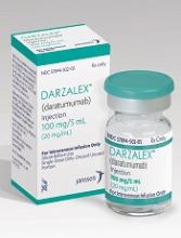
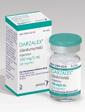
The European Commission (EC) has expanded the approved use of the anti-CD38 antibody daratumumab (Darzalex®).
Daratumumab is now approved for use in combination with lenalidomide and dexamethasone or bortezomib and dexamethasone for the treatment of adults with multiple myeloma (MM) who have received at least 1 prior therapy.
The EC previously granted conditional approval for daratumumab as monotherapy for the treatment of adults with relapsed or refractory MM whose prior treatment included a proteasome inhibitor and an immunomodulatory agent and who demonstrated disease progression on their last therapy.
Now, the EC has granted daratumumab full approval for this indication. The conditional approval was contingent upon Janssen, the company developing daratumumab, providing additional data from the phase 3 POLLUX and CASTOR trials.
As these data have been provided, the EC said the specific obligations associated with the conditional approval have been fulfilled, allowing the switch from conditional to full approval.
The EC’s decision to grant daratumumab approval in combination with lenalidomide and dexamethasone or bortezomib and dexamethasone was also based on data from the POLLUX and CASTOR trials.
In the POLLUX trial, researchers compared treatment with lenalidomide and dexamethasone to treatment with daratumumab, lenalidomide, and dexamethasone in patients with relapsed or refractory MM.
Patients who received daratumumab in combination had a significantly higher response rate and longer progression-free survival than patients who received the 2-drug combination.
However, treatment with daratumumab was associated with infusion-related reactions and a higher incidence of neutropenia.
Results from this trial were published in NEJM in October 2016.
In the CASTOR trial, researchers compared treatment with bortezomib and dexamethasone to treatment with daratumumab, bortezomib, and dexamethasone in patients with previously treated MM.
Patients who received the 3-drug combination had a higher response rate, longer progression-free survival, and a higher incidence of grade 3/4 adverse events than those who received the 2-drug combination.
Results from this trial were published in NEJM in August 2016. ![]()


