Drug reaction or metastatic lung cancer?

Article Type
Changed
Fri, 12/01/2017 - 10:00
Display Headline
Drug reaction or metastatic lung cancer?

A 76-year-old man with ulcerative colitis presented with a 1-week history of low-grade fever and progressive dyspnea. He was taking infliximab for the ulcerative colitis. He was known to be negative for human immunodeficiency virus.

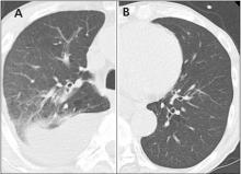
Computed tomography at the time of presentation showed infiltrates and pleural effusion in the upper lobe of the right lung (A). The left lung appeared normal (B).
Figure 1. Computed tomography at the time of presentation showed infiltrates and pleural effusion in the upper lobe of the right lung (A). The left lung appeared normal (B).
Computed tomography (CT) of the chest revealed infiltrates in the upper lobe of the right lung and right pleural effusion (Figure 1). A sputum smear was positive for acid-fast bacilli, and Mycobacterium tuberculosis was isolated from both sputum and the pleural effusion. He was diagnosed with pulmonary tuberculosis and pleuritis. Infliximab treatment was discontinued and induction chemotherapy for tuberculosis was started.

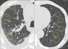
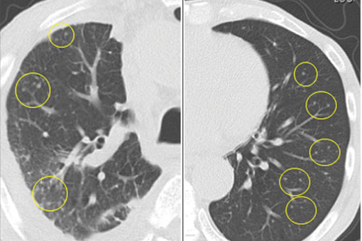
Computed tomography after 3 months of tuberculosis treatment showed randomly distributed nodules in both lungs that resembled metastatic lung cancer.
Figure 2. Computed tomography after 3 months of tuberculosis treatment showed randomly distributed nodules in both lungs that resembled metastatic lung cancer.
After 2 months of combination therapy with isoniazid, rifampicin, ethambutol, and pyrazinamide, maintenance therapy with isoniazid and rifampicin was started. However, after 1 month on maintenance therapy (ie, a total of 3 months of therapy), the patient still had low-grade fever, and repeat chest CT showed randomly distributed nodules mimicking metastatic lung cancer throughout both lungs (Figure 2).

Since the M tuberculosis cultured from his lung proved to be sensitive to the antituberculosis drugs, we suspected that the nodules were a paradoxical reaction to the drug therapy, and thus we continued the treatment because of the continued low-grade fever. After 9 months of therapy, the fever had resolved and the nodules had disappeared, confirming our suspicion of a paradoxical reaction. The number of lymphocytes gradually increased during drug therapy.

Paradoxical reaction during tuberculosis treatment is defined as a worsening of pre-existing lesions or as the emergence of new lesions during appropriate therapy.1,2 The diagnosis is sometimes difficult, since new lesions can resemble other lung diseases. However, a paradoxical reaction involving randomly distributed nodules is rare and radiographically resembles metastatic lung cancer. Clinicians should be aware of this type of reaction in patients on tuberculosis therapy.

References
  1. Cheng SL, Wang HC, Yang PC. Paradoxical response during anti-tuberculosis treatment in HIV-negative patients with pulmonary tuberculosis. Int J Tuberc Lung Dis 2007; 11:1290–1295.
  2. Narita M, Ashkin D, Hollender ES, Pitchenik AE. Paradoxical worsening of tuberculosis following antiretroviral therapy in patients with AIDS. Am J Respir Crit Care Med 1998; 158:157–161.
Article PDF
Author and Disclosure Information

Hiromichi Hara, MD, PhD
Division of Respiratory Diseases, Department of Internal Medicine, Jikei University School of Medicine, Minatoku, Tokyo, Japan

Hirofumi Utsumi, MD, PhD
Division of Respiratory Diseases, Department of Internal Medicine, Jikei University School of Medicine, Minatoku, Tokyo, Japan

Hiroshi Wakui, MD, PhD
Division of Respiratory Diseases, Department of Internal Medicine, Jikei University School of Medicine, Minatoku, Tokyo, Japan

Haruhiko Yanagisawa, MD, PhD
Division of Respiratory Diseases, Department of Internal Medicine, Jikei University School of Medicine, Minatoku, Tokyo, Japan

Mitsuo Hashimoto, MD, PhD
Division of Respiratory Diseases, Department of Internal Medicine, Jikei University School of Medicine, Minatoku, Tokyo, Japan

Kazuyoshi Kuwano, MD, PhD
Division of Respiratory Diseases, Department of Internal Medicine, Jikei University School of Medicine, Minatoku, Tokyo, Japan

Address: Hiromichi Hara, MD, PhD, Division of Respiratory Diseases, Department of Internal Medicine, Jikei University School of Medicine, 3-19-18 Nishishimbashi, Minatoku, Tokyo, Japan; hirohara@jikei.ac.jp

Issue
Cleveland Clinic Journal of Medicine - 84(12)
Publications
Topics
Page Number
914-915
Legacy Keywords
ulcerative colitis, infliximab, tuberculosis, lung cancer, computed tomography, CT, paradoxical reaction, lung nodules, Hiromichi Hara, Hirofumi Utsumi, Hiroshi Wakui, Haruhiko Yanagisawa, Mitsuo Hashimoto, Kazuyoshi Kuwano
Sections
Author and Disclosure Information

Hiromichi Hara, MD, PhD
Division of Respiratory Diseases, Department of Internal Medicine, Jikei University School of Medicine, Minatoku, Tokyo, Japan

Hirofumi Utsumi, MD, PhD
Division of Respiratory Diseases, Department of Internal Medicine, Jikei University School of Medicine, Minatoku, Tokyo, Japan

Hiroshi Wakui, MD, PhD
Division of Respiratory Diseases, Department of Internal Medicine, Jikei University School of Medicine, Minatoku, Tokyo, Japan

Haruhiko Yanagisawa, MD, PhD
Division of Respiratory Diseases, Department of Internal Medicine, Jikei University School of Medicine, Minatoku, Tokyo, Japan

Mitsuo Hashimoto, MD, PhD
Division of Respiratory Diseases, Department of Internal Medicine, Jikei University School of Medicine, Minatoku, Tokyo, Japan

Kazuyoshi Kuwano, MD, PhD
Division of Respiratory Diseases, Department of Internal Medicine, Jikei University School of Medicine, Minatoku, Tokyo, Japan

Address: Hiromichi Hara, MD, PhD, Division of Respiratory Diseases, Department of Internal Medicine, Jikei University School of Medicine, 3-19-18 Nishishimbashi, Minatoku, Tokyo, Japan; hirohara@jikei.ac.jp

Article PDF
Article PDF
Related Articles

A 76-year-old man with ulcerative colitis presented with a 1-week history of low-grade fever and progressive dyspnea. He was taking infliximab for the ulcerative colitis. He was known to be negative for human immunodeficiency virus.

Computed tomography at the time of presentation showed infiltrates and pleural effusion in the upper lobe of the right lung (A). The left lung appeared normal (B).
Figure 1. Computed tomography at the time of presentation showed infiltrates and pleural effusion in the upper lobe of the right lung (A). The left lung appeared normal (B).
Computed tomography (CT) of the chest revealed infiltrates in the upper lobe of the right lung and right pleural effusion (Figure 1). A sputum smear was positive for acid-fast bacilli, and Mycobacterium tuberculosis was isolated from both sputum and the pleural effusion. He was diagnosed with pulmonary tuberculosis and pleuritis. Infliximab treatment was discontinued and induction chemotherapy for tuberculosis was started.

Computed tomography after 3 months of tuberculosis treatment showed randomly distributed nodules in both lungs that resembled metastatic lung cancer.
Figure 2. Computed tomography after 3 months of tuberculosis treatment showed randomly distributed nodules in both lungs that resembled metastatic lung cancer.
After 2 months of combination therapy with isoniazid, rifampicin, ethambutol, and pyrazinamide, maintenance therapy with isoniazid and rifampicin was started. However, after 1 month on maintenance therapy (ie, a total of 3 months of therapy), the patient still had low-grade fever, and repeat chest CT showed randomly distributed nodules mimicking metastatic lung cancer throughout both lungs (Figure 2).

Since the M tuberculosis cultured from his lung proved to be sensitive to the antituberculosis drugs, we suspected that the nodules were a paradoxical reaction to the drug therapy, and thus we continued the treatment because of the continued low-grade fever. After 9 months of therapy, the fever had resolved and the nodules had disappeared, confirming our suspicion of a paradoxical reaction. The number of lymphocytes gradually increased during drug therapy.

Paradoxical reaction during tuberculosis treatment is defined as a worsening of pre-existing lesions or as the emergence of new lesions during appropriate therapy.1,2 The diagnosis is sometimes difficult, since new lesions can resemble other lung diseases. However, a paradoxical reaction involving randomly distributed nodules is rare and radiographically resembles metastatic lung cancer. Clinicians should be aware of this type of reaction in patients on tuberculosis therapy.

References
  1. Cheng SL, Wang HC, Yang PC. Paradoxical response during anti-tuberculosis treatment in HIV-negative patients with pulmonary tuberculosis. Int J Tuberc Lung Dis 2007; 11:1290–1295.
  2. Narita M, Ashkin D, Hollender ES, Pitchenik AE. Paradoxical worsening of tuberculosis following antiretroviral therapy in patients with AIDS. Am J Respir Crit Care Med 1998; 158:157–161.
Issue
Cleveland Clinic Journal of Medicine - 84(12)
Issue
Cleveland Clinic Journal of Medicine - 84(12)
Page Number
914-915
Page Number
914-915
Publications
Publications
Topics
Article Type
Display Headline
Drug reaction or metastatic lung cancer?
Display Headline
Drug reaction or metastatic lung cancer?
Legacy Keywords
ulcerative colitis, infliximab, tuberculosis, lung cancer, computed tomography, CT, paradoxical reaction, lung nodules, Hiromichi Hara, Hirofumi Utsumi, Hiroshi Wakui, Haruhiko Yanagisawa, Mitsuo Hashimoto, Kazuyoshi Kuwano
Legacy Keywords
ulcerative colitis, infliximab, tuberculosis, lung cancer, computed tomography, CT, paradoxical reaction, lung nodules, Hiromichi Hara, Hirofumi Utsumi, Hiroshi Wakui, Haruhiko Yanagisawa, Mitsuo Hashimoto, Kazuyoshi Kuwano
Sections
Disallow All Ads
Content Gating
No Gating (article Unlocked/Free)
Alternative CME
Disqus Comments
Default
Article PDF Media