User login
Chronic lesions on legs
A 62-YEAR-OLD MAN came into our facility for a skin exam at the urging of his wife. They were both concerned about a rash on his legs that was asymptomatic and had gradually developed over the past few years.
The patient, who worked as a laborer, was not taking any prescription medications, vitamins, or herbal products. He didn’t smoke, drink alcohol, or use recreational drugs and denied having any fevers, chills, arthralgias, or night sweats. He indicated that his father had a basal cell carcinoma.
On physical exam, we noted some seborrheic keratoses and mild acne rosacea in addition to the rash on his legs. The rash itself consisted of nonpalpable, nonblanchable macules on the lower shins, ankles, and dorsal feet with sparing of the soles (FIGURE).
FIGURE
Nonpalpable rash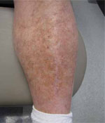
The rash on this 62-year-old man’s legs developed over several years. The nonblanchable macules on the lower shins, ankles, and dorsal feet spared the soles.
WHAT IS YOUR DIAGNOSIS?
HOW WOULD YOU TREAT THIS PATIENT?
Diagnosis: Schamberg’s disease
Schamberg’s disease is a form of progressive pigmented purpuric dermatosis. This type of nonpalpable purpura consists of reddish-brown macules that are usually located on the lower extremities and ankles, and spare the soles of the feet. These petechiae appear due to capillary leakage and breakdown of blood near the skin surface, leaving behind hemosiderin deposits.
Clinically, the lesions are asymptomatic and may persist for months or years. They are characteristically referred to as punctate "cayenne pepper" spots, and can vary in size and shape. Occasionally, mild erythema and scaling can cause slight itching.
Although these lesions have a vasculitic appearance, there is no hematologic or other internal disease association. Schamberg’s purpura may resemble stasis dermatitis, because both processes include inflammation, dilation of capillaries, extravasation of erythrocytes, and hemosiderin deposits. However, lesions due to venous insufficiency extend deep into the dermis and have more pronounced epidermal changes and dermal fibrosis.
An unknown cause. Although no one knows what causes Schamberg’s disease, a cellular immune reaction may be at work. Early endothelial expression of certain intracellular adhesion receptors has shown a common pericapillary infiltrate pattern in cases of Schamberg’s disease.1 Recent viral infection and allergic reactions to medications, such as aspirin, have also been associated with Schamberg’s disease.2
One of several pigmented purpuric dermatoses
Schamberg’s disease fits under the inclusive term of pigmented purpuric dermatoses (PPD), as a macular purpuric reddish-brown dermatosis. Other subtypes of PPD can be distinguished from Schamberg’s disease clinically. These subtypes include lichen aureus, which appears as golden patches; Majocchi’s purpura, which forms an annular pattern with telangiectasias; Gougerot-Blum purpura, which is associated with lichenoid dermatitis; and Doucas and Kapentanakis purpura, which is an eczematous variant. Other forms of purpura can be distinguished from Schamberg’s disease histologically (TABLE).3
Platelet abnormalities can also produce red, flat, nonblanchable petechiae, and may occur in patients who have taken certain prescribed drugs, over-the-counter medications, or herbal remedies; those who have received recent vaccinations; and in those who have (or have had) severe viral infections, beta-hemolytic streptococcal infections, leukemia, lupus erythematosus, or idiopathic thrombocytopenic purpura.
Scurvy can also cause perifollicular petechiae that are distributed primarily on the lower extremities and are symmetrical. Gums may be swollen or hemorrhagic, and patients will have a history of vitamin C deficiency, myalgia, and fatigue. Children may have bone tenderness, epistaxis, and hematuria. A diagnosis can be made by looking for low ascorbic acid in the serum. Prompt correction with vitamin C supplementation is also diagnostic.
Dysproteinemia can cause crops of petechiae and occasionally ecchymoses. Lesions appear on the lower extremities and sometimes on the ears and the tip of the nose. Cryoglobulinemia is associated with Raynaud’s phenomenon, cold urticaria, dizziness, and epistaxis; it is also associated with hepatitis C infection in >90% of cases.4 Macroglobulinemia is associated with dilated vessels and hemorrhage in the optic fundi, mental confusion, anemia, weight loss, hepatosplenomegaly, and lymphadenopathy. Hyperglobulinemic purpura may be associated with arthritis, xerostomia, anemia, and hepatosplenomegaly. These various dysproteinemias can be diagnosed by finding abnormal proteins through serum electrophoresis. Skin biopsy of the petechiae reveals thrombi in dermal vessels, which may be associated with leukocytoclastic vasculitis.4
Many viral infections may result in a petechial rash, including measles, rubella, hepatitis, cytomegalovirus, Coxsackievirus, and respiratory syncytial virus. Petechial rashes associated with eczema or seborrheic dermatitis-like lesions can also be seen in children with Langerhans cell histiocytosis
TABLE
Histologic spectrum of purpura3
| Histologic pattern | Diseases |
|---|---|
| Erythrocyte extravasation without perivascular inflammation or fibrin deposition | Thrombocytopenic purpura, senile purpura, steroid purpura, scurvy |
| Erythrocyte extravasation with lymphocytic perivascular infiltrate; no fibrin deposition | Lichen aureus; Schamberg’s, Majocchi’s, Gougerot-Blum, and Doucas and Kapentanakis purpuras |
| Erythrocyte extravasation with vascular damage, neutrophilic infiltrate, and fibrin deposition | Leukocytoclastic vasculitis, mixed cryoglobulinemia |
Biopsy confirms diagnosis
Platelet and clotting studies are usually normal with Schamberg’s purpura. The definitive diagnosis can be confirmed through skin biopsy, which shows capillaritis of dermal vessels. Perivascular inflammatory infiltrates with extravasations of blood cells and hemosiderin-laden macrophages are seen on histologic evaluation.
Treat mild itching with corticosteroids
These lesions pose mostly a cosmetic problem, but mild itching and scaling occasionally occur. These symptoms can be treated with topical corticosteroid therapy.
Treatment with an 8-week course of pentoxifylline (Trental)—400 mg daily—has shown resolution of lesions in some patients, but no benefit in others.5,6 In some patient’s, psoralen plus ultraviolet light therapy has been shown to provide modest improvement.7 Treatment with aminaphtone 75 mg bid for 4 weeks has recently shown improvements in patients with longstanding Schamberg’s purpura.8 A review of medications may point to a possible etiology for some patients.9
Although lesions can persist for years, the lesions may eventually clear and patients should be assured that there is no definitive underlying systemic disease associated with Schamberg’s disease.10
Educating our patient
Our patient required no treatment, because his rash was asymptomatic. We told him that the pigmentation can last for years, but could be covered with cosmetics. We also reassured him that no underlying systemic disease had caused his rash.
CORRESPONDENCE Jay Shubrook, DO, Castrop Center, O’Bleness Health System, 75 Hospital Drive, Athens, OH 45701; shubrook@ohio.edu
1. Ghersetich I, Lotti T, Bacci S, et al. Cell infiltrate in progressive pigmented purpura (Schamberg’s disease): immunophenotype, adhesion receptors, and intercellular relationships. Int J Dermatol. 1995;34:846-850.
2. Abeck D, Gross GE, Kuwert C, et al. Acetaminophen-induced progressive pigmentary purpura (Schamberg’s disease). J Am Acad Dermatol. 1992;27:123-124.
3. Elder D, Elenitsas R, Jaworsky C, et al. Lever’s Histopathology of the Skin. 8th ed. Philadelphia, Pa: Lippincott-Raven Publishers; 1997.
4. James WD, Berger TG, Elston DM. Andrews’ Diseases of the Skin: Clinical Dermatology. 10th ed. New York, NY: Elsevier; 2006.
5. Gandhi V, Singal A, Sachdeva B, et al. Treatment of Schamberg’s disease with pentoxifylline–therapeutic trial. Indian J Dermatol Venereol Leprol. 2003;69:25-26.
6. Kano Y, Hirayama K, Orihara M, et al. Successful treatment of Schamberg’s disease with pentoxifylline. J Am Acad Dermatol. 1997;36:827-830.
7. Seckin D, Yazici Z, Senol A, et al. A case of Schamberg’s disease responding dramatically to PUVA treatment. Photodermatol Photoimmunol Photomed. 2008;24:95-96.
8. de Godoy JM, Batigalia F. Aminaphtone in the control of Schamberg’s disease. Thromb J. 2009;7:8.-
9. Ratnam KV, Su WP, Peters MS. Purpura simplex (inflammatory purpura without vasculitis): a clinicopathologic study of 174 cases. J Am Acad Dermatol. 1991;25:642-647.
10. Habif TP. Clinical Dermatology: A Color Guide to Diagnosis and Therapy. 4th ed. Philadelphia, Pa: Mosby, Inc; 2003.
A 62-YEAR-OLD MAN came into our facility for a skin exam at the urging of his wife. They were both concerned about a rash on his legs that was asymptomatic and had gradually developed over the past few years.
The patient, who worked as a laborer, was not taking any prescription medications, vitamins, or herbal products. He didn’t smoke, drink alcohol, or use recreational drugs and denied having any fevers, chills, arthralgias, or night sweats. He indicated that his father had a basal cell carcinoma.
On physical exam, we noted some seborrheic keratoses and mild acne rosacea in addition to the rash on his legs. The rash itself consisted of nonpalpable, nonblanchable macules on the lower shins, ankles, and dorsal feet with sparing of the soles (FIGURE).
FIGURE
Nonpalpable rash
The rash on this 62-year-old man’s legs developed over several years. The nonblanchable macules on the lower shins, ankles, and dorsal feet spared the soles.
WHAT IS YOUR DIAGNOSIS?
HOW WOULD YOU TREAT THIS PATIENT?
Diagnosis: Schamberg’s disease
Schamberg’s disease is a form of progressive pigmented purpuric dermatosis. This type of nonpalpable purpura consists of reddish-brown macules that are usually located on the lower extremities and ankles, and spare the soles of the feet. These petechiae appear due to capillary leakage and breakdown of blood near the skin surface, leaving behind hemosiderin deposits.
Clinically, the lesions are asymptomatic and may persist for months or years. They are characteristically referred to as punctate "cayenne pepper" spots, and can vary in size and shape. Occasionally, mild erythema and scaling can cause slight itching.
Although these lesions have a vasculitic appearance, there is no hematologic or other internal disease association. Schamberg’s purpura may resemble stasis dermatitis, because both processes include inflammation, dilation of capillaries, extravasation of erythrocytes, and hemosiderin deposits. However, lesions due to venous insufficiency extend deep into the dermis and have more pronounced epidermal changes and dermal fibrosis.
An unknown cause. Although no one knows what causes Schamberg’s disease, a cellular immune reaction may be at work. Early endothelial expression of certain intracellular adhesion receptors has shown a common pericapillary infiltrate pattern in cases of Schamberg’s disease.1 Recent viral infection and allergic reactions to medications, such as aspirin, have also been associated with Schamberg’s disease.2
One of several pigmented purpuric dermatoses
Schamberg’s disease fits under the inclusive term of pigmented purpuric dermatoses (PPD), as a macular purpuric reddish-brown dermatosis. Other subtypes of PPD can be distinguished from Schamberg’s disease clinically. These subtypes include lichen aureus, which appears as golden patches; Majocchi’s purpura, which forms an annular pattern with telangiectasias; Gougerot-Blum purpura, which is associated with lichenoid dermatitis; and Doucas and Kapentanakis purpura, which is an eczematous variant. Other forms of purpura can be distinguished from Schamberg’s disease histologically (TABLE).3
Platelet abnormalities can also produce red, flat, nonblanchable petechiae, and may occur in patients who have taken certain prescribed drugs, over-the-counter medications, or herbal remedies; those who have received recent vaccinations; and in those who have (or have had) severe viral infections, beta-hemolytic streptococcal infections, leukemia, lupus erythematosus, or idiopathic thrombocytopenic purpura.
Scurvy can also cause perifollicular petechiae that are distributed primarily on the lower extremities and are symmetrical. Gums may be swollen or hemorrhagic, and patients will have a history of vitamin C deficiency, myalgia, and fatigue. Children may have bone tenderness, epistaxis, and hematuria. A diagnosis can be made by looking for low ascorbic acid in the serum. Prompt correction with vitamin C supplementation is also diagnostic.
Dysproteinemia can cause crops of petechiae and occasionally ecchymoses. Lesions appear on the lower extremities and sometimes on the ears and the tip of the nose. Cryoglobulinemia is associated with Raynaud’s phenomenon, cold urticaria, dizziness, and epistaxis; it is also associated with hepatitis C infection in >90% of cases.4 Macroglobulinemia is associated with dilated vessels and hemorrhage in the optic fundi, mental confusion, anemia, weight loss, hepatosplenomegaly, and lymphadenopathy. Hyperglobulinemic purpura may be associated with arthritis, xerostomia, anemia, and hepatosplenomegaly. These various dysproteinemias can be diagnosed by finding abnormal proteins through serum electrophoresis. Skin biopsy of the petechiae reveals thrombi in dermal vessels, which may be associated with leukocytoclastic vasculitis.4
Many viral infections may result in a petechial rash, including measles, rubella, hepatitis, cytomegalovirus, Coxsackievirus, and respiratory syncytial virus. Petechial rashes associated with eczema or seborrheic dermatitis-like lesions can also be seen in children with Langerhans cell histiocytosis
TABLE
Histologic spectrum of purpura3
| Histologic pattern | Diseases |
|---|---|
| Erythrocyte extravasation without perivascular inflammation or fibrin deposition | Thrombocytopenic purpura, senile purpura, steroid purpura, scurvy |
| Erythrocyte extravasation with lymphocytic perivascular infiltrate; no fibrin deposition | Lichen aureus; Schamberg’s, Majocchi’s, Gougerot-Blum, and Doucas and Kapentanakis purpuras |
| Erythrocyte extravasation with vascular damage, neutrophilic infiltrate, and fibrin deposition | Leukocytoclastic vasculitis, mixed cryoglobulinemia |
Biopsy confirms diagnosis
Platelet and clotting studies are usually normal with Schamberg’s purpura. The definitive diagnosis can be confirmed through skin biopsy, which shows capillaritis of dermal vessels. Perivascular inflammatory infiltrates with extravasations of blood cells and hemosiderin-laden macrophages are seen on histologic evaluation.
Treat mild itching with corticosteroids
These lesions pose mostly a cosmetic problem, but mild itching and scaling occasionally occur. These symptoms can be treated with topical corticosteroid therapy.
Treatment with an 8-week course of pentoxifylline (Trental)—400 mg daily—has shown resolution of lesions in some patients, but no benefit in others.5,6 In some patient’s, psoralen plus ultraviolet light therapy has been shown to provide modest improvement.7 Treatment with aminaphtone 75 mg bid for 4 weeks has recently shown improvements in patients with longstanding Schamberg’s purpura.8 A review of medications may point to a possible etiology for some patients.9
Although lesions can persist for years, the lesions may eventually clear and patients should be assured that there is no definitive underlying systemic disease associated with Schamberg’s disease.10
Educating our patient
Our patient required no treatment, because his rash was asymptomatic. We told him that the pigmentation can last for years, but could be covered with cosmetics. We also reassured him that no underlying systemic disease had caused his rash.
CORRESPONDENCE Jay Shubrook, DO, Castrop Center, O’Bleness Health System, 75 Hospital Drive, Athens, OH 45701; shubrook@ohio.edu
1. Ghersetich I, Lotti T, Bacci S, et al. Cell infiltrate in progressive pigmented purpura (Schamberg’s disease): immunophenotype, adhesion receptors, and intercellular relationships. Int J Dermatol. 1995;34:846-850.
2. Abeck D, Gross GE, Kuwert C, et al. Acetaminophen-induced progressive pigmentary purpura (Schamberg’s disease). J Am Acad Dermatol. 1992;27:123-124.
3. Elder D, Elenitsas R, Jaworsky C, et al. Lever’s Histopathology of the Skin. 8th ed. Philadelphia, Pa: Lippincott-Raven Publishers; 1997.
4. James WD, Berger TG, Elston DM. Andrews’ Diseases of the Skin: Clinical Dermatology. 10th ed. New York, NY: Elsevier; 2006.
5. Gandhi V, Singal A, Sachdeva B, et al. Treatment of Schamberg’s disease with pentoxifylline–therapeutic trial. Indian J Dermatol Venereol Leprol. 2003;69:25-26.
6. Kano Y, Hirayama K, Orihara M, et al. Successful treatment of Schamberg’s disease with pentoxifylline. J Am Acad Dermatol. 1997;36:827-830.
7. Seckin D, Yazici Z, Senol A, et al. A case of Schamberg’s disease responding dramatically to PUVA treatment. Photodermatol Photoimmunol Photomed. 2008;24:95-96.
8. de Godoy JM, Batigalia F. Aminaphtone in the control of Schamberg’s disease. Thromb J. 2009;7:8.-
9. Ratnam KV, Su WP, Peters MS. Purpura simplex (inflammatory purpura without vasculitis): a clinicopathologic study of 174 cases. J Am Acad Dermatol. 1991;25:642-647.
10. Habif TP. Clinical Dermatology: A Color Guide to Diagnosis and Therapy. 4th ed. Philadelphia, Pa: Mosby, Inc; 2003.
Bluish pigmentation of face and sclera
A 67-YEAR-OLD CAUCASIAN WOMAN came to the diabetes center for a routine check-up. She indicated that her face and eyes had become discolored, and that the discoloration had occurred gradually. She denied pruritus, erythema, edema, or pain at the site, and reported no vision changes, dry eyes, or other constitutional signs and symptoms. She did not smoke.
Th e patient’s past medical history was significant for type 2 diabetes, hypertension, depression, dyslipidemia, and rosacea. The medications she was taking included pioglitazone, metformin, coral calcium, glyburide, atorvastatin, fosinopril, and minocycline.
On physical exam, there was a bluish pigmentation of the left lateral forehead and cheek (FIGURE 1A) and bluish pigmentation of the sclera (FIGURE 1B). The rest of her exam was normal.
We were concerned about venous dilation, and ordered a computed tomography scan of the chest and an ultrasound of the neck. Both were normal, thus ruling out a thoracic outlet obstruction.
FIGURE 1
Unusual pigmentation of forehead and eyes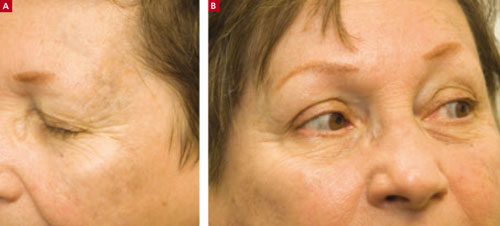
This 67-year-old woman said that the bluish discoloration of her face and eyes had come on gradually. She had no problems with her vision and no pain in her eyes.
WHAT IS YOUR DIAGNOSIS?
HOW WOULD YOU TREAT THIS PATIENT?
Diagnosis: Minocycline-induced hyperpigmentation
We referred the patient to a dermatologist, who diagnosed minocycline-induced hyperpigmentation of the skin and sclera.
Minocycline hydrochloride, a derivative of tetracycline, is a broad-spectrum antibiotic that is often used to treat rosacea (which our patient had) and acne vulgaris. Minocycline’s lipid-soluble properties enable penetration into sebaceous glands and subsequent clearing of acne in many cases.1
Despite minocycline’s therapeutic effect, it can lead to a non–dose-dependent pigmentation of the skin (typically a blue-gray pigmentation on the legs), nails, sclera, bones, oral mucosa, teeth, and thyroid.1,2
Rare serious adverse effects include systemic lupus erythematosus (antinuclear antibody positive, DNA antibody negative), pseudotumor cerebri syndrome, and an autoimmune drug reaction leading to hepatitis. Resolution of these conditions occurs slowly once minocycline is discontinued.1 However, scleral hyperpigmentation may be permanent. This pigmentation appears blue-gray.3
Hyperpigmentation falls into 4 categories
Although the etiology of the minocycline-induced hyperpigmentation is unclear, iron and melanin-staining granules identified in dermal dendrocytes and macrophages are likely responsible for producing the blue-gray pigmentation of the skin.2,4
The hyperpigmentation of minocycline is classified into 4 different types, based on clinical features, light and electron microscopy, and energy dispersive X-ray analysis:
Type I is a blue-gray discoloration that appears on the face at sites of inflammation or scarring.
Type II is characterized by blue-gray pigmentation of normal skin of the anterior lower legs.
In Type III, the skin has a muddy brown appearance at sun-exposed sites.2
Type IV occurs in scars, giving them a blue-gray appearance.5
Differential Dx includes Addison’s disease
Addison’s disease, hemochromatosis, and melanoma all make up the differential.
The hyperpigmentation of Addison’s disease is a characteristic brown or bronzing of the skin, most notably in the creases of the hands. Mucous membranes may develop a bluish pigmentation.
Patients with hemochromatosis develop bronzing of the skin caused by iron deposits in the dermis. This pigmentation is characterized by a generalized metallic or slate gray coloration of the face, neck, upper extremities, lower legs, and genitals. This coloration also appears in scars.
Melanoma was less likely in this case because of the uniformity of pigmentation on the skin and sclera, as well as the symmetrical pigmentation of the sclera. A skin biopsy would be helpful in ruling out melanoma.6
A biopsy of this lesion would show granules within macrophages that stain positive for iron and melanin, confirming the clinical diagnosis of minocycline-induced hyperpigmentation.2
Patients may not want to wait for discoloration to go away
Some patients find the benign hyperpigmentation cosmetically undesirable and may not want to wait to see if the discoloration goes away. The Q-switched ruby laser has been successful in removing minocycline-induced hyperpigmentation of the skin and oral mucosa.2
Our patient stopped the drug, and saw (some) improvement
Once the diagnosis was made, our patient stopped taking minocycline—which she’d been taking at a low daily dose for 4 years. She was started on metronidazole for her rosacea. Three years later, the patient had a slight clinical improvement in the bluish appearance of her face and sclera.
Minocycline is still a good option for the treatment of acne and rosacea. If you prescribe it, though, be sure to monitor for hyperpigmentation on a regular basis.
CORRESPONDENCE Jay Shubrook, DO, associate professor of family medicine, director of clinical research, Cornwell Center, O’Bleness Memorial Hospital, 65 Hospital Drive, Athens, OH 45701; shubrook@ohio.edu.
1. Habif TP. Clinical Dermatology: A Color Guide to Diagnosis and Therapy. 4th ed. Philadelphia, Pa: Mosby Inc; 2004:181–182.
2. James W, Berger T, Elston D. Andrews’ Diseases of the Skin: Clinical Dermatology. 10th ed. Philadelphia, Pa: Elsevier Inc; 2006:125–126.
3. Fraundfelder FT, Randall JA. Minocycline-induced scleral pigmentation. Ophthalmology. 1997;104:936-938.
4. Altman DA, Fiveson DP, Lee MW. Minocycline hyperpigmentation: model for in situ phagocytic activity of factor XIIa dermal dendrocytes. J Cutan Pathol. 1992;19:340-345.
5. Mouton RW, Jordann HF, Schneider JW. A new type of minocycline-induced cutaneous hyperpigmentation. Clin Exper Dermatol. 2004;29:8-14.
6. Kasper DL, Braunwald E, Fauci AS, et al. Harrison’s Principles of Internal Medicine. 16th ed. New York, NY: McGraw-Hill Companies, Inc; 2005:2142, 2300.
A 67-YEAR-OLD CAUCASIAN WOMAN came to the diabetes center for a routine check-up. She indicated that her face and eyes had become discolored, and that the discoloration had occurred gradually. She denied pruritus, erythema, edema, or pain at the site, and reported no vision changes, dry eyes, or other constitutional signs and symptoms. She did not smoke.
Th e patient’s past medical history was significant for type 2 diabetes, hypertension, depression, dyslipidemia, and rosacea. The medications she was taking included pioglitazone, metformin, coral calcium, glyburide, atorvastatin, fosinopril, and minocycline.
On physical exam, there was a bluish pigmentation of the left lateral forehead and cheek (FIGURE 1A) and bluish pigmentation of the sclera (FIGURE 1B). The rest of her exam was normal.
We were concerned about venous dilation, and ordered a computed tomography scan of the chest and an ultrasound of the neck. Both were normal, thus ruling out a thoracic outlet obstruction.
FIGURE 1
Unusual pigmentation of forehead and eyes
This 67-year-old woman said that the bluish discoloration of her face and eyes had come on gradually. She had no problems with her vision and no pain in her eyes.
WHAT IS YOUR DIAGNOSIS?
HOW WOULD YOU TREAT THIS PATIENT?
Diagnosis: Minocycline-induced hyperpigmentation
We referred the patient to a dermatologist, who diagnosed minocycline-induced hyperpigmentation of the skin and sclera.
Minocycline hydrochloride, a derivative of tetracycline, is a broad-spectrum antibiotic that is often used to treat rosacea (which our patient had) and acne vulgaris. Minocycline’s lipid-soluble properties enable penetration into sebaceous glands and subsequent clearing of acne in many cases.1
Despite minocycline’s therapeutic effect, it can lead to a non–dose-dependent pigmentation of the skin (typically a blue-gray pigmentation on the legs), nails, sclera, bones, oral mucosa, teeth, and thyroid.1,2
Rare serious adverse effects include systemic lupus erythematosus (antinuclear antibody positive, DNA antibody negative), pseudotumor cerebri syndrome, and an autoimmune drug reaction leading to hepatitis. Resolution of these conditions occurs slowly once minocycline is discontinued.1 However, scleral hyperpigmentation may be permanent. This pigmentation appears blue-gray.3
Hyperpigmentation falls into 4 categories
Although the etiology of the minocycline-induced hyperpigmentation is unclear, iron and melanin-staining granules identified in dermal dendrocytes and macrophages are likely responsible for producing the blue-gray pigmentation of the skin.2,4
The hyperpigmentation of minocycline is classified into 4 different types, based on clinical features, light and electron microscopy, and energy dispersive X-ray analysis:
Type I is a blue-gray discoloration that appears on the face at sites of inflammation or scarring.
Type II is characterized by blue-gray pigmentation of normal skin of the anterior lower legs.
In Type III, the skin has a muddy brown appearance at sun-exposed sites.2
Type IV occurs in scars, giving them a blue-gray appearance.5
Differential Dx includes Addison’s disease
Addison’s disease, hemochromatosis, and melanoma all make up the differential.
The hyperpigmentation of Addison’s disease is a characteristic brown or bronzing of the skin, most notably in the creases of the hands. Mucous membranes may develop a bluish pigmentation.
Patients with hemochromatosis develop bronzing of the skin caused by iron deposits in the dermis. This pigmentation is characterized by a generalized metallic or slate gray coloration of the face, neck, upper extremities, lower legs, and genitals. This coloration also appears in scars.
Melanoma was less likely in this case because of the uniformity of pigmentation on the skin and sclera, as well as the symmetrical pigmentation of the sclera. A skin biopsy would be helpful in ruling out melanoma.6
A biopsy of this lesion would show granules within macrophages that stain positive for iron and melanin, confirming the clinical diagnosis of minocycline-induced hyperpigmentation.2
Patients may not want to wait for discoloration to go away
Some patients find the benign hyperpigmentation cosmetically undesirable and may not want to wait to see if the discoloration goes away. The Q-switched ruby laser has been successful in removing minocycline-induced hyperpigmentation of the skin and oral mucosa.2
Our patient stopped the drug, and saw (some) improvement
Once the diagnosis was made, our patient stopped taking minocycline—which she’d been taking at a low daily dose for 4 years. She was started on metronidazole for her rosacea. Three years later, the patient had a slight clinical improvement in the bluish appearance of her face and sclera.
Minocycline is still a good option for the treatment of acne and rosacea. If you prescribe it, though, be sure to monitor for hyperpigmentation on a regular basis.
CORRESPONDENCE Jay Shubrook, DO, associate professor of family medicine, director of clinical research, Cornwell Center, O’Bleness Memorial Hospital, 65 Hospital Drive, Athens, OH 45701; shubrook@ohio.edu.
1. Habif TP. Clinical Dermatology: A Color Guide to Diagnosis and Therapy. 4th ed. Philadelphia, Pa: Mosby Inc; 2004:181–182.
2. James W, Berger T, Elston D. Andrews’ Diseases of the Skin: Clinical Dermatology. 10th ed. Philadelphia, Pa: Elsevier Inc; 2006:125–126.
3. Fraundfelder FT, Randall JA. Minocycline-induced scleral pigmentation. Ophthalmology. 1997;104:936-938.
4. Altman DA, Fiveson DP, Lee MW. Minocycline hyperpigmentation: model for in situ phagocytic activity of factor XIIa dermal dendrocytes. J Cutan Pathol. 1992;19:340-345.
5. Mouton RW, Jordann HF, Schneider JW. A new type of minocycline-induced cutaneous hyperpigmentation. Clin Exper Dermatol. 2004;29:8-14.
6. Kasper DL, Braunwald E, Fauci AS, et al. Harrison’s Principles of Internal Medicine. 16th ed. New York, NY: McGraw-Hill Companies, Inc; 2005:2142, 2300.