User login
Bluish pigmentation of face and sclera
A 67-YEAR-OLD CAUCASIAN WOMAN came to the diabetes center for a routine check-up. She indicated that her face and eyes had become discolored, and that the discoloration had occurred gradually. She denied pruritus, erythema, edema, or pain at the site, and reported no vision changes, dry eyes, or other constitutional signs and symptoms. She did not smoke.
Th e patient’s past medical history was significant for type 2 diabetes, hypertension, depression, dyslipidemia, and rosacea. The medications she was taking included pioglitazone, metformin, coral calcium, glyburide, atorvastatin, fosinopril, and minocycline.
On physical exam, there was a bluish pigmentation of the left lateral forehead and cheek (FIGURE 1A) and bluish pigmentation of the sclera (FIGURE 1B). The rest of her exam was normal.
We were concerned about venous dilation, and ordered a computed tomography scan of the chest and an ultrasound of the neck. Both were normal, thus ruling out a thoracic outlet obstruction.
FIGURE 1
Unusual pigmentation of forehead and eyes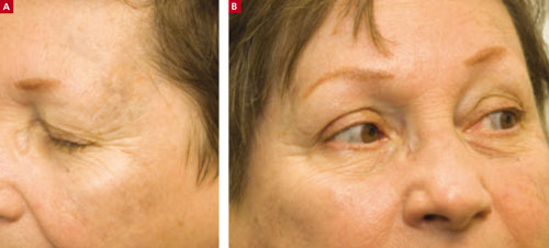
This 67-year-old woman said that the bluish discoloration of her face and eyes had come on gradually. She had no problems with her vision and no pain in her eyes.
WHAT IS YOUR DIAGNOSIS?
HOW WOULD YOU TREAT THIS PATIENT?
Diagnosis: Minocycline-induced hyperpigmentation
We referred the patient to a dermatologist, who diagnosed minocycline-induced hyperpigmentation of the skin and sclera.
Minocycline hydrochloride, a derivative of tetracycline, is a broad-spectrum antibiotic that is often used to treat rosacea (which our patient had) and acne vulgaris. Minocycline’s lipid-soluble properties enable penetration into sebaceous glands and subsequent clearing of acne in many cases.1
Despite minocycline’s therapeutic effect, it can lead to a non–dose-dependent pigmentation of the skin (typically a blue-gray pigmentation on the legs), nails, sclera, bones, oral mucosa, teeth, and thyroid.1,2
Rare serious adverse effects include systemic lupus erythematosus (antinuclear antibody positive, DNA antibody negative), pseudotumor cerebri syndrome, and an autoimmune drug reaction leading to hepatitis. Resolution of these conditions occurs slowly once minocycline is discontinued.1 However, scleral hyperpigmentation may be permanent. This pigmentation appears blue-gray.3
Hyperpigmentation falls into 4 categories
Although the etiology of the minocycline-induced hyperpigmentation is unclear, iron and melanin-staining granules identified in dermal dendrocytes and macrophages are likely responsible for producing the blue-gray pigmentation of the skin.2,4
The hyperpigmentation of minocycline is classified into 4 different types, based on clinical features, light and electron microscopy, and energy dispersive X-ray analysis:
Type I is a blue-gray discoloration that appears on the face at sites of inflammation or scarring.
Type II is characterized by blue-gray pigmentation of normal skin of the anterior lower legs.
In Type III, the skin has a muddy brown appearance at sun-exposed sites.2
Type IV occurs in scars, giving them a blue-gray appearance.5
Differential Dx includes Addison’s disease
Addison’s disease, hemochromatosis, and melanoma all make up the differential.
The hyperpigmentation of Addison’s disease is a characteristic brown or bronzing of the skin, most notably in the creases of the hands. Mucous membranes may develop a bluish pigmentation.
Patients with hemochromatosis develop bronzing of the skin caused by iron deposits in the dermis. This pigmentation is characterized by a generalized metallic or slate gray coloration of the face, neck, upper extremities, lower legs, and genitals. This coloration also appears in scars.
Melanoma was less likely in this case because of the uniformity of pigmentation on the skin and sclera, as well as the symmetrical pigmentation of the sclera. A skin biopsy would be helpful in ruling out melanoma.6
A biopsy of this lesion would show granules within macrophages that stain positive for iron and melanin, confirming the clinical diagnosis of minocycline-induced hyperpigmentation.2
Patients may not want to wait for discoloration to go away
Some patients find the benign hyperpigmentation cosmetically undesirable and may not want to wait to see if the discoloration goes away. The Q-switched ruby laser has been successful in removing minocycline-induced hyperpigmentation of the skin and oral mucosa.2
Our patient stopped the drug, and saw (some) improvement
Once the diagnosis was made, our patient stopped taking minocycline—which she’d been taking at a low daily dose for 4 years. She was started on metronidazole for her rosacea. Three years later, the patient had a slight clinical improvement in the bluish appearance of her face and sclera.
Minocycline is still a good option for the treatment of acne and rosacea. If you prescribe it, though, be sure to monitor for hyperpigmentation on a regular basis.
CORRESPONDENCE Jay Shubrook, DO, associate professor of family medicine, director of clinical research, Cornwell Center, O’Bleness Memorial Hospital, 65 Hospital Drive, Athens, OH 45701; shubrook@ohio.edu.
1. Habif TP. Clinical Dermatology: A Color Guide to Diagnosis and Therapy. 4th ed. Philadelphia, Pa: Mosby Inc; 2004:181–182.
2. James W, Berger T, Elston D. Andrews’ Diseases of the Skin: Clinical Dermatology. 10th ed. Philadelphia, Pa: Elsevier Inc; 2006:125–126.
3. Fraundfelder FT, Randall JA. Minocycline-induced scleral pigmentation. Ophthalmology. 1997;104:936-938.
4. Altman DA, Fiveson DP, Lee MW. Minocycline hyperpigmentation: model for in situ phagocytic activity of factor XIIa dermal dendrocytes. J Cutan Pathol. 1992;19:340-345.
5. Mouton RW, Jordann HF, Schneider JW. A new type of minocycline-induced cutaneous hyperpigmentation. Clin Exper Dermatol. 2004;29:8-14.
6. Kasper DL, Braunwald E, Fauci AS, et al. Harrison’s Principles of Internal Medicine. 16th ed. New York, NY: McGraw-Hill Companies, Inc; 2005:2142, 2300.
A 67-YEAR-OLD CAUCASIAN WOMAN came to the diabetes center for a routine check-up. She indicated that her face and eyes had become discolored, and that the discoloration had occurred gradually. She denied pruritus, erythema, edema, or pain at the site, and reported no vision changes, dry eyes, or other constitutional signs and symptoms. She did not smoke.
Th e patient’s past medical history was significant for type 2 diabetes, hypertension, depression, dyslipidemia, and rosacea. The medications she was taking included pioglitazone, metformin, coral calcium, glyburide, atorvastatin, fosinopril, and minocycline.
On physical exam, there was a bluish pigmentation of the left lateral forehead and cheek (FIGURE 1A) and bluish pigmentation of the sclera (FIGURE 1B). The rest of her exam was normal.
We were concerned about venous dilation, and ordered a computed tomography scan of the chest and an ultrasound of the neck. Both were normal, thus ruling out a thoracic outlet obstruction.
FIGURE 1
Unusual pigmentation of forehead and eyes
This 67-year-old woman said that the bluish discoloration of her face and eyes had come on gradually. She had no problems with her vision and no pain in her eyes.
WHAT IS YOUR DIAGNOSIS?
HOW WOULD YOU TREAT THIS PATIENT?
Diagnosis: Minocycline-induced hyperpigmentation
We referred the patient to a dermatologist, who diagnosed minocycline-induced hyperpigmentation of the skin and sclera.
Minocycline hydrochloride, a derivative of tetracycline, is a broad-spectrum antibiotic that is often used to treat rosacea (which our patient had) and acne vulgaris. Minocycline’s lipid-soluble properties enable penetration into sebaceous glands and subsequent clearing of acne in many cases.1
Despite minocycline’s therapeutic effect, it can lead to a non–dose-dependent pigmentation of the skin (typically a blue-gray pigmentation on the legs), nails, sclera, bones, oral mucosa, teeth, and thyroid.1,2
Rare serious adverse effects include systemic lupus erythematosus (antinuclear antibody positive, DNA antibody negative), pseudotumor cerebri syndrome, and an autoimmune drug reaction leading to hepatitis. Resolution of these conditions occurs slowly once minocycline is discontinued.1 However, scleral hyperpigmentation may be permanent. This pigmentation appears blue-gray.3
Hyperpigmentation falls into 4 categories
Although the etiology of the minocycline-induced hyperpigmentation is unclear, iron and melanin-staining granules identified in dermal dendrocytes and macrophages are likely responsible for producing the blue-gray pigmentation of the skin.2,4
The hyperpigmentation of minocycline is classified into 4 different types, based on clinical features, light and electron microscopy, and energy dispersive X-ray analysis:
Type I is a blue-gray discoloration that appears on the face at sites of inflammation or scarring.
Type II is characterized by blue-gray pigmentation of normal skin of the anterior lower legs.
In Type III, the skin has a muddy brown appearance at sun-exposed sites.2
Type IV occurs in scars, giving them a blue-gray appearance.5
Differential Dx includes Addison’s disease
Addison’s disease, hemochromatosis, and melanoma all make up the differential.
The hyperpigmentation of Addison’s disease is a characteristic brown or bronzing of the skin, most notably in the creases of the hands. Mucous membranes may develop a bluish pigmentation.
Patients with hemochromatosis develop bronzing of the skin caused by iron deposits in the dermis. This pigmentation is characterized by a generalized metallic or slate gray coloration of the face, neck, upper extremities, lower legs, and genitals. This coloration also appears in scars.
Melanoma was less likely in this case because of the uniformity of pigmentation on the skin and sclera, as well as the symmetrical pigmentation of the sclera. A skin biopsy would be helpful in ruling out melanoma.6
A biopsy of this lesion would show granules within macrophages that stain positive for iron and melanin, confirming the clinical diagnosis of minocycline-induced hyperpigmentation.2
Patients may not want to wait for discoloration to go away
Some patients find the benign hyperpigmentation cosmetically undesirable and may not want to wait to see if the discoloration goes away. The Q-switched ruby laser has been successful in removing minocycline-induced hyperpigmentation of the skin and oral mucosa.2
Our patient stopped the drug, and saw (some) improvement
Once the diagnosis was made, our patient stopped taking minocycline—which she’d been taking at a low daily dose for 4 years. She was started on metronidazole for her rosacea. Three years later, the patient had a slight clinical improvement in the bluish appearance of her face and sclera.
Minocycline is still a good option for the treatment of acne and rosacea. If you prescribe it, though, be sure to monitor for hyperpigmentation on a regular basis.
CORRESPONDENCE Jay Shubrook, DO, associate professor of family medicine, director of clinical research, Cornwell Center, O’Bleness Memorial Hospital, 65 Hospital Drive, Athens, OH 45701; shubrook@ohio.edu.
1. Habif TP. Clinical Dermatology: A Color Guide to Diagnosis and Therapy. 4th ed. Philadelphia, Pa: Mosby Inc; 2004:181–182.
2. James W, Berger T, Elston D. Andrews’ Diseases of the Skin: Clinical Dermatology. 10th ed. Philadelphia, Pa: Elsevier Inc; 2006:125–126.
3. Fraundfelder FT, Randall JA. Minocycline-induced scleral pigmentation. Ophthalmology. 1997;104:936-938.
4. Altman DA, Fiveson DP, Lee MW. Minocycline hyperpigmentation: model for in situ phagocytic activity of factor XIIa dermal dendrocytes. J Cutan Pathol. 1992;19:340-345.
5. Mouton RW, Jordann HF, Schneider JW. A new type of minocycline-induced cutaneous hyperpigmentation. Clin Exper Dermatol. 2004;29:8-14.
6. Kasper DL, Braunwald E, Fauci AS, et al. Harrison’s Principles of Internal Medicine. 16th ed. New York, NY: McGraw-Hill Companies, Inc; 2005:2142, 2300.