User login
Always Test for Other Causes of Osteoporosis
SAN FRANCISCO – Before initiating osteoporosis therapy on the basis of a T score, investigate any correctable cases of secondary osteoporosis, urged Dr. Steven T. Harris of the University of California, San Francisco.
Screening for secondary causes of low bone mineral density (BMD) that starts with a careful history and examination, plus laboratory tests, identifies roughly 90% of new diagnoses of secondary osteoporosis at modest cost, he said at a meeting on osteoporosis sponsored by the university.

The differential diagnosis of low BMD includes a "hopelessly bewildering" list of problems that can cause secondary osteoporosis in adults, but these can be narrowed down to relatively common causes, including vitamin D deficiency, hypercalciuria, hypogonadism, malabsorption, chronic obstructive pulmonary disease, rheumatoid arthritis, and myeloma. Drug-induced causes – including secondary osteoporosis related to taking steroid therapy, antiepileptics, GnRH agonists, Depo-Provera, aromatase inhibitors, and excess thyroxine – also make the short list.
Neither age nor disease identifies patients who are most likely to have an occult disorder that’s causing osteoporosis. "All patients deserve at least a limited laboratory evaluation prior to [initiating] treatment," he said. Persistent, additional testing is warranted if BMD decreases significantly in patients who are on therapy for primary osteoporosis.
Patients with low z scores (indicating that they have BMD that is lower than expected for their age) require extra scrutiny because they’re more likely to have an occult disease as the cause, and thus deserve closer attention and laboratory testing for secondary causes. "There is no research evidence to support that, but it’s my clinical bias," Dr. Harris added.
Regarding lab tests, he orders a complete blood count to look for myeloma or malabsorption of iron, vitamin B12, and folate. He also advises checking the serum 25-hydroxyvitamin D level for vitamin D deficiency. He gets a 24-hour urine calcium and creatinine screen to check for hypercalciuria or malabsorption.
In a serum chemistry panel, the albumin level may point to malabsorption or malnutrition. Globulin results screen for myeloma. Alkaline phosphatase results help identify malignancy, cirrhosis, or vitamin D deficiency. Calcium levels may suggest hyperparathyroidism or malabsorption. Phosphate results can suggest malnutrition or osteomalacia. Creatinine or BUN results may point to renal disease.
He orders thyroid function testing if the patient is on thyroid replacement therapy or if symptoms warrant it.
Other tests to consider (based on symptoms and results of the laboratory tests) include parathyroid hormone levels if the urine or serum calcium level is abnormally high or low. He orders serum protein electrophoresis if the CBC is abnormal, and he tests for celiac disease if the patient has low 24-hour urine calcium or anemia.
Getting a 24-hour urine calcium is particularly important because it effectively identifies hypercalciuria or malabsorption, two disorders that are associated with higher rates of bone loss. Without a 24-hour urine calcium test, 38% of new diagnoses of hypercalciuria or malabsorption would be missed, data suggest. "Spot urine calcium does not detect malabsorption," he said.
Secondary causes of low BMD are common, multiple studies show. In one study of 664 consecutive postmenopausal women with a T score of –2.5 or below, 54% had known secondary causes of osteoporosis. Laboratory evaluations in 173 women without known secondary causes or prior laboratory abnormalities showed that 32% (55) had a previously unknown secondary cause of low BMD (J. Clin. Endocrinol. Metab. 2002;87:4431-7). A reanalysis of the data suggested that 44% of the 173 had secondary causes of low BMD, most commonly low vitamin D levels, Dr. Harris said.
The prevalence of occult secondary osteoporosis has been estimated at 37%-63% in women and men at various ages, at 60%-80% in patients after hip fracture, and at 50% or more in patients on pharmacologic therapy. The estimates are based on studies with varying criteria for inclusion, the extent of testing, and the definition of vitamin D deficiency. There have been no large, population-based studies of the prevalence of occult disorders causing osteoporosis, he said.
Dr. Harris disclosed financial relationships with Amgen, Eli Lilly & Co., Genentech, Gilead Sciences, Merck, Novartis, Roche, Sanofi-Aventis, and Warner Chilcott.
SAN FRANCISCO – Before initiating osteoporosis therapy on the basis of a T score, investigate any correctable cases of secondary osteoporosis, urged Dr. Steven T. Harris of the University of California, San Francisco.
Screening for secondary causes of low bone mineral density (BMD) that starts with a careful history and examination, plus laboratory tests, identifies roughly 90% of new diagnoses of secondary osteoporosis at modest cost, he said at a meeting on osteoporosis sponsored by the university.
The differential diagnosis of low BMD includes a "hopelessly bewildering" list of problems that can cause secondary osteoporosis in adults, but these can be narrowed down to relatively common causes, including vitamin D deficiency, hypercalciuria, hypogonadism, malabsorption, chronic obstructive pulmonary disease, rheumatoid arthritis, and myeloma. Drug-induced causes – including secondary osteoporosis related to taking steroid therapy, antiepileptics, GnRH agonists, Depo-Provera, aromatase inhibitors, and excess thyroxine – also make the short list.
Neither age nor disease identifies patients who are most likely to have an occult disorder that’s causing osteoporosis. "All patients deserve at least a limited laboratory evaluation prior to [initiating] treatment," he said. Persistent, additional testing is warranted if BMD decreases significantly in patients who are on therapy for primary osteoporosis.
Patients with low z scores (indicating that they have BMD that is lower than expected for their age) require extra scrutiny because they’re more likely to have an occult disease as the cause, and thus deserve closer attention and laboratory testing for secondary causes. "There is no research evidence to support that, but it’s my clinical bias," Dr. Harris added.
Regarding lab tests, he orders a complete blood count to look for myeloma or malabsorption of iron, vitamin B12, and folate. He also advises checking the serum 25-hydroxyvitamin D level for vitamin D deficiency. He gets a 24-hour urine calcium and creatinine screen to check for hypercalciuria or malabsorption.
In a serum chemistry panel, the albumin level may point to malabsorption or malnutrition. Globulin results screen for myeloma. Alkaline phosphatase results help identify malignancy, cirrhosis, or vitamin D deficiency. Calcium levels may suggest hyperparathyroidism or malabsorption. Phosphate results can suggest malnutrition or osteomalacia. Creatinine or BUN results may point to renal disease.
He orders thyroid function testing if the patient is on thyroid replacement therapy or if symptoms warrant it.
Other tests to consider (based on symptoms and results of the laboratory tests) include parathyroid hormone levels if the urine or serum calcium level is abnormally high or low. He orders serum protein electrophoresis if the CBC is abnormal, and he tests for celiac disease if the patient has low 24-hour urine calcium or anemia.
Getting a 24-hour urine calcium is particularly important because it effectively identifies hypercalciuria or malabsorption, two disorders that are associated with higher rates of bone loss. Without a 24-hour urine calcium test, 38% of new diagnoses of hypercalciuria or malabsorption would be missed, data suggest. "Spot urine calcium does not detect malabsorption," he said.
Secondary causes of low BMD are common, multiple studies show. In one study of 664 consecutive postmenopausal women with a T score of –2.5 or below, 54% had known secondary causes of osteoporosis. Laboratory evaluations in 173 women without known secondary causes or prior laboratory abnormalities showed that 32% (55) had a previously unknown secondary cause of low BMD (J. Clin. Endocrinol. Metab. 2002;87:4431-7). A reanalysis of the data suggested that 44% of the 173 had secondary causes of low BMD, most commonly low vitamin D levels, Dr. Harris said.
The prevalence of occult secondary osteoporosis has been estimated at 37%-63% in women and men at various ages, at 60%-80% in patients after hip fracture, and at 50% or more in patients on pharmacologic therapy. The estimates are based on studies with varying criteria for inclusion, the extent of testing, and the definition of vitamin D deficiency. There have been no large, population-based studies of the prevalence of occult disorders causing osteoporosis, he said.
Dr. Harris disclosed financial relationships with Amgen, Eli Lilly & Co., Genentech, Gilead Sciences, Merck, Novartis, Roche, Sanofi-Aventis, and Warner Chilcott.
EXPERT ANALYSIS FROM A MEETING ON OSTEOPOROSIS SPONSORED BY THE UNIVERSITY OF CALIFORNIA, SAN FRANCISCO
ACCORD Mined for Clues About Cardiovascular Deaths
When a landmark trial was stopped early due to a higher death rate in patients randomized to intensive glycemic control, the key question was the medical equivalent of the murder-mystery line, "Who done it?" What caused the higher death rate with intensive control compared with standard glycemic targets?
The randomized controlled trial, Action to Control Cardiovascular Risk in Diabetes (ACCORD), involving 10,251 patients with diabetes who were at high risk for cardiovascular disease, showed a 22% higher all-cause mortality rate among the 5,128 patients whose treatment arm aimed for a hemoglobin A1c level of less than 6% compared with patients whose target HbA1c was in the range of 7%-7.9% (N. Engl. J. Med. 2008;358:2545-59).
Subsequent analyses showed that a rapid drop in glucose wasn’t the cause. Neither was a low HbA1c level in and of itself. "It’s not a problem to get a lower A1c if you’re trying to," explained Dr. Richard M. Bergenstal of the ACCORD study team.

Neither could weight gain or the use of thiazolidinediones or other medications explain the higher mortality risk with intensive glycemic control.
Episodes of severe hypoglycemia increased the risk for death in both the intensive- and standard-therapy groups, but the general consensus is that severe hypoglycemia did not explain the difference in death rates between those groups, according to Dr. Bergenstal, executive director of the International Diabetes Center at Park Nicollet Health Services, Minneapolis.
Mild hypoglycemia was also investigated as a possible factor in the higher death rates. To get a profile of the potential risk associated with mild hypoglycemia, the ACCORD researchers analyzed more than 9.4 million data points from self-monitored blood glucose tests recorded over an average 2-year period by 5,347 patients, approximately half of the ACCORD cohort, he said at the annual meeting of the American Diabetes Association in San Diego.
What they found was surprising. It wasn’t a high or low HbA1c per se that was most associated with the increased mortality risk in the intensive control group, but unintended high or low blood sugars.
"The more you diverge from what you’re trying to achieve in glycemic targets, the higher the risk of mortality," Dr. Bergenstal said.
In Dr. Bergenstal’s opinion, this implies that clinicians should set a goal not only for HbA1c levels but for blood glucose levels, and they should monitor and evaluate patients’ blood glucose profiles.
"If you’re not achieving the target you set, be careful, and think about whether you want to intensify further if you’re diverging from that," he suggested.
Of the patients with self-monitored blood glucose data, the 2,691 patients in the intensive control group tested their blood sugar levels a mean of 2.7 times per day, compared with 2 times per day for the 2,656 patients with data in the standard control group.
Those who tested more frequently tended to have lower HbA1c levels. In the intensive control group, those who tested blood glucose once per day had a mean HbA1c of 6.9% and those who tested five or more times per day had a mean HbA1c of 6.5%. In the standard control group, those who tested blood glucose once per day had a mean HbA1c of 7.8% and those who tested five or more times per day had a mean HbA1c of 7.3%.
When the investigators analyzed the blood glucose monitoring data by 2-hour intervals, a profile emerged of rising blood sugar levels during the day and a significant drop in glucose levels overnight, in both the intensive and standard control groups.
"The more steep both of those trajectories are, the worse they did," Dr. Bergenstal said.
This indicates that medication approaches are needed to help smooth out this type of curve, he said.
A modal day profile of patients with one or more severe hypoglycemic reactions produced the same pattern but with more instability – a sharper curve of blood glucose values going up during the day, and a sharper curve down overnight. Among patients who died, the modal day profile featured even steeper increases in blood glucose values during the day and sharper drops overnight.
"I can’t say we’ve had the database open long enough to go back and say, ‘At 8 p.m. on March 22, what happened to this person?’ But we will, trust me. We will look even closer at these numbers over time," he said. "I think they’re already giving a sense that it’s probably not a good thing to be going up and down with such velocity."
Difficulties in Controlling Blood Glucose Values
Patients in the intensive-control group recorded three times as many low blood glucose levels as did patients in the standard-control group. That might lead one to think that excess hypoglycemia was the cause of excess mortality in the intensive group, but that wasn’t the case. In the intensive control group, patients who died had more high blood glucose readings than did patients who survived, but not more low blood glucose readings.
The point is that patients who died diverged from their targets, Dr. Bergenstal said. Investigators expected to see low glucose levels in the intensive control group. Patients who died had a lot of blood glucose readings of 200 mg/dL or 300 mg/dL.
Patients in the standard-control group recorded twice as many high blood sugar readings as did patients in the intensive-control group. Patients in the standard group who died were more likely to have high or low blood glucose values compared with patients who survived.
"Again, they diverged" from the goal. "We were trying to keep these people about in the middle," he said.
The findings imply that clinicians should relax the intensity of therapy, he added. The HbA1c and self-monitored blood glucose values diverged significantly from targets, despite reasonably intensive efforts at control.
High HbA1c levels were associated with both dangerously high glucose values and dangerously low glucose values. High levels were dangerous because of hyperglycemia but also because people who had some of the worst hypoglycemia had high HbA1c levels, he said.
Severe hypoglycemia increased risk for death in both patient groups.
And mild to moderate hypoglycemia? "We want to minimize it, particularly in those patients for whom we relax our A1c and self-monitored blood glucose targets, such as the frail elderly," Dr. Bergenstal advised. "Be very careful about even mild to moderate hypoglycemia in those individuals."
Characteristics of patients with self-monitored blood glucose data were similar to those without the data, so this sample was fairly representative of all patients in ACCORD, he said.
The investigators are working to distill the findings into a practical message about how to quantify stability or variability in self-monitored blood glucose levels.
Dr. Bergenstal holds stock in Merck. He has received research support from, or been an adviser or consultant to, many companies including Abbot Diabetes Care, Amylin Pharmaceuticals, Bayer Health Care, Biodel, Calibra Medical, Eli Lilly, Hygieia, Intuity Medical, Lifescan, MannKind Corp., Medtronic, Novo Nordisk, Pfizer, ResMed, Roche Diagnostics, Roche Pharmaceuticals, Sanofi-Aventis, Takeda Pharmaceutical Company, Valeritas, United Health Group/i3 Statprobe, Dexcom, and Intarcia Therapeutics.
The ACCORD trial was sponsored by Abbot Laboratories, Amylin Pharmaceuticals, AstraZeneca Pharmaceuticals, Bayer HealthCare, Closer Healthcare, GlaxoSmithKline Pharmaceuticals, King Pharmaceuticals, Merck, Novartis Pharmaceuticals, Novo Nordisk, Omron Healthcare, Sanofi U.S., Takeda Pharmaceuticals, the National Institute of Diabetes and Digestive and Kidney Diseases, the National Eye Institute, the National Institute on Aging, and the Centers for Disease Control and Prevention.
When a landmark trial was stopped early due to a higher death rate in patients randomized to intensive glycemic control, the key question was the medical equivalent of the murder-mystery line, "Who done it?" What caused the higher death rate with intensive control compared with standard glycemic targets?
The randomized controlled trial, Action to Control Cardiovascular Risk in Diabetes (ACCORD), involving 10,251 patients with diabetes who were at high risk for cardiovascular disease, showed a 22% higher all-cause mortality rate among the 5,128 patients whose treatment arm aimed for a hemoglobin A1c level of less than 6% compared with patients whose target HbA1c was in the range of 7%-7.9% (N. Engl. J. Med. 2008;358:2545-59).
Subsequent analyses showed that a rapid drop in glucose wasn’t the cause. Neither was a low HbA1c level in and of itself. "It’s not a problem to get a lower A1c if you’re trying to," explained Dr. Richard M. Bergenstal of the ACCORD study team.
Neither could weight gain or the use of thiazolidinediones or other medications explain the higher mortality risk with intensive glycemic control.
Episodes of severe hypoglycemia increased the risk for death in both the intensive- and standard-therapy groups, but the general consensus is that severe hypoglycemia did not explain the difference in death rates between those groups, according to Dr. Bergenstal, executive director of the International Diabetes Center at Park Nicollet Health Services, Minneapolis.
Mild hypoglycemia was also investigated as a possible factor in the higher death rates. To get a profile of the potential risk associated with mild hypoglycemia, the ACCORD researchers analyzed more than 9.4 million data points from self-monitored blood glucose tests recorded over an average 2-year period by 5,347 patients, approximately half of the ACCORD cohort, he said at the annual meeting of the American Diabetes Association in San Diego.
What they found was surprising. It wasn’t a high or low HbA1c per se that was most associated with the increased mortality risk in the intensive control group, but unintended high or low blood sugars.
"The more you diverge from what you’re trying to achieve in glycemic targets, the higher the risk of mortality," Dr. Bergenstal said.
In Dr. Bergenstal’s opinion, this implies that clinicians should set a goal not only for HbA1c levels but for blood glucose levels, and they should monitor and evaluate patients’ blood glucose profiles.
"If you’re not achieving the target you set, be careful, and think about whether you want to intensify further if you’re diverging from that," he suggested.
Of the patients with self-monitored blood glucose data, the 2,691 patients in the intensive control group tested their blood sugar levels a mean of 2.7 times per day, compared with 2 times per day for the 2,656 patients with data in the standard control group.
Those who tested more frequently tended to have lower HbA1c levels. In the intensive control group, those who tested blood glucose once per day had a mean HbA1c of 6.9% and those who tested five or more times per day had a mean HbA1c of 6.5%. In the standard control group, those who tested blood glucose once per day had a mean HbA1c of 7.8% and those who tested five or more times per day had a mean HbA1c of 7.3%.
When the investigators analyzed the blood glucose monitoring data by 2-hour intervals, a profile emerged of rising blood sugar levels during the day and a significant drop in glucose levels overnight, in both the intensive and standard control groups.
"The more steep both of those trajectories are, the worse they did," Dr. Bergenstal said.
This indicates that medication approaches are needed to help smooth out this type of curve, he said.
A modal day profile of patients with one or more severe hypoglycemic reactions produced the same pattern but with more instability – a sharper curve of blood glucose values going up during the day, and a sharper curve down overnight. Among patients who died, the modal day profile featured even steeper increases in blood glucose values during the day and sharper drops overnight.
"I can’t say we’ve had the database open long enough to go back and say, ‘At 8 p.m. on March 22, what happened to this person?’ But we will, trust me. We will look even closer at these numbers over time," he said. "I think they’re already giving a sense that it’s probably not a good thing to be going up and down with such velocity."
Difficulties in Controlling Blood Glucose Values
Patients in the intensive-control group recorded three times as many low blood glucose levels as did patients in the standard-control group. That might lead one to think that excess hypoglycemia was the cause of excess mortality in the intensive group, but that wasn’t the case. In the intensive control group, patients who died had more high blood glucose readings than did patients who survived, but not more low blood glucose readings.
The point is that patients who died diverged from their targets, Dr. Bergenstal said. Investigators expected to see low glucose levels in the intensive control group. Patients who died had a lot of blood glucose readings of 200 mg/dL or 300 mg/dL.
Patients in the standard-control group recorded twice as many high blood sugar readings as did patients in the intensive-control group. Patients in the standard group who died were more likely to have high or low blood glucose values compared with patients who survived.
"Again, they diverged" from the goal. "We were trying to keep these people about in the middle," he said.
The findings imply that clinicians should relax the intensity of therapy, he added. The HbA1c and self-monitored blood glucose values diverged significantly from targets, despite reasonably intensive efforts at control.
High HbA1c levels were associated with both dangerously high glucose values and dangerously low glucose values. High levels were dangerous because of hyperglycemia but also because people who had some of the worst hypoglycemia had high HbA1c levels, he said.
Severe hypoglycemia increased risk for death in both patient groups.
And mild to moderate hypoglycemia? "We want to minimize it, particularly in those patients for whom we relax our A1c and self-monitored blood glucose targets, such as the frail elderly," Dr. Bergenstal advised. "Be very careful about even mild to moderate hypoglycemia in those individuals."
Characteristics of patients with self-monitored blood glucose data were similar to those without the data, so this sample was fairly representative of all patients in ACCORD, he said.
The investigators are working to distill the findings into a practical message about how to quantify stability or variability in self-monitored blood glucose levels.
Dr. Bergenstal holds stock in Merck. He has received research support from, or been an adviser or consultant to, many companies including Abbot Diabetes Care, Amylin Pharmaceuticals, Bayer Health Care, Biodel, Calibra Medical, Eli Lilly, Hygieia, Intuity Medical, Lifescan, MannKind Corp., Medtronic, Novo Nordisk, Pfizer, ResMed, Roche Diagnostics, Roche Pharmaceuticals, Sanofi-Aventis, Takeda Pharmaceutical Company, Valeritas, United Health Group/i3 Statprobe, Dexcom, and Intarcia Therapeutics.
The ACCORD trial was sponsored by Abbot Laboratories, Amylin Pharmaceuticals, AstraZeneca Pharmaceuticals, Bayer HealthCare, Closer Healthcare, GlaxoSmithKline Pharmaceuticals, King Pharmaceuticals, Merck, Novartis Pharmaceuticals, Novo Nordisk, Omron Healthcare, Sanofi U.S., Takeda Pharmaceuticals, the National Institute of Diabetes and Digestive and Kidney Diseases, the National Eye Institute, the National Institute on Aging, and the Centers for Disease Control and Prevention.
Bariatric Surgery Can Lead to Bone Loss
SAN FRANCISCO – Bariatric surgery can be beneficial for obese people, but it also can lead to significant bone loss.
The limited data so far suggest that decreased bone mineral density after bariatric surgery is a real problem that increases the risk for fracture, Dr. Anne Schafer said at a meeting on osteoporosis sponsored by the University of California, San Francisco.
The extent of bone loss within a year after the most common bariatric surgery, Roux-en-Y gastric bypass, can be equivalent to "what you would expect in the first 5 years of menopause" in some women, said Dr. Schafer of the division of endocrinology at the University of California, San Francisco.
A 2011 study not yet published by the Mayo Clinic, Rochester, Minn., compared fracture rates in 277 patients undergoing bariatric surgery with local age- and sex-matched fracture rates. The surgeries occurred in 1985-2004, and 94% were gastric bypasses. The retrospective chart study found 138 fractures in 82 patients since the surgery, with a standardized incidence ratio of 2.1 for any fracture and 1.9 for fractures of the hip, spine, wrist, or arm after bariatric surgery, she said.
Dr. Schafer incorporated her own clinical experience with recommendations from the Endocrine Society and from Tufts University in advising clinicians to take the following steps in managing patients undergoing bariatric surgery.
Prior to surgery, check serum 25-hydroxyvitamin D (25[OH]D) levels and prescribe preoperative treatment to augment vitamin D in patients with low levels.
After surgery, all patients should take two multivitamins per day to make sure their micronutrient needs are met.
After malabsorptive bariatric surgery, such as gastric bypass, patients also should take calcium supplements, though there aren’t enough data to pinpoint the best dose or to identify which patients might most need the calcium, Dr. Schafer said. She recommended 1,200-2,000 mg/day (preferably in citrate form) after malabsorptive surgery and possibly after restrictive bariatric surgery such as adjustable gastric banding.
Based on the preoperative vitamin D level, prescribe 800-2,000 IU/day of vitamin D3 supplementation after malabsorptive surgery and possibly after restrictive bariatric surgery. "I’ve had people who need more" than that dose range, she added.
For postoperative surveillance, check calcium homeostasis laboratory tests every 6 months for the first 2 years and then annually after malabsorptive surgery and possibly after any bariatric surgery. The tests include calcium, albumin, phosphate, creatinine, 24(OH)D, and parathyroid hormone.
If the parathyroid hormone level is high, but the 25(OH)D level is low, treat with vitamin D supplementation. If the parathyroid hormone level is high and the 25(OH)D level is ideal, check the patient’s 24-hour urinary calcium, and if that is low, increase calcium intake.
Because some of the etiology of bariatric surgery–induced bone loss may be the preferential loss of lean mass over fat mass, or changes in fat distribution, encourage patients to consume protein and to exercise, she said.
The Endocrine Society recommends dual-energy x-ray absorptiometry (DXA) at baseline and annually in people who are undergoing malabsorptive bariatric surgery. No data show that such monitoring actually improves outcomes, "but I do think that you should consider it for any people who can fit on the DXA scan before the operation," Dr. Schafer said. The weight limit for the scanner is approximately 275-350 pounds.
Dr. Schafer also said that she advises a DXA scan 1-2 years postoperatively. Incorporate those results into "your clinical judgment and other risk factors like age or prior history of fractures to set up an individualized plan for monitoring bone density from there."
In general, high body mass index has been associated with high bone mineral density, and either voluntary or involuntary weight loss is associated with bone loss and increased fracture risk. Bariatric surgery leads to loss of bone mass for multiple reasons, she said, including nutritional deficiencies from malabsorption, the body’s signals about decreased skeletal loading with weight loss, and changes in fat-secreted hormone.
Most of the data on bone loss after bariatric surgery are for Roux-en-Y gastric bypass, which induces early and sustained increases in bone turnover and decreases in bone mineral density. Fewer data are available on other procedures, but a handful of studies suggest that another malabsorptive procedure, biliopancreatic diversion, may produce effects similar to those of gastric bypass, and that adjustable gastric banding may have less of an impact on bone, she said.
For gastric bypass, one study of 15 patients reported an 8% decrease in total hip bone mineral density within 9 months (J. Clin. Endocrinol. Metab. 2004;89:1061-5). Femoral neck bone density decreased by 9% within 1 year of gastric bypass in a separate study of 23 patients (J. Clin. Endocrinol. Metab. 2008;93:3735-40). A third study of 42 patients reported a 7% decrease in spine bone density and a 10% decrease in total hip bone density a year after gastric bypass (Obes. Surg.2009;19:41-6).
Vitamin D deficiency can be a problem after bariatric surgery because many patients have low vitamin D levels before surgery, some of the surgeries are designed to create malabsorption, and patients eat less food and different kinds of food after surgery. In the worst cases, patients may develop secondary hyperparathyroidism or bone loss, and there have been case reports of osteomalacia.
All the studies used DXA scans to assess bone density after bariatric surgery, but DXA assessment may be biased in the setting of marked weight loss because of changes in soft tissue surrounding the bones. The informal consensus among experts is that the bone density losses reported by studies are real, "but we need nonbiased methods of assessing bone mineral density" for future studies of bariatric surgery’s effects, she said.
Dr. Schafer said that she has no relevant conflicts of interest.
EXPERT ANALYSIS FROM A MEETING ON OSTEOPOROSIS SPONSORED BY THE UNIVERSITY OF CALIFORNIA, SAN FRANCISCO
Compare Drugs for Treating Steroid-induced Osteoporosis
SAN FRANCISCO – Physicians know that corticosteroids are bad for bones. So bad that rheumatologist Dr. Jonathan D. Graf calls them "a case of skeletal cruelty."
Given the bad reputation of chronic steroid use when it comes to bone health, one might expect physicians to be aware of the medical evidence for choosing a drug to prevent or treat glucocorticoid-induced osteoporosis. Many physicians may be unaware of the evidence, however, judging by a poll Dr. Graf conducted at a meeting on osteoporosis sponsored by the University of California, San Francisco.

Nearly everyone present believed that there have been no comparative head-to-head studies of different medications to manage glucocorticoid-induced osteoporosis.
In reality, two of the approved drugs for managing glucocorticoid-induced osteoporosis – alendronate and risedronate – were shown to be effective in only placebo-controlled trials. But two others – zoledronic acid and teriparatide – showed some advantages over risedronate or alendronate, respectively, in head-to-head comparisons, said Dr. Graf of San Francisco General Hospital.
• Alendronate: A 48-week study randomized 477 patients who were taking at least 7.5 mg/day of prednisone (or the equivalent) to treatment with alendronate 5 mg/day or 10 mg/day or placebo. The 10 mg alendronate group showed significantly improved lumbar bone mineral density at 48 weeks, compared with the placebo group. Bone density benefits were less impressive but statistically significant for the femoral neck, trochanter, and total body, compared with placebo (New Engl. J. Med. 1998;339:292-9).
The bone density improvements on 10 mg alendronate were seen in all subgroups of patients but especially in postmenopausal women who were not taking estrogen. The risk for fractures did not differ significantly between groups for the cohort as a whole, but postmenopausal women had a significantly lower risk of fracture if they were on 10 mg alendronate, compared with placebo (4% vs. 13%).
In an extension study that followed 208 of the patients on their same regimens for another 12 months, the difference in fracture risk became significant for the cohort as a whole at 2 years: 1% in the alendronate groups and 7% on placebo. All patients in the extension study received calcium and vitamin D supplementation (Arthritis Rheum. 2001;44:202-11).
• Risedronate: Two separate multicenter, double-blind studies randomized patients to 2.5 mg/day or 5 mg/day risedronate or placebo, and all received calcium and vitamin D supplementation.
In one study of 224 adults starting long-term glucocorticoid therapy, patients on risedronate maintained or improved bone mineral density, which decreased significantly in patients on placebo. There was a trend toward a lower rate of new vertebral fractures at 1 year on risedronate 5 mg (6%) than on placebo (17%), but the difference was not statistically significant (Arthritis Rheum. 1999;42:2309-18).
In a separate study of 290 adults who already had been using at least 7.5 mg/day of prednisone for at least 6 months, the rate of new vertebral fractures was significantly lower at 1 year in the combined risedronate groups (5%), compared with the placebo group (15%), while rates of adverse events did not differ significantly. The risedronate groups also showed significantly higher bone mineral density at the hip and spine, compared with the placebo group (J. Bone Miner. Res. 2000;15:1006-13).
• Zoledronic acid: A multicenter double-blind, double-dummy trial randomized 833 patients who were on steroid therapy to either a single intravenous infusion of 5 mg zoledronic acid or oral risedronate 5 mg/day. The cohort was "very representative of patients that I see" in practice, Dr. Graf said. Most were on more than 7.5 mg/day or more of prednisone daily, mostly for rheumatologic disorders.
Patients receiving zoledronic acid showed significantly better bone density at the lumbar spine after 1 year, compared with patients on risedronate (Lancet 2009;373:1253-63). The advantage was true both for preventing osteoporosis in "new" steroid users (less than 3 months use) and for treating chronic steroid users (more than 3 months).
"There are a whole bunch of issues with toxicity with zoledronic acid, compared with the other bisphosphonates, and there are cost issues and infusion issues," Dr. Graf said. "Whether or not you should use it in your practice is your choice, but I think you have to be aware of the fact that this drug has been studied head to head."
The study did not assess fracture risk. "Clinically speaking, we really don’t know if this improves fracture risk, but we do know that there is a superior effect on bone mineral density," he said.
• Teriparatide: A 3-year double-blinded trial randomized 428 adults who had been on the equivalent of 5 mg/day of prednisone for at least 3 months to treatment with 20 mcg/day of teriparatide or 10 mg/day of alendronate. These were high-risk patients with baseline bone mineral densities less than 2.0 or less than 1.0 with a history of fragility fracture. In all, 20% in each group had a history of nonvertebral fragility fracture. The reason for taking glucocorticoids was rheumatologic disease in 75% of each group.
A quarter of patients in each group dropped out of the study. In intent-to-treat analyses, bone mineral densities were higher in the teriparatide group than on alendronate at 18 months at 36 months. A highly significant difference emerged in radiographically identified vertebral fractures in the teriparatide group (1%), compared with the alendronate group (6%) at the interim 18-month analysis (New Engl. J. Med. 2007;357:2028-39).
By 36 months, the teriparatide group showed significantly lower rates of radiographic vertebral fractures (2%) and clinical vertebral fractures (0%), compared with the alendronate group (8% and 12%). No significant differences in nonvertebral fractures were seen at either time point.
"So, this primarily is of clinical benefit in the spine," Dr. Graf said, "but the overall rate of clinical fractures is low in both groups."
He suggested that clinicians consider teriparatide for patients with the most severe glucocorticoid-induced osteoporosis or patients at highest risk for fracture, such as those with previous fragility fractures.
• Denosumab: Although this drug is not yet approved for managing glucocorticoid-induced osteoporosis, a subgroup analysis from phase II trial data on 218 patients who were taking either glucocorticoids or bisphosphonates showed that adding denosumab significantly improved lumbar bone mineral density "on top of what you would normally see from the bisphosphonate," Dr. Graf said (Ann. Rheum. Dis. 2010;69:872-5).
Dr. Graf said he has no conflicts of interest.
SAN FRANCISCO – Physicians know that corticosteroids are bad for bones. So bad that rheumatologist Dr. Jonathan D. Graf calls them "a case of skeletal cruelty."
Given the bad reputation of chronic steroid use when it comes to bone health, one might expect physicians to be aware of the medical evidence for choosing a drug to prevent or treat glucocorticoid-induced osteoporosis. Many physicians may be unaware of the evidence, however, judging by a poll Dr. Graf conducted at a meeting on osteoporosis sponsored by the University of California, San Francisco.
Nearly everyone present believed that there have been no comparative head-to-head studies of different medications to manage glucocorticoid-induced osteoporosis.
In reality, two of the approved drugs for managing glucocorticoid-induced osteoporosis – alendronate and risedronate – were shown to be effective in only placebo-controlled trials. But two others – zoledronic acid and teriparatide – showed some advantages over risedronate or alendronate, respectively, in head-to-head comparisons, said Dr. Graf of San Francisco General Hospital.
• Alendronate: A 48-week study randomized 477 patients who were taking at least 7.5 mg/day of prednisone (or the equivalent) to treatment with alendronate 5 mg/day or 10 mg/day or placebo. The 10 mg alendronate group showed significantly improved lumbar bone mineral density at 48 weeks, compared with the placebo group. Bone density benefits were less impressive but statistically significant for the femoral neck, trochanter, and total body, compared with placebo (New Engl. J. Med. 1998;339:292-9).
The bone density improvements on 10 mg alendronate were seen in all subgroups of patients but especially in postmenopausal women who were not taking estrogen. The risk for fractures did not differ significantly between groups for the cohort as a whole, but postmenopausal women had a significantly lower risk of fracture if they were on 10 mg alendronate, compared with placebo (4% vs. 13%).
In an extension study that followed 208 of the patients on their same regimens for another 12 months, the difference in fracture risk became significant for the cohort as a whole at 2 years: 1% in the alendronate groups and 7% on placebo. All patients in the extension study received calcium and vitamin D supplementation (Arthritis Rheum. 2001;44:202-11).
• Risedronate: Two separate multicenter, double-blind studies randomized patients to 2.5 mg/day or 5 mg/day risedronate or placebo, and all received calcium and vitamin D supplementation.
In one study of 224 adults starting long-term glucocorticoid therapy, patients on risedronate maintained or improved bone mineral density, which decreased significantly in patients on placebo. There was a trend toward a lower rate of new vertebral fractures at 1 year on risedronate 5 mg (6%) than on placebo (17%), but the difference was not statistically significant (Arthritis Rheum. 1999;42:2309-18).
In a separate study of 290 adults who already had been using at least 7.5 mg/day of prednisone for at least 6 months, the rate of new vertebral fractures was significantly lower at 1 year in the combined risedronate groups (5%), compared with the placebo group (15%), while rates of adverse events did not differ significantly. The risedronate groups also showed significantly higher bone mineral density at the hip and spine, compared with the placebo group (J. Bone Miner. Res. 2000;15:1006-13).
• Zoledronic acid: A multicenter double-blind, double-dummy trial randomized 833 patients who were on steroid therapy to either a single intravenous infusion of 5 mg zoledronic acid or oral risedronate 5 mg/day. The cohort was "very representative of patients that I see" in practice, Dr. Graf said. Most were on more than 7.5 mg/day or more of prednisone daily, mostly for rheumatologic disorders.
Patients receiving zoledronic acid showed significantly better bone density at the lumbar spine after 1 year, compared with patients on risedronate (Lancet 2009;373:1253-63). The advantage was true both for preventing osteoporosis in "new" steroid users (less than 3 months use) and for treating chronic steroid users (more than 3 months).
"There are a whole bunch of issues with toxicity with zoledronic acid, compared with the other bisphosphonates, and there are cost issues and infusion issues," Dr. Graf said. "Whether or not you should use it in your practice is your choice, but I think you have to be aware of the fact that this drug has been studied head to head."
The study did not assess fracture risk. "Clinically speaking, we really don’t know if this improves fracture risk, but we do know that there is a superior effect on bone mineral density," he said.
• Teriparatide: A 3-year double-blinded trial randomized 428 adults who had been on the equivalent of 5 mg/day of prednisone for at least 3 months to treatment with 20 mcg/day of teriparatide or 10 mg/day of alendronate. These were high-risk patients with baseline bone mineral densities less than 2.0 or less than 1.0 with a history of fragility fracture. In all, 20% in each group had a history of nonvertebral fragility fracture. The reason for taking glucocorticoids was rheumatologic disease in 75% of each group.
A quarter of patients in each group dropped out of the study. In intent-to-treat analyses, bone mineral densities were higher in the teriparatide group than on alendronate at 18 months at 36 months. A highly significant difference emerged in radiographically identified vertebral fractures in the teriparatide group (1%), compared with the alendronate group (6%) at the interim 18-month analysis (New Engl. J. Med. 2007;357:2028-39).
By 36 months, the teriparatide group showed significantly lower rates of radiographic vertebral fractures (2%) and clinical vertebral fractures (0%), compared with the alendronate group (8% and 12%). No significant differences in nonvertebral fractures were seen at either time point.
"So, this primarily is of clinical benefit in the spine," Dr. Graf said, "but the overall rate of clinical fractures is low in both groups."
He suggested that clinicians consider teriparatide for patients with the most severe glucocorticoid-induced osteoporosis or patients at highest risk for fracture, such as those with previous fragility fractures.
• Denosumab: Although this drug is not yet approved for managing glucocorticoid-induced osteoporosis, a subgroup analysis from phase II trial data on 218 patients who were taking either glucocorticoids or bisphosphonates showed that adding denosumab significantly improved lumbar bone mineral density "on top of what you would normally see from the bisphosphonate," Dr. Graf said (Ann. Rheum. Dis. 2010;69:872-5).
Dr. Graf said he has no conflicts of interest.
EXPERT ANALYSIS FROM A MEETING ON OSTEOPOROSIS
Risk of Hearing Loss Doubled In Patients With Diabetes
Major Finding: Adults with diabetes are more than twice as likely as those without diabetes to develop hearing loss.
Data Source: Meta-analysis of 13 studies comparing the prevalence of hearing impairment in people with or without diabetes, nine of which studied the general population and four of which studied hospital-based populations.
Disclosures: The investigators reported having no conflicts of interest.
SAN DIEGO – Hearing impairment was more than twice as likely in adults with diabetes compared with adults without diabetes in a meta-analysis of 13 observational studies.
Screening for hearing impairment for patients with diabetes may improve their ability to communicate and their quality of life, the investigators suggested.
Age-related hearing loss had been found associated with diabetes, but the study may be the first to quantify the strength of that association, Dr. Hirohito Sone, Chika Horikawa, and their associates reported at the meeting.
The findings suggest that diabetes contributes to the progression of hearing impairment, though the biological or pathological mechanisms of this are not known, said Dr. Sone, professor of medicine at the University of Tsukuba Mito Medical Center, Ibaraki, Japan, where Ms. Horikawa also is based. They speculated that neuropathy or vascular disease may contribute to the hearing loss.
Eight of the 13 studies were conducted in Asia and five in Western countries. A total of 20,061 participants were included, 7,797 of whom had diabetes. Nine studies focused on the general population and four focused on hospital-based patients.
The studies looked for nonidiopathic progressive hearing impairment that was not due to noise or heredity. Hearing impairment was defined by cutoff values of pure-tone thresholds measured at a frequency range that included 2,000 Hz. Three of the studies matched patients with and without diabetes by age and sex.
Overall, diabetes was associated with a doubling in the odds of developing hearing impairment, the meta-analysis found. The risk was higher for younger adults or hospital-based patients.
In subjects younger than 60 years, hearing impairment was three times more common in those with diabetes than in those without diabetes. Hospital-based patients had nearly a fourfold increased risk for hearing impairment if they had diabetes.
In all subsets of participants, there was a positive association between diabetes and hearing impairment.
Further research might focus on potential relationships between diabetes severity or duration and the likelihood of hearing impairment, whether people with diabetes have an earlier onset of age-related hearing impairment, and whether good glycemic control might prevent the progression of hearing loss.
From the Annual Scientific Sessions of the American Diabetes Association
Sleep Apnea Tied to Retinopathy, Neuropathy
Major Finding: In adults with diabetes, the presence of obstructive sleep apnea predicted a 3.6-fold greater risk for sight-threatening retinopathy and a threefold greater risk for diabetic peripheral neuropathy, compared with patients without obstructive sleep apnea.
Data Source: Two separate prospective, observational cross-sectional studies of 224 patients in the retinopathy study and 231 patients in the neuropathy study who were recruited from a hospital-based, outpatient diabetes clinic.
Disclosures: Dr. Tahrani reported having no conflicts of interest.
SAN DIEGO – Obstructive sleep apnea in patients with type 2 diabetes predicts a three- to fourfold higher risk for diabetic peripheral neuropathy or sight-threatening retinopathy, separate analyses have found.
The results suggest that obstructive sleep apnea may play a role in the development of peripheral neuropathy and sight-threatening retinopathy in people with diabetes, Dr. Abd Tahrani and his associates reported at the meeting.
Ongoing studies are exploring the possible mechanisms involved.
Further research also is warranted on the possibility that treating obstructive sleep apnea might affect the development or progression of retinopathy or neuropathy, added Dr. Tahrani of the University of Birmingham (U.K.), where he is a research fellow for the U.K. National Institute for Health Research.
The prospective studies recruited random patients from a hospital-based, outpatient diabetes clinic in the United Kingdom. Individuals were excluded if they had a known respiratory disorder, including obstructive sleep apnea.
Patients had a mean age of 59 years and a mean 11-year history of diabetes, and 48% were white.
Participants underwent one night of home-based multichannel respiratory monitoring, and were considered to have obstructive sleep apnea if they had an apnea-hypopnea index of at least 5 events per hour.
In 224 patients who also were assessed for sight-threatening retinopathy, 63% had obstructive sleep apnea and 38% had sight-threatening retinopathy. Patients with obstructive sleep apnea were significantly more likely to have sight-threatening retinopathy (48%) than were patients without obstructive sleep apnea (20%).
The study defined sight-threatening retinopathy as the presence of preproliferative or proliferative retinopathy, maculopathy, or the need for laser treatment.
After adjustment for a wide range of possible confounders, patients with obstructive sleep apnea were 3.6 times more likely to have sight-threatening retinopathy, 5 times more likely to have advanced diabetic retinopathy, and 4.4 times more likely to have maculopathy than were patients without obstructive sleep apnea.
In a separate study by the same investigators involving 231 patients who were assessed for both obstructive sleep apnea and peripheral neuropathy, 65% had obstructive sleep apnea and 45% had diabetic peripheral neuropathy.
In that study, patients with obstructive sleep apnea reported more neuropathic symptoms.
Among patients with obstructive sleep apnea, 60% had diabetic peripheral neuropathy, compared with 27% of patients without sleep apnea.
Obstructive sleep apnea conferred a significant threefold higher risk for peripheral neuropathy after adjustment for a wide variety of potentially confounding variables, Dr. Tahrani reported. The severity of peripheral neuropathy correlated with the severity of obstructive sleep apnea.
Obstructive sleep apnea was prevalent in 75% and 52% of white and South Asian patients, respectively. Likewise, diabetic peripheral neuropathy was more prevalent in whites (56% vs. 40%). Both differences were significant.
The lower prevalence of obstructive sleep apnea in the South Asian patients might be one reason for the lower prevalence of diabetic peripheral neuropathy, the investigators suggested.
View on the News
Take Sleep Apnea Seriously
These two studies definitely are helpful to clinicians caring for patients with diabetes. The relationships that these studies show between obstructive sleep apnea and retinopathy or neuropathy are not ones that have been reported previously.
It makes a lot of sense to me that this would happen. Obstructive sleep apnea and type 2 diabetes complications share common mechanisms of oxidative stress and inflammation.
The findings point to the significance of managing obstructive sleep apnea or any other sleep disorder. People need to take it much more seriously than they have in the past.
All people with diabetes should be assessed for the quantity and quality of their sleep. In our area, some patients are assessed, and some are not. Some are assessed only for sleepiness, but in mild to moderate obstructive sleep apnea, they may not be sleepy, so that's not a good indicator of risk.
CHERYL TANNAS, R.N., is a diabetes educator at the University of Michigan, Ann Arbor, who provided these comments in an interview at the meeting. She is doing a doctoral dissertation on sleep disturbances in diabetes.
From the Annual Scientific Sessions of the American Diabetes Association
Sleep Apnea Associated With Diabetic Retinopathy, Neuropathy
SAN DIEGO – Obstructive sleep apnea in patients with type 2 diabetes predicted a three- to fourfold higher risk for diabetic peripheral neuropathy or sight-threatening retinopathy, separate analyses have found.
The results suggest that obstructive sleep apnea may play a role in the development of peripheral neuropathy and sight-threatening retinopathy in people with diabetes, Dr. Abd Tahrani and his associates reported at the annual scientific sessions of the American Diabetes Association. Ongoing studies are exploring the possible mechanisms involved.
Further research also is warranted on the possibility that treating obstructive sleep apnea might affect the development or progression of retinopathy or neuropathy, added Dr. Tahrani of the University of Birmingham (U.K.), where he is a research fellow for the U.K. National Institute for Health Research.
The prospective studies recruited random patients from a hospital-based, outpatient diabetes clinic in the United Kingdom. Individuals were excluded if they had a known respiratory disorder, including obstructive sleep apnea. Patients had a mean age of 59 years and a mean 11-year history of diabetes, and 48% were white.
Participants underwent one night of home-based multichannel respiratory monitoring, and were considered to have obstructive sleep apnea if they had an apnea-hypopnea index of at least 5 events per hour.
In 224 patients who also were assessed for sight-threatening retinopathy, 63% had obstructive sleep apnea and 38% had sight-threatening retinopathy. Patients with obstructive sleep apnea were significantly more likely to have sight-threatening retinopathy (48%) than were patients without obstructive sleep apnea (20%).
The study defined sight-threatening retinopathy as the presence of preproliferative or proliferative retinopathy, maculopathy, or the need for laser treatment.
After adjustment for a wide range of possible confounders, patients with obstructive sleep apnea were 3.6 times more likely to have sight-threatening retinopathy, 5 times more likely to have advanced diabetic retinopathy, and 4.4 times more likely to have maculopathy than were patients without obstructive sleep apnea.
In a separate study by the same investigators involving 231 patients who were assessed for both obstructive sleep apnea and peripheral neuropathy, 65% had obstructive sleep apnea and 45% had diabetic peripheral neuropathy. Patients with obstructive sleep apnea reported more neuropathic symptoms.
Among patients with obstructive sleep apnea, 60% had diabetic peripheral neuropathy, compared with 27% of patients without sleep apnea.
Obstructive sleep apnea conferred a significant threefold higher risk for peripheral neuropathy after adjustment for a wide variety of potentially confounding variables, Dr. Tahrani reported. The severity of peripheral neuropathy correlated with the severity of obstructive sleep apnea.
Obstructive sleep apnea was prevalent in 75% and 52% of white and South Asian patients, respectively. Likewise, diabetic peripheral neuropathy was more prevalent in whites (56% vs. 40%). Both differences were significant.
The lower prevalence of obstructive sleep apnea in the South Asian patients might be one reason for the lower prevalence of diabetic peripheral neuropathy, the investigators suggested.
Dr. Tahrani reported having no conflicts of interest.
These two studies definitely are helpful to clinicians caring for patients with diabetes. The relationships that these studies show between obstructive sleep apnea and retinopathy or neuropathy are not ones that have been reported previously.
It makes a lot of sense to me that this would happen. Obstructive sleep apnea and type 2 diabetes complications share common mechanisms of oxidative stress and inflammation.
The findings point to the significance of managing obstructive sleep apnea or any other sleep disorders. People need to take it much more seriously than they have.
All people with diabetes should be assessed for the quantity and quality of their sleep. In our area, some patients are assessed, and some are not. Some are assessed only for sleepiness, but in mild to moderate obstructive sleep apnea, they may not be sleepy, so that’s not a good indicator of risk.
Cheryl Tannas is a diabetes educator at the University of Michigan, Ann Arbor, who provided these comments in an interview at the meeting. She is doing a doctoral dissertation on sleep disturbances in diabetes.
SAN DIEGO – Obstructive sleep apnea in patients with type 2 diabetes predicted a three- to fourfold higher risk for diabetic peripheral neuropathy or sight-threatening retinopathy, separate analyses have found.
The results suggest that obstructive sleep apnea may play a role in the development of peripheral neuropathy and sight-threatening retinopathy in people with diabetes, Dr. Abd Tahrani and his associates reported at the annual scientific sessions of the American Diabetes Association. Ongoing studies are exploring the possible mechanisms involved.
Further research also is warranted on the possibility that treating obstructive sleep apnea might affect the development or progression of retinopathy or neuropathy, added Dr. Tahrani of the University of Birmingham (U.K.), where he is a research fellow for the U.K. National Institute for Health Research.
The prospective studies recruited random patients from a hospital-based, outpatient diabetes clinic in the United Kingdom. Individuals were excluded if they had a known respiratory disorder, including obstructive sleep apnea. Patients had a mean age of 59 years and a mean 11-year history of diabetes, and 48% were white.
Participants underwent one night of home-based multichannel respiratory monitoring, and were considered to have obstructive sleep apnea if they had an apnea-hypopnea index of at least 5 events per hour.
In 224 patients who also were assessed for sight-threatening retinopathy, 63% had obstructive sleep apnea and 38% had sight-threatening retinopathy. Patients with obstructive sleep apnea were significantly more likely to have sight-threatening retinopathy (48%) than were patients without obstructive sleep apnea (20%).
The study defined sight-threatening retinopathy as the presence of preproliferative or proliferative retinopathy, maculopathy, or the need for laser treatment.
After adjustment for a wide range of possible confounders, patients with obstructive sleep apnea were 3.6 times more likely to have sight-threatening retinopathy, 5 times more likely to have advanced diabetic retinopathy, and 4.4 times more likely to have maculopathy than were patients without obstructive sleep apnea.
In a separate study by the same investigators involving 231 patients who were assessed for both obstructive sleep apnea and peripheral neuropathy, 65% had obstructive sleep apnea and 45% had diabetic peripheral neuropathy. Patients with obstructive sleep apnea reported more neuropathic symptoms.
Among patients with obstructive sleep apnea, 60% had diabetic peripheral neuropathy, compared with 27% of patients without sleep apnea.
Obstructive sleep apnea conferred a significant threefold higher risk for peripheral neuropathy after adjustment for a wide variety of potentially confounding variables, Dr. Tahrani reported. The severity of peripheral neuropathy correlated with the severity of obstructive sleep apnea.
Obstructive sleep apnea was prevalent in 75% and 52% of white and South Asian patients, respectively. Likewise, diabetic peripheral neuropathy was more prevalent in whites (56% vs. 40%). Both differences were significant.
The lower prevalence of obstructive sleep apnea in the South Asian patients might be one reason for the lower prevalence of diabetic peripheral neuropathy, the investigators suggested.
Dr. Tahrani reported having no conflicts of interest.
FROM THE ANNUAL SCIENTIFIC SESSIONS OF THE AMERICAN DIABETES ASSOCIATION
Major Finding: In adults with diabetes, the presence of obstructive sleep apnea predicted a 3.6-fold greater risk for sight-threatening retinopathy and a threefold greater risk for diabetic peripheral neuropathy, compared with patients without obstructive sleep apnea.
Data Source: Two separate prospective, observational cross-sectional studies of 224 patients in the retinopathy study and 231 patients in the neuropathy study who were recruited from a hospital-based, outpatient diabetes clinic.
Disclosures: Dr. Tahrani reported having no conflicts of interest.
First Pediatric Stroke Severity Scale Validated
LOS ANGELES – For the first time, a pediatric stroke severity scale has been validated in a prospective clinical trial.
The study in 15 North American medical centers showed excellent interrater reliability when neurologists used a pediatric version of the National Institutes of Health Stroke Scale for adults to examine children aged 2-18 years with acute arterial ischemic stroke.
The neurologists used the Pediatric NIH Stroke Scale (PedNIHSS) on 113 patients who were examined daily from admission to discharge, or through day 7 of hospitalization.
Interrater reliability was tested in a subset of 25 patients who underwent simultaneous examinations by two pediatric neurologists.
Characteristics of the subgroup were similar to those of the entire cohort, Dr. Rebecca N. Ichord reported at the International Stroke Conference.
The simultaneous raters’ scores were identical in 60% of ratings and were within a 1-point difference in 84% of ratings (Stroke 2011;42:613-7).
Research into potential ways of preventing or treating childhood stroke has been stymied in the past by the lack of a validated and reliable pediatric stroke scale.
The PedNIHSS provides a way to index the severity of a child’s stroke, to make comparisons across treatment groups, and to get a baseline for predicting outcome, said Dr. Ichord, director of the pediatric stroke program at the Children’s Hospital of Philadelphia.
Clinicians, too, have been hungering for such a scale. "I have been asked over and over again [for a pediatric stroke scale] by clinicians who want to have a method of describing the severity of a child’s stroke," she said at the meeting, which was sponsored by the American Heart Association.
"It helps them with clinical decision making. It helps them to prepare parents and counsel parents. It helps to guide them as to how quickly and how urgently the child’s care should move forward. [Such a scale] is absolutely needed and wanted right now by clinicians on the front line," according to Dr. Ichord.
Characteristics of the patients and the strokes in the study were similar to those reported in previous pediatric stroke cohort studies, which suggests the current findings are generalizable and the PedNIHSS should be applicable in other settings.
Because all of the raters in the current study were pediatric neurologists or trainees, a separate study is needed to assess the PedNIHSS in the hands of other specialists, Dr. Ichord said.
The interrater reliability in the study compared favorably with that seen in studies of the adult NIHSS.
The pediatric version also found good interrater reliability for facial weakness, dysarthria, and ataxia, which was not seen with the adult stroke scale. The reasons for this difference are unclear.
The PedNIHSS was drafted by a consensus panel of pediatric and adult stroke experts. They limited the use of the pediatric scale to ages 2-18 years because younger children have limited language abilities, which are needed for use of the PedNIHSS.
Neonates and children younger than 2 years of age with acute ischemic stroke also frequently present without focal deficits, and so may require a scale with less emphasis on focal sensorimotor deficits, the investigators suggested.
A study is underway to compare PedNIHSS scores with infarct volume and with functional outcomes at 3 and 12 months.
Stroke affects 25 in 100,000 newborns and 12 in 100,000 children under 18 years of age. It is the sixth leading cause of death in children, according to the PedNIHSS Web site.
Ischemic stroke, in particular, is one of the top 10 causes of death among children, affecting 1.2-7.9 per 100,000 children aged 1 month to 18 years in North America and Europe, the investigators noted. Some 40%-60% of survivors are left with long-term motor and cognitive deficits that interfere with function.
Dr. Ichord and one of her associates in the study are on the clinical event committee for the Berlin Heart Trial for pediatric ventricular assist devices.
Risk of Hearing Loss Doubled with Diabetes
SAN DIEGO – Hearing impairment was more than twice as likely in adults with diabetes, compared with adults without diabetes in a meta-analysis of 13 observational studies.
Screening for hearing impairment for patients with diabetes may improve their ability to communicate and their quality of life, the investigators suggested.
Age-related hearing loss had been found associated with diabetes, but the study may be the first to quantify the strength of that association, Dr. Hirohito Sone, Chika Horikawa, and their associates reported at the annual Scientific Sessions of the American Diabetes Association.
The findings suggest that diabetes contributes to the progression of hearing impairment, though the biological or pathological mechanisms of this are not known, said Dr. Sone, professor of medicine at the University of Tsukuba Mito Medical Center, Ibaraki, Japan, where Ms. Horikawa also is based. They speculated that neuropathy or vascular disease may contribute to the hearing loss.
Eight of the 13 studies were conducted in Asia and five in Western countries. A total of 20,061 participants were included, 7,797 of whom had diabetes. Nine studies focused on the general population and four focused on hospital-based patients.
The studies looked for nonidiopathic progressive hearing impairment that was not due to noise or heredity. Hearing impairment was defined by cutoff values of pure-tone thresholds measured at a frequency range that included 2,000 Hz. Three of the studies matched patients with and without diabetes by age and sex.
Overall, diabetes was associated with a doubling in the odds of developing hearing impairment, the meta-analysis found. The risk was higher for younger adults or hospital-based patients.
Hearing impairment was three times more common in adults younger than 60 years with diabetes, compared with those without diabetes. Hospital-based patients had nearly a fourfold increased risk for hearing impairment if they had diabetes.
In all subsets of participants, there was a positive association between diabetes and hearing impairment.
Further research is needed to determine whether hearing impairment is a complication of diabetes. Research might focus on potential relationships between diabetes severity or duration and the likelihood of hearing impairment, whether people with diabetes have an earlier onset of age-related hearing impairment, and whether good glycemic control might prevent the progression of hearing loss.
The investigators reported having no conflicts of interest.
FROM THE ANNUAL SCIENTIFIC SESSIONS OF THE AMERICAN DIABETES ASSOCIATION
Major Finding: Adults with diabetes are more than twice as likely as those without diabetes to develop hearing loss.
Data Source: Meta-analysis of 13 studies comparing the prevalence of hearing impairment in people with or without diabetes, nine of which studied the general population and four of which studied hospital-based populations.
Disclosures: The investigators reported having no conflicts of interest.
Purse String Stitch Handy for Lip Defects
LAS VEGAS – A simple purse string stitch can provide an elegant closure of challenging lip defects after Mohs surgery.
"It's quite easy to perform. It's useful for numerous areas of the lip. It's low-risk, with low morbidity, and requires little down time," yet provides an excellent aesthetic outcome, Dr. Kenny J. Omlin said at the annual meeting of the American College of Mohs Surgery.
The keys to reconstruction using the purse string stitch start with undermining the entire surgical wound in the subdermal plane to decrease sheering forces.
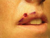
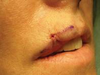
Next, uniformly place an absorbable suture in the deep dermis using a small needle, with circumferential tissue advancement to distribute the tension uniformly, explained Dr. Omlin, chief of Mohs surgery for Kaiser Permanente Napa-Solano County, Vacaville, Calif.
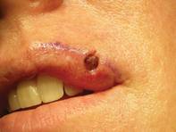
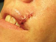
"There is a uniform stitch all the way around the perimeter" of the wound with particular attention to precisely aligning the vermilion/cutaneous lip junction, he said.
The purse string stitch creates a trestle-like framework that supports normal wound healing.
As with any reconstructions on the lower cosmetic subunits of the face, he tells patients to practice a "ventriloquist act" while healing and not talk much or move their mouths much.
One of his patients provided an excellent case-control comparison of wound closures. She initially presented with a basal cell carcinoma that intersected both the cutaneous and vermilion margins of her left upper lip.
After Mohs surgery, Dr. Omlin did a standard, complex linear closure, followed later by two pulsed-dye laser treatments. At 1-year follow-up, the patient was satisfied with an acceptable cosmetic outcome.
Six months later, she presented with a nearly identical basal cell carcinoma on the right upper lip. This time, Dr. Omlin used a purse string stitch after Mohs surgery. "It takes all of 5-10 minutes," he noted. The aesthetic result was "nearly perfect" a month later, said Dr. Omlin, also of the University of California, Davis.
On the central upper lip, "a lot of our older patients tend to have absent philtral columns or an absent Cupid's bow. Again, this is an excellent place for a purse string stitch," he said.
For patients on warfarin, the purse string stitch is great for hemostasis when repairing Mohs defects of the lip, Dr. Omlin added.
He also likes to use it for defects at the oral commissure. "Sure, you can use an elaborate cross-lip commissuroplasty or other elaborate techniques," but a simple purse string stitch reproduces the accordion-like structure of the oral commissure.
After wound granulation and healing, at 3 months it's hard to tell that a defect was ever there, he said.
In an interview after his presentation, he said he was pleased by the enthusiasm expressed by other attendees at the meeting for his simple surgical pearl.
Dr. Omlin said he has no relevant financial disclosures.
LAS VEGAS – A simple purse string stitch can provide an elegant closure of challenging lip defects after Mohs surgery.
"It's quite easy to perform. It's useful for numerous areas of the lip. It's low-risk, with low morbidity, and requires little down time," yet provides an excellent aesthetic outcome, Dr. Kenny J. Omlin said at the annual meeting of the American College of Mohs Surgery.
The keys to reconstruction using the purse string stitch start with undermining the entire surgical wound in the subdermal plane to decrease sheering forces.
Next, uniformly place an absorbable suture in the deep dermis using a small needle, with circumferential tissue advancement to distribute the tension uniformly, explained Dr. Omlin, chief of Mohs surgery for Kaiser Permanente Napa-Solano County, Vacaville, Calif.
"There is a uniform stitch all the way around the perimeter" of the wound with particular attention to precisely aligning the vermilion/cutaneous lip junction, he said.
The purse string stitch creates a trestle-like framework that supports normal wound healing.
As with any reconstructions on the lower cosmetic subunits of the face, he tells patients to practice a "ventriloquist act" while healing and not talk much or move their mouths much.
One of his patients provided an excellent case-control comparison of wound closures. She initially presented with a basal cell carcinoma that intersected both the cutaneous and vermilion margins of her left upper lip.
After Mohs surgery, Dr. Omlin did a standard, complex linear closure, followed later by two pulsed-dye laser treatments. At 1-year follow-up, the patient was satisfied with an acceptable cosmetic outcome.
Six months later, she presented with a nearly identical basal cell carcinoma on the right upper lip. This time, Dr. Omlin used a purse string stitch after Mohs surgery. "It takes all of 5-10 minutes," he noted. The aesthetic result was "nearly perfect" a month later, said Dr. Omlin, also of the University of California, Davis.
On the central upper lip, "a lot of our older patients tend to have absent philtral columns or an absent Cupid's bow. Again, this is an excellent place for a purse string stitch," he said.
For patients on warfarin, the purse string stitch is great for hemostasis when repairing Mohs defects of the lip, Dr. Omlin added.
He also likes to use it for defects at the oral commissure. "Sure, you can use an elaborate cross-lip commissuroplasty or other elaborate techniques," but a simple purse string stitch reproduces the accordion-like structure of the oral commissure.
After wound granulation and healing, at 3 months it's hard to tell that a defect was ever there, he said.
In an interview after his presentation, he said he was pleased by the enthusiasm expressed by other attendees at the meeting for his simple surgical pearl.
Dr. Omlin said he has no relevant financial disclosures.
EXPERT ANALYSIS FROM THE ANNUAL MEETING OF THE AMERICAN COLLEGE OF MOHS SURGERY