User login
Children Help Themselves by Helping Others
SAN FRANCISCO – Look around at the next black-tie fundraiser for your hospital. See any children?
Dr. Ronald Marino, director of general pediatrics at Winthrop-University Hospital, Mineola, N.Y., saw only adults at hospital benefit events. He realized that involving children in supporting his department could be a win-win-win for the participants, the patients, and the community.
He picked one of his passions – swimming – as a focus for a new event and launched an annual swim-a-thon that has raised more than $120,000 over the past 9 years. The funds benefit the hospital's Child Life Program, which gives pediatric patients the opportunity to talk at length with hospital physicians and staff about the tests and procedures they're facing in an effort to dispel some of their fears.
The money is a small part of the benefits produced by the swim-a-thon, he said.
The children who help organize and participate in the event learn ways to become involved in their community. They gain leadership and planning skills, get public speaking experience as they promote the event, and learn about health care careers from their adult co-organizers. In addition, the children design the event logo and are involved in creating publicity materials, gaining practical experience. Dr. Marino said he has seen boosts to the children's pride and self-esteem, and the focus on swimming promotes their health and well-being.
A month or so after the day of swimming, an awards celebration for all who were involved features public recognition for participants, food, music, and a clown or superhero entertainer, followed by a tour of the hospital and its pediatric services.
“Kids really love seeing what nurseries look like, what the pediatric wards look like. They love seeing where their money goes,” Dr. Marino said.
The event also has inspired some of the hospital's doctors, nurses, and staff to support children's health and development outside of the clinical setting. One of the nurse participants this year decided to mentor a child with Down syndrome separately from the swim-a-thon, to teach the child how to swim.
Swimming itself is a great equalizer, Dr. Marino added. Some children with disabilities that severely limit their motion on land become more graceful in the pool. “So often in our society we're separated from people with disabilities,” he noted.
Plus, because the usual signs of socioeconomic status are left in the locker room when swimmers shed their clothing for swimsuits, “it demonstrates that we're all the same,” Dr. Marino said. In addition to getting 95 swimmers to take part this year, targeted outreach to the community has drawn people of all ages and races to participate in many different roles.
The swim-a-thon has attracted community support from corporations, lifeguards, swim coaches, bands that play during the event, raffle donors, the medical school's pediatrics club, and many volunteers who contribute in their own ways. One teenager who didn't swim made a slideshow to promote the swim-a-thon. Another nonswimmer started his own nonprofit organization to auction off sports memorabilia and donate the proceeds to charity, including the hospital.
Swimmers are grouped in four categories by age, ranging from 4 years to more than 60 years of age. The minimal expenses of the event – for towels, pool rental, and some promotion – keep overhead low. Swimmers pay a small fee varying by age (from $3 to $10).
Physicians who want to start a similar event in their communities should pick one of their own passions as a focus, Dr. Marino advised: “For me, it was children, swimming, and community service. I put them all together.” Try to make it a grassroots effort. Cast a wide net for organizers and participants, and build relationships in the process, he said. Start small, and be patient. “You'll find that it just takes off on its own once it gets going.”
Events like the swim-a-thon help realize several aspects of the five key promises that our society should give to its children, Dr. Marino said – caring adults, safe places, a healthy start, an effective education, and opportunities to help others.
“Empowering kids strengthens our communities and ensures a brighter future,” he said.
Dr. Marino said he had no pertinent conflicts of interest.
Cast a wide net for organizers and participants. 'You'll find that it just takes off on its own once it gets going.'
Source DR. MARINO
Hospital staff Christian Burdick and Nicole Almeida, with a medical school volunteer (right), help run the swim-a-thon.
Richard Obiol and his sons Jack and Justin participated in the swim-a-thon to raise funds for the Child Life Program.
Source Photos courtesy Anne Lucas
Inhaled Drug Relieved Migraine Even if Taken Long After Onset
Major Finding: An investigational, inhaled form of dihydroergotamine was significantly more likely than placebo to relieve pain within 2 hours in patients who took treatment more than 8 hours after headache onset (92% vs. 52%, respectively) and in patients who took treatment earlier.
Data Source: Post hoc analysis of data from a randomized, double-blind trial of 771 patients who treated a single moderate to severe migraine.
Disclosures: Dr. Tepper and each of his associates in the study has been a speaker or consultant for, or received funding from, MAP Pharmaceuticals Inc., which hopes to market the inhaled formulation of dihydroergotamine.
LOS ANGELES – An experimental inhaled form of dihydroergotamine appears to be effective in reducing migraine pain even if taken as late as 8 hours or more after the start of the headache, a post hoc analysis of a phase III clinical trial suggests.
Investigators analyzed data from the randomized, double-blind, placebo-controlled trial known as the FREEDOM 301 study. Among 771 patients who treated a moderate to severe migraine and recorded both efficacy and the time from onset of headache to treatment, patients randomized to inhaled dihydroergotamine were significantly more likely than those given placebo to report being pain free 2 hours after treatment if they initiated treatment within an hour of migraine onset, 1-4 hours after onset, or 4-8 hours after migraine onset, Dr. Stewart J. Tepper and his associates reported.
Rates of freedom from pain were not significantly higher with the drug, compared with placebo, in patients who took treatment more than 8 hours after the migraine started. Reports of pain relief, however, were significantly higher in the inhaled dihydroergotamine subgroups regardless of how long after headache onset they took treatment, he said at the meeting.
In the real world, patients give a number of reasons for delaying treatment for migraines, noted Dr. Tepper of the Cleveland Clinic. Some want to be sure they have a migraine, or that they need triptan therapy. Some patients hoard medicine for cost reasons. Others just don't want to take a strong medicine if it's not needed.
Inhaled dihydroergotamine may offer an alternative for patients who delay starting migraine treatment, if the formulation wins approval, he said.
Freedom from pain at 2 hours after treatment was reported by 34% of 112 patients randomized to inhaled dihydroergotamine and 11% of 118 on placebo who took treatment within an hour of headache onset. In those who took treatment after an hour but within 4 hours of migraine onset, 18% of 152 patients on inhaled dihydroergotamine and 6% of 169 on placebo were pain free 2 hours later. Among patients who treated their migraine after 4 hours but within 8 hours of onset, 22 of 68 (32%) on inhaled dihydroergotamine and 8 of 53 (15%) on placebo were pain free 2 hours later.
For patients who started treatment more than 8 hours after migraine onset, 19 of 53 (36%) on inhaled dihydroergotamine and 9 of 46 (20%) on placebo were pain free 2 hours later. Although those rates were not significantly different, pain relief 2 hours after treatment was reported by 49 on inhaled dihydroergotamine (92%) and 24 on placebo (52%), a significant difference between groups.
“That's a very dramatic finding,” Dr. Tepper said.
Pain relief rates in patients who started treatment within an hour of migraine onset were 60% with inhaled dihydroergotamine and 35% with placebo. Among those who started treatment after an hour but within 4 hours of migraine onset, 37% on inhaled dihydroergotamine and 21% on placebo reported pain relief 2 hours later. Pain relief also occurred in 53 patients on inhaled dihydroergotamine (78%) and 30 on placebo (57%) who took treatment after 4 hours but within 8 hours of migraine onset.
Data on adverse events in 404 patients in the inhaled dihydroergotamine group and 401 in the placebo group suggest that the drug is well tolerated, Dr. Tepper said. Symptoms typically associated with triptan use, such as chest discomfort or chest pain, occurred rarely and at similar rates in both groups. There were no drug-related serious adverse events and no clinically meaningful change in lung function in this single-dose study. The most common adverse events that occurred more often with inhaled dihydroergotamine than with placebo were bad taste (in 6% and 2%, respectively), nausea (in 4% and 2%, respectively), and cough or vomiting (both in 2% and 1%, respectively).
The main FREEDOM 301 trial included 792 patients in an intent-to-treat analysis, and showed significantly increased likelihood of pain relief 2 hours after treatment in all patients on inhaled dihydroergotamine (59%), compared with patients on placebo (35%). Pain relief rates were significantly different between groups within an hour of treatment and remained significantly different after 24 and 48 hours.
“I can think of many clinical situations where this will be useful,” he said. “We hope this device will be able to be used at home.”
In Skin and Soft-Tissue Infections, Think MRSA
PASADENA, CALIF. – Clinicians should assume community-acquired skin and soft-tissue infections are due to methicillin-resistant Staphylococcus aureus infection unless proved otherwise, according to Dr. Paul D. Holtom.
For years, most hospital-associated S. aureus infections have been resistant to methicillin, and now the same has been found for community-acquired S. aureus in studies done mostly in adults, Dr. Holtom of the University of Southern California, Los Angeles, said at the meeting.
At his institution, he said, 70% of people presenting to the emergency department with skin and soft-tissue infections have community-acquired MRSA. And a multistate study of 422 patients seen in EDs for skin and soft-tissue infections found MRSA in 59%, with rates ranging from 32% to 74% in various states, except for an inexplicably low outlier rate of 15% in New York (N. Engl. J. Med. 2006;355:666-74).
Enough risk factors have been identified for community-acquired MRSA that “you might say that almost everyone is now at risk,” he said. Risk factors include intravenous drug use, men having sex with men, residence in correctional institutions, being homeless or marginally housed, various athletic sports, and postinfluenza pneumonia.
A study of 812 U.S. soldiers found that 28% had nasal colonization of methicillin-susceptible S. aureus (MSSA) and 3% had MRSA in their nares. Those colonized with MRSA, however, were significantly more likely to develop soft tissue infection – 9 of 24 soldiers (38%), compared with infections in 8 of 229 soldiers (3%) with MSSA colonization (Clin. Infect. Dis. 2004;39:971-9).
After a “very serious outbreak” of MRSA infections in 928 of 165,000 inmates in the Los Angeles County Jail in 2002, 66 inmates were hospitalized, most with skin and soft-tissue infections, and 10 had invasive disease. Subsequent implementation of preventive measures was ineffective, Dr. Holtom said. The number of MRSA infections increased to 1,849 in 2003 and 2,480 in 2004. “It's not only being spread in the jail, but it's being brought in. It is throughout the community,” he said.
There have been many reports of MRSA infections being spread among competitive athletes, including wrestlers, fencers, and collegiate football players. “This has continued to be a problem. It's not only collegiate teams but now has moved to high school teams as well,” Dr. Holtom said.
Assume that skin and soft-tissue infections are due to MRSA, he advised, and get culture and sensitivity testing if you want to understand the epidemiology in your area. When appropriate, treat with surgical drainage of the infection site. Studies suggest that adding antibiotics for patients treated with irrigation and drainage does not improve rates of healing but may help the abscesses heal faster, he said.
When treating suspected S. aureus infection with empiric antibiotics, choose carefully, he added. The infection most likely is due to MRSA, so drugs like cephalexin and dicloxacillin probably will not be effective.
“The good news is that unlike hospital-associated MRSA, community-associated MRSA is frequently sensitive to multiple old-fashioned, inexpensive drugs,” including trimethoprim/sulfamethoxazole (TMP/SMX), tetracyclines, or clindamycin, Dr. Holtom said.
TMP/SMX is not very active against Streptococcus pyogenes, the other most likely cause of skin and soft-tissue infections and abscesses, so many clinicians combine TMP/SMX with rifampin for synergistic activity against S. aureus and activity against S. pyogenes. Others use TMP/SMX and cephalexin, he said.
The tetracycline drugs doxycycline and minocycline are active against S. aureus. Clindamycin also is a popular choice, but rates of resistance are increasing. At Dr. Holtom's institution, he said, 8%-10% of S. aureus infections are now resistant to clindamycin.
Dr. Holtom reported having no disclosures or conflicts of interest.
Inhaled Drug Limits Prolonged Migraine
Major Finding: An investigational, inhaled form of dihydroergotamine was significantly more likely than was placebo to relieve pain within 2 hours in patients who took treatment more than 8 hours after headache onset (92% vs. 52%, respectively) and in patients who took treatment earlier.
Data Source: Post hoc analysis of data from a randomized, double-blind trial of 771 patients who treated a single moderate to severe migraine.
Disclosures: Dr. Tepper and each of his associates in the study has been a speaker or consultant for, or received funding from, MAP Pharmaceuticals Inc., which hopes to market the inhaled formulation of dihydroergotamine.
LOS ANGELES – An experimental inhaled form of dihydroergotamine appears to be effective in reducing migraine pain even if taken as late as 8 hours or more after the start of the headache, a post hoc analysis of a phase III clinical trial suggests.
Investigators analyzed data from the randomized, double-blind, placebo-controlled FREEDOM 301 study. Among 771 patients who treated a moderate to severe migraine and recorded both efficacy and the time from onset of headache to treatment, patients randomized to inhaled dihydroergotamine were significantly more likely than were those given placebo to report being pain -free 2 hours after treatment if they took the drug within an hour of migraine onset, 1-4 hours after onset, or 4-8 hours aftermonset, Dr. Stewart J. Tepper and his associates reported.
Rates of freedom from pain were not significantly higher with the drug compared with placebo in patients who took treatment more than 8 hours after the migraine started. Reports of pain relief, however, were significantly higher in the inhaled dihydroergotamine subgroups regardless of how long after headache onset they took treatment, he said at the meeting.
Triptans are known to provide their best relief when taken early in a migraine attack and to have reduced efficacy when treatment is delayed, said Dr. Tepper of the Cleveland Clinic. Inhaled dihydroergotamine may beoan alternative for patients who delay starting migraine treatment, if the formulation wins approval, he added.
Freedom from pain at 2 hours post treatment was reported by 34% of 112 patients randomized to inhaled dihydroergotamine and 11% of 118 on placebo who took treatment within an hour of headache onset. In those who took treatment after an hour but within 4 hours of migraine onset, 18% of 152 patients on inhaled dihydroergotamine and 6% of 169 on placebo were pain free 2 hours later. Among patients who treated theimigraine after 4 hours but within 8 hours of onset, 22 of 68 (32%) on inhaled dihydroergotamine and 8 of 53 (15%) on placebo were pain free 2 hours later.
For patients who started treatment more than 8 hours after migraine onset, 19 of 53 (36%) on inhaled dihydroergotamine and 9 of 46 (20%) on placebo were pain free 2 hours later. Although those rates were not significantly different, pain relief 2 hours after treatment was reported by 49 on inhaled dihydroergotamine (92%) and 24 on placebo (52%), a significant difference between groups.
“That's a very dramatic finding,” Dr. Tepper said.
Pain relief rates in patients who started treatment within an hour of migraine onset were 60% with inhaled dihydroergotamine and 35% with placebo. Among those who started treatment after an hour but within 4 hours of migraine onset, 37% on inhaled dihydroergotamine and 21% on placebo reported pain relief 2 hours later. Pain relief also occurred in 53 patients on inhaled dihydroergotamine (78%) and 30 on placebo (57%) who took treatment after 4 hours but within 8 hours of migraine onset.
Data on adverse events in 404 patients in the inhaled dihydroergotamine group and 401 in the placebo group suggest that the drug is well tolerated, Dr. Tepper said. Typical triptan-related symptoms such as chest discomfort or chest pain, occurred rarely and at similar rates in both groups. There were no drug-related serious adverse events and no clinically meaningful change in lung function in this single-dose study. The most common adverse events that occurred more often with inhaled dihydroergotamine than with placebo were bad taste (in 6% and 2%, respectively), nausea (4% and 2%), and cough or vomiting (both in 2% and 1%.
The main FREEDOM 301 trial included 792 patients in an intent-to-treat analysis, and showed significantly increased likelihood of pain relief 2 hours after treatment in all patients on inhaled dihydroergotamine (59%), compared with patients on placebo (35%). Pain relief rates were significantly different between groups within an hour of treatment and remained significantly different after 24 and 48 hours.
Dihydroergotamine has been used in both an oral form and as an infusion for migraine. The inhaled version may work more quickly by passing directly into the bloodstream through the lungs.
Teams Help Reduce Psychoactive Drug Doses
LONG BEACH, CALIF. – Using interdisciplinary teams in a systematic way can help nursing homes meet requirements that they stop excessive use of psychoactive medications in residents.
Three reports from the meeting showed how facilities are using interdisciplinary teams to comply with Federal Health Regulations for Long-Term Care Facilities rule 329 (F-Tag 329), which mandates that “each resident's medication regimen must be free from unnecessary drugs,” and to comply with state regulations such as California's State Operations Manual Appendix PP, which requires consideration of gradual dose reductions to avoid unnecessary medications.
At a 230-bed long-term care facility in San Antonio, an interdisciplinary team approach reduced antipsychotic use by 74%, anxiolytic use by 23%, and stimulant use by 13% within 6 months, Dr. Kunle Adedeji and associates reported in a poster presentation.
The facility had been contracting with a psychiatry group to provide mental health services and collaborate with the medical director on all psychoactive prescriptions. The new interdisciplinary team included the facility's medical director, consulting pharmacist, director of nursing, nurse manager, Minimum Data Set coordinator, two social service representatives, and the consulting psychiatry group's nurse practitioner.
The team met monthly to review the cases of all residents who were prescribed psychoactive drugs within the preceding month. Members discussed concerns about each patient, made recommendations for reducing or discontinuing medications, and made sure that each psychoactive drug prescription had a specific diagnosis linked to it.
The use of antipsychotics had been slightly higher in the facility than state and national averages before the multidisciplinary team formed. But 6 months later, usage was down to 6% of residents, far below those averages, reported Dr. Adedeji of the University of Texas Health Science Center, San Antonio.
The team's efforts were supplemented by systematic implementation of nonpharmacologic treatments such as offering residents “busy boxes” or gardening to reduce agitation. The team implemented protocols for discontinuation of “as needed” medications for anxiety and sleep management. Floor staff underwent training in managing residents with dementia and behavioral problems.
Psychoactive Drug Doses Reduced
At a 150-bed skilled nursing facility in Hendersonville, N.C., 6 months of an interdisciplinary team approach reduced use of antipsychotics by 54%, reduced anxiolytic use by 54%, reduced the use of hypnotics more than twice per week by 64%, and decreased psychiatric discharges to hospitals by 72%, Mark Coggins, Pharm.D., and his associates reported in a separate poster presentation.
The interdisciplinary team included a consultant pharmacist, nurse, social worker, dietician, therapy staff, and activity staff. They met twice weekly to discuss individual needs and interventions for residents who were on psychoactive drugs or whom staff identified as having weight loss, disruptive behaviors, pressure ulcers, or falls.
The attending physicians accepted the team's recommendations 93% of the time, said Dr. Coggins of Golden Living Centers, Inman, S.C.
The incidence of untreated depression dropped by 47%, and the proportion of residents experiencing increased symptoms of depression or anxiety fell by 10%.
Better Documentation, Better Surveys
Even without measuring outcomes, using a multidisciplinary team approach can help nursing homes stay in compliance with requirements for regular consideration of gradual dose reductions, geriatrician Jay S. Luxenberg said in an oral presentation.
At the 430-bed Jewish Home, San Francisco, where Dr. Luxenberg serves as medical director, an interdisciplinary team doing weekly “drug rounds” reviews residents on psychoactive drugs, so that each of those residents is reviewed at least every 6 months.
The team – the medical director, two psychiatrists, a nurse practitioner, a pharmacist, and usually representatives of the floor unit's social workers or nurses – assesses drug indications, documentation, consent, efficacy parameters, side effects, nonpharmacologic strategies, and previous or ongoing gradual dose reductions.
It then presents recommendations to the attending physician about whether a gradual dose reduction would be appropriate, and the attending must document agreement or give a rationale for not accepting the recommendation, according to Dr. Luxenberg.
“So far, that has helped us tremendously” in improving documentation and avoiding deficiencies on annual surveys by regulators, Dr. Luxenberg said. “Surveyers are very generous when you have documented well. We need to step away from the defense that the person is doing well, so we don't want to change anything. A lot of our physicians have that as a gut feeling. The surveyors won't buy that.”
The speakers and investigators said they have no conflicts of interest.
TDr. Jay S. Luxenberg, of the Jewish Home in San Francisco, says the team approach has helped improve documentation.
Source ©Caught in the Moment 2010. All Rights Reserved.
Use Risk Communication to Answer Parents' Environmental Questions
SAN FRANCISCO – The principles of "risk communication" can help physicians answer parents’ questions about environmental risks to health.
Dr. Maida P. Galvez employed these principles to demonstrate how she addresses a common parental question these days: Are plastics dangerous? Whether discussing plastics or other topics, it’s helpful to craft clear and concise messages in advance using straightforward language, she said at the annual meeting of the American Academy of Pediatrics.
Develop a maximum of three key messages. For each of those messages, prepare three supporting facts, advised Dr. Galvez of Mount Sinai School of Medicine’s Center for Children’s Environmental Health and Disease Prevention Research, New York.
In the three key messages, first define the exposure to the potential environmental risk, then explain what is known about potential health effects, and finally offer action items for families, she said.
Many parents have heard of potential health risks from phthalates or bisphenol A in plastics. They may ask physicians how to tell if toys contain phthalates. Are bottles with bisphenol A harmful? What health effects should they look for? What alternatives can their children use to avoid phthalates or bisphenol A?
Employing the lessons in risk communication, a physician might first define the exposure to these substances by saying that phthalates and bisphenol A are plasticizers that are added to common products because they add flexibility and durability, Dr. Galvez said.
Summarizing what is known about potential health effects, the physician might then say that concerns have been raised about the potential for health effects based on animal studies and growing evidence that the U.S. population is universally exposed to phthalates and bisphenol A.
If families want to take action, given the concerns raised by animal studies and limited human studies, they can take a precautionary approach and choose alternatives to products that may contain these plasticizers, she said.
That simple one-two-three messaging can be fleshed out with the supporting facts if there’s time and interest from the parents.
Phthalates are found in personal hygiene products like cosmetics, shampoos, fragrances, and nail polish and in food packaging, medical tubing, children’s toys, and vinyl products. Bisphenol A is found in hard plastic items like sports water bottles, baby bottles, canned foods, and dental sealants. These two plasticizers can leach from the products, exposing humans through ingestion, inhalation, or dermal absorption.
Phthalates and bisphenol A are known to have hormonal activity, and thus are often referred to as endocrine disruptors. Studies suggest that they are ubiquitous in the U.S. population, with higher exposure levels in children and adolescents than in adults.
In animal studies, exposure to these plasticizers can affect birth outcomes, especially the male reproductive tract. Human studies are assessing potential links between plasticizers and early puberty, obesity, asthma, and male infertility.
What can parents do? "Keep it simple," Dr. Galvez said. "Less is more. Especially in pregnancy, simplify your routine, and simplify the number of products you’re using."
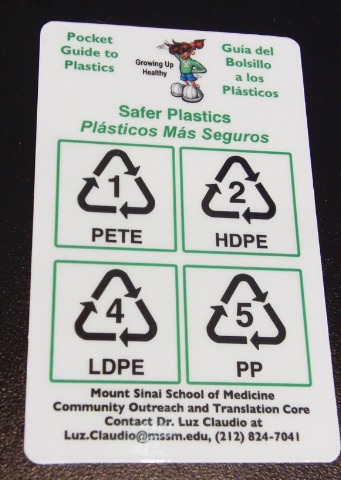
Look for products free of phthalates and bisphenol A. If labels don’t provide the needed information, look at recycling symbols: numbers 3, 6, and 7 should be avoided, but numbers 1, 2, 4, and 5 should be okay, she said. Her institution created a 2-inch by 3.5-inch"Pocket Guide to Plastics" that physicians may want to distribute to patients. Copies can be requested by e-mailing Dr. Claudio Luz at Luz.Claudio@mssm.edu. Similar fact sheets can be downloaded from the Web site of the Pediatric Environmental Health Specialty Units.
To avoid plasticizers, don’t put plastic items in the microwave or dishwasher because heat promotes leaching. When possible, choose safer alternatives to plasticizer-containing products, such as fresh fruits and vegetables instead of canned food, breast milk instead of canned infant formulas, foods in glass containers instead of plastic, and water bottles made of stainless steel, Dr. Galvez recommended.
The antiandrogen effects of phthalates syndrome can cause hypospadias, undescended testes, fetal germ cell effects, infertility, and decreased anogenital distance, according to rodent research (Int. J. Androl. 2006;29:140-7).
Bisphenol A is a weak estrogen, and in animal studies has been linked to adverse birth outcomes, effects on the male reproductive tract, early puberty, and increased body size. Limited human studies suggest it may affect neurodevelopment or liver function and may be associated with cardiovascular disease or type 2 diabetes, she said.
Dr. Galvez said that she has no pertinent conflicts of interest.
SAN FRANCISCO – The principles of "risk communication" can help physicians answer parents’ questions about environmental risks to health.
Dr. Maida P. Galvez employed these principles to demonstrate how she addresses a common parental question these days: Are plastics dangerous? Whether discussing plastics or other topics, it’s helpful to craft clear and concise messages in advance using straightforward language, she said at the annual meeting of the American Academy of Pediatrics.
Develop a maximum of three key messages. For each of those messages, prepare three supporting facts, advised Dr. Galvez of Mount Sinai School of Medicine’s Center for Children’s Environmental Health and Disease Prevention Research, New York.
In the three key messages, first define the exposure to the potential environmental risk, then explain what is known about potential health effects, and finally offer action items for families, she said.
Many parents have heard of potential health risks from phthalates or bisphenol A in plastics. They may ask physicians how to tell if toys contain phthalates. Are bottles with bisphenol A harmful? What health effects should they look for? What alternatives can their children use to avoid phthalates or bisphenol A?
Employing the lessons in risk communication, a physician might first define the exposure to these substances by saying that phthalates and bisphenol A are plasticizers that are added to common products because they add flexibility and durability, Dr. Galvez said.
Summarizing what is known about potential health effects, the physician might then say that concerns have been raised about the potential for health effects based on animal studies and growing evidence that the U.S. population is universally exposed to phthalates and bisphenol A.
If families want to take action, given the concerns raised by animal studies and limited human studies, they can take a precautionary approach and choose alternatives to products that may contain these plasticizers, she said.
That simple one-two-three messaging can be fleshed out with the supporting facts if there’s time and interest from the parents.
Phthalates are found in personal hygiene products like cosmetics, shampoos, fragrances, and nail polish and in food packaging, medical tubing, children’s toys, and vinyl products. Bisphenol A is found in hard plastic items like sports water bottles, baby bottles, canned foods, and dental sealants. These two plasticizers can leach from the products, exposing humans through ingestion, inhalation, or dermal absorption.
Phthalates and bisphenol A are known to have hormonal activity, and thus are often referred to as endocrine disruptors. Studies suggest that they are ubiquitous in the U.S. population, with higher exposure levels in children and adolescents than in adults.
In animal studies, exposure to these plasticizers can affect birth outcomes, especially the male reproductive tract. Human studies are assessing potential links between plasticizers and early puberty, obesity, asthma, and male infertility.
What can parents do? "Keep it simple," Dr. Galvez said. "Less is more. Especially in pregnancy, simplify your routine, and simplify the number of products you’re using."
Look for products free of phthalates and bisphenol A. If labels don’t provide the needed information, look at recycling symbols: numbers 3, 6, and 7 should be avoided, but numbers 1, 2, 4, and 5 should be okay, she said. Her institution created a 2-inch by 3.5-inch"Pocket Guide to Plastics" that physicians may want to distribute to patients. Copies can be requested by e-mailing Dr. Claudio Luz at Luz.Claudio@mssm.edu. Similar fact sheets can be downloaded from the Web site of the Pediatric Environmental Health Specialty Units.
To avoid plasticizers, don’t put plastic items in the microwave or dishwasher because heat promotes leaching. When possible, choose safer alternatives to plasticizer-containing products, such as fresh fruits and vegetables instead of canned food, breast milk instead of canned infant formulas, foods in glass containers instead of plastic, and water bottles made of stainless steel, Dr. Galvez recommended.
The antiandrogen effects of phthalates syndrome can cause hypospadias, undescended testes, fetal germ cell effects, infertility, and decreased anogenital distance, according to rodent research (Int. J. Androl. 2006;29:140-7).
Bisphenol A is a weak estrogen, and in animal studies has been linked to adverse birth outcomes, effects on the male reproductive tract, early puberty, and increased body size. Limited human studies suggest it may affect neurodevelopment or liver function and may be associated with cardiovascular disease or type 2 diabetes, she said.
Dr. Galvez said that she has no pertinent conflicts of interest.
Use Risk Communication to Answer Parents' Environmental Questions
SAN FRANCISCO – The principles of "risk communication" can help physicians answer parents’ questions about environmental risks to health.
Dr. Maida P. Galvez employed these principles to demonstrate how she addresses a common parental question these days: Are plastics dangerous? Whether discussing plastics or other topics, it’s helpful to craft clear and concise messages in advance using straightforward language, she said at the annual meeting of the American Academy of Pediatrics.
Develop a maximum of three key messages. For each of those messages, prepare three supporting facts, advised Dr. Galvez of Mount Sinai School of Medicine’s Center for Children’s Environmental Health and Disease Prevention Research, New York.
In the three key messages, first define the exposure to the potential environmental risk, then explain what is known about potential health effects, and finally offer action items for families, she said.
Many parents have heard of potential health risks from phthalates or bisphenol A in plastics. They may ask physicians how to tell if toys contain phthalates. Are bottles with bisphenol A harmful? What health effects should they look for? What alternatives can their children use to avoid phthalates or bisphenol A?
Employing the lessons in risk communication, a physician might first define the exposure to these substances by saying that phthalates and bisphenol A are plasticizers that are added to common products because they add flexibility and durability, Dr. Galvez said.
Summarizing what is known about potential health effects, the physician might then say that concerns have been raised about the potential for health effects based on animal studies and growing evidence that the U.S. population is universally exposed to phthalates and bisphenol A.
If families want to take action, given the concerns raised by animal studies and limited human studies, they can take a precautionary approach and choose alternatives to products that may contain these plasticizers, she said.
That simple one-two-three messaging can be fleshed out with the supporting facts if there’s time and interest from the parents.
Phthalates are found in personal hygiene products like cosmetics, shampoos, fragrances, and nail polish and in food packaging, medical tubing, children’s toys, and vinyl products. Bisphenol A is found in hard plastic items like sports water bottles, baby bottles, canned foods, and dental sealants. These two plasticizers can leach from the products, exposing humans through ingestion, inhalation, or dermal absorption.
Phthalates and bisphenol A are known to have hormonal activity, and thus are often referred to as endocrine disruptors. Studies suggest that they are ubiquitous in the U.S. population, with higher exposure levels in children and adolescents than in adults.
In animal studies, exposure to these plasticizers can affect birth outcomes, especially the male reproductive tract. Human studies are assessing potential links between plasticizers and early puberty, obesity, asthma, and male infertility.
What can parents do? "Keep it simple," Dr. Galvez said. "Less is more. Especially in pregnancy, simplify your routine, and simplify the number of products you’re using."
Look for products free of phthalates and bisphenol A. If labels don’t provide the needed information, look at recycling symbols: numbers 3, 6, and 7 should be avoided, but numbers 1, 2, 4, and 5 should be okay, she said. Her institution created a 2-inch by 3.5-inch"Pocket Guide to Plastics" that physicians may want to distribute to patients. Copies can be requested by e-mailing Dr. Claudio Luz at Luz.Claudio@mssm.edu. Similar fact sheets can be downloaded from the Web site of the Pediatric Environmental Health Specialty Units.
To avoid plasticizers, don’t put plastic items in the microwave or dishwasher because heat promotes leaching. When possible, choose safer alternatives to plasticizer-containing products, such as fresh fruits and vegetables instead of canned food, breast milk instead of canned infant formulas, foods in glass containers instead of plastic, and water bottles made of stainless steel, Dr. Galvez recommended.
The antiandrogen effects of phthalates syndrome can cause hypospadias, undescended testes, fetal germ cell effects, infertility, and decreased anogenital distance, according to rodent research (Int. J. Androl. 2006;29:140-7).
Bisphenol A is a weak estrogen, and in animal studies has been linked to adverse birth outcomes, effects on the male reproductive tract, early puberty, and increased body size. Limited human studies suggest it may affect neurodevelopment or liver function and may be associated with cardiovascular disease or type 2 diabetes, she said.
Dr. Galvez said that she has no pertinent conflicts of interest.
Criteria for Cardiac CT Updated
SAN FRANCISCO – A new report compiled by eight cardiology and imaging specialty organizations updates 4-year-old recommendations on when to use (or not use) cardiac CT imaging.
The eight societies hope that the recommendations not only will help inform clinicians and patients who are considering cardiac CT but will also guide insurers and third-party payers in setting rational reimbursement policies for cardiac CT.
The report, released by the American College of Cardiology, was endorsed by the ACC Foundation, the Society of Cardiovascular Computed Tomography, the American College of Radiology, the American Heart Association, the American Society of Echocardiography, the American Society of Nuclear Cardiology, the Society for Cardiovascular Angiography and Interventions, and the Society for Cardiovascular Magnetic Resonance.
The full report was published online Oct. 25 in the Journal of the American College of Cardiology, and is available at www.cardiosource.org. It will also be published in Circulation and in the Journal of Cardiovascular Computed Tomography.
The appropriate use criteria cover two tests: cardiac CT angiography using contrast, x-ray, or dye; and noncontrast CT scanning for calcium scoring, used to detect calcium deposits in the arteries.
In general, CT angiography was considered appropriate for diagnosis and risk assessment in patients with symptoms of possible heart disease who have a low to intermediate risk of a heart problem, or in situations where the diagnosis of heart disease is uncertain after other tests are performed.
Calcium scanning was considered appropriate in patients without heart symptoms who have an intermediate risk of heart disease, or in selected patients with low risk (especially women or younger men) with a family history of heart problems.
Cardiac CT would not be appropriate for general screening in asymptomatic patients, or in patients with known heart problems or a high risk for heart disease, or for routine repeat testing, the report concludes. Adding the test when patients have high risk for heart disease or existing heart problems does not add any useful clinical information, said Dr. Allen J. Taylor in a statement released by the ACC. Dr. Taylor is chair of the report’s writing committee and professor of medicine at Georgetown University, Washington.
The report also judged the usefulness of cardiac CT to be “uncertain” in some clinical scenarios, and the authors emphasized repeatedly that this does not mean that the test is inappropriate or that insurers should not reimburse for its use in these situations. An “uncertain” indication may require individual physician judgment and understanding of the patient to decide whether cardiac CT might help.
Tables in the report list 60 indications deemed to be appropriate, 52 rated as uncertain, and 55 indications that were considered inappropriate for cardiac CT. Clinical scenarios included acute and chronic chest pain, testing in symptomatic and asymptomatic patients, heart failure, preoperative risk assessment before either cardiac or noncardiac surgery, testing in the setting of prior test results (such as exercise testing, stress imaging procedures, or coronary calcium scores), prior revascularization, and evaluation of cardiac structure and function.
The document replaces the original 2006 criteria that were created when cardiac CT was relatively new (J. Am. Coll. Cardiol. 2006;48:1475-97).
The process that was used to create the new criteria combined evidence-based medicine and practice experience. A seven-member writing group developed clinical scenarios that were scored by a 19-member technical panel on a 1-9 scale to reflect their judgments of appropriate use of cardiac CT, inappropriate use, or uncertainty about the appropriateness of use.
In the real world, no physicians or facilities will have 100% of their cardiac CT procedure fall within the “appropriate” indications, the report notes. But if a physician or facility has a higher rate of inappropriate procedures than the national average, they may want to examine their patterns of care.
For the first time, the report considered CT angiography in patients with heart failure and normal, as well as abnormal, left ventricular ejection fraction (LVEF), with ratings of appropriate or uncertain. The only appropriate scenarios covered patients with reduced LVEF who had low or intermediate pretest probability of coronary artery disease.
CT angiography was considered a potential option as part of preoperative evaluations for patients undergoing heart surgery for noncoronary indications such as valve replacement, and was considered appropriate in patients with intermediate pretest risk for coronary artery disease, and of uncertain appropriateness if the pretest risk was low. Coronary CT angiography was never considered appropriate for evaluations before noncardiac surgery.
Imaging for evaluation of left main coronary stents was deemed appropriate, and was considered uncertain for any coronary stents measuring 3 mm in diameter or larger that had been in place at least 2 years.
The evaluation of cardiac structure and function is considered a strength of cardiac CT imaging. For the first time, the report rated cardiac CT as appropriate in patients with suspected arrhythmogenic right ventricular dysplasia, and as uncertain for evaluation of myocardial viability when other imaging modalities are inadequate or contraindicated.
Using cardiac CT before electrophysiologic procedures for anatomical mapping, or prior to repeat sternotomy in reoperative cardiac surgery, also was rated appropriate.
Disagreement among panelists over two clinical scenarios in particular left these two in the uncertain category: using cardiac CT to detect coronary artery disease in patients with a low probability of coronary artery disease when the ECG is interpretable and the patient is able to exercise, and using cardiac CT for coronary assessment prior to noncoronary cardiac surgery in patients with a low probability for coronary artery disease.
The report attempts to align its language and definitions with those in the ACC’s 2009 appropriate use criteria for cardiac radionuclide imaging (J. Am. Coll. Cardiol. 2009;53:2201-29).
Besides considering appropriate use of cardiac CT, clinicians should consider balancing the use of radiation dose–reduction techniques with the preservation of image quality, the report notes. A separate 2010 expert consensus document addressed issues of balancing those competing demands (J. Am. Coll. Cardiol. 2010;55:2663-99).
Creation of the report was funded by the American College of Cardiology Foundation and by the other professional societies. Dr. Taylor reported having no pertinent conflicts of interest. He has been a consultant to Abbott Laboratories and has received research funds form Abbott and Resverlogix Corp. Others on the writing or technical committees and a panel of reviewers involved in the report declared potential conflicts of interest that are listed in the report.
Dr. Rita F. Redberg: Cardiac CT Not Proven Useful
There aren’t any clinical trial data to suggest any benefit to use of cardiac CT in terms of clinical outcomes. The report’s authors didn’t spend a lot of time on the data. They spent a lot of time on various scenarios. There certainly are small studies that have looked at selected populations and the sensitivity and specificity of cardiac CT in those populations. And there are some clinical trials that are now starting to recruit participants. I think we’ll have results in a few years from those, but at this time, we have no outcomes data.
Cardiac CT is being used more, and so they’re trying to put out guidance for it. There are certainly a lot of CT scanners around. That does drive its use.
I’m really driven a lot more by evidence and clinical trials than by reading this report. I don’t think it will change my practice.
Any potential effect on reimbursements depends on the insurer’s criteria. A lot of insurers look for evidence as well. The ACC and the Society of Cardiovascular Computed Tomography are making a good faith effort to give guidance in this area, but I think that in the absence of data, it’s best to wait until the data are collected. There’s no big rush to start doing CT. We have ways to diagnose heart disease that work pretty well. There are no data suggesting that cardiac CT is a better way. There are risks to CT. There’s certainly a lot of radiation, there’s contrast, and dye. I think it’s prudent to wait for the data showing that this is good for patients before changing my practice.
The appropriate-use criteria is a good process, but I think that, like any process, it needs to be driven by evidence. Evidence is certainly accumulating, but we’re not there yet. I think we’re looking at another few years before we’ll really have outcomes data for cardiac CT.
Rita F. Redberg, M.D. is a professor of cardiology at the University of California, San Francisco who was not involved in the 2010 report but was involved in the 2006 report. She provided these comments in a phone interview, and said she has no pertinent conflicts of interest.
|

At our institution, we will be incorporating the new practices and sharing them with our referring physicians. We will be advocating more calcium scoring in asymptomatic intermediate-risk patients, as this indication is now considered appropriate based upon more available science and validation studies. In addition, the criteria expand to stents and bypass grafts, and this will open doors for patients and clinicians to expand their practice and increase this tool as an important part of their imaging armamentarium.
Cardiologists will continue to increase their use of cardiac CT because of the very high negative predictive power of cardiac CT, whereby a negative test effectively rules out obstructive coronary artery disease. This obviates the need in these cases for the more expensive options of both nuclear imaging and invasive angiography. Using cardiac CT first (or early) in the course of patient management has been shown to be a more cost-effective algorithm for patient treatment. Large HMOs like Kaiser are also incorporating cardiac CT into their practices, expediting cardiac work-ups with a more accurate and less expensive test.
I think this report certainly helps the case for reimbursement, since many radiology benefit managers who control approvals for certain payers (such as Blue Cross/Blue Shield) can incorporate these criteria into their approval process. These criteria are specific for different cases and presentations, so it is very pertinent to payers who can choose to pay for these specific cases.
Matthew J. Budoff, M.D., is president of the Society of Cardiovascular Computed Tomography, which helped develop the report. He is a professor of medicine at the University of California, Los Angeles, and director of cardiac CT at Harbor-UCLA Medical Center, Torrance, Calif. Dr. Budoff has been a speaker for General Electric and an expert witness in CT scanning.
|
At our institution, we will be incorporating the new practices and sharing them with our referring physicians. We will be advocating more calcium scoring in asymptomatic intermediate-risk patients, as this indication is now considered appropriate based upon more available science and validation studies. In addition, the criteria expand to stents and bypass grafts, and this will open doors for patients and clinicians to expand their practice and increase this tool as an important part of their imaging armamentarium.
Cardiologists will continue to increase their use of cardiac CT because of the very high negative predictive power of cardiac CT, whereby a negative test effectively rules out obstructive coronary artery disease. This obviates the need in these cases for the more expensive options of both nuclear imaging and invasive angiography. Using cardiac CT first (or early) in the course of patient management has been shown to be a more cost-effective algorithm for patient treatment. Large HMOs like Kaiser are also incorporating cardiac CT into their practices, expediting cardiac work-ups with a more accurate and less expensive test.
I think this report certainly helps the case for reimbursement, since many radiology benefit managers who control approvals for certain payers (such as Blue Cross/Blue Shield) can incorporate these criteria into their approval process. These criteria are specific for different cases and presentations, so it is very pertinent to payers who can choose to pay for these specific cases.
Matthew J. Budoff, M.D., is president of the Society of Cardiovascular Computed Tomography, which helped develop the report. He is a professor of medicine at the University of California, Los Angeles, and director of cardiac CT at Harbor-UCLA Medical Center, Torrance, Calif. Dr. Budoff has been a speaker for General Electric and an expert witness in CT scanning.
SAN FRANCISCO – A new report compiled by eight cardiology and imaging specialty organizations updates 4-year-old recommendations on when to use (or not use) cardiac CT imaging.
The eight societies hope that the recommendations not only will help inform clinicians and patients who are considering cardiac CT but will also guide insurers and third-party payers in setting rational reimbursement policies for cardiac CT.
The report, released by the American College of Cardiology, was endorsed by the ACC Foundation, the Society of Cardiovascular Computed Tomography, the American College of Radiology, the American Heart Association, the American Society of Echocardiography, the American Society of Nuclear Cardiology, the Society for Cardiovascular Angiography and Interventions, and the Society for Cardiovascular Magnetic Resonance.
The full report was published online Oct. 25 in the Journal of the American College of Cardiology, and is available at www.cardiosource.org. It will also be published in Circulation and in the Journal of Cardiovascular Computed Tomography.
The appropriate use criteria cover two tests: cardiac CT angiography using contrast, x-ray, or dye; and noncontrast CT scanning for calcium scoring, used to detect calcium deposits in the arteries.
In general, CT angiography was considered appropriate for diagnosis and risk assessment in patients with symptoms of possible heart disease who have a low to intermediate risk of a heart problem, or in situations where the diagnosis of heart disease is uncertain after other tests are performed.
Calcium scanning was considered appropriate in patients without heart symptoms who have an intermediate risk of heart disease, or in selected patients with low risk (especially women or younger men) with a family history of heart problems.
Cardiac CT would not be appropriate for general screening in asymptomatic patients, or in patients with known heart problems or a high risk for heart disease, or for routine repeat testing, the report concludes. Adding the test when patients have high risk for heart disease or existing heart problems does not add any useful clinical information, said Dr. Allen J. Taylor in a statement released by the ACC. Dr. Taylor is chair of the report’s writing committee and professor of medicine at Georgetown University, Washington.
The report also judged the usefulness of cardiac CT to be “uncertain” in some clinical scenarios, and the authors emphasized repeatedly that this does not mean that the test is inappropriate or that insurers should not reimburse for its use in these situations. An “uncertain” indication may require individual physician judgment and understanding of the patient to decide whether cardiac CT might help.
Tables in the report list 60 indications deemed to be appropriate, 52 rated as uncertain, and 55 indications that were considered inappropriate for cardiac CT. Clinical scenarios included acute and chronic chest pain, testing in symptomatic and asymptomatic patients, heart failure, preoperative risk assessment before either cardiac or noncardiac surgery, testing in the setting of prior test results (such as exercise testing, stress imaging procedures, or coronary calcium scores), prior revascularization, and evaluation of cardiac structure and function.
The document replaces the original 2006 criteria that were created when cardiac CT was relatively new (J. Am. Coll. Cardiol. 2006;48:1475-97).
The process that was used to create the new criteria combined evidence-based medicine and practice experience. A seven-member writing group developed clinical scenarios that were scored by a 19-member technical panel on a 1-9 scale to reflect their judgments of appropriate use of cardiac CT, inappropriate use, or uncertainty about the appropriateness of use.
In the real world, no physicians or facilities will have 100% of their cardiac CT procedure fall within the “appropriate” indications, the report notes. But if a physician or facility has a higher rate of inappropriate procedures than the national average, they may want to examine their patterns of care.
For the first time, the report considered CT angiography in patients with heart failure and normal, as well as abnormal, left ventricular ejection fraction (LVEF), with ratings of appropriate or uncertain. The only appropriate scenarios covered patients with reduced LVEF who had low or intermediate pretest probability of coronary artery disease.
CT angiography was considered a potential option as part of preoperative evaluations for patients undergoing heart surgery for noncoronary indications such as valve replacement, and was considered appropriate in patients with intermediate pretest risk for coronary artery disease, and of uncertain appropriateness if the pretest risk was low. Coronary CT angiography was never considered appropriate for evaluations before noncardiac surgery.
Imaging for evaluation of left main coronary stents was deemed appropriate, and was considered uncertain for any coronary stents measuring 3 mm in diameter or larger that had been in place at least 2 years.
The evaluation of cardiac structure and function is considered a strength of cardiac CT imaging. For the first time, the report rated cardiac CT as appropriate in patients with suspected arrhythmogenic right ventricular dysplasia, and as uncertain for evaluation of myocardial viability when other imaging modalities are inadequate or contraindicated.
Using cardiac CT before electrophysiologic procedures for anatomical mapping, or prior to repeat sternotomy in reoperative cardiac surgery, also was rated appropriate.
Disagreement among panelists over two clinical scenarios in particular left these two in the uncertain category: using cardiac CT to detect coronary artery disease in patients with a low probability of coronary artery disease when the ECG is interpretable and the patient is able to exercise, and using cardiac CT for coronary assessment prior to noncoronary cardiac surgery in patients with a low probability for coronary artery disease.
The report attempts to align its language and definitions with those in the ACC’s 2009 appropriate use criteria for cardiac radionuclide imaging (J. Am. Coll. Cardiol. 2009;53:2201-29).
Besides considering appropriate use of cardiac CT, clinicians should consider balancing the use of radiation dose–reduction techniques with the preservation of image quality, the report notes. A separate 2010 expert consensus document addressed issues of balancing those competing demands (J. Am. Coll. Cardiol. 2010;55:2663-99).
Creation of the report was funded by the American College of Cardiology Foundation and by the other professional societies. Dr. Taylor reported having no pertinent conflicts of interest. He has been a consultant to Abbott Laboratories and has received research funds form Abbott and Resverlogix Corp. Others on the writing or technical committees and a panel of reviewers involved in the report declared potential conflicts of interest that are listed in the report.
Dr. Rita F. Redberg: Cardiac CT Not Proven Useful
There aren’t any clinical trial data to suggest any benefit to use of cardiac CT in terms of clinical outcomes. The report’s authors didn’t spend a lot of time on the data. They spent a lot of time on various scenarios. There certainly are small studies that have looked at selected populations and the sensitivity and specificity of cardiac CT in those populations. And there are some clinical trials that are now starting to recruit participants. I think we’ll have results in a few years from those, but at this time, we have no outcomes data.
Cardiac CT is being used more, and so they’re trying to put out guidance for it. There are certainly a lot of CT scanners around. That does drive its use.
I’m really driven a lot more by evidence and clinical trials than by reading this report. I don’t think it will change my practice.
Any potential effect on reimbursements depends on the insurer’s criteria. A lot of insurers look for evidence as well. The ACC and the Society of Cardiovascular Computed Tomography are making a good faith effort to give guidance in this area, but I think that in the absence of data, it’s best to wait until the data are collected. There’s no big rush to start doing CT. We have ways to diagnose heart disease that work pretty well. There are no data suggesting that cardiac CT is a better way. There are risks to CT. There’s certainly a lot of radiation, there’s contrast, and dye. I think it’s prudent to wait for the data showing that this is good for patients before changing my practice.
The appropriate-use criteria is a good process, but I think that, like any process, it needs to be driven by evidence. Evidence is certainly accumulating, but we’re not there yet. I think we’re looking at another few years before we’ll really have outcomes data for cardiac CT.
Rita F. Redberg, M.D. is a professor of cardiology at the University of California, San Francisco who was not involved in the 2010 report but was involved in the 2006 report. She provided these comments in a phone interview, and said she has no pertinent conflicts of interest.
Criteria for Cardiac CT Updated
SAN FRANCISCO – A new report compiled by eight cardiology and imaging specialty organizations updates 4-year-old recommendations on when to use (or not use) cardiac CT imaging.
The eight societies hope that the recommendations not only will help inform clinicians and patients who are considering cardiac CT but will also guide insurers and third-party payers in setting rational reimbursement policies for cardiac CT.
The report, released by the American College of Cardiology, was endorsed by the ACC Foundation, the Society of Cardiovascular Computed Tomography, the American College of Radiology, the American Heart Association, the American Society of Echocardiography, the American Society of Nuclear Cardiology, the Society for Cardiovascular Angiography and Interventions, and the Society for Cardiovascular Magnetic Resonance.
The full report was published online Oct. 25 in the Journal of the American College of Cardiology, and is available at www.cardiosource.org. It will also be published in Circulation and in the Journal of Cardiovascular Computed Tomography.
The appropriate use criteria cover two tests: cardiac CT angiography using contrast, x-ray, or dye; and noncontrast CT scanning for calcium scoring, used to detect calcium deposits in the arteries.
In general, CT angiography was considered appropriate for diagnosis and risk assessment in patients with symptoms of possible heart disease who have a low to intermediate risk of a heart problem, or in situations where the diagnosis of heart disease is uncertain after other tests are performed.
Calcium scanning was considered appropriate in patients without heart symptoms who have an intermediate risk of heart disease, or in selected patients with low risk (especially women or younger men) with a family history of heart problems.
Cardiac CT would not be appropriate for general screening in asymptomatic patients, or in patients with known heart problems or a high risk for heart disease, or for routine repeat testing, the report concludes. Adding the test when patients have high risk for heart disease or existing heart problems does not add any useful clinical information, said Dr. Allen J. Taylor in a statement released by the ACC. Dr. Taylor is chair of the report’s writing committee and professor of medicine at Georgetown University, Washington.
The report also judged the usefulness of cardiac CT to be “uncertain” in some clinical scenarios, and the authors emphasized repeatedly that this does not mean that the test is inappropriate or that insurers should not reimburse for its use in these situations. An “uncertain” indication may require individual physician judgment and understanding of the patient to decide whether cardiac CT might help.
Tables in the report list 60 indications deemed to be appropriate, 52 rated as uncertain, and 55 indications that were considered inappropriate for cardiac CT. Clinical scenarios included acute and chronic chest pain, testing in symptomatic and asymptomatic patients, heart failure, preoperative risk assessment before either cardiac or noncardiac surgery, testing in the setting of prior test results (such as exercise testing, stress imaging procedures, or coronary calcium scores), prior revascularization, and evaluation of cardiac structure and function.
The document replaces the original 2006 criteria that were created when cardiac CT was relatively new (J. Am. Coll. Cardiol. 2006;48:1475-97).
The process that was used to create the new criteria combined evidence-based medicine and practice experience. A seven-member writing group developed clinical scenarios that were scored by a 19-member technical panel on a 1-9 scale to reflect their judgments of appropriate use of cardiac CT, inappropriate use, or uncertainty about the appropriateness of use.
In the real world, no physicians or facilities will have 100% of their cardiac CT procedure fall within the “appropriate” indications, the report notes. But if a physician or facility has a higher rate of inappropriate procedures than the national average, they may want to examine their patterns of care.
For the first time, the report considered CT angiography in patients with heart failure and normal, as well as abnormal, left ventricular ejection fraction (LVEF), with ratings of appropriate or uncertain. The only appropriate scenarios covered patients with reduced LVEF who had low or intermediate pretest probability of coronary artery disease.
CT angiography was considered a potential option as part of preoperative evaluations for patients undergoing heart surgery for noncoronary indications such as valve replacement, and was considered appropriate in patients with intermediate pretest risk for coronary artery disease, and of uncertain appropriateness if the pretest risk was low. Coronary CT angiography was never considered appropriate for evaluations before noncardiac surgery.
Imaging for evaluation of left main coronary stents was deemed appropriate, and was considered uncertain for any coronary stents measuring 3 mm in diameter or larger that had been in place at least 2 years.
The evaluation of cardiac structure and function is considered a strength of cardiac CT imaging. For the first time, the report rated cardiac CT as appropriate in patients with suspected arrhythmogenic right ventricular dysplasia, and as uncertain for evaluation of myocardial viability when other imaging modalities are inadequate or contraindicated.
Using cardiac CT before electrophysiologic procedures for anatomical mapping, or prior to repeat sternotomy in reoperative cardiac surgery, also was rated appropriate.
Disagreement among panelists over two clinical scenarios in particular left these two in the uncertain category: using cardiac CT to detect coronary artery disease in patients with a low probability of coronary artery disease when the ECG is interpretable and the patient is able to exercise, and using cardiac CT for coronary assessment prior to noncoronary cardiac surgery in patients with a low probability for coronary artery disease.
The report attempts to align its language and definitions with those in the ACC’s 2009 appropriate use criteria for cardiac radionuclide imaging (J. Am. Coll. Cardiol. 2009;53:2201-29).
Besides considering appropriate use of cardiac CT, clinicians should consider balancing the use of radiation dose–reduction techniques with the preservation of image quality, the report notes. A separate 2010 expert consensus document addressed issues of balancing those competing demands (J. Am. Coll. Cardiol. 2010;55:2663-99).
Creation of the report was funded by the American College of Cardiology Foundation and by the other professional societies. Dr. Taylor reported having no pertinent conflicts of interest. He has been a consultant to Abbott Laboratories and has received research funds form Abbott and Resverlogix Corp. Others on the writing or technical committees and a panel of reviewers involved in the report declared potential conflicts of interest that are listed in the report.
Dr. Rita F. Redberg: Cardiac CT Not Proven Useful
There aren’t any clinical trial data to suggest any benefit to use of cardiac CT in terms of clinical outcomes. The report’s authors didn’t spend a lot of time on the data. They spent a lot of time on various scenarios. There certainly are small studies that have looked at selected populations and the sensitivity and specificity of cardiac CT in those populations. And there are some clinical trials that are now starting to recruit participants. I think we’ll have results in a few years from those, but at this time, we have no outcomes data.
Cardiac CT is being used more, and so they’re trying to put out guidance for it. There are certainly a lot of CT scanners around. That does drive its use.
I’m really driven a lot more by evidence and clinical trials than by reading this report. I don’t think it will change my practice.
Any potential effect on reimbursements depends on the insurer’s criteria. A lot of insurers look for evidence as well. The ACC and the Society of Cardiovascular Computed Tomography are making a good faith effort to give guidance in this area, but I think that in the absence of data, it’s best to wait until the data are collected. There’s no big rush to start doing CT. We have ways to diagnose heart disease that work pretty well. There are no data suggesting that cardiac CT is a better way. There are risks to CT. There’s certainly a lot of radiation, there’s contrast, and dye. I think it’s prudent to wait for the data showing that this is good for patients before changing my practice.
The appropriate-use criteria is a good process, but I think that, like any process, it needs to be driven by evidence. Evidence is certainly accumulating, but we’re not there yet. I think we’re looking at another few years before we’ll really have outcomes data for cardiac CT.
Rita F. Redberg, M.D. is a professor of cardiology at the University of California, San Francisco who was not involved in the 2010 report but was involved in the 2006 report. She provided these comments in a phone interview, and said she has no pertinent conflicts of interest.
|
At our institution, we will be incorporating the new practices and sharing them with our referring physicians. We will be advocating more calcium scoring in asymptomatic intermediate-risk patients, as this indication is now considered appropriate based upon more available science and validation studies. In addition, the criteria expand to stents and bypass grafts, and this will open doors for patients and clinicians to expand their practice and increase this tool as an important part of their imaging armamentarium.
Cardiologists will continue to increase their use of cardiac CT because of the very high negative predictive power of cardiac CT, whereby a negative test effectively rules out obstructive coronary artery disease. This obviates the need in these cases for the more expensive options of both nuclear imaging and invasive angiography. Using cardiac CT first (or early) in the course of patient management has been shown to be a more cost-effective algorithm for patient treatment. Large HMOs like Kaiser are also incorporating cardiac CT into their practices, expediting cardiac work-ups with a more accurate and less expensive test.
I think this report certainly helps the case for reimbursement, since many radiology benefit managers who control approvals for certain payers (such as Blue Cross/Blue Shield) can incorporate these criteria into their approval process. These criteria are specific for different cases and presentations, so it is very pertinent to payers who can choose to pay for these specific cases.
Matthew J. Budoff, M.D., is president of the Society of Cardiovascular Computed Tomography, which helped develop the report. He is a professor of medicine at the University of California, Los Angeles, and director of cardiac CT at Harbor-UCLA Medical Center, Torrance, Calif. Dr. Budoff has been a speaker for General Electric and an expert witness in CT scanning.
SAN FRANCISCO – A new report compiled by eight cardiology and imaging specialty organizations updates 4-year-old recommendations on when to use (or not use) cardiac CT imaging.
The eight societies hope that the recommendations not only will help inform clinicians and patients who are considering cardiac CT but will also guide insurers and third-party payers in setting rational reimbursement policies for cardiac CT.
The report, released by the American College of Cardiology, was endorsed by the ACC Foundation, the Society of Cardiovascular Computed Tomography, the American College of Radiology, the American Heart Association, the American Society of Echocardiography, the American Society of Nuclear Cardiology, the Society for Cardiovascular Angiography and Interventions, and the Society for Cardiovascular Magnetic Resonance.
The full report was published online Oct. 25 in the Journal of the American College of Cardiology, and is available at www.cardiosource.org. It will also be published in Circulation and in the Journal of Cardiovascular Computed Tomography.
The appropriate use criteria cover two tests: cardiac CT angiography using contrast, x-ray, or dye; and noncontrast CT scanning for calcium scoring, used to detect calcium deposits in the arteries.
In general, CT angiography was considered appropriate for diagnosis and risk assessment in patients with symptoms of possible heart disease who have a low to intermediate risk of a heart problem, or in situations where the diagnosis of heart disease is uncertain after other tests are performed.
Calcium scanning was considered appropriate in patients without heart symptoms who have an intermediate risk of heart disease, or in selected patients with low risk (especially women or younger men) with a family history of heart problems.
Cardiac CT would not be appropriate for general screening in asymptomatic patients, or in patients with known heart problems or a high risk for heart disease, or for routine repeat testing, the report concludes. Adding the test when patients have high risk for heart disease or existing heart problems does not add any useful clinical information, said Dr. Allen J. Taylor in a statement released by the ACC. Dr. Taylor is chair of the report’s writing committee and professor of medicine at Georgetown University, Washington.
The report also judged the usefulness of cardiac CT to be “uncertain” in some clinical scenarios, and the authors emphasized repeatedly that this does not mean that the test is inappropriate or that insurers should not reimburse for its use in these situations. An “uncertain” indication may require individual physician judgment and understanding of the patient to decide whether cardiac CT might help.
Tables in the report list 60 indications deemed to be appropriate, 52 rated as uncertain, and 55 indications that were considered inappropriate for cardiac CT. Clinical scenarios included acute and chronic chest pain, testing in symptomatic and asymptomatic patients, heart failure, preoperative risk assessment before either cardiac or noncardiac surgery, testing in the setting of prior test results (such as exercise testing, stress imaging procedures, or coronary calcium scores), prior revascularization, and evaluation of cardiac structure and function.
The document replaces the original 2006 criteria that were created when cardiac CT was relatively new (J. Am. Coll. Cardiol. 2006;48:1475-97).
The process that was used to create the new criteria combined evidence-based medicine and practice experience. A seven-member writing group developed clinical scenarios that were scored by a 19-member technical panel on a 1-9 scale to reflect their judgments of appropriate use of cardiac CT, inappropriate use, or uncertainty about the appropriateness of use.
In the real world, no physicians or facilities will have 100% of their cardiac CT procedure fall within the “appropriate” indications, the report notes. But if a physician or facility has a higher rate of inappropriate procedures than the national average, they may want to examine their patterns of care.
For the first time, the report considered CT angiography in patients with heart failure and normal, as well as abnormal, left ventricular ejection fraction (LVEF), with ratings of appropriate or uncertain. The only appropriate scenarios covered patients with reduced LVEF who had low or intermediate pretest probability of coronary artery disease.
CT angiography was considered a potential option as part of preoperative evaluations for patients undergoing heart surgery for noncoronary indications such as valve replacement, and was considered appropriate in patients with intermediate pretest risk for coronary artery disease, and of uncertain appropriateness if the pretest risk was low. Coronary CT angiography was never considered appropriate for evaluations before noncardiac surgery.
Imaging for evaluation of left main coronary stents was deemed appropriate, and was considered uncertain for any coronary stents measuring 3 mm in diameter or larger that had been in place at least 2 years.
The evaluation of cardiac structure and function is considered a strength of cardiac CT imaging. For the first time, the report rated cardiac CT as appropriate in patients with suspected arrhythmogenic right ventricular dysplasia, and as uncertain for evaluation of myocardial viability when other imaging modalities are inadequate or contraindicated.
Using cardiac CT before electrophysiologic procedures for anatomical mapping, or prior to repeat sternotomy in reoperative cardiac surgery, also was rated appropriate.
Disagreement among panelists over two clinical scenarios in particular left these two in the uncertain category: using cardiac CT to detect coronary artery disease in patients with a low probability of coronary artery disease when the ECG is interpretable and the patient is able to exercise, and using cardiac CT for coronary assessment prior to noncoronary cardiac surgery in patients with a low probability for coronary artery disease.
The report attempts to align its language and definitions with those in the ACC’s 2009 appropriate use criteria for cardiac radionuclide imaging (J. Am. Coll. Cardiol. 2009;53:2201-29).
Besides considering appropriate use of cardiac CT, clinicians should consider balancing the use of radiation dose–reduction techniques with the preservation of image quality, the report notes. A separate 2010 expert consensus document addressed issues of balancing those competing demands (J. Am. Coll. Cardiol. 2010;55:2663-99).
Creation of the report was funded by the American College of Cardiology Foundation and by the other professional societies. Dr. Taylor reported having no pertinent conflicts of interest. He has been a consultant to Abbott Laboratories and has received research funds form Abbott and Resverlogix Corp. Others on the writing or technical committees and a panel of reviewers involved in the report declared potential conflicts of interest that are listed in the report.
Dr. Rita F. Redberg: Cardiac CT Not Proven Useful
There aren’t any clinical trial data to suggest any benefit to use of cardiac CT in terms of clinical outcomes. The report’s authors didn’t spend a lot of time on the data. They spent a lot of time on various scenarios. There certainly are small studies that have looked at selected populations and the sensitivity and specificity of cardiac CT in those populations. And there are some clinical trials that are now starting to recruit participants. I think we’ll have results in a few years from those, but at this time, we have no outcomes data.
Cardiac CT is being used more, and so they’re trying to put out guidance for it. There are certainly a lot of CT scanners around. That does drive its use.
I’m really driven a lot more by evidence and clinical trials than by reading this report. I don’t think it will change my practice.
Any potential effect on reimbursements depends on the insurer’s criteria. A lot of insurers look for evidence as well. The ACC and the Society of Cardiovascular Computed Tomography are making a good faith effort to give guidance in this area, but I think that in the absence of data, it’s best to wait until the data are collected. There’s no big rush to start doing CT. We have ways to diagnose heart disease that work pretty well. There are no data suggesting that cardiac CT is a better way. There are risks to CT. There’s certainly a lot of radiation, there’s contrast, and dye. I think it’s prudent to wait for the data showing that this is good for patients before changing my practice.
The appropriate-use criteria is a good process, but I think that, like any process, it needs to be driven by evidence. Evidence is certainly accumulating, but we’re not there yet. I think we’re looking at another few years before we’ll really have outcomes data for cardiac CT.
Rita F. Redberg, M.D. is a professor of cardiology at the University of California, San Francisco who was not involved in the 2010 report but was involved in the 2006 report. She provided these comments in a phone interview, and said she has no pertinent conflicts of interest.
Lack of Vitamin D, Ped Headache Linked
LOS ANGELES – Vitamin D deficiency was detected in 37% of 497 children and adolescents presenting to a tertiary care center for recurrent headache that required preventive treatment. Vitamin D insufficiency was found in 87%.
These results of routinely measuring baseline serum 25-hydroxyvitamin D (25[OH]D) levels at new or follow-up visits suggest that pediatric patients with recurrent headaches may be at increased risk for vitamin D insufficiency or deficiency compared with the general healthy population, Dr. Hope L. O'Brien and her associates reported in a poster presentation at the meeting.
Vitamin D deficiency was defined as a serum 25(OH)D level of less than 20 ng/mL. Vitamin D insufficiency was defined as a level below 30 ng/mL. Serum 25(OH)D levels averaged 24 ng/mL in patients presenting with episodic migraine and 23 ng/mL in those with chronic migraine, reported Dr. O'Brien of the University of Cincinnati.
A few studies have suggested a possible link between low vitamin D levels and migraine or chronic tension-type headache in adults, but this may be the first study to assess the association in children and adolescents. Patients in the current study had a mean age of 14 years (range 4-25 years).
The finding's implications are unclear, but vitamin D supplementation might help improve headaches and overall health, Dr. O'Brien said. Studies in adults have linked low vitamin D levels with various chronic medical problems such as heart disease, diabetes, cancer, autoimmune disease, chronic pain, and osteoporosis.
Dr. O'Brien did not list any disclosures and did not respond to attempts to contact her.