User login
CCJM delivers practical clinical articles relevant to internists, cardiologists, endocrinologists, and other specialists, all written by known experts.
Copyright © 2019 Cleveland Clinic. All rights reserved. The information provided is for educational purposes only. Use of this website is subject to the disclaimer and privacy policy.
gambling
compulsive behaviors
ammunition
assault rifle
black jack
Boko Haram
bondage
child abuse
cocaine
Daech
drug paraphernalia
explosion
gun
human trafficking
ISIL
ISIS
Islamic caliphate
Islamic state
mixed martial arts
MMA
molestation
national rifle association
NRA
nsfw
pedophile
pedophilia
poker
porn
pornography
psychedelic drug
recreational drug
sex slave rings
slot machine
terrorism
terrorist
Texas hold 'em
UFC
substance abuse
abuseed
abuseer
abusees
abuseing
abusely
abuses
aeolus
aeolused
aeoluser
aeoluses
aeolusing
aeolusly
aeoluss
ahole
aholeed
aholeer
aholees
aholeing
aholely
aholes
alcohol
alcoholed
alcoholer
alcoholes
alcoholing
alcoholly
alcohols
allman
allmaned
allmaner
allmanes
allmaning
allmanly
allmans
alted
altes
alting
altly
alts
analed
analer
anales
analing
anally
analprobe
analprobeed
analprobeer
analprobees
analprobeing
analprobely
analprobes
anals
anilingus
anilingused
anilinguser
anilinguses
anilingusing
anilingusly
anilinguss
anus
anused
anuser
anuses
anusing
anusly
anuss
areola
areolaed
areolaer
areolaes
areolaing
areolaly
areolas
areole
areoleed
areoleer
areolees
areoleing
areolely
areoles
arian
arianed
arianer
arianes
arianing
arianly
arians
aryan
aryaned
aryaner
aryanes
aryaning
aryanly
aryans
asiaed
asiaer
asiaes
asiaing
asialy
asias
ass
ass hole
ass lick
ass licked
ass licker
ass lickes
ass licking
ass lickly
ass licks
assbang
assbanged
assbangeded
assbangeder
assbangedes
assbangeding
assbangedly
assbangeds
assbanger
assbanges
assbanging
assbangly
assbangs
assbangsed
assbangser
assbangses
assbangsing
assbangsly
assbangss
assed
asser
asses
assesed
asseser
asseses
assesing
assesly
assess
assfuck
assfucked
assfucker
assfuckered
assfuckerer
assfuckeres
assfuckering
assfuckerly
assfuckers
assfuckes
assfucking
assfuckly
assfucks
asshat
asshated
asshater
asshates
asshating
asshatly
asshats
assholeed
assholeer
assholees
assholeing
assholely
assholes
assholesed
assholeser
assholeses
assholesing
assholesly
assholess
assing
assly
assmaster
assmastered
assmasterer
assmasteres
assmastering
assmasterly
assmasters
assmunch
assmunched
assmuncher
assmunches
assmunching
assmunchly
assmunchs
asss
asswipe
asswipeed
asswipeer
asswipees
asswipeing
asswipely
asswipes
asswipesed
asswipeser
asswipeses
asswipesing
asswipesly
asswipess
azz
azzed
azzer
azzes
azzing
azzly
azzs
babeed
babeer
babees
babeing
babely
babes
babesed
babeser
babeses
babesing
babesly
babess
ballsac
ballsaced
ballsacer
ballsaces
ballsacing
ballsack
ballsacked
ballsacker
ballsackes
ballsacking
ballsackly
ballsacks
ballsacly
ballsacs
ballsed
ballser
ballses
ballsing
ballsly
ballss
barf
barfed
barfer
barfes
barfing
barfly
barfs
bastard
bastarded
bastarder
bastardes
bastarding
bastardly
bastards
bastardsed
bastardser
bastardses
bastardsing
bastardsly
bastardss
bawdy
bawdyed
bawdyer
bawdyes
bawdying
bawdyly
bawdys
beaner
beanered
beanerer
beaneres
beanering
beanerly
beaners
beardedclam
beardedclamed
beardedclamer
beardedclames
beardedclaming
beardedclamly
beardedclams
beastiality
beastialityed
beastialityer
beastialityes
beastialitying
beastialityly
beastialitys
beatch
beatched
beatcher
beatches
beatching
beatchly
beatchs
beater
beatered
beaterer
beateres
beatering
beaterly
beaters
beered
beerer
beeres
beering
beerly
beeyotch
beeyotched
beeyotcher
beeyotches
beeyotching
beeyotchly
beeyotchs
beotch
beotched
beotcher
beotches
beotching
beotchly
beotchs
biatch
biatched
biatcher
biatches
biatching
biatchly
biatchs
big tits
big titsed
big titser
big titses
big titsing
big titsly
big titss
bigtits
bigtitsed
bigtitser
bigtitses
bigtitsing
bigtitsly
bigtitss
bimbo
bimboed
bimboer
bimboes
bimboing
bimboly
bimbos
bisexualed
bisexualer
bisexuales
bisexualing
bisexually
bisexuals
bitch
bitched
bitcheded
bitcheder
bitchedes
bitcheding
bitchedly
bitcheds
bitcher
bitches
bitchesed
bitcheser
bitcheses
bitchesing
bitchesly
bitchess
bitching
bitchly
bitchs
bitchy
bitchyed
bitchyer
bitchyes
bitchying
bitchyly
bitchys
bleached
bleacher
bleaches
bleaching
bleachly
bleachs
blow job
blow jobed
blow jober
blow jobes
blow jobing
blow jobly
blow jobs
blowed
blower
blowes
blowing
blowjob
blowjobed
blowjober
blowjobes
blowjobing
blowjobly
blowjobs
blowjobsed
blowjobser
blowjobses
blowjobsing
blowjobsly
blowjobss
blowly
blows
boink
boinked
boinker
boinkes
boinking
boinkly
boinks
bollock
bollocked
bollocker
bollockes
bollocking
bollockly
bollocks
bollocksed
bollockser
bollockses
bollocksing
bollocksly
bollockss
bollok
bolloked
bolloker
bollokes
bolloking
bollokly
bolloks
boner
bonered
bonerer
boneres
bonering
bonerly
boners
bonersed
bonerser
bonerses
bonersing
bonersly
bonerss
bong
bonged
bonger
bonges
bonging
bongly
bongs
boob
boobed
boober
boobes
boobies
boobiesed
boobieser
boobieses
boobiesing
boobiesly
boobiess
boobing
boobly
boobs
boobsed
boobser
boobses
boobsing
boobsly
boobss
booby
boobyed
boobyer
boobyes
boobying
boobyly
boobys
booger
boogered
boogerer
boogeres
boogering
boogerly
boogers
bookie
bookieed
bookieer
bookiees
bookieing
bookiely
bookies
bootee
booteeed
booteeer
booteees
booteeing
booteely
bootees
bootie
bootieed
bootieer
bootiees
bootieing
bootiely
booties
booty
bootyed
bootyer
bootyes
bootying
bootyly
bootys
boozeed
boozeer
boozees
boozeing
boozely
boozer
boozered
boozerer
boozeres
boozering
boozerly
boozers
boozes
boozy
boozyed
boozyer
boozyes
boozying
boozyly
boozys
bosomed
bosomer
bosomes
bosoming
bosomly
bosoms
bosomy
bosomyed
bosomyer
bosomyes
bosomying
bosomyly
bosomys
bugger
buggered
buggerer
buggeres
buggering
buggerly
buggers
bukkake
bukkakeed
bukkakeer
bukkakees
bukkakeing
bukkakely
bukkakes
bull shit
bull shited
bull shiter
bull shites
bull shiting
bull shitly
bull shits
bullshit
bullshited
bullshiter
bullshites
bullshiting
bullshitly
bullshits
bullshitsed
bullshitser
bullshitses
bullshitsing
bullshitsly
bullshitss
bullshitted
bullshitteded
bullshitteder
bullshittedes
bullshitteding
bullshittedly
bullshitteds
bullturds
bullturdsed
bullturdser
bullturdses
bullturdsing
bullturdsly
bullturdss
bung
bunged
bunger
bunges
bunging
bungly
bungs
busty
bustyed
bustyer
bustyes
bustying
bustyly
bustys
butt
butt fuck
butt fucked
butt fucker
butt fuckes
butt fucking
butt fuckly
butt fucks
butted
buttes
buttfuck
buttfucked
buttfucker
buttfuckered
buttfuckerer
buttfuckeres
buttfuckering
buttfuckerly
buttfuckers
buttfuckes
buttfucking
buttfuckly
buttfucks
butting
buttly
buttplug
buttpluged
buttpluger
buttpluges
buttpluging
buttplugly
buttplugs
butts
caca
cacaed
cacaer
cacaes
cacaing
cacaly
cacas
cahone
cahoneed
cahoneer
cahonees
cahoneing
cahonely
cahones
cameltoe
cameltoeed
cameltoeer
cameltoees
cameltoeing
cameltoely
cameltoes
carpetmuncher
carpetmunchered
carpetmuncherer
carpetmuncheres
carpetmunchering
carpetmuncherly
carpetmunchers
cawk
cawked
cawker
cawkes
cawking
cawkly
cawks
chinc
chinced
chincer
chinces
chincing
chincly
chincs
chincsed
chincser
chincses
chincsing
chincsly
chincss
chink
chinked
chinker
chinkes
chinking
chinkly
chinks
chode
chodeed
chodeer
chodees
chodeing
chodely
chodes
chodesed
chodeser
chodeses
chodesing
chodesly
chodess
clit
clited
cliter
clites
cliting
clitly
clitoris
clitorised
clitoriser
clitorises
clitorising
clitorisly
clitoriss
clitorus
clitorused
clitoruser
clitoruses
clitorusing
clitorusly
clitoruss
clits
clitsed
clitser
clitses
clitsing
clitsly
clitss
clitty
clittyed
clittyer
clittyes
clittying
clittyly
clittys
cocain
cocaine
cocained
cocaineed
cocaineer
cocainees
cocaineing
cocainely
cocainer
cocaines
cocaining
cocainly
cocains
cock
cock sucker
cock suckered
cock suckerer
cock suckeres
cock suckering
cock suckerly
cock suckers
cockblock
cockblocked
cockblocker
cockblockes
cockblocking
cockblockly
cockblocks
cocked
cocker
cockes
cockholster
cockholstered
cockholsterer
cockholsteres
cockholstering
cockholsterly
cockholsters
cocking
cockknocker
cockknockered
cockknockerer
cockknockeres
cockknockering
cockknockerly
cockknockers
cockly
cocks
cocksed
cockser
cockses
cocksing
cocksly
cocksmoker
cocksmokered
cocksmokerer
cocksmokeres
cocksmokering
cocksmokerly
cocksmokers
cockss
cocksucker
cocksuckered
cocksuckerer
cocksuckeres
cocksuckering
cocksuckerly
cocksuckers
coital
coitaled
coitaler
coitales
coitaling
coitally
coitals
commie
commieed
commieer
commiees
commieing
commiely
commies
condomed
condomer
condomes
condoming
condomly
condoms
coon
cooned
cooner
coones
cooning
coonly
coons
coonsed
coonser
coonses
coonsing
coonsly
coonss
corksucker
corksuckered
corksuckerer
corksuckeres
corksuckering
corksuckerly
corksuckers
cracked
crackwhore
crackwhoreed
crackwhoreer
crackwhorees
crackwhoreing
crackwhorely
crackwhores
crap
craped
craper
crapes
craping
craply
crappy
crappyed
crappyer
crappyes
crappying
crappyly
crappys
cum
cumed
cumer
cumes
cuming
cumly
cummin
cummined
cumminer
cummines
cumming
cumminged
cumminger
cumminges
cumminging
cummingly
cummings
cummining
cumminly
cummins
cums
cumshot
cumshoted
cumshoter
cumshotes
cumshoting
cumshotly
cumshots
cumshotsed
cumshotser
cumshotses
cumshotsing
cumshotsly
cumshotss
cumslut
cumsluted
cumsluter
cumslutes
cumsluting
cumslutly
cumsluts
cumstain
cumstained
cumstainer
cumstaines
cumstaining
cumstainly
cumstains
cunilingus
cunilingused
cunilinguser
cunilinguses
cunilingusing
cunilingusly
cunilinguss
cunnilingus
cunnilingused
cunnilinguser
cunnilinguses
cunnilingusing
cunnilingusly
cunnilinguss
cunny
cunnyed
cunnyer
cunnyes
cunnying
cunnyly
cunnys
cunt
cunted
cunter
cuntes
cuntface
cuntfaceed
cuntfaceer
cuntfacees
cuntfaceing
cuntfacely
cuntfaces
cunthunter
cunthuntered
cunthunterer
cunthunteres
cunthuntering
cunthunterly
cunthunters
cunting
cuntlick
cuntlicked
cuntlicker
cuntlickered
cuntlickerer
cuntlickeres
cuntlickering
cuntlickerly
cuntlickers
cuntlickes
cuntlicking
cuntlickly
cuntlicks
cuntly
cunts
cuntsed
cuntser
cuntses
cuntsing
cuntsly
cuntss
dago
dagoed
dagoer
dagoes
dagoing
dagoly
dagos
dagosed
dagoser
dagoses
dagosing
dagosly
dagoss
dammit
dammited
dammiter
dammites
dammiting
dammitly
dammits
damn
damned
damneded
damneder
damnedes
damneding
damnedly
damneds
damner
damnes
damning
damnit
damnited
damniter
damnites
damniting
damnitly
damnits
damnly
damns
dick
dickbag
dickbaged
dickbager
dickbages
dickbaging
dickbagly
dickbags
dickdipper
dickdippered
dickdipperer
dickdipperes
dickdippering
dickdipperly
dickdippers
dicked
dicker
dickes
dickface
dickfaceed
dickfaceer
dickfacees
dickfaceing
dickfacely
dickfaces
dickflipper
dickflippered
dickflipperer
dickflipperes
dickflippering
dickflipperly
dickflippers
dickhead
dickheaded
dickheader
dickheades
dickheading
dickheadly
dickheads
dickheadsed
dickheadser
dickheadses
dickheadsing
dickheadsly
dickheadss
dicking
dickish
dickished
dickisher
dickishes
dickishing
dickishly
dickishs
dickly
dickripper
dickrippered
dickripperer
dickripperes
dickrippering
dickripperly
dickrippers
dicks
dicksipper
dicksippered
dicksipperer
dicksipperes
dicksippering
dicksipperly
dicksippers
dickweed
dickweeded
dickweeder
dickweedes
dickweeding
dickweedly
dickweeds
dickwhipper
dickwhippered
dickwhipperer
dickwhipperes
dickwhippering
dickwhipperly
dickwhippers
dickzipper
dickzippered
dickzipperer
dickzipperes
dickzippering
dickzipperly
dickzippers
diddle
diddleed
diddleer
diddlees
diddleing
diddlely
diddles
dike
dikeed
dikeer
dikees
dikeing
dikely
dikes
dildo
dildoed
dildoer
dildoes
dildoing
dildoly
dildos
dildosed
dildoser
dildoses
dildosing
dildosly
dildoss
diligaf
diligafed
diligafer
diligafes
diligafing
diligafly
diligafs
dillweed
dillweeded
dillweeder
dillweedes
dillweeding
dillweedly
dillweeds
dimwit
dimwited
dimwiter
dimwites
dimwiting
dimwitly
dimwits
dingle
dingleed
dingleer
dinglees
dingleing
dinglely
dingles
dipship
dipshiped
dipshiper
dipshipes
dipshiping
dipshiply
dipships
dizzyed
dizzyer
dizzyes
dizzying
dizzyly
dizzys
doggiestyleed
doggiestyleer
doggiestylees
doggiestyleing
doggiestylely
doggiestyles
doggystyleed
doggystyleer
doggystylees
doggystyleing
doggystylely
doggystyles
dong
donged
donger
donges
donging
dongly
dongs
doofus
doofused
doofuser
doofuses
doofusing
doofusly
doofuss
doosh
dooshed
doosher
dooshes
dooshing
dooshly
dooshs
dopeyed
dopeyer
dopeyes
dopeying
dopeyly
dopeys
douchebag
douchebaged
douchebager
douchebages
douchebaging
douchebagly
douchebags
douchebagsed
douchebagser
douchebagses
douchebagsing
douchebagsly
douchebagss
doucheed
doucheer
douchees
doucheing
douchely
douches
douchey
doucheyed
doucheyer
doucheyes
doucheying
doucheyly
doucheys
drunk
drunked
drunker
drunkes
drunking
drunkly
drunks
dumass
dumassed
dumasser
dumasses
dumassing
dumassly
dumasss
dumbass
dumbassed
dumbasser
dumbasses
dumbassesed
dumbasseser
dumbasseses
dumbassesing
dumbassesly
dumbassess
dumbassing
dumbassly
dumbasss
dummy
dummyed
dummyer
dummyes
dummying
dummyly
dummys
dyke
dykeed
dykeer
dykees
dykeing
dykely
dykes
dykesed
dykeser
dykeses
dykesing
dykesly
dykess
erotic
eroticed
eroticer
erotices
eroticing
eroticly
erotics
extacy
extacyed
extacyer
extacyes
extacying
extacyly
extacys
extasy
extasyed
extasyer
extasyes
extasying
extasyly
extasys
fack
facked
facker
fackes
facking
fackly
facks
fag
faged
fager
fages
fagg
fagged
faggeded
faggeder
faggedes
faggeding
faggedly
faggeds
fagger
fagges
fagging
faggit
faggited
faggiter
faggites
faggiting
faggitly
faggits
faggly
faggot
faggoted
faggoter
faggotes
faggoting
faggotly
faggots
faggs
faging
fagly
fagot
fagoted
fagoter
fagotes
fagoting
fagotly
fagots
fags
fagsed
fagser
fagses
fagsing
fagsly
fagss
faig
faiged
faiger
faiges
faiging
faigly
faigs
faigt
faigted
faigter
faigtes
faigting
faigtly
faigts
fannybandit
fannybandited
fannybanditer
fannybandites
fannybanditing
fannybanditly
fannybandits
farted
farter
fartes
farting
fartknocker
fartknockered
fartknockerer
fartknockeres
fartknockering
fartknockerly
fartknockers
fartly
farts
felch
felched
felcher
felchered
felcherer
felcheres
felchering
felcherly
felchers
felches
felching
felchinged
felchinger
felchinges
felchinging
felchingly
felchings
felchly
felchs
fellate
fellateed
fellateer
fellatees
fellateing
fellately
fellates
fellatio
fellatioed
fellatioer
fellatioes
fellatioing
fellatioly
fellatios
feltch
feltched
feltcher
feltchered
feltcherer
feltcheres
feltchering
feltcherly
feltchers
feltches
feltching
feltchly
feltchs
feom
feomed
feomer
feomes
feoming
feomly
feoms
fisted
fisteded
fisteder
fistedes
fisteding
fistedly
fisteds
fisting
fistinged
fistinger
fistinges
fistinging
fistingly
fistings
fisty
fistyed
fistyer
fistyes
fistying
fistyly
fistys
floozy
floozyed
floozyer
floozyes
floozying
floozyly
floozys
foad
foaded
foader
foades
foading
foadly
foads
fondleed
fondleer
fondlees
fondleing
fondlely
fondles
foobar
foobared
foobarer
foobares
foobaring
foobarly
foobars
freex
freexed
freexer
freexes
freexing
freexly
freexs
frigg
frigga
friggaed
friggaer
friggaes
friggaing
friggaly
friggas
frigged
frigger
frigges
frigging
friggly
friggs
fubar
fubared
fubarer
fubares
fubaring
fubarly
fubars
fuck
fuckass
fuckassed
fuckasser
fuckasses
fuckassing
fuckassly
fuckasss
fucked
fuckeded
fuckeder
fuckedes
fuckeding
fuckedly
fuckeds
fucker
fuckered
fuckerer
fuckeres
fuckering
fuckerly
fuckers
fuckes
fuckface
fuckfaceed
fuckfaceer
fuckfacees
fuckfaceing
fuckfacely
fuckfaces
fuckin
fuckined
fuckiner
fuckines
fucking
fuckinged
fuckinger
fuckinges
fuckinging
fuckingly
fuckings
fuckining
fuckinly
fuckins
fuckly
fucknugget
fucknuggeted
fucknuggeter
fucknuggetes
fucknuggeting
fucknuggetly
fucknuggets
fucknut
fucknuted
fucknuter
fucknutes
fucknuting
fucknutly
fucknuts
fuckoff
fuckoffed
fuckoffer
fuckoffes
fuckoffing
fuckoffly
fuckoffs
fucks
fucksed
fuckser
fuckses
fucksing
fucksly
fuckss
fucktard
fucktarded
fucktarder
fucktardes
fucktarding
fucktardly
fucktards
fuckup
fuckuped
fuckuper
fuckupes
fuckuping
fuckuply
fuckups
fuckwad
fuckwaded
fuckwader
fuckwades
fuckwading
fuckwadly
fuckwads
fuckwit
fuckwited
fuckwiter
fuckwites
fuckwiting
fuckwitly
fuckwits
fudgepacker
fudgepackered
fudgepackerer
fudgepackeres
fudgepackering
fudgepackerly
fudgepackers
fuk
fuked
fuker
fukes
fuking
fukly
fuks
fvck
fvcked
fvcker
fvckes
fvcking
fvckly
fvcks
fxck
fxcked
fxcker
fxckes
fxcking
fxckly
fxcks
gae
gaeed
gaeer
gaees
gaeing
gaely
gaes
gai
gaied
gaier
gaies
gaiing
gaily
gais
ganja
ganjaed
ganjaer
ganjaes
ganjaing
ganjaly
ganjas
gayed
gayer
gayes
gaying
gayly
gays
gaysed
gayser
gayses
gaysing
gaysly
gayss
gey
geyed
geyer
geyes
geying
geyly
geys
gfc
gfced
gfcer
gfces
gfcing
gfcly
gfcs
gfy
gfyed
gfyer
gfyes
gfying
gfyly
gfys
ghay
ghayed
ghayer
ghayes
ghaying
ghayly
ghays
ghey
gheyed
gheyer
gheyes
gheying
gheyly
gheys
gigolo
gigoloed
gigoloer
gigoloes
gigoloing
gigololy
gigolos
goatse
goatseed
goatseer
goatsees
goatseing
goatsely
goatses
godamn
godamned
godamner
godamnes
godamning
godamnit
godamnited
godamniter
godamnites
godamniting
godamnitly
godamnits
godamnly
godamns
goddam
goddamed
goddamer
goddames
goddaming
goddamly
goddammit
goddammited
goddammiter
goddammites
goddammiting
goddammitly
goddammits
goddamn
goddamned
goddamner
goddamnes
goddamning
goddamnly
goddamns
goddams
goldenshower
goldenshowered
goldenshowerer
goldenshoweres
goldenshowering
goldenshowerly
goldenshowers
gonad
gonaded
gonader
gonades
gonading
gonadly
gonads
gonadsed
gonadser
gonadses
gonadsing
gonadsly
gonadss
gook
gooked
gooker
gookes
gooking
gookly
gooks
gooksed
gookser
gookses
gooksing
gooksly
gookss
gringo
gringoed
gringoer
gringoes
gringoing
gringoly
gringos
gspot
gspoted
gspoter
gspotes
gspoting
gspotly
gspots
gtfo
gtfoed
gtfoer
gtfoes
gtfoing
gtfoly
gtfos
guido
guidoed
guidoer
guidoes
guidoing
guidoly
guidos
handjob
handjobed
handjober
handjobes
handjobing
handjobly
handjobs
hard on
hard oned
hard oner
hard ones
hard oning
hard only
hard ons
hardknight
hardknighted
hardknighter
hardknightes
hardknighting
hardknightly
hardknights
hebe
hebeed
hebeer
hebees
hebeing
hebely
hebes
heeb
heebed
heeber
heebes
heebing
heebly
heebs
hell
helled
heller
helles
helling
hellly
hells
hemp
hemped
hemper
hempes
hemping
hemply
hemps
heroined
heroiner
heroines
heroining
heroinly
heroins
herp
herped
herper
herpes
herpesed
herpeser
herpeses
herpesing
herpesly
herpess
herping
herply
herps
herpy
herpyed
herpyer
herpyes
herpying
herpyly
herpys
hitler
hitlered
hitlerer
hitleres
hitlering
hitlerly
hitlers
hived
hiver
hives
hiving
hivly
hivs
hobag
hobaged
hobager
hobages
hobaging
hobagly
hobags
homey
homeyed
homeyer
homeyes
homeying
homeyly
homeys
homo
homoed
homoer
homoes
homoey
homoeyed
homoeyer
homoeyes
homoeying
homoeyly
homoeys
homoing
homoly
homos
honky
honkyed
honkyer
honkyes
honkying
honkyly
honkys
hooch
hooched
hoocher
hooches
hooching
hoochly
hoochs
hookah
hookahed
hookaher
hookahes
hookahing
hookahly
hookahs
hooker
hookered
hookerer
hookeres
hookering
hookerly
hookers
hoor
hoored
hoorer
hoores
hooring
hoorly
hoors
hootch
hootched
hootcher
hootches
hootching
hootchly
hootchs
hooter
hootered
hooterer
hooteres
hootering
hooterly
hooters
hootersed
hooterser
hooterses
hootersing
hootersly
hooterss
horny
hornyed
hornyer
hornyes
hornying
hornyly
hornys
houstoned
houstoner
houstones
houstoning
houstonly
houstons
hump
humped
humpeded
humpeder
humpedes
humpeding
humpedly
humpeds
humper
humpes
humping
humpinged
humpinger
humpinges
humpinging
humpingly
humpings
humply
humps
husbanded
husbander
husbandes
husbanding
husbandly
husbands
hussy
hussyed
hussyer
hussyes
hussying
hussyly
hussys
hymened
hymener
hymenes
hymening
hymenly
hymens
inbred
inbreded
inbreder
inbredes
inbreding
inbredly
inbreds
incest
incested
incester
incestes
incesting
incestly
incests
injun
injuned
injuner
injunes
injuning
injunly
injuns
jackass
jackassed
jackasser
jackasses
jackassing
jackassly
jackasss
jackhole
jackholeed
jackholeer
jackholees
jackholeing
jackholely
jackholes
jackoff
jackoffed
jackoffer
jackoffes
jackoffing
jackoffly
jackoffs
jap
japed
japer
japes
japing
japly
japs
japsed
japser
japses
japsing
japsly
japss
jerkoff
jerkoffed
jerkoffer
jerkoffes
jerkoffing
jerkoffly
jerkoffs
jerks
jism
jismed
jismer
jismes
jisming
jismly
jisms
jiz
jized
jizer
jizes
jizing
jizly
jizm
jizmed
jizmer
jizmes
jizming
jizmly
jizms
jizs
jizz
jizzed
jizzeded
jizzeder
jizzedes
jizzeding
jizzedly
jizzeds
jizzer
jizzes
jizzing
jizzly
jizzs
junkie
junkieed
junkieer
junkiees
junkieing
junkiely
junkies
junky
junkyed
junkyer
junkyes
junkying
junkyly
junkys
kike
kikeed
kikeer
kikees
kikeing
kikely
kikes
kikesed
kikeser
kikeses
kikesing
kikesly
kikess
killed
killer
killes
killing
killly
kills
kinky
kinkyed
kinkyer
kinkyes
kinkying
kinkyly
kinkys
kkk
kkked
kkker
kkkes
kkking
kkkly
kkks
klan
klaned
klaner
klanes
klaning
klanly
klans
knobend
knobended
knobender
knobendes
knobending
knobendly
knobends
kooch
kooched
koocher
kooches
koochesed
koocheser
koocheses
koochesing
koochesly
koochess
kooching
koochly
koochs
kootch
kootched
kootcher
kootches
kootching
kootchly
kootchs
kraut
krauted
krauter
krautes
krauting
krautly
krauts
kyke
kykeed
kykeer
kykees
kykeing
kykely
kykes
lech
leched
lecher
leches
leching
lechly
lechs
leper
lepered
leperer
leperes
lepering
leperly
lepers
lesbiansed
lesbianser
lesbianses
lesbiansing
lesbiansly
lesbianss
lesbo
lesboed
lesboer
lesboes
lesboing
lesboly
lesbos
lesbosed
lesboser
lesboses
lesbosing
lesbosly
lesboss
lez
lezbianed
lezbianer
lezbianes
lezbianing
lezbianly
lezbians
lezbiansed
lezbianser
lezbianses
lezbiansing
lezbiansly
lezbianss
lezbo
lezboed
lezboer
lezboes
lezboing
lezboly
lezbos
lezbosed
lezboser
lezboses
lezbosing
lezbosly
lezboss
lezed
lezer
lezes
lezing
lezly
lezs
lezzie
lezzieed
lezzieer
lezziees
lezzieing
lezziely
lezzies
lezziesed
lezzieser
lezzieses
lezziesing
lezziesly
lezziess
lezzy
lezzyed
lezzyer
lezzyes
lezzying
lezzyly
lezzys
lmaoed
lmaoer
lmaoes
lmaoing
lmaoly
lmaos
lmfao
lmfaoed
lmfaoer
lmfaoes
lmfaoing
lmfaoly
lmfaos
loined
loiner
loines
loining
loinly
loins
loinsed
loinser
loinses
loinsing
loinsly
loinss
lubeed
lubeer
lubees
lubeing
lubely
lubes
lusty
lustyed
lustyer
lustyes
lustying
lustyly
lustys
massa
massaed
massaer
massaes
massaing
massaly
massas
masterbate
masterbateed
masterbateer
masterbatees
masterbateing
masterbately
masterbates
masterbating
masterbatinged
masterbatinger
masterbatinges
masterbatinging
masterbatingly
masterbatings
masterbation
masterbationed
masterbationer
masterbationes
masterbationing
masterbationly
masterbations
masturbate
masturbateed
masturbateer
masturbatees
masturbateing
masturbately
masturbates
masturbating
masturbatinged
masturbatinger
masturbatinges
masturbatinging
masturbatingly
masturbatings
masturbation
masturbationed
masturbationer
masturbationes
masturbationing
masturbationly
masturbations
methed
mether
methes
mething
methly
meths
militaryed
militaryer
militaryes
militarying
militaryly
militarys
mofo
mofoed
mofoer
mofoes
mofoing
mofoly
mofos
molest
molested
molester
molestes
molesting
molestly
molests
moolie
moolieed
moolieer
mooliees
moolieing
mooliely
moolies
moron
moroned
moroner
morones
moroning
moronly
morons
motherfucka
motherfuckaed
motherfuckaer
motherfuckaes
motherfuckaing
motherfuckaly
motherfuckas
motherfucker
motherfuckered
motherfuckerer
motherfuckeres
motherfuckering
motherfuckerly
motherfuckers
motherfucking
motherfuckinged
motherfuckinger
motherfuckinges
motherfuckinging
motherfuckingly
motherfuckings
mtherfucker
mtherfuckered
mtherfuckerer
mtherfuckeres
mtherfuckering
mtherfuckerly
mtherfuckers
mthrfucker
mthrfuckered
mthrfuckerer
mthrfuckeres
mthrfuckering
mthrfuckerly
mthrfuckers
mthrfucking
mthrfuckinged
mthrfuckinger
mthrfuckinges
mthrfuckinging
mthrfuckingly
mthrfuckings
muff
muffdiver
muffdivered
muffdiverer
muffdiveres
muffdivering
muffdiverly
muffdivers
muffed
muffer
muffes
muffing
muffly
muffs
murdered
murderer
murderes
murdering
murderly
murders
muthafuckaz
muthafuckazed
muthafuckazer
muthafuckazes
muthafuckazing
muthafuckazly
muthafuckazs
muthafucker
muthafuckered
muthafuckerer
muthafuckeres
muthafuckering
muthafuckerly
muthafuckers
mutherfucker
mutherfuckered
mutherfuckerer
mutherfuckeres
mutherfuckering
mutherfuckerly
mutherfuckers
mutherfucking
mutherfuckinged
mutherfuckinger
mutherfuckinges
mutherfuckinging
mutherfuckingly
mutherfuckings
muthrfucking
muthrfuckinged
muthrfuckinger
muthrfuckinges
muthrfuckinging
muthrfuckingly
muthrfuckings
nad
naded
nader
nades
nading
nadly
nads
nadsed
nadser
nadses
nadsing
nadsly
nadss
nakeded
nakeder
nakedes
nakeding
nakedly
nakeds
napalm
napalmed
napalmer
napalmes
napalming
napalmly
napalms
nappy
nappyed
nappyer
nappyes
nappying
nappyly
nappys
nazi
nazied
nazier
nazies
naziing
nazily
nazis
nazism
nazismed
nazismer
nazismes
nazisming
nazismly
nazisms
negro
negroed
negroer
negroes
negroing
negroly
negros
nigga
niggaed
niggaer
niggaes
niggah
niggahed
niggaher
niggahes
niggahing
niggahly
niggahs
niggaing
niggaly
niggas
niggased
niggaser
niggases
niggasing
niggasly
niggass
niggaz
niggazed
niggazer
niggazes
niggazing
niggazly
niggazs
nigger
niggered
niggerer
niggeres
niggering
niggerly
niggers
niggersed
niggerser
niggerses
niggersing
niggersly
niggerss
niggle
niggleed
niggleer
nigglees
niggleing
nigglely
niggles
niglet
nigleted
nigleter
nigletes
nigleting
nigletly
niglets
nimrod
nimroded
nimroder
nimrodes
nimroding
nimrodly
nimrods
ninny
ninnyed
ninnyer
ninnyes
ninnying
ninnyly
ninnys
nooky
nookyed
nookyer
nookyes
nookying
nookyly
nookys
nuccitelli
nuccitellied
nuccitellier
nuccitellies
nuccitelliing
nuccitellily
nuccitellis
nympho
nymphoed
nymphoer
nymphoes
nymphoing
nympholy
nymphos
opium
opiumed
opiumer
opiumes
opiuming
opiumly
opiums
orgies
orgiesed
orgieser
orgieses
orgiesing
orgiesly
orgiess
orgy
orgyed
orgyer
orgyes
orgying
orgyly
orgys
paddy
paddyed
paddyer
paddyes
paddying
paddyly
paddys
paki
pakied
pakier
pakies
pakiing
pakily
pakis
pantie
pantieed
pantieer
pantiees
pantieing
pantiely
panties
pantiesed
pantieser
pantieses
pantiesing
pantiesly
pantiess
panty
pantyed
pantyer
pantyes
pantying
pantyly
pantys
pastie
pastieed
pastieer
pastiees
pastieing
pastiely
pasties
pasty
pastyed
pastyer
pastyes
pastying
pastyly
pastys
pecker
peckered
peckerer
peckeres
peckering
peckerly
peckers
pedo
pedoed
pedoer
pedoes
pedoing
pedoly
pedophile
pedophileed
pedophileer
pedophilees
pedophileing
pedophilely
pedophiles
pedophilia
pedophiliac
pedophiliaced
pedophiliacer
pedophiliaces
pedophiliacing
pedophiliacly
pedophiliacs
pedophiliaed
pedophiliaer
pedophiliaes
pedophiliaing
pedophilialy
pedophilias
pedos
penial
penialed
penialer
peniales
penialing
penially
penials
penile
penileed
penileer
penilees
penileing
penilely
peniles
penis
penised
peniser
penises
penising
penisly
peniss
perversion
perversioned
perversioner
perversiones
perversioning
perversionly
perversions
peyote
peyoteed
peyoteer
peyotees
peyoteing
peyotely
peyotes
phuck
phucked
phucker
phuckes
phucking
phuckly
phucks
pillowbiter
pillowbitered
pillowbiterer
pillowbiteres
pillowbitering
pillowbiterly
pillowbiters
pimp
pimped
pimper
pimpes
pimping
pimply
pimps
pinko
pinkoed
pinkoer
pinkoes
pinkoing
pinkoly
pinkos
pissed
pisseded
pisseder
pissedes
pisseding
pissedly
pisseds
pisser
pisses
pissing
pissly
pissoff
pissoffed
pissoffer
pissoffes
pissoffing
pissoffly
pissoffs
pisss
polack
polacked
polacker
polackes
polacking
polackly
polacks
pollock
pollocked
pollocker
pollockes
pollocking
pollockly
pollocks
poon
pooned
pooner
poones
pooning
poonly
poons
poontang
poontanged
poontanger
poontanges
poontanging
poontangly
poontangs
porn
porned
porner
pornes
porning
pornly
porno
pornoed
pornoer
pornoes
pornography
pornographyed
pornographyer
pornographyes
pornographying
pornographyly
pornographys
pornoing
pornoly
pornos
porns
prick
pricked
pricker
prickes
pricking
prickly
pricks
prig
priged
priger
priges
priging
prigly
prigs
prostitute
prostituteed
prostituteer
prostitutees
prostituteing
prostitutely
prostitutes
prude
prudeed
prudeer
prudees
prudeing
prudely
prudes
punkass
punkassed
punkasser
punkasses
punkassing
punkassly
punkasss
punky
punkyed
punkyer
punkyes
punkying
punkyly
punkys
puss
pussed
pusser
pusses
pussies
pussiesed
pussieser
pussieses
pussiesing
pussiesly
pussiess
pussing
pussly
pusss
pussy
pussyed
pussyer
pussyes
pussying
pussyly
pussypounder
pussypoundered
pussypounderer
pussypounderes
pussypoundering
pussypounderly
pussypounders
pussys
puto
putoed
putoer
putoes
putoing
putoly
putos
queaf
queafed
queafer
queafes
queafing
queafly
queafs
queef
queefed
queefer
queefes
queefing
queefly
queefs
queer
queered
queerer
queeres
queering
queerly
queero
queeroed
queeroer
queeroes
queeroing
queeroly
queeros
queers
queersed
queerser
queerses
queersing
queersly
queerss
quicky
quickyed
quickyer
quickyes
quickying
quickyly
quickys
quim
quimed
quimer
quimes
quiming
quimly
quims
racy
racyed
racyer
racyes
racying
racyly
racys
rape
raped
rapeded
rapeder
rapedes
rapeding
rapedly
rapeds
rapeed
rapeer
rapees
rapeing
rapely
raper
rapered
raperer
raperes
rapering
raperly
rapers
rapes
rapist
rapisted
rapister
rapistes
rapisting
rapistly
rapists
raunch
raunched
rauncher
raunches
raunching
raunchly
raunchs
rectus
rectused
rectuser
rectuses
rectusing
rectusly
rectuss
reefer
reefered
reeferer
reeferes
reefering
reeferly
reefers
reetard
reetarded
reetarder
reetardes
reetarding
reetardly
reetards
reich
reiched
reicher
reiches
reiching
reichly
reichs
retard
retarded
retardeded
retardeder
retardedes
retardeding
retardedly
retardeds
retarder
retardes
retarding
retardly
retards
rimjob
rimjobed
rimjober
rimjobes
rimjobing
rimjobly
rimjobs
ritard
ritarded
ritarder
ritardes
ritarding
ritardly
ritards
rtard
rtarded
rtarder
rtardes
rtarding
rtardly
rtards
rum
rumed
rumer
rumes
ruming
rumly
rump
rumped
rumper
rumpes
rumping
rumply
rumprammer
rumprammered
rumprammerer
rumprammeres
rumprammering
rumprammerly
rumprammers
rumps
rums
ruski
ruskied
ruskier
ruskies
ruskiing
ruskily
ruskis
sadism
sadismed
sadismer
sadismes
sadisming
sadismly
sadisms
sadist
sadisted
sadister
sadistes
sadisting
sadistly
sadists
scag
scaged
scager
scages
scaging
scagly
scags
scantily
scantilyed
scantilyer
scantilyes
scantilying
scantilyly
scantilys
schlong
schlonged
schlonger
schlonges
schlonging
schlongly
schlongs
scrog
scroged
scroger
scroges
scroging
scrogly
scrogs
scrot
scrote
scroted
scroteed
scroteer
scrotees
scroteing
scrotely
scroter
scrotes
scroting
scrotly
scrots
scrotum
scrotumed
scrotumer
scrotumes
scrotuming
scrotumly
scrotums
scrud
scruded
scruder
scrudes
scruding
scrudly
scruds
scum
scumed
scumer
scumes
scuming
scumly
scums
seaman
seamaned
seamaner
seamanes
seamaning
seamanly
seamans
seamen
seamened
seamener
seamenes
seamening
seamenly
seamens
seduceed
seduceer
seducees
seduceing
seducely
seduces
semen
semened
semener
semenes
semening
semenly
semens
shamedame
shamedameed
shamedameer
shamedamees
shamedameing
shamedamely
shamedames
shit
shite
shiteater
shiteatered
shiteaterer
shiteateres
shiteatering
shiteaterly
shiteaters
shited
shiteed
shiteer
shitees
shiteing
shitely
shiter
shites
shitface
shitfaceed
shitfaceer
shitfacees
shitfaceing
shitfacely
shitfaces
shithead
shitheaded
shitheader
shitheades
shitheading
shitheadly
shitheads
shithole
shitholeed
shitholeer
shitholees
shitholeing
shitholely
shitholes
shithouse
shithouseed
shithouseer
shithousees
shithouseing
shithousely
shithouses
shiting
shitly
shits
shitsed
shitser
shitses
shitsing
shitsly
shitss
shitt
shitted
shitteded
shitteder
shittedes
shitteding
shittedly
shitteds
shitter
shittered
shitterer
shitteres
shittering
shitterly
shitters
shittes
shitting
shittly
shitts
shitty
shittyed
shittyer
shittyes
shittying
shittyly
shittys
shiz
shized
shizer
shizes
shizing
shizly
shizs
shooted
shooter
shootes
shooting
shootly
shoots
sissy
sissyed
sissyer
sissyes
sissying
sissyly
sissys
skag
skaged
skager
skages
skaging
skagly
skags
skank
skanked
skanker
skankes
skanking
skankly
skanks
slave
slaveed
slaveer
slavees
slaveing
slavely
slaves
sleaze
sleazeed
sleazeer
sleazees
sleazeing
sleazely
sleazes
sleazy
sleazyed
sleazyer
sleazyes
sleazying
sleazyly
sleazys
slut
slutdumper
slutdumpered
slutdumperer
slutdumperes
slutdumpering
slutdumperly
slutdumpers
sluted
sluter
slutes
sluting
slutkiss
slutkissed
slutkisser
slutkisses
slutkissing
slutkissly
slutkisss
slutly
sluts
slutsed
slutser
slutses
slutsing
slutsly
slutss
smegma
smegmaed
smegmaer
smegmaes
smegmaing
smegmaly
smegmas
smut
smuted
smuter
smutes
smuting
smutly
smuts
smutty
smuttyed
smuttyer
smuttyes
smuttying
smuttyly
smuttys
snatch
snatched
snatcher
snatches
snatching
snatchly
snatchs
sniper
snipered
sniperer
sniperes
snipering
sniperly
snipers
snort
snorted
snorter
snortes
snorting
snortly
snorts
snuff
snuffed
snuffer
snuffes
snuffing
snuffly
snuffs
sodom
sodomed
sodomer
sodomes
sodoming
sodomly
sodoms
spic
spiced
spicer
spices
spicing
spick
spicked
spicker
spickes
spicking
spickly
spicks
spicly
spics
spik
spoof
spoofed
spoofer
spoofes
spoofing
spoofly
spoofs
spooge
spoogeed
spoogeer
spoogees
spoogeing
spoogely
spooges
spunk
spunked
spunker
spunkes
spunking
spunkly
spunks
steamyed
steamyer
steamyes
steamying
steamyly
steamys
stfu
stfued
stfuer
stfues
stfuing
stfuly
stfus
stiffy
stiffyed
stiffyer
stiffyes
stiffying
stiffyly
stiffys
stoneded
stoneder
stonedes
stoneding
stonedly
stoneds
stupided
stupider
stupides
stupiding
stupidly
stupids
suckeded
suckeder
suckedes
suckeding
suckedly
suckeds
sucker
suckes
sucking
suckinged
suckinger
suckinges
suckinging
suckingly
suckings
suckly
sucks
sumofabiatch
sumofabiatched
sumofabiatcher
sumofabiatches
sumofabiatching
sumofabiatchly
sumofabiatchs
tard
tarded
tarder
tardes
tarding
tardly
tards
tawdry
tawdryed
tawdryer
tawdryes
tawdrying
tawdryly
tawdrys
teabagging
teabagginged
teabagginger
teabagginges
teabagginging
teabaggingly
teabaggings
terd
terded
terder
terdes
terding
terdly
terds
teste
testee
testeed
testeeed
testeeer
testeees
testeeing
testeely
testeer
testees
testeing
testely
testes
testesed
testeser
testeses
testesing
testesly
testess
testicle
testicleed
testicleer
testiclees
testicleing
testiclely
testicles
testis
testised
testiser
testises
testising
testisly
testiss
thrusted
thruster
thrustes
thrusting
thrustly
thrusts
thug
thuged
thuger
thuges
thuging
thugly
thugs
tinkle
tinkleed
tinkleer
tinklees
tinkleing
tinklely
tinkles
tit
tited
titer
tites
titfuck
titfucked
titfucker
titfuckes
titfucking
titfuckly
titfucks
titi
titied
titier
tities
titiing
titily
titing
titis
titly
tits
titsed
titser
titses
titsing
titsly
titss
tittiefucker
tittiefuckered
tittiefuckerer
tittiefuckeres
tittiefuckering
tittiefuckerly
tittiefuckers
titties
tittiesed
tittieser
tittieses
tittiesing
tittiesly
tittiess
titty
tittyed
tittyer
tittyes
tittyfuck
tittyfucked
tittyfucker
tittyfuckered
tittyfuckerer
tittyfuckeres
tittyfuckering
tittyfuckerly
tittyfuckers
tittyfuckes
tittyfucking
tittyfuckly
tittyfucks
tittying
tittyly
tittys
toke
tokeed
tokeer
tokees
tokeing
tokely
tokes
toots
tootsed
tootser
tootses
tootsing
tootsly
tootss
tramp
tramped
tramper
trampes
tramping
tramply
tramps
transsexualed
transsexualer
transsexuales
transsexualing
transsexually
transsexuals
trashy
trashyed
trashyer
trashyes
trashying
trashyly
trashys
tubgirl
tubgirled
tubgirler
tubgirles
tubgirling
tubgirlly
tubgirls
turd
turded
turder
turdes
turding
turdly
turds
tush
tushed
tusher
tushes
tushing
tushly
tushs
twat
twated
twater
twates
twating
twatly
twats
twatsed
twatser
twatses
twatsing
twatsly
twatss
undies
undiesed
undieser
undieses
undiesing
undiesly
undiess
unweded
unweder
unwedes
unweding
unwedly
unweds
uzi
uzied
uzier
uzies
uziing
uzily
uzis
vag
vaged
vager
vages
vaging
vagly
vags
valium
valiumed
valiumer
valiumes
valiuming
valiumly
valiums
venous
virgined
virginer
virgines
virgining
virginly
virgins
vixen
vixened
vixener
vixenes
vixening
vixenly
vixens
vodkaed
vodkaer
vodkaes
vodkaing
vodkaly
vodkas
voyeur
voyeured
voyeurer
voyeures
voyeuring
voyeurly
voyeurs
vulgar
vulgared
vulgarer
vulgares
vulgaring
vulgarly
vulgars
wang
wanged
wanger
wanges
wanging
wangly
wangs
wank
wanked
wanker
wankered
wankerer
wankeres
wankering
wankerly
wankers
wankes
wanking
wankly
wanks
wazoo
wazooed
wazooer
wazooes
wazooing
wazooly
wazoos
wedgie
wedgieed
wedgieer
wedgiees
wedgieing
wedgiely
wedgies
weeded
weeder
weedes
weeding
weedly
weeds
weenie
weenieed
weenieer
weeniees
weenieing
weeniely
weenies
weewee
weeweeed
weeweeer
weeweees
weeweeing
weeweely
weewees
weiner
weinered
weinerer
weineres
weinering
weinerly
weiners
weirdo
weirdoed
weirdoer
weirdoes
weirdoing
weirdoly
weirdos
wench
wenched
wencher
wenches
wenching
wenchly
wenchs
wetback
wetbacked
wetbacker
wetbackes
wetbacking
wetbackly
wetbacks
whitey
whiteyed
whiteyer
whiteyes
whiteying
whiteyly
whiteys
whiz
whized
whizer
whizes
whizing
whizly
whizs
whoralicious
whoralicioused
whoraliciouser
whoraliciouses
whoraliciousing
whoraliciously
whoraliciouss
whore
whorealicious
whorealicioused
whorealiciouser
whorealiciouses
whorealiciousing
whorealiciously
whorealiciouss
whored
whoreded
whoreder
whoredes
whoreding
whoredly
whoreds
whoreed
whoreer
whorees
whoreface
whorefaceed
whorefaceer
whorefacees
whorefaceing
whorefacely
whorefaces
whorehopper
whorehoppered
whorehopperer
whorehopperes
whorehoppering
whorehopperly
whorehoppers
whorehouse
whorehouseed
whorehouseer
whorehousees
whorehouseing
whorehousely
whorehouses
whoreing
whorely
whores
whoresed
whoreser
whoreses
whoresing
whoresly
whoress
whoring
whoringed
whoringer
whoringes
whoringing
whoringly
whorings
wigger
wiggered
wiggerer
wiggeres
wiggering
wiggerly
wiggers
woody
woodyed
woodyer
woodyes
woodying
woodyly
woodys
wop
woped
woper
wopes
woping
woply
wops
wtf
wtfed
wtfer
wtfes
wtfing
wtfly
wtfs
xxx
xxxed
xxxer
xxxes
xxxing
xxxly
xxxs
yeasty
yeastyed
yeastyer
yeastyes
yeastying
yeastyly
yeastys
yobbo
yobboed
yobboer
yobboes
yobboing
yobboly
yobbos
zoophile
zoophileed
zoophileer
zoophilees
zoophileing
zoophilely
zoophiles
anal
ass
ass lick
balls
ballsac
bisexual
bleach
causas
cheap
cost of miracles
cunt
display network stats
fart
fda and death
fda AND warn
fda AND warning
fda AND warns
feom
fuck
gfc
humira AND expensive
illegal
madvocate
masturbation
nuccitelli
overdose
porn
shit
snort
texarkana
direct\-acting antivirals
assistance
ombitasvir
support path
harvoni
abbvie
direct-acting antivirals
paritaprevir
advocacy
ledipasvir
vpak
ritonavir with dasabuvir
program
gilead
greedy
financial
needy
fake-ovir
viekira pak
v pak
sofosbuvir
support
oasis
discount
dasabuvir
protest
ritonavir
section[contains(@class, 'nav-hidden')]
footer[@id='footer']
div[contains(@class, 'pane-pub-article-cleveland-clinic')]
div[contains(@class, 'pane-pub-home-cleveland-clinic')]
div[contains(@class, 'pane-pub-topic-cleveland-clinic')]
div[contains(@class, 'panel-panel-inner')]
div[contains(@class, 'pane-node-field-article-topics')]
section[contains(@class, 'footer-nav-section-wrapper')]
The role of adjuvant chemotherapy in early-stage and locally advanced non–small cell lung cancer
Despite surgery, 40% to 75% of patients with stage I to IIIA non–small cell lung cancer (NSCLC) will die within 5 years. After multiple trials showed no survival advantage to chemotherapy in the adjuvant setting for the treatment of locally advanced NSCLC, the first hint of benefit came in 1995 with the publication of a meta-analysis of 14 clinical trials, which showed a nonsignificant 5% improvement in 5-year survival with chemotherapy after surgery.1
A second meta-analysis, this one conducted by the Lung Adjuvant Cisplatin Evaluation (LACE) Collaborative Group, demonstrated a hazard ratio (HR) of 0.89 (P = .005) on the end point of overall survival with the use of postoperative cisplatin-based chemotherapy in patients with NSCLC; this translates to a 5-year absolute improvement of 5.4% from chemotherapy.2 The survival benefit was confined to patients with stage II and stage III disease.
Post hoc exploratory subgroup analyses of the Cancer and Leukemia Group B (CALGB) 96333 and Adjuvant Navelbine International Trialist Association (ANITA)4 trials revealed a significant survival benefit to four cycles of cisplatin-based adjuvant chemotherapy in patients with stage Ib disease who had tumors 4 cm or larger.
BIOMARKERS
Prognostic and predictive biomarkers beyond cancer stage are needed, as only 10% to 15% of patients with resected NSCLC who receive chemotherapy derive a benefit. Predictive markers can be used to guide therapeutic decision-making, and prognostic markers permit estimation of patient outcome independent of treatment modality.
As with ERCC1, expression of the DNA mismatch repair protein mutS homolog 2 (MSH2)7 is both prognostic and predictive after surgery. In a separate biomarker analysis from the IALT study, approximately two-thirds of patients with NSCLC had MSH2-negative tumors by immunohistochemistry, indicating lack of expression of MSH2 in tumors. Patients with expression of MSH2, who have intact mismatch repair, had a better prognosis and benefitted less from systemic chemotherapy than those with an absence of MSH2 expression.8
Individually, ERCC1 and MSH2 have similar power in predicting benefit from adjuvant chemotherapy in NSCLC; the HR for death was similar in patients with low expression of either gene.8 The two biomarkers combined, however, were more powerful than either alone in their ability to predict a survival advantage with chemotherapy.8 In an evaluation of 658 patients with NSCLC for whom both biomarkers were available, patients who expressed low tumor levels of both ERCC1 and MSH2 had an HR for death that was 35% lower with adjuvant chemotherapy compared with surgery alone after median follow-up of 7.5 years; the presence of two positive biomarkers was associated with an increase in the HR for death by 32%. Validation of these findings in a phase 3 setting will be necessary before these biomarkers can be used in the clinical setting.
The Southwest Oncology Group (SWOG) is conducting a trial (SWOG 0720) in patients with stage I NSCLC to determine whether a subset based on ERCC1 and ribonucleotide reductase M1 (RRM1) status will derive benefit from adjuvant therapy with gemcitabine together with cisplatin. Ribonucleotide reductase subunit 1 is the regulatory subunit of ribonuclease reductase, which is an enzyme that catalyzes the deoxynucleotide production required for DNA repair.
Two other clinical trials, under way but not completed, testing various forms of chemotherapy and targeted therapy based on ERCC1 and epidermal growth factor receptor (EGFR) mutation status are the Tailored Post-Surgical Therapy in Early Stage NSCLC (TASTE) and the International Tailored Chemotherapy Adjuvant (ITACA) trials. The TASTE trial is comparing standard chemotherapy (cisplatin plus pemetrexed) with customized adjuvant treatment based on EGFR and ERCC1 status in patients with stage II or IIIa nonsquamous NSCLC. The ITACA trial is a phase 3 study of pemetrexed, cisplatin, and radiotherapy determined by thymidylate synthase (TS) and ERCC1 gene expression levels in patients with stage II to III completely resected NSCLC. TS is an enzyme responsible for maintaining intracellular levels of thymidine, important for DNA synthesis and repair, and may serve as a predictor of response to pemetrexed.
GENE EXPRESSION PROFILING
Gene expression profiles, already used to predict benefit from chemotherapy in early-stage breast cancer, may inform treatment decisions in lung cancer as well. A15-gene signature that could predict risk of recurrence and death after surgery alone for stage Ib or II NSCLC was identified using fresh frozen tissue of patients from the National Cancer Institute of Canada (NCIC) Clinical Trials Group (CTG) JBR.10 trial of vinorelbine/cisplatin.9 The risk profile was subsequently validated with reverse transcriptase-quantitative polymerase chain reaction (RT-qPCR) in the same cases and in four independent sets of patients.9
Among those patients with stage Ib disease, the gene expression profile was both prognostic (HR of 13.22 for disease-specific survival in the high- vs low-risk population) and predictive (HR of 0.44 for the use of adjuvant chemotherapy in the high-risk patients but no survival benefit observed with chemotherapy in low-risk patients).9
USE OF BIOMARKERS TO SELECT TREATMENT
As alluded to earlier, the use of biomarker expression to guide treatment selection is an area of intense investigation. In the metastatic setting, therapy targeted to the EGFR mutation has proven to be remarkably beneficial in patients with EGFR-activating mutations. In the adjuvant setting, the NCIC CTG BR.19 trial enrolled an unselected population of patients with completely resected stage Ib to IIIa NSCLC; the patients were randomized to 2 years of treatment with the tyrosine kinase inhibitor gefitinib, which targets EGFR, or placebo. Tissue samples from trial participants were collected and revealed KRAS mutation in 27%, a high EGFR gene copy number by fluorescence in situ hybridization (FISH) in 41%, and an activating EGFR mutation in 21%.
The NCIC CTG BR.19 trial was greatly underpowered because enrollment was stopped at 503 patients when, in 2008, the SWOG 0023 investigators reported a worse overall median survival with maintenance gefitinib after definitive chemoradiation in patients with stage III NSCLC.10 As a result of the early termination of patient accrual, the median duration of adjuvant gefitinib in NCIC CTG BR.19 was less than 5 months. Further, only 20% were exposed to chemotherapy and only 21% of the final study population had an EGFR mutation. In the overall study population, the HR for overall survival among gefitinib recipients was 1.23, indicating harm, and there was a trend in favor of placebo on the end point of disease-free survival. Neither KRAS nor EGFR copy number was predictive or prognostic, and EGFR mutation status was not prognostic.11 Patients with wild-type EGFR had a trend toward detriment with maintenance gefitinib that was similar to that of the overall population, and those with EGFR mutation experienced no benefit with maintenance gefitinib.
In the Randomized Double-Blind Trial in Adjuvant NSCLC with Tarceva (RADIANT), patients with resected stage I to IIIa NSCLC, with the option for postoperative chemotherapy, were assessed for EGFR expression by immunohistochemistry or FISH and then randomized to erlotinib or placebo for 2 years. The trial completed accrual in 2010 and results are expected in 4 to 5 years.
Cleveland Clinic is currently accruing patients for a phase 2 trial of patients with resected stage I to IIIa NSCLC. All patients will have their tumors screened for activating EGFR mutations; those with activating mutations will receive adjuvant erlotinib for 2 years. starting within 6 months of surgery.
SUMMARY
Although adjuvant chemotherapy has been well established for patients with early-stage NSCLC, stage alone is not an ideal biomarker to predict the utility of chemotherapy, as the vast majority of patients derive no benefit from chemotherapy.
Biomarkers have been poorly validated and therefore are inappropriate for clinical use at this time. Validation of gene arrays has been disappointingly slow in lung cancer because of the absence of large tumor banks that are available in breast cancer and colon cancer.
It remains unclear whether targeted therapies improve outcomes over traditional chemotherapy in the adjuvant setting in NSCLC, as tumors in the metastatic and adjuvant settings are not the same.
- Non-Small-Cell Lung Cancer Collaborative Group. Chemotherapy in non-small cell lung cancer: a meta-analysis using updated data on individual patients from 52 randomised clinical trials. BMJ 1995; 311:899–909.
- Pignon J-P, Tribodet H, Scagliotti GV, et al. Lung adjuvant cisplatin evaluation: a pooled analysis by the LACE Collaborative Group. J Clin Oncol 2008; 26:3552–3559.
- Strauss GM, Herndon JE, Maddaus ME, et al. Adjuvant paclitaxel plus carboplatin compared with observation in stage IB non–small-cell lung cancer: CALGB 9633 with the Cancer and Leukemia Group B, Radiation Therapy Oncology Group, and North Central Cancer Treatment Group Study Groups. J Clin Oncol 2008; 26:5043–5051.
- Douillard JY, Rosell R, De Lena M, et al. Adjuvant vinorelbine plus cisplatin versus observation in patients with completely resected stage IB-IIIA non-small-cell lung cancer (Adjuvant Navelbine International Trialist Association [ANITA]): a randomised controlled trial. Lancet Oncol 2006; 7:719–727.
- Gazdar AF. DNA repair and survival in lung cancer—the two faces of Janus. N Engl J Med 2007; 356:771–773.
- Olaussen KA, Dunant A, Fouret P, et al. DNA repair by ERCC1 in non-small-cell lung cancer and cisplatin-based adjuvant chemotherapy. N Engl J Med 2006; 355:983–991.
- Vilar E, Gruber SB. Microsatellite instability in colorectal cancer—the stable evidence. Nat Rev Clin Oncol 2010; 7:153–162.
- Fouret P, Planchard D, Mendiboure J, et al. MSH2 and adjuvant cisplatin-based chemotherapy in non-small cell lung cancer [ASCO abstract CRA7502]. J Clin Oncol 2009; 27 (suppl).
- Zhu C-Q, Ding K, Strumpf D, et al. Prognostic and predictive gene signature for adjuvant chemotherapy in resected non-small-cell lung cancer. J Clin Oncol 2010; 28:4417–4424.
- Kelly K, Chansky K, Gaspar LE, et al. Updated analysis of SWOG 0023: a randomized phase III trial of gefitinib versus placebo maintenance after definitive chemoradiation followed by docetaxel in patients with locally advanced stage III nonsmall cell lung cancer [ASCO abstract 7513]. J Clin Oncol 2007; 25 (suppl).
- Goss GD, Lorimer I, Taso S, et al. A phase III randomized, double-blind, placebo-controlled trial of the epidermal growth factor inhibitor gefitinib in completely resected stage IB-IIIA non-small cell lung cancer (NSCLC): NCIC CTG BR.19 [ASCO abstract LBA7005]. J Clin Oncol 2010; 28 (suppl).
Despite surgery, 40% to 75% of patients with stage I to IIIA non–small cell lung cancer (NSCLC) will die within 5 years. After multiple trials showed no survival advantage to chemotherapy in the adjuvant setting for the treatment of locally advanced NSCLC, the first hint of benefit came in 1995 with the publication of a meta-analysis of 14 clinical trials, which showed a nonsignificant 5% improvement in 5-year survival with chemotherapy after surgery.1
A second meta-analysis, this one conducted by the Lung Adjuvant Cisplatin Evaluation (LACE) Collaborative Group, demonstrated a hazard ratio (HR) of 0.89 (P = .005) on the end point of overall survival with the use of postoperative cisplatin-based chemotherapy in patients with NSCLC; this translates to a 5-year absolute improvement of 5.4% from chemotherapy.2 The survival benefit was confined to patients with stage II and stage III disease.
Post hoc exploratory subgroup analyses of the Cancer and Leukemia Group B (CALGB) 96333 and Adjuvant Navelbine International Trialist Association (ANITA)4 trials revealed a significant survival benefit to four cycles of cisplatin-based adjuvant chemotherapy in patients with stage Ib disease who had tumors 4 cm or larger.
BIOMARKERS
Prognostic and predictive biomarkers beyond cancer stage are needed, as only 10% to 15% of patients with resected NSCLC who receive chemotherapy derive a benefit. Predictive markers can be used to guide therapeutic decision-making, and prognostic markers permit estimation of patient outcome independent of treatment modality.
As with ERCC1, expression of the DNA mismatch repair protein mutS homolog 2 (MSH2)7 is both prognostic and predictive after surgery. In a separate biomarker analysis from the IALT study, approximately two-thirds of patients with NSCLC had MSH2-negative tumors by immunohistochemistry, indicating lack of expression of MSH2 in tumors. Patients with expression of MSH2, who have intact mismatch repair, had a better prognosis and benefitted less from systemic chemotherapy than those with an absence of MSH2 expression.8
Individually, ERCC1 and MSH2 have similar power in predicting benefit from adjuvant chemotherapy in NSCLC; the HR for death was similar in patients with low expression of either gene.8 The two biomarkers combined, however, were more powerful than either alone in their ability to predict a survival advantage with chemotherapy.8 In an evaluation of 658 patients with NSCLC for whom both biomarkers were available, patients who expressed low tumor levels of both ERCC1 and MSH2 had an HR for death that was 35% lower with adjuvant chemotherapy compared with surgery alone after median follow-up of 7.5 years; the presence of two positive biomarkers was associated with an increase in the HR for death by 32%. Validation of these findings in a phase 3 setting will be necessary before these biomarkers can be used in the clinical setting.
The Southwest Oncology Group (SWOG) is conducting a trial (SWOG 0720) in patients with stage I NSCLC to determine whether a subset based on ERCC1 and ribonucleotide reductase M1 (RRM1) status will derive benefit from adjuvant therapy with gemcitabine together with cisplatin. Ribonucleotide reductase subunit 1 is the regulatory subunit of ribonuclease reductase, which is an enzyme that catalyzes the deoxynucleotide production required for DNA repair.
Two other clinical trials, under way but not completed, testing various forms of chemotherapy and targeted therapy based on ERCC1 and epidermal growth factor receptor (EGFR) mutation status are the Tailored Post-Surgical Therapy in Early Stage NSCLC (TASTE) and the International Tailored Chemotherapy Adjuvant (ITACA) trials. The TASTE trial is comparing standard chemotherapy (cisplatin plus pemetrexed) with customized adjuvant treatment based on EGFR and ERCC1 status in patients with stage II or IIIa nonsquamous NSCLC. The ITACA trial is a phase 3 study of pemetrexed, cisplatin, and radiotherapy determined by thymidylate synthase (TS) and ERCC1 gene expression levels in patients with stage II to III completely resected NSCLC. TS is an enzyme responsible for maintaining intracellular levels of thymidine, important for DNA synthesis and repair, and may serve as a predictor of response to pemetrexed.
GENE EXPRESSION PROFILING
Gene expression profiles, already used to predict benefit from chemotherapy in early-stage breast cancer, may inform treatment decisions in lung cancer as well. A15-gene signature that could predict risk of recurrence and death after surgery alone for stage Ib or II NSCLC was identified using fresh frozen tissue of patients from the National Cancer Institute of Canada (NCIC) Clinical Trials Group (CTG) JBR.10 trial of vinorelbine/cisplatin.9 The risk profile was subsequently validated with reverse transcriptase-quantitative polymerase chain reaction (RT-qPCR) in the same cases and in four independent sets of patients.9
Among those patients with stage Ib disease, the gene expression profile was both prognostic (HR of 13.22 for disease-specific survival in the high- vs low-risk population) and predictive (HR of 0.44 for the use of adjuvant chemotherapy in the high-risk patients but no survival benefit observed with chemotherapy in low-risk patients).9
USE OF BIOMARKERS TO SELECT TREATMENT
As alluded to earlier, the use of biomarker expression to guide treatment selection is an area of intense investigation. In the metastatic setting, therapy targeted to the EGFR mutation has proven to be remarkably beneficial in patients with EGFR-activating mutations. In the adjuvant setting, the NCIC CTG BR.19 trial enrolled an unselected population of patients with completely resected stage Ib to IIIa NSCLC; the patients were randomized to 2 years of treatment with the tyrosine kinase inhibitor gefitinib, which targets EGFR, or placebo. Tissue samples from trial participants were collected and revealed KRAS mutation in 27%, a high EGFR gene copy number by fluorescence in situ hybridization (FISH) in 41%, and an activating EGFR mutation in 21%.
The NCIC CTG BR.19 trial was greatly underpowered because enrollment was stopped at 503 patients when, in 2008, the SWOG 0023 investigators reported a worse overall median survival with maintenance gefitinib after definitive chemoradiation in patients with stage III NSCLC.10 As a result of the early termination of patient accrual, the median duration of adjuvant gefitinib in NCIC CTG BR.19 was less than 5 months. Further, only 20% were exposed to chemotherapy and only 21% of the final study population had an EGFR mutation. In the overall study population, the HR for overall survival among gefitinib recipients was 1.23, indicating harm, and there was a trend in favor of placebo on the end point of disease-free survival. Neither KRAS nor EGFR copy number was predictive or prognostic, and EGFR mutation status was not prognostic.11 Patients with wild-type EGFR had a trend toward detriment with maintenance gefitinib that was similar to that of the overall population, and those with EGFR mutation experienced no benefit with maintenance gefitinib.
In the Randomized Double-Blind Trial in Adjuvant NSCLC with Tarceva (RADIANT), patients with resected stage I to IIIa NSCLC, with the option for postoperative chemotherapy, were assessed for EGFR expression by immunohistochemistry or FISH and then randomized to erlotinib or placebo for 2 years. The trial completed accrual in 2010 and results are expected in 4 to 5 years.
Cleveland Clinic is currently accruing patients for a phase 2 trial of patients with resected stage I to IIIa NSCLC. All patients will have their tumors screened for activating EGFR mutations; those with activating mutations will receive adjuvant erlotinib for 2 years. starting within 6 months of surgery.
SUMMARY
Although adjuvant chemotherapy has been well established for patients with early-stage NSCLC, stage alone is not an ideal biomarker to predict the utility of chemotherapy, as the vast majority of patients derive no benefit from chemotherapy.
Biomarkers have been poorly validated and therefore are inappropriate for clinical use at this time. Validation of gene arrays has been disappointingly slow in lung cancer because of the absence of large tumor banks that are available in breast cancer and colon cancer.
It remains unclear whether targeted therapies improve outcomes over traditional chemotherapy in the adjuvant setting in NSCLC, as tumors in the metastatic and adjuvant settings are not the same.
- Non-Small-Cell Lung Cancer Collaborative Group. Chemotherapy in non-small cell lung cancer: a meta-analysis using updated data on individual patients from 52 randomised clinical trials. BMJ 1995; 311:899–909.
- Pignon J-P, Tribodet H, Scagliotti GV, et al. Lung adjuvant cisplatin evaluation: a pooled analysis by the LACE Collaborative Group. J Clin Oncol 2008; 26:3552–3559.
- Strauss GM, Herndon JE, Maddaus ME, et al. Adjuvant paclitaxel plus carboplatin compared with observation in stage IB non–small-cell lung cancer: CALGB 9633 with the Cancer and Leukemia Group B, Radiation Therapy Oncology Group, and North Central Cancer Treatment Group Study Groups. J Clin Oncol 2008; 26:5043–5051.
- Douillard JY, Rosell R, De Lena M, et al. Adjuvant vinorelbine plus cisplatin versus observation in patients with completely resected stage IB-IIIA non-small-cell lung cancer (Adjuvant Navelbine International Trialist Association [ANITA]): a randomised controlled trial. Lancet Oncol 2006; 7:719–727.
- Gazdar AF. DNA repair and survival in lung cancer—the two faces of Janus. N Engl J Med 2007; 356:771–773.
- Olaussen KA, Dunant A, Fouret P, et al. DNA repair by ERCC1 in non-small-cell lung cancer and cisplatin-based adjuvant chemotherapy. N Engl J Med 2006; 355:983–991.
- Vilar E, Gruber SB. Microsatellite instability in colorectal cancer—the stable evidence. Nat Rev Clin Oncol 2010; 7:153–162.
- Fouret P, Planchard D, Mendiboure J, et al. MSH2 and adjuvant cisplatin-based chemotherapy in non-small cell lung cancer [ASCO abstract CRA7502]. J Clin Oncol 2009; 27 (suppl).
- Zhu C-Q, Ding K, Strumpf D, et al. Prognostic and predictive gene signature for adjuvant chemotherapy in resected non-small-cell lung cancer. J Clin Oncol 2010; 28:4417–4424.
- Kelly K, Chansky K, Gaspar LE, et al. Updated analysis of SWOG 0023: a randomized phase III trial of gefitinib versus placebo maintenance after definitive chemoradiation followed by docetaxel in patients with locally advanced stage III nonsmall cell lung cancer [ASCO abstract 7513]. J Clin Oncol 2007; 25 (suppl).
- Goss GD, Lorimer I, Taso S, et al. A phase III randomized, double-blind, placebo-controlled trial of the epidermal growth factor inhibitor gefitinib in completely resected stage IB-IIIA non-small cell lung cancer (NSCLC): NCIC CTG BR.19 [ASCO abstract LBA7005]. J Clin Oncol 2010; 28 (suppl).
Selection of chemotherapy for patients with advanced non–small cell lung cancer
Despite enthusiasm for the use of molecular testing and molecularly targeted agents in patients with advanced non–small cell lung cancer (NSCLC), most patients are not candidates for upfront treatment with molecular agents. Chemotherapy therefore remains the backbone of treatment for this patient population.
This article presents the best available evidence for selection of chemotherapy for patients with advanced NSCLC and examines the controversy surrounding maintenance therapy.
EVOLUTION OF CHEMOTHERAPY IN NSCLC
The first evidence that chemotherapy produced a significant survival benefit in patients with advanced NSCLC came in 1995 when a meta-analysis showed that platinum-based chemotherapy conferred a 2-month improvement in median survival over best supportive care.1
This finding led to a decade of randomized phase 3 clinical trials that compared different platinum-based regimens. The quintessential trial in this regard was Eastern Cooperative Oncology Group (ECOG) 1594, in which three platinum doublets were compared with cisplatin and paclitaxel on the end point of overall survival (OS) in patients with advanced NSCLC. No significant differences were found among the regimens tested.2
Bevacizumab adds to platinum doublet in nonsquamous NSCLC
With the introduction of bevacizumab, an antibody against vascular endothelial growth factor, knowledge of NSCLC histology became important. In the ECOG 4599 trial, published in 2006, bevacizumab added to platinum doublet chemotherapy in patients with advanced nonsquamous NSCLC significantly improved the response rate, progression-free survival (PFS), and median OS compared with platinum doublet chemotherapy alone.3 This trial was limited to patients with nonsquamous NSCLC because its predecessor trial had revealed an excess of life-threatening pulmonary hemorrhage in association with bevacizumab in patients with squamous cell carcinoma.
Pemetrexed superior to docetaxel in nonsquamous histology
In 2004, Hanna et al4 demonstrated pemetrexed to be noninferior to docetaxel on efficacy outcomes as second-line therapy in advanced NSCLC. Pemetrexed had a significantly better toxicity profile, however, which led to its approval for this indication. Post hoc analyses of this trial suggested a differential effect of pemetrexed based on histology. In the pemetrexed arm, patients with nonsquamous histology appeared to have superior survival compared with patients who had squamous histology, whereas in the docetaxel arm, histology did not affect outcome.5 Further, the nonsquamous histologic subgroup had superior OS with pemetrexed compared with docetaxel.6
Pemetrexed and gemcitabine perform differently based on histology
A phase 3 trial with a noninferiority design compared cisplatin/pemetrexed with cisplatin/gemcitabine as first-line therapy on OS. The noninferiority criteria were met, with no difference in median OS between the two groups (median OS: 10.3 months in both arms).7
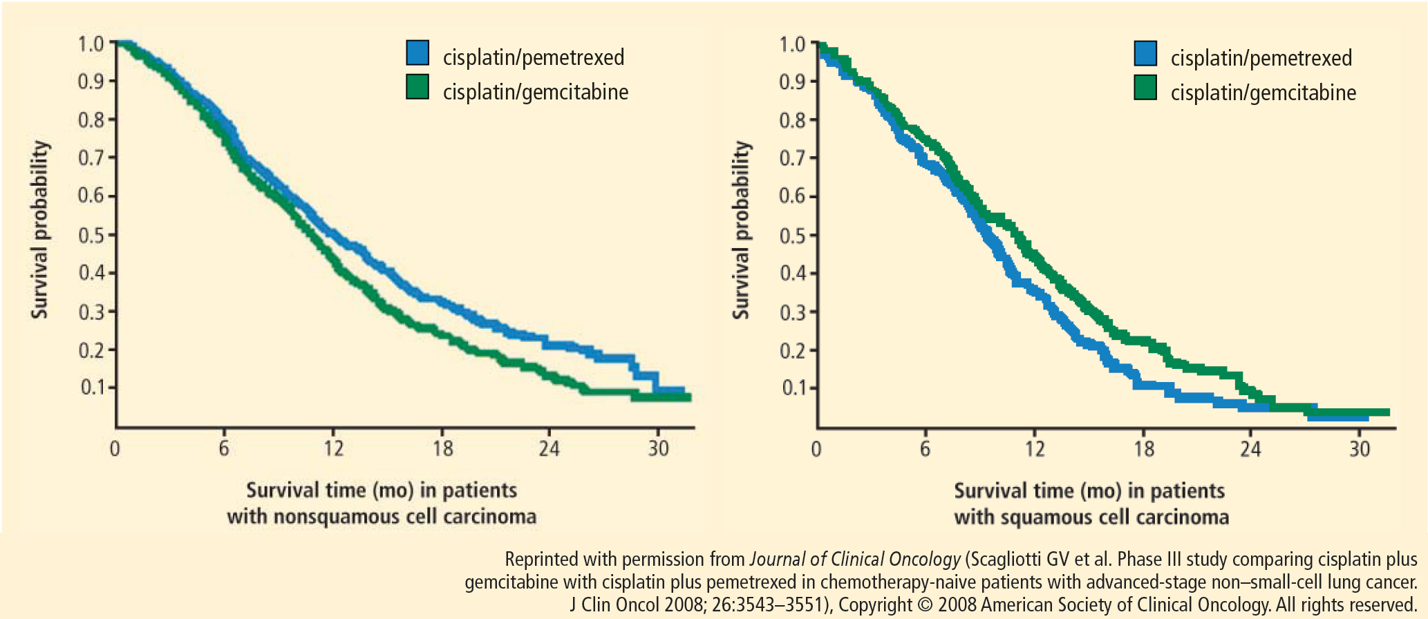
Based on this evidence, NSCLC is no longer an adequate pathologic diagnosis. Pathologists must differentiate squamous from nonsquamous histology to take full advantage of the safety of angiogenesis inhibitors and the efficacy of pemetrexed.
OPTIMAL FIRST-LINE REGIMEN FOR NONSQUAMOUS NSCLC
Both cisplatin/pemetrexed and carboplatin/paclitaxel plus bevacizumab have level 1 evidence to support their use as first-line treatment of NSCLC with nonsquamous histology. Carboplatin/pemetrexed/bevacizumab is also being used in the community despite the absence of randomized trial evidence to support its use for this indication.
A single-arm phase 2 trial of carboplatin/pemetrexed/bevacizumab followed by maintenance pemetrexed/bevacizumab in 49 patients produced a response rate of 55%, PFS of 7.8 months, and OS of 14 months.10 Although the results are impressive, they should be considered hypothesis-generating rather than treatment-changing in light of the small number of patients enrolled and the single-arm design.
An open-label randomized phase 3 trial, Point-Break, is comparing two regimens in patients who have advanced nonsquamous NSCLC: (1) carboplatin/pemetrexed/bevacizumab followed by maintenance pemetrexed/bevacizumab and (2) carboplatin/paclitaxel/bevacizumab followed by bevacizumab.11 Although potentially practice-changing, PointBreak will not answer whether bevacizumab adds benefit to cisplatin and pemetrexed, nor will it determine which first-line regimen is superior because of the different maintenance regimens.
SQUAMOUS NSCLC: PLATINUM DOUBLET OPTIMAL
No agents are currently approved specifically for the treatment of squamous cell carcinoma, which appears to have a high level of expression of insulin-like growth factor receptor (IGF-1R). A 64% response rate observed with an IGF-1R antagonist added to paclitaxel/carboplatin in patients with NSCLC of squamous cell histology in a phase 2 trial led to the design of a phase 3 trial in which patients were randomized to carboplatin/paclitaxel with or without the IGF-1R antagonist figitumumab. The trial ended prematurely in 2009 because of an imbalance of deaths in the experimental arm.12 As expected, hyperglycemia was more common in the experimental arm. Unexpectedly, the incidences of grade 5 infections and cardiovascular events were also significantly higher in the experimental arm.
A randomized phase 3 trial of carboplatin/paclitaxel compared with carboplatin and nanoparticle albumin-bound (nab) paclitaxel was conducted in 1,052 patients with stage IIIb/IV NSCLC with the primary end point being overall response rate (ORR).13 The rationale for substituting nab-paclitaxel for paclitaxel was that paclitaxel is dissolved in polyoxyethylated castor oil. This decreases the efficacy of paclitaxel and contributes to its toxicities, including hypersensitivity reactions and neuropathy. In metastatic breast cancer, nab-paclitaxel was shown to be more efficacious than solvent-based paclitaxel.14
The nab-paclitaxel trial met its primary end point of superior response rate: 33% in the nab-paclitaxel arm versus 25% in the standard paclitaxel arm. However, on final analysis there was no difference in PFS or OS between the two arms, making the difference in ORR of little clinical significance. No hypersensitivity reactions occurred in the nab-paclitaxel arm, while three occurred in the paclitaxel arm. Grade 3 sensory neuropathy occurred significantly less often in the group assigned to nab-paclitaxel compared with paclitaxel (3 vs 10, respectively; P < .001). Although there appears to be no efficacy advantage of nab-paclitaxel over standard paclitaxel for advanced NSCLC patients, use of nab-paclitaxel might be considered in patients with stage IV NSCLC who have poorly controlled diabetes or who already suffer significant neuropathy.
Although not a prespecified end point, the response rate in patients with squamous cell histology nearly doubled among those treated with nab-paclitaxel compared with standard paclitaxel. However, this did not translate into significant differences in PFS or OS in this subgroup, and the use of nab-paclitaxel in this patient population specifically is not advised.
In light of the data, the standard treatment for squamous NSCLC remains a platinum doublet other than pemetrexed. A phase 3 clinical trial, ECLIPSE, is currently enrolling patients at the Cleveland Clinic. The trial will randomize chemotherapy-naïve patients with stage IIIb/IV NSCLC and a Karnofsky performance status of 0 or 1 to receive carboplatin and gemcitabine with or without the polyadenosine diphosphate–ribose polymerase inhibitor iniparib.
MAINTENANCE THERAPY
The utility of maintenance therapy—the uninterrupted continuation of therapy for patients who do not progress after completing first-line chemotherapy—in patients with advanced NSCLC is controversial. Two kinds of maintenance therapy have emerged.
Switch maintenance, also known as early second-line therapy, is so termed because patients are immediately switched to a second-line agent different from the first-line doublet therapy.
Continuation maintenance is the continuation of one or more drugs from the induction regimen, the best example being continuation of single-agent evacizumab in the ECOG 4599 regimen.
Five major trials of successful maintenance therapy for nonprogressors after first-line chemotherapy have been presented over the past 4 years, and these have led to new indications for the maintenance drugs.8,15–18 PFS in favor of active maintenance has been documented in trials of early versus delayed maintenance docetaxel,15 pemetrexed versus placebo,8 erlotinib versus placebo,16 bevacizumab/erlotinib versus bevacizumab/placebo (ATLAS trial),17 and gemcitabine or erlotinib versus placebo (IFCT-GFPC 0502).18 The magnitude of improved PFS associated with each treatment has been similar. As a result, improved of PFS with maintenance therapy is now widely accepted.
To improve survival: maintenance therapy or better second-line therapy?
The utility of maintenance therapy can be confounded by the lack of a predefined second-line treatment. Some argue that the benefit to maintenance may also be realized by appropriate use of the same agent as salvage therapy in the case of disease progression.
Overall survival improved with maintenance therapy in only two trials; one compared pemetrexed with placebo and the other compared erlotinib with placebo.8,16 The validity of these findings, however, remains in question. In the trial of pemetrexed, only 19% of the patients in the control arm ever received pemetrexed, and in the erlotinib trial, only 21% of the patients in the control arm ever received erlotinib after progression. Whether maintenance therapy was responsible for an improvement in OS or whether ineffective second-line therapy dampened survival in patients in the control arms is unknown.
In the IFCT-GFPC 0502 phase 3 study, patients received four cycles of cisplatin/gemcitabine.19 If patients did not progress, they were randomized to observation, continuation maintenance with gemcitabine, or switch maintenance to erlotinib. Predefined second-line therapy in all arms was pemetrexed. The primary end point chosen was PFS, even though maintenance therapy had already been established to extend PFS. The median PFS in the gemcitabine maintenance arm was 3.8 months, compared with 1.9 months in the observation arm. The hazard ratio (HR) of 0.55 (P < .0001) was similar to that observed with pemetrexed in the maintenance trial noted above.19 Subgroup analysis showed improvement in PFS with the gemcitabine maintenance group compared with observation in all subgroups, including those based on histology. The HR in the erlotinib maintenance arm was 0.82 (P = .002), similar to that observed with erlotinib in the Sequential Tarceva in Unresectable NSCLC (SATURN) trial.16
In the IFCT-GFPC 0502 study, 60.4% of patients in the gemcitabine arm, 63.2 % of those in the erlotinib arm, and 76.1% in the observation arm received postmaintenance treatment with pemetrexed.19 Nearly one-half (49.6%) of patients in the observation arm received third-line treatment with erlotinib.
In the overall study population, a trend toward improved OS was observed with maintenance gemcitabine or erlotinib compared with observation, but did not achieve statistical significance, possibly because the trial lacked adequate power to detect a difference on this end point.
A subgroup analysis of patients who received second-line pemetrexed showed a significant improvement in OS in the maintenance arms compared with the observation arm. About 25% of the patients never advanced to second-line pemetrexed. Because patients who never received second-line pemetrexed may represent the sickest patients, the relevance of this finding to the overall population of patients with stage IV NSCLC is unknown.
Ongoing maintenance trials
Two ongoing clinical trials of maintenance are exploring bevacizumab with or without pemetrexed as maintenance following first-line cisplatin/pemetrexed/bevacizumab (AVAPERL) and bevacizumab alone, pemetrexed alone, or a combination of the two as maintenance following first-line therapy with carboplatin/paclitaxel/bevacizumab (Eastern Cooperative Oncology Group). Preliminary results from the AVAPERL study were reported recently and support the use of pemetrexed and bevacizumab as maintenance therapy compared with bevacizumab alone.20
CONCLUSIONS ABOUT MAINTENANCE THERAPY
Pemetrexed and erlotinib significantly prolong OS survival compared with placebo when used as maintenance therapy in advanced NSCLC patients who do not progress after four cycles of first-line chemotherapy. Whether this improvement in OS can be attributed to maintenance therapy or more effective second-line therapy is open to debate.
Maintenance chemotherapy should be discussed with all patients whose tumors do not progress after four cycles of first-line chemotherapy. The use of maintenance therapy may be most reasonable in very symptomatic patients who receive palliative benefit from chemotherapy, or as a means of encouraging noncompliant patients to return for care.
- Non-small Cell Lung Cancer Collaborative Group. Chemotherapy in non-small cell lung cancer: a meta-analysis using updated data on individual patients from 52 randomised clinical trials. BMJ 1995; 311:899–909.
- Schiller JH, Harrington D, Belani C, et al. Comparison of four chemotherapy regimens for advanced non-small-cell lung cancer. N Engl J Med 2002; 346:92–98.
- Sandler A, Gray R, Perry MC, et al. Paclitaxel-carboplatin alone or with bevacizumab for non-small-cell lung cancer. N Engl J Med 2006; 355:2542–2450.
- Hanna N, Shepherd FA, Fossella FV, et al. Randomized phase III trial of pemetrexed versus docetaxel in patients with non-small-cell lung cancer previously treated with chemotherapy. J Clin Oncol 2004; 22:1589–1597.
- Peterson P, Park K, Fossella F, Gatzmeier U, John W, Scagliotti G. Is pemetrexed more effective in adenocarcinoma and large cell lung cancer than in squamous cell carcinoma? A retrospective analysis of a phase III trial of pemetrexed vs docetaxel in previously treated patients with advanced non-small cell lung cancer (NSCLC) [12th World Congress on Lung Cancer abstract P2-328]. J Thorac Oncol 2007; 2 (suppl 4): S851.
- Scagliotti G, Hanna N, Fossella F, et al. The differential efficacy of pemetrexed according to NSCLC histology: a review of two phase III studies. Oncologist 2009; 14:253–263.
- Scagliotti GV, Parikh P, von Pawel J, et al. Phase III study comparing cisplatin plus gemcitabine with cisplatin plus pemetrexed in chemotherapy-naïve patients with advanced-stage non–small-cell lung cancer. J Clin Oncol 2008; 26:3543–3551.
- Ciuleanu T, Brodowicz T, Zielinski C, et al. Maintenance pemetrexed plus best supportive care versus placebo plus best supportive care for non-small-cell lung cancer: a randomised, double-blind, phase 3 study. Lancet 2009; 374:1432–1440.
- Belani CP, Brodowicz T, Ciuleanu T, et al. Maintenance pemetrexed (Pem) plus best supportive care (BSC) versus placebo (Plac) plus BSC: a randomized phase III study in advanced nonsmall cell lung cancer (NSCLC) [ASCO abstract CRA8000]. J Clin Oncol 2009; 27 (suppl).
- Patel JD, Hensing TA, Rademaker A, et al. Phase II study of pemetrexed and carboplatin plus bevacizumab with maintenance pemetrexed and bevacizumab as first-line therapy for nonsquamous non–small-cell lung cancer. J Clin Oncol 2009; 27:3284–3289.
- Patel JD, Bonomi P, Socinski MA, et al. Treatment rationale and study design for the PointBreak study: a randomized, open-label phase III study of pemetrexed/carboplatin/bevacizumab followed by maintenance pemetrexed/bevacizumab versus paclitaxel/carboplatin/bevacizumab followed by maintenance bevacizumab in patients with stage IIIB or IV nonsquamous non–small-cell lung cancer. Clin Lung Cancer 2009; 10:252–256.
- Jassem J, Langer CJ, Karp DD, et al. Randomized, open label, phase III trial of figitumumab in combination with paclitaxel and carboplatin versus paclitaxel and carboplatin in patients with nonsmall cell lung cancer (NSCLC). J Clin Oncol 2010; 28 (suppl): abstr 7500.
- Socinski MA, Bondarenko IN, Karaseva NA, et al. Survival results of a randomized, phase III trial of nab-paclitaxel and carboplatin compared with cremophor-based paclitaxel and carboplatin as first-line therapy in advanced non-small cell lung cancer [ASCO abstract 7551]. J Clin Oncol 2011; 29 (suppl).
- Gradishar WJ, Tjulandin S, Davidson N, et al. Phase III trial of nanoparticle albumin-bound paclitaxel compared with polyethylated castor oil-based paclitaxel in women with breast cancer. J Clin Oncol 2005; 23:7794–7803.
- Fidias PM, Dakhil SR, Lyss AP, et al Phase III study of immediate compared with delayed docetaxel after front-line therapy with gemcitabine plus carboplatin in advanced non–small-cell lung cancer. J Clin Oncol 2008; 27:591–598.
- Cappuzzo F, Ciuleanu T, Stelmakh L, et al. Erlotinib as maintenance treatment in advanced non-small-cell lung cancer: a multicentre, randomised, placebo-controlled phase 3 study. Lancet Oncol 2010; 11:521–529.
- Miller VA, O’Connor P, Soh C, Kabbinavar F; ATLAS investigators. A randomized, double-blind, placebo-controlled, phase IIIb trial (ATLAS) comparing bevacizumab (B) therapy with or without erlotinib (E) after completion of chemotherapy with B for first-line treatment of locally advanced, recurrent, or metastatic non-small cell lung cancer (NSCLC) [ASCO abstract LBA8002]. J Clin Oncol 2009; 27 (suppl).
- Perol M, Zalcman G, Monnet I, et al. Final results from the IFCTGFPC 0502 phase III study: Maintenance therapy in advanced NSCLC with either gemcitabine (G) or erlotinib (E) versus observation (O) after cisplatin-gemcitabine induction chemotherapy (CT), with a predefined second-line treatment [ESMO abstract 370PD]. Ann Oncol 2010; 21 (suppl 8): viii124.
- Perol M, Chouaid C, Milleron J, et al. Maintenance with either gemcitabine or erlotinib versus observation with predefined second-line treatment after cisplatin-gemcitabine induction chemotherapy in advanced NSCLC: IFCT-GFPC 0502 phase III study [ASCO abstract 7507]. J Clin Oncol 2010; 28 (suppl).
- Barlesi F, de Castro J, Dvornichencko V, et al. AVAPERL (MO22089): Final efficacy outcomes for patients with advanced non-squamous non-small cell lung cancer randomized to continuation maintenance with bevacizumab (bev) or bev + pemetrexed (pem) after first-line bev-cisplatin-pem treatment. Paper presented at: The European Multidiscipinary Cancer Congress; September 24, 2011; Stockholm, Sweden.
Despite enthusiasm for the use of molecular testing and molecularly targeted agents in patients with advanced non–small cell lung cancer (NSCLC), most patients are not candidates for upfront treatment with molecular agents. Chemotherapy therefore remains the backbone of treatment for this patient population.
This article presents the best available evidence for selection of chemotherapy for patients with advanced NSCLC and examines the controversy surrounding maintenance therapy.
EVOLUTION OF CHEMOTHERAPY IN NSCLC
The first evidence that chemotherapy produced a significant survival benefit in patients with advanced NSCLC came in 1995 when a meta-analysis showed that platinum-based chemotherapy conferred a 2-month improvement in median survival over best supportive care.1
This finding led to a decade of randomized phase 3 clinical trials that compared different platinum-based regimens. The quintessential trial in this regard was Eastern Cooperative Oncology Group (ECOG) 1594, in which three platinum doublets were compared with cisplatin and paclitaxel on the end point of overall survival (OS) in patients with advanced NSCLC. No significant differences were found among the regimens tested.2
Bevacizumab adds to platinum doublet in nonsquamous NSCLC
With the introduction of bevacizumab, an antibody against vascular endothelial growth factor, knowledge of NSCLC histology became important. In the ECOG 4599 trial, published in 2006, bevacizumab added to platinum doublet chemotherapy in patients with advanced nonsquamous NSCLC significantly improved the response rate, progression-free survival (PFS), and median OS compared with platinum doublet chemotherapy alone.3 This trial was limited to patients with nonsquamous NSCLC because its predecessor trial had revealed an excess of life-threatening pulmonary hemorrhage in association with bevacizumab in patients with squamous cell carcinoma.
Pemetrexed superior to docetaxel in nonsquamous histology
In 2004, Hanna et al4 demonstrated pemetrexed to be noninferior to docetaxel on efficacy outcomes as second-line therapy in advanced NSCLC. Pemetrexed had a significantly better toxicity profile, however, which led to its approval for this indication. Post hoc analyses of this trial suggested a differential effect of pemetrexed based on histology. In the pemetrexed arm, patients with nonsquamous histology appeared to have superior survival compared with patients who had squamous histology, whereas in the docetaxel arm, histology did not affect outcome.5 Further, the nonsquamous histologic subgroup had superior OS with pemetrexed compared with docetaxel.6
Pemetrexed and gemcitabine perform differently based on histology
A phase 3 trial with a noninferiority design compared cisplatin/pemetrexed with cisplatin/gemcitabine as first-line therapy on OS. The noninferiority criteria were met, with no difference in median OS between the two groups (median OS: 10.3 months in both arms).7
Based on this evidence, NSCLC is no longer an adequate pathologic diagnosis. Pathologists must differentiate squamous from nonsquamous histology to take full advantage of the safety of angiogenesis inhibitors and the efficacy of pemetrexed.
OPTIMAL FIRST-LINE REGIMEN FOR NONSQUAMOUS NSCLC
Both cisplatin/pemetrexed and carboplatin/paclitaxel plus bevacizumab have level 1 evidence to support their use as first-line treatment of NSCLC with nonsquamous histology. Carboplatin/pemetrexed/bevacizumab is also being used in the community despite the absence of randomized trial evidence to support its use for this indication.
A single-arm phase 2 trial of carboplatin/pemetrexed/bevacizumab followed by maintenance pemetrexed/bevacizumab in 49 patients produced a response rate of 55%, PFS of 7.8 months, and OS of 14 months.10 Although the results are impressive, they should be considered hypothesis-generating rather than treatment-changing in light of the small number of patients enrolled and the single-arm design.
An open-label randomized phase 3 trial, Point-Break, is comparing two regimens in patients who have advanced nonsquamous NSCLC: (1) carboplatin/pemetrexed/bevacizumab followed by maintenance pemetrexed/bevacizumab and (2) carboplatin/paclitaxel/bevacizumab followed by bevacizumab.11 Although potentially practice-changing, PointBreak will not answer whether bevacizumab adds benefit to cisplatin and pemetrexed, nor will it determine which first-line regimen is superior because of the different maintenance regimens.
SQUAMOUS NSCLC: PLATINUM DOUBLET OPTIMAL
No agents are currently approved specifically for the treatment of squamous cell carcinoma, which appears to have a high level of expression of insulin-like growth factor receptor (IGF-1R). A 64% response rate observed with an IGF-1R antagonist added to paclitaxel/carboplatin in patients with NSCLC of squamous cell histology in a phase 2 trial led to the design of a phase 3 trial in which patients were randomized to carboplatin/paclitaxel with or without the IGF-1R antagonist figitumumab. The trial ended prematurely in 2009 because of an imbalance of deaths in the experimental arm.12 As expected, hyperglycemia was more common in the experimental arm. Unexpectedly, the incidences of grade 5 infections and cardiovascular events were also significantly higher in the experimental arm.
A randomized phase 3 trial of carboplatin/paclitaxel compared with carboplatin and nanoparticle albumin-bound (nab) paclitaxel was conducted in 1,052 patients with stage IIIb/IV NSCLC with the primary end point being overall response rate (ORR).13 The rationale for substituting nab-paclitaxel for paclitaxel was that paclitaxel is dissolved in polyoxyethylated castor oil. This decreases the efficacy of paclitaxel and contributes to its toxicities, including hypersensitivity reactions and neuropathy. In metastatic breast cancer, nab-paclitaxel was shown to be more efficacious than solvent-based paclitaxel.14
The nab-paclitaxel trial met its primary end point of superior response rate: 33% in the nab-paclitaxel arm versus 25% in the standard paclitaxel arm. However, on final analysis there was no difference in PFS or OS between the two arms, making the difference in ORR of little clinical significance. No hypersensitivity reactions occurred in the nab-paclitaxel arm, while three occurred in the paclitaxel arm. Grade 3 sensory neuropathy occurred significantly less often in the group assigned to nab-paclitaxel compared with paclitaxel (3 vs 10, respectively; P < .001). Although there appears to be no efficacy advantage of nab-paclitaxel over standard paclitaxel for advanced NSCLC patients, use of nab-paclitaxel might be considered in patients with stage IV NSCLC who have poorly controlled diabetes or who already suffer significant neuropathy.
Although not a prespecified end point, the response rate in patients with squamous cell histology nearly doubled among those treated with nab-paclitaxel compared with standard paclitaxel. However, this did not translate into significant differences in PFS or OS in this subgroup, and the use of nab-paclitaxel in this patient population specifically is not advised.
In light of the data, the standard treatment for squamous NSCLC remains a platinum doublet other than pemetrexed. A phase 3 clinical trial, ECLIPSE, is currently enrolling patients at the Cleveland Clinic. The trial will randomize chemotherapy-naïve patients with stage IIIb/IV NSCLC and a Karnofsky performance status of 0 or 1 to receive carboplatin and gemcitabine with or without the polyadenosine diphosphate–ribose polymerase inhibitor iniparib.
MAINTENANCE THERAPY
The utility of maintenance therapy—the uninterrupted continuation of therapy for patients who do not progress after completing first-line chemotherapy—in patients with advanced NSCLC is controversial. Two kinds of maintenance therapy have emerged.
Switch maintenance, also known as early second-line therapy, is so termed because patients are immediately switched to a second-line agent different from the first-line doublet therapy.
Continuation maintenance is the continuation of one or more drugs from the induction regimen, the best example being continuation of single-agent evacizumab in the ECOG 4599 regimen.
Five major trials of successful maintenance therapy for nonprogressors after first-line chemotherapy have been presented over the past 4 years, and these have led to new indications for the maintenance drugs.8,15–18 PFS in favor of active maintenance has been documented in trials of early versus delayed maintenance docetaxel,15 pemetrexed versus placebo,8 erlotinib versus placebo,16 bevacizumab/erlotinib versus bevacizumab/placebo (ATLAS trial),17 and gemcitabine or erlotinib versus placebo (IFCT-GFPC 0502).18 The magnitude of improved PFS associated with each treatment has been similar. As a result, improved of PFS with maintenance therapy is now widely accepted.
To improve survival: maintenance therapy or better second-line therapy?
The utility of maintenance therapy can be confounded by the lack of a predefined second-line treatment. Some argue that the benefit to maintenance may also be realized by appropriate use of the same agent as salvage therapy in the case of disease progression.
Overall survival improved with maintenance therapy in only two trials; one compared pemetrexed with placebo and the other compared erlotinib with placebo.8,16 The validity of these findings, however, remains in question. In the trial of pemetrexed, only 19% of the patients in the control arm ever received pemetrexed, and in the erlotinib trial, only 21% of the patients in the control arm ever received erlotinib after progression. Whether maintenance therapy was responsible for an improvement in OS or whether ineffective second-line therapy dampened survival in patients in the control arms is unknown.
In the IFCT-GFPC 0502 phase 3 study, patients received four cycles of cisplatin/gemcitabine.19 If patients did not progress, they were randomized to observation, continuation maintenance with gemcitabine, or switch maintenance to erlotinib. Predefined second-line therapy in all arms was pemetrexed. The primary end point chosen was PFS, even though maintenance therapy had already been established to extend PFS. The median PFS in the gemcitabine maintenance arm was 3.8 months, compared with 1.9 months in the observation arm. The hazard ratio (HR) of 0.55 (P < .0001) was similar to that observed with pemetrexed in the maintenance trial noted above.19 Subgroup analysis showed improvement in PFS with the gemcitabine maintenance group compared with observation in all subgroups, including those based on histology. The HR in the erlotinib maintenance arm was 0.82 (P = .002), similar to that observed with erlotinib in the Sequential Tarceva in Unresectable NSCLC (SATURN) trial.16
In the IFCT-GFPC 0502 study, 60.4% of patients in the gemcitabine arm, 63.2 % of those in the erlotinib arm, and 76.1% in the observation arm received postmaintenance treatment with pemetrexed.19 Nearly one-half (49.6%) of patients in the observation arm received third-line treatment with erlotinib.
In the overall study population, a trend toward improved OS was observed with maintenance gemcitabine or erlotinib compared with observation, but did not achieve statistical significance, possibly because the trial lacked adequate power to detect a difference on this end point.
A subgroup analysis of patients who received second-line pemetrexed showed a significant improvement in OS in the maintenance arms compared with the observation arm. About 25% of the patients never advanced to second-line pemetrexed. Because patients who never received second-line pemetrexed may represent the sickest patients, the relevance of this finding to the overall population of patients with stage IV NSCLC is unknown.
Ongoing maintenance trials
Two ongoing clinical trials of maintenance are exploring bevacizumab with or without pemetrexed as maintenance following first-line cisplatin/pemetrexed/bevacizumab (AVAPERL) and bevacizumab alone, pemetrexed alone, or a combination of the two as maintenance following first-line therapy with carboplatin/paclitaxel/bevacizumab (Eastern Cooperative Oncology Group). Preliminary results from the AVAPERL study were reported recently and support the use of pemetrexed and bevacizumab as maintenance therapy compared with bevacizumab alone.20
CONCLUSIONS ABOUT MAINTENANCE THERAPY
Pemetrexed and erlotinib significantly prolong OS survival compared with placebo when used as maintenance therapy in advanced NSCLC patients who do not progress after four cycles of first-line chemotherapy. Whether this improvement in OS can be attributed to maintenance therapy or more effective second-line therapy is open to debate.
Maintenance chemotherapy should be discussed with all patients whose tumors do not progress after four cycles of first-line chemotherapy. The use of maintenance therapy may be most reasonable in very symptomatic patients who receive palliative benefit from chemotherapy, or as a means of encouraging noncompliant patients to return for care.
- Non-small Cell Lung Cancer Collaborative Group. Chemotherapy in non-small cell lung cancer: a meta-analysis using updated data on individual patients from 52 randomised clinical trials. BMJ 1995; 311:899–909.
- Schiller JH, Harrington D, Belani C, et al. Comparison of four chemotherapy regimens for advanced non-small-cell lung cancer. N Engl J Med 2002; 346:92–98.
- Sandler A, Gray R, Perry MC, et al. Paclitaxel-carboplatin alone or with bevacizumab for non-small-cell lung cancer. N Engl J Med 2006; 355:2542–2450.
- Hanna N, Shepherd FA, Fossella FV, et al. Randomized phase III trial of pemetrexed versus docetaxel in patients with non-small-cell lung cancer previously treated with chemotherapy. J Clin Oncol 2004; 22:1589–1597.
- Peterson P, Park K, Fossella F, Gatzmeier U, John W, Scagliotti G. Is pemetrexed more effective in adenocarcinoma and large cell lung cancer than in squamous cell carcinoma? A retrospective analysis of a phase III trial of pemetrexed vs docetaxel in previously treated patients with advanced non-small cell lung cancer (NSCLC) [12th World Congress on Lung Cancer abstract P2-328]. J Thorac Oncol 2007; 2 (suppl 4): S851.
- Scagliotti G, Hanna N, Fossella F, et al. The differential efficacy of pemetrexed according to NSCLC histology: a review of two phase III studies. Oncologist 2009; 14:253–263.
- Scagliotti GV, Parikh P, von Pawel J, et al. Phase III study comparing cisplatin plus gemcitabine with cisplatin plus pemetrexed in chemotherapy-naïve patients with advanced-stage non–small-cell lung cancer. J Clin Oncol 2008; 26:3543–3551.
- Ciuleanu T, Brodowicz T, Zielinski C, et al. Maintenance pemetrexed plus best supportive care versus placebo plus best supportive care for non-small-cell lung cancer: a randomised, double-blind, phase 3 study. Lancet 2009; 374:1432–1440.
- Belani CP, Brodowicz T, Ciuleanu T, et al. Maintenance pemetrexed (Pem) plus best supportive care (BSC) versus placebo (Plac) plus BSC: a randomized phase III study in advanced nonsmall cell lung cancer (NSCLC) [ASCO abstract CRA8000]. J Clin Oncol 2009; 27 (suppl).
- Patel JD, Hensing TA, Rademaker A, et al. Phase II study of pemetrexed and carboplatin plus bevacizumab with maintenance pemetrexed and bevacizumab as first-line therapy for nonsquamous non–small-cell lung cancer. J Clin Oncol 2009; 27:3284–3289.
- Patel JD, Bonomi P, Socinski MA, et al. Treatment rationale and study design for the PointBreak study: a randomized, open-label phase III study of pemetrexed/carboplatin/bevacizumab followed by maintenance pemetrexed/bevacizumab versus paclitaxel/carboplatin/bevacizumab followed by maintenance bevacizumab in patients with stage IIIB or IV nonsquamous non–small-cell lung cancer. Clin Lung Cancer 2009; 10:252–256.
- Jassem J, Langer CJ, Karp DD, et al. Randomized, open label, phase III trial of figitumumab in combination with paclitaxel and carboplatin versus paclitaxel and carboplatin in patients with nonsmall cell lung cancer (NSCLC). J Clin Oncol 2010; 28 (suppl): abstr 7500.
- Socinski MA, Bondarenko IN, Karaseva NA, et al. Survival results of a randomized, phase III trial of nab-paclitaxel and carboplatin compared with cremophor-based paclitaxel and carboplatin as first-line therapy in advanced non-small cell lung cancer [ASCO abstract 7551]. J Clin Oncol 2011; 29 (suppl).
- Gradishar WJ, Tjulandin S, Davidson N, et al. Phase III trial of nanoparticle albumin-bound paclitaxel compared with polyethylated castor oil-based paclitaxel in women with breast cancer. J Clin Oncol 2005; 23:7794–7803.
- Fidias PM, Dakhil SR, Lyss AP, et al Phase III study of immediate compared with delayed docetaxel after front-line therapy with gemcitabine plus carboplatin in advanced non–small-cell lung cancer. J Clin Oncol 2008; 27:591–598.
- Cappuzzo F, Ciuleanu T, Stelmakh L, et al. Erlotinib as maintenance treatment in advanced non-small-cell lung cancer: a multicentre, randomised, placebo-controlled phase 3 study. Lancet Oncol 2010; 11:521–529.
- Miller VA, O’Connor P, Soh C, Kabbinavar F; ATLAS investigators. A randomized, double-blind, placebo-controlled, phase IIIb trial (ATLAS) comparing bevacizumab (B) therapy with or without erlotinib (E) after completion of chemotherapy with B for first-line treatment of locally advanced, recurrent, or metastatic non-small cell lung cancer (NSCLC) [ASCO abstract LBA8002]. J Clin Oncol 2009; 27 (suppl).
- Perol M, Zalcman G, Monnet I, et al. Final results from the IFCTGFPC 0502 phase III study: Maintenance therapy in advanced NSCLC with either gemcitabine (G) or erlotinib (E) versus observation (O) after cisplatin-gemcitabine induction chemotherapy (CT), with a predefined second-line treatment [ESMO abstract 370PD]. Ann Oncol 2010; 21 (suppl 8): viii124.
- Perol M, Chouaid C, Milleron J, et al. Maintenance with either gemcitabine or erlotinib versus observation with predefined second-line treatment after cisplatin-gemcitabine induction chemotherapy in advanced NSCLC: IFCT-GFPC 0502 phase III study [ASCO abstract 7507]. J Clin Oncol 2010; 28 (suppl).
- Barlesi F, de Castro J, Dvornichencko V, et al. AVAPERL (MO22089): Final efficacy outcomes for patients with advanced non-squamous non-small cell lung cancer randomized to continuation maintenance with bevacizumab (bev) or bev + pemetrexed (pem) after first-line bev-cisplatin-pem treatment. Paper presented at: The European Multidiscipinary Cancer Congress; September 24, 2011; Stockholm, Sweden.
The emerging role of palliative medicine in the treatment of lung cancer patients
Several important developments in the palliative care of patients with lung cancer have occurred over the past few years, including publication of a landmark study comparing early with as-needed palliative care, the release of new data on the treatment of cancer-related anorexia, elucidation of new mechanisms and treatment options for dyspnea, and the availability of buprenorphine. This article reviews these emerging concepts.
LUNG CANCER SYMPTOMS: COMMON AND SEVERE
The symptom burden of lung cancer is usually great. At least 80% of patients experience fatigue, 65% suffer loss of appetite, 77% have cough, 73% report dyspnea (both from local symptoms and weight loss), 57% have chest pain, and 17% have hemoptysis.1
When symptoms are present, they are usually severe. Thirty-eight percent of the patients who report fatigue have severe fatigue, 47% have inadequate appetite to the point of requiring intervention, and more than one-half of patients who have chest pain require opioids for relief.1
Symptom frequency and severity are worse in individuals who survive 3 months or less.1 Increasing symptom burden is therefore prognostically important, particularly in patients with advanced stages of lung cancer. As a result, self-assessment of quality of life has a significant ability to predict survival in patients with advanced non–small cell lung cancer (NSCLC).2
Patients with lung cancer tend to suffer from groups of symptoms or symptom clusters. Lutz et al1 found that 79% of patients reported three or more symptoms; these results were similar to the findings of a study by Hollen et al,3 in which 81% of patients suffered from three or more symptoms, all them severe except for cough.
EARLY PALLIATIVE CARE HAS CLINICAL BENEFITS
A landmark study by Temel et al4 examined the benefits of early palliative care integrated with standard oncologic care versus standard oncologic care and palliative care only “as needed” on patient-reported outcomes, the use of health services, and the quality of end-of-life care among patients with metastatic NSCLC. The study was a prospective, nonblinded, randomized, controlled trial of outpatients conducted at a single center. The intervention was based on guidelines from the National Consensus Project for Quality Palliative Care, with specific attention to symptom management, goals of care, decision-making regarding treatment, and coordination of care. Patients assigned to the intervention met monthly with both a palliative care service and an oncologist, and 90% of the patients randomized to intervention complied with at least 50% of the visits.
Measures of health-related quality of life and mood were obtained using the Functional Assessment of Cancer Therapy-Lung (FACT-L), the Hospital Anxiety and Depression Scale, and the 9-item depression scale of the Patient Health Questionnaire.
Measures of health care service utilization included use of antitumor therapy within 14 days of death, late or no referral to hospice, hospital admissions, and emergency room visits. Patients were considered to have received aggressive care if they met any one of the following three criteria: chemotherapy within 14 days of death, no hospice care, or admission to hospice within 3 days of death.
Compared with standard care, early palliative care was associated with an increase in the number of advance directives, earlier hospice referral (11 days vs 4 days), fewer hospitalizations and emergency room visits, and fewer instances of inappropriate oncologic care (defined as chemotherapy within 14 days of death). The percentage of patients with depressed mood was also lower among those assigned to early palliative care versus standard care (16% vs 38%).
A 2.7-month difference in median survival (P = .02) in favor of the group assigned to early palliative care was also observed, although survival was not a primary end point of the trial. This outcome needs to be validated in future studies.
CANCER-RELATED ANOREXIA AND CACHEXIA: TREATMENT IMPROVES APPETITE
The main hallmark of cancer-related anorexia and cachexia is weight loss; this symptom cluster is most often associated with hypophagia. The coexistence of anorexia and appetite-related anhedonia is common in lung cancer patients, such that 25% of lung cancer patients with anorexia report no distress with not eating, nor do they derive pleasure from eating. Others report that early satiety and changes in taste dramatically affect appetite. To some, anorexia is a distressful reminder of progression of their cancer.
Megestrol acetate and medroxyprogesterone acetate at least partially improve appetite in a subset of anorectic cancer patients. The use of medroxyprogesterone acetate has resulted in weight gain but not muscle mass in some patients with cancer-related anorexia, but has had less effect on fatigue and quality of life in these patients.
Olanzapine is an atypical antipsychotic with an affinity for multiple neurotransmitter receptors. Several of these, such as the serotonin receptors 5-HT2 and 5-HT3, histamine receptors, and dopamine receptors, are implicated in anorexia, nausea, and vomiting. Case reports suggest that olanzapine has antiemetic activity in patients with advanced cancer and usefulness as prophylaxis against chemotherapy-related nausea and vomiting.5 Reduced risk of extrapyramidal symptoms compared with standard antiemetics enhances the value of olanzapine for prevention of cancer-related anorexia.
Navari et al6 conducted a randomized trial to determine the effectiveness of megestrol acetate and olanzapine for the treatment of cancer-related anorexia. Eighty patients were randomized to receive oral megestrol acetate 800 mg/d, or oral megestrol acetate 800 mg/d plus olanzapine 5 mg once nightly, for 8 weeks. Patients were removed from the study if they did not take the study medication for a 48-hour period or if intolerable toxicity developed that was attributable to the study agents.
The MD Anderson Symptom Inventory (MDASI) was completed weekly to assess key symptom outcome variables. A change of 3 cm on the visual analog scale over two separate time periods for a symptom was considered sufficient to define a change in the symptom.
Quality of life was measured using a valid 28-item self-reported instrument (Functional Assessment of Cancer Therapy-General). Patients were examined by their physicians every 2 weeks.
In the group assigned to megestrol acetate, 15 patients had a weight gain of at least 5%—a change that was considered significant. Appetite improved in two patients, nausea decreased in three patients, and quality of life improved in five patients at both 4 weeks and 8 weeks. The improvements in appetite, nausea, and quality of life for the whole group on megestrol acetate alone were not significant, and there was no improvement in mean symptom scores measured by the MDASI.
There were incremental improvements of all measures in patients randomized to megestrol acetate plus olanzapine. Among patients receiving the combination, 33 had a weight gain of at least 5%; 25 reported an improvement in appetite, 21 experienced a reduction in nausea, and 23 had an improvement in quality of life at both 4 weeks and 8 weeks. All outcome variables were improved on the MDASI
CANCER AND DYSPNEA: NUMEROUS INTERVENTIONS HAVE BEEN ASSESSED
Reduced inspiratory capacity caused by weakened inspiratory muscles results in an increased Borg rating of perceived exertion (RPE) relative to oxygen levels. Both central nervous system activation of muscle and loss of muscle tissue contribute to dyspnea and fatigue in lung cancer patients.7 Cancer fatigue, also measured by the Borg RPE scale, appears to be a “central” mechanism that stems from a mismatch between efferent output for afferent inputs in the setting of ventilatory muscle weakness, thereby increasing the perception of dyspnea. Several interventions have been used to relieve dyspnea, ranging from oxygen therapy to treatment with opioids.
Oxygen saturation
The association between hypoxemia and dyspnea is poor.8 In a randomized prospective trial, Abernethy et al9 found no benefit to oxygen therapy compared with medical air without added supplemental oxygen in individuals who had normal oxygen saturation but symptomatic dyspnea.
Bilevel positive airway pressure
Bilevel positive airway pressure has been shown to reduce the need for invasive ventilation; improve oxygen saturation; and reduce dynamic hyperinflation, thus relieving dyspnea.10 It has been effective in dyspneic patients with motor neuron disease, cancer, heart failure, status asthmaticus, stroke, drug overdose, and interstitial lung disease.
Indwelling pleural catheters
Tunneled pleural catheters reduce the severity of dyspnea in 95% of patients.11 These catheters are inserted on an outpatient basis, allowing for outpatient drainage. Autopleurodesis occurs in about 45% of patients, in which case the catheter can be removed. Adverse reactions are few (incidence < 10%), but consist of empyema, pneumothorax, cellulitis, or catheter obstruction. The disadvantage is the expense of catheter maintenance.
Nebulized furosemide
Case reports suggest that inhalation of nebulized furosemide, 20 mg four times daily, dramatically improves dyspnea in patients with advanced cancer and severe shortness of breath that is unresponsive to opioids.12 Nebulized furosemide appears to have a direct effect on either pulmonary stretch receptors or irritant receptors in the airways; it also has a diuretic effect. Response occurs quickly with an onset of effect in 20 to 30 minutes.
B-type natriuretic peptide
The level of N-terminal precursor of B-type natriuretic peptide (NT-pro-BNP) can predict response to sunitinib in renal cancer,13 and the BNP level predicts 30-day mortality in pulmonary embolism.14 Measurement of BNP to detect dyspnea in patients with lung cancer is not useful, however, because the BNP level increases with cardiac and pericardial metastases. The BNP level is also persistently elevated after chest radiation therapy, and it increases with anthracycline cardiotoxicity. It is not a useful marker for distinguishing pulmonary from nonpulmonary or cardiac from noncardiac causes of dyspnea.
Lung ultrasound
Portable diagnostic lung ultrasound can be used to detect pneumonia, pleural effusions, pulmonary emboli, pneumothorax, atelectasis, and lung abscesses as potential causes of dyspnea.15–18 In addition to the advantage of portability, there is no radiation exposure and the technology permits echocardiography to be conducted.
Opioids
Evidence supports opioids for pharmacologic relief of dyspnea in the palliative care of patients with chronic obstructive pulmonary disease and cancer. Studies have been conducted with morphine sulfate, hydromorphone, dihydrocodeine, intranasal and transmucosal fentanyl, oxycodone, and diamorphine.19–21
The response to opioids is unrelated to the severity of dyspnea.22 Responses and safe administration occur even in patients with reduced oxygen saturation or elevated carbon dioxide partial pressure.20 Opioids can be used safely in the opioid-naïve population.20 Recommended dosages in these patients are 2.5 to 5.0 mg of morphine sulfate every 4 hours, 5 mg of oxycodone every 4 hours as needed, and 1 mg of hydromorphone every 4 hours in the opioid-naïve. In opioid-tolerant patients, it is recommended that therapy start with these doses and then be increased in 25% increments every 24 hours, as needed.
BUPRENORPHINE: UNIQUE OPIOID
Buprenorphine is a mu- and nociceptin (ORL-1)-receptor partial agonist with intravenous, subcutaneous, sublingual, transdermal, and intranasal routes of delivery.23 An agent that acts as an ORL-1 agonist can induce analgesia by blocking nociceptive responses at the level of the spinal cord. It is a kappa antagonist (depending upon the kappa ligand used in the assay), which may contribute to its antihyperalgesia. The parent drug has a high affinity and low intrinsic efficacy for the mu receptor. The main metabolite, norbuprenorphine, is a delta opioid-receptor agonist.
Secondary hyperalgesia is an increased sensitivity to painful stimuli around an area of injury and occurs frequently following injury. The increased pain sensation is a result of central sensitization derived from brainstem neurons that facilitate pain; it is not derived from afferent signals from the primary site. Secondary hyperalgesia is less responsive to opioids than primary hyperalgesia at the site of injury.
Pain is improved with buprenorphine predominantly through modulation of central sensitization and less so at the primary site. Koppert et al25 demonstrated in human volunteers that buprenorphine reduced the area and duration of secondary hyperalgesia more than pain at the site of injury (half-life of 171 minutes vs 288 minutes, respectively). Buprenorphine had a much greater antihyperalgesic effect than analgesic effect compared with potent opioids such as fentanyl. In contrast, the analgesic effects with fentanyl and alfentanil were much greater than their antihyperalgesic effects (Figure), suggesting the possibility of a combination of opioid therapy for superior pain relief or choices based on pain phenotype (eg, secondary or primary hyperalgesia).
SUMMARY
Early palliative care improves quality of life and decision-making in patients with advanced lung cancer and may improve survival, although survival data need to be confirmed. Olanzapine and megestrol acetate are superior to megestrol acetate alone for the treatment of anorexia. Oxygen is no better than medical air in the management of dyspnea associated with normal oxygen saturation. Buprenorphine is a unique opioid that has value for pharmacologic relief in patients at risk for respiratory depression.
- Lutz S, Norrell R, Bertucio C, et al. Symptom frequency and severity in patients with metastatic or locally recurrent lung cancer: a prospective study using the Lung Cancer Symptom Scale in a community hospital. J Palliat Med 2001; 4:157–165.
- Perrone F, Gridelli C, Cigolari S, et al. Baseline assessment of quality of life (QOL) is a strong prognostic factor for survival of elderly patients with advanced non-small cell lung cancer (NSCLC): a secondary analysis of the MILES study [ASCO abstract 1346]. Proc Am Soc Clin Oncol 2002;21.
- Hollen PJ, Gralla RJ, Kris MG, et al. Measurement of quality of life in patients with lung cancer in multicenter trials of new therapies: psychometric assessment of the Lung Cancer Symptom Scale. Cancer 1994; 73:2087–2098.
- Temel JS, Greer JA, Muzikansky A, et al. Early palliative care for patients with metastatic non–small-cell lung cancer. N Engl J Med 2010; 363:733–742.
- Navari RM, Einhorn LH, Loehrer PJ, et al. A phase II trial of olanzapine, dexamethasone, and palonosetron for the prevention of chemotherapy-induced nausea and vomiting: a Hoosier oncology group study [published online ahead of print March 21, 2007]. Support Care Cancer 2007; 15:1285–1291. doi: 10.1007/s00520-007-0248-5
- Navari RM, Brenner MC. Treatment of cancer-related anorexia with olanzapine and megestrol acetate: a randomized trial. Support Care Cancer 2010; 18:951–956.
- Travers J, Dudgeon DJ, Amjadi K, et al. Mechanisms of exertional dyspnea in patients with cancer. J Appl Physiol 2008; 104:57–66.
- Galbraith S, Fagan P, Perkins P, Lynch A, Booth S. Does the use of a handheld fan improve chronic dyspnea? A randomized, controlled, crossover trial. J Pain Symptom Manage 2010; 39:831–838.
- Abernethy AP, McDonald CF, Frith PA, et al. Effect of palliative oxygen versus room air in relief of breathlessness in patients with refractory dyspnoea: a double-blind, randomised controlled trial. Lancet 2010; 376:784–793.
- Sellner-Pogány T, Lahrmann H. BIPAP-mask-ventilation in terminal amyotrophic lateral sclerosis (ALS) [in German]. Wien Med Wochenschr 2009; 159:604–607.
- Van Meter ME, McKee KY, Kohlwes RJ. Efficacy and safety of tunneled pleural catheters in adults with malignant pleural effusions: a systematic review [published online ahead of print August 10, 2010]. J Gen Intern Med 2011; 26:70–76. doi: 10.1007/s11606-010-1472-0
- Shimoyama N, Shimoyama M. Nebulized furosemide as a novel treatment for dyspnea in terminal cancer patients. J Pain Symptom Manage 2002; 23:73–76.
- Papazisis KT, Kontovinis LF, Papandreou CN, et al. Brain natriuretic peptide precursor (NT-pro-BNP) levels predict for clinical benefit to sunitinib treatment in patients with metastatic renal cell carcinoma. BMC Cancer 2010; 10:489.
- Cavallazzi R, Nair A, Vasu T, Marik PE. Natriuretic peptides in acute pulmonary embolism: a systematic review [published online ahead of print July 15, 2008]. Intensive Care Med 2008; 34:2147–2156. doi: 10.1007/s00134-008-1214-5
- Chen H-J, Yu Y-H, Tu C-Y, Chen C-H, Hsia T-C, Tsai K-D. Ultrasound in peripheral pulmonary air-fluid lesions: color Doppler imaging as an aid in differentiating empyema and abscess. Chest 2009; 135:1426–1432.
- Lichtenstein DA, Mezière GA, Lagoueyte J-F, Biderman P, Goldstein I, Gepner A. A-lines and B-lines: lung ultrasound as a bedside tool for predicting pulmonary artery occlusion pressure in the critically ill. Chest 2009; 136:1014–1020.
- Lichtenstein DA. Ultrasound examination of the lungs in the intensive care unit. Pediatr Crit Care Med 2009; 10:693–698.
- Reissig A, Heyne J-P, Kroegel C. Sonography of lung and pleura in pulmonary embolism: sonomorphologic characterization and comparison with spiral CT scanning. Chest 2001; 120:1977–1983.
- Kallet RH. The role of inhaled opioids and furosemide for the treatment of dyspnea. Respir Care 2007; 52:900–910.
- Clemens KE, Quednau I, Klaschik E. Is there a higher risk of respiratory depression in opioid-naïve palliative care patients during symptomatic therapy of dyspnea with strong opioids? J Palliat Med 2008; 11:204–216.
- Varkey B. Opioids for palliation of refractory dyspnea in chronic obstructive pulmonary disease patients. Curr Opin Pulm Med 2010; 16:150–154.
- Currow DC, Plummer J, Frith P, Abernethy AP. Can we predict which patients with refractory dyspnea will respond to opioids? J Palliat Med 2007; 10:1031–1036.
- Davis MP. Buprenorphine in cancer pain. Support Care Cancer 2005; 13:878–887.
- Watson PJQ, McQuay HJ, Bullingham RES, Allen MC, Moore RA. Single-dose comparison of buprenorphine 0.3 and 0.6 mg i.v. given after operation: clinical effects and plasma concentrations. Br J Anaesth 1982; 54:37–43.
- Koppert W, Ihmsen H, Körber N, et al. Different profiles of buprenorphine-induced analgesia and antihyperalgesia in a human pain model. Pain 2005; 118:15–22.
- Sittl R, Likar R, Nautrup BP. Equipotent doses of transdermal fentanyl and transdermal buprenorphine in patients with cancer and noncancer pain: results of a retrospective cohort study. Clin Ther 2005; 27:225–237.
- Sittl R. Transdermal buprenorphine in cancer pain and palliative care. Palliat Med 2006; 20 (suppl 1):s25–s30.
Several important developments in the palliative care of patients with lung cancer have occurred over the past few years, including publication of a landmark study comparing early with as-needed palliative care, the release of new data on the treatment of cancer-related anorexia, elucidation of new mechanisms and treatment options for dyspnea, and the availability of buprenorphine. This article reviews these emerging concepts.
LUNG CANCER SYMPTOMS: COMMON AND SEVERE
The symptom burden of lung cancer is usually great. At least 80% of patients experience fatigue, 65% suffer loss of appetite, 77% have cough, 73% report dyspnea (both from local symptoms and weight loss), 57% have chest pain, and 17% have hemoptysis.1
When symptoms are present, they are usually severe. Thirty-eight percent of the patients who report fatigue have severe fatigue, 47% have inadequate appetite to the point of requiring intervention, and more than one-half of patients who have chest pain require opioids for relief.1
Symptom frequency and severity are worse in individuals who survive 3 months or less.1 Increasing symptom burden is therefore prognostically important, particularly in patients with advanced stages of lung cancer. As a result, self-assessment of quality of life has a significant ability to predict survival in patients with advanced non–small cell lung cancer (NSCLC).2
Patients with lung cancer tend to suffer from groups of symptoms or symptom clusters. Lutz et al1 found that 79% of patients reported three or more symptoms; these results were similar to the findings of a study by Hollen et al,3 in which 81% of patients suffered from three or more symptoms, all them severe except for cough.
EARLY PALLIATIVE CARE HAS CLINICAL BENEFITS
A landmark study by Temel et al4 examined the benefits of early palliative care integrated with standard oncologic care versus standard oncologic care and palliative care only “as needed” on patient-reported outcomes, the use of health services, and the quality of end-of-life care among patients with metastatic NSCLC. The study was a prospective, nonblinded, randomized, controlled trial of outpatients conducted at a single center. The intervention was based on guidelines from the National Consensus Project for Quality Palliative Care, with specific attention to symptom management, goals of care, decision-making regarding treatment, and coordination of care. Patients assigned to the intervention met monthly with both a palliative care service and an oncologist, and 90% of the patients randomized to intervention complied with at least 50% of the visits.
Measures of health-related quality of life and mood were obtained using the Functional Assessment of Cancer Therapy-Lung (FACT-L), the Hospital Anxiety and Depression Scale, and the 9-item depression scale of the Patient Health Questionnaire.
Measures of health care service utilization included use of antitumor therapy within 14 days of death, late or no referral to hospice, hospital admissions, and emergency room visits. Patients were considered to have received aggressive care if they met any one of the following three criteria: chemotherapy within 14 days of death, no hospice care, or admission to hospice within 3 days of death.
Compared with standard care, early palliative care was associated with an increase in the number of advance directives, earlier hospice referral (11 days vs 4 days), fewer hospitalizations and emergency room visits, and fewer instances of inappropriate oncologic care (defined as chemotherapy within 14 days of death). The percentage of patients with depressed mood was also lower among those assigned to early palliative care versus standard care (16% vs 38%).
A 2.7-month difference in median survival (P = .02) in favor of the group assigned to early palliative care was also observed, although survival was not a primary end point of the trial. This outcome needs to be validated in future studies.
CANCER-RELATED ANOREXIA AND CACHEXIA: TREATMENT IMPROVES APPETITE
The main hallmark of cancer-related anorexia and cachexia is weight loss; this symptom cluster is most often associated with hypophagia. The coexistence of anorexia and appetite-related anhedonia is common in lung cancer patients, such that 25% of lung cancer patients with anorexia report no distress with not eating, nor do they derive pleasure from eating. Others report that early satiety and changes in taste dramatically affect appetite. To some, anorexia is a distressful reminder of progression of their cancer.
Megestrol acetate and medroxyprogesterone acetate at least partially improve appetite in a subset of anorectic cancer patients. The use of medroxyprogesterone acetate has resulted in weight gain but not muscle mass in some patients with cancer-related anorexia, but has had less effect on fatigue and quality of life in these patients.
Olanzapine is an atypical antipsychotic with an affinity for multiple neurotransmitter receptors. Several of these, such as the serotonin receptors 5-HT2 and 5-HT3, histamine receptors, and dopamine receptors, are implicated in anorexia, nausea, and vomiting. Case reports suggest that olanzapine has antiemetic activity in patients with advanced cancer and usefulness as prophylaxis against chemotherapy-related nausea and vomiting.5 Reduced risk of extrapyramidal symptoms compared with standard antiemetics enhances the value of olanzapine for prevention of cancer-related anorexia.
Navari et al6 conducted a randomized trial to determine the effectiveness of megestrol acetate and olanzapine for the treatment of cancer-related anorexia. Eighty patients were randomized to receive oral megestrol acetate 800 mg/d, or oral megestrol acetate 800 mg/d plus olanzapine 5 mg once nightly, for 8 weeks. Patients were removed from the study if they did not take the study medication for a 48-hour period or if intolerable toxicity developed that was attributable to the study agents.
The MD Anderson Symptom Inventory (MDASI) was completed weekly to assess key symptom outcome variables. A change of 3 cm on the visual analog scale over two separate time periods for a symptom was considered sufficient to define a change in the symptom.
Quality of life was measured using a valid 28-item self-reported instrument (Functional Assessment of Cancer Therapy-General). Patients were examined by their physicians every 2 weeks.
In the group assigned to megestrol acetate, 15 patients had a weight gain of at least 5%—a change that was considered significant. Appetite improved in two patients, nausea decreased in three patients, and quality of life improved in five patients at both 4 weeks and 8 weeks. The improvements in appetite, nausea, and quality of life for the whole group on megestrol acetate alone were not significant, and there was no improvement in mean symptom scores measured by the MDASI.
There were incremental improvements of all measures in patients randomized to megestrol acetate plus olanzapine. Among patients receiving the combination, 33 had a weight gain of at least 5%; 25 reported an improvement in appetite, 21 experienced a reduction in nausea, and 23 had an improvement in quality of life at both 4 weeks and 8 weeks. All outcome variables were improved on the MDASI
CANCER AND DYSPNEA: NUMEROUS INTERVENTIONS HAVE BEEN ASSESSED
Reduced inspiratory capacity caused by weakened inspiratory muscles results in an increased Borg rating of perceived exertion (RPE) relative to oxygen levels. Both central nervous system activation of muscle and loss of muscle tissue contribute to dyspnea and fatigue in lung cancer patients.7 Cancer fatigue, also measured by the Borg RPE scale, appears to be a “central” mechanism that stems from a mismatch between efferent output for afferent inputs in the setting of ventilatory muscle weakness, thereby increasing the perception of dyspnea. Several interventions have been used to relieve dyspnea, ranging from oxygen therapy to treatment with opioids.
Oxygen saturation
The association between hypoxemia and dyspnea is poor.8 In a randomized prospective trial, Abernethy et al9 found no benefit to oxygen therapy compared with medical air without added supplemental oxygen in individuals who had normal oxygen saturation but symptomatic dyspnea.
Bilevel positive airway pressure
Bilevel positive airway pressure has been shown to reduce the need for invasive ventilation; improve oxygen saturation; and reduce dynamic hyperinflation, thus relieving dyspnea.10 It has been effective in dyspneic patients with motor neuron disease, cancer, heart failure, status asthmaticus, stroke, drug overdose, and interstitial lung disease.
Indwelling pleural catheters
Tunneled pleural catheters reduce the severity of dyspnea in 95% of patients.11 These catheters are inserted on an outpatient basis, allowing for outpatient drainage. Autopleurodesis occurs in about 45% of patients, in which case the catheter can be removed. Adverse reactions are few (incidence < 10%), but consist of empyema, pneumothorax, cellulitis, or catheter obstruction. The disadvantage is the expense of catheter maintenance.
Nebulized furosemide
Case reports suggest that inhalation of nebulized furosemide, 20 mg four times daily, dramatically improves dyspnea in patients with advanced cancer and severe shortness of breath that is unresponsive to opioids.12 Nebulized furosemide appears to have a direct effect on either pulmonary stretch receptors or irritant receptors in the airways; it also has a diuretic effect. Response occurs quickly with an onset of effect in 20 to 30 minutes.
B-type natriuretic peptide
The level of N-terminal precursor of B-type natriuretic peptide (NT-pro-BNP) can predict response to sunitinib in renal cancer,13 and the BNP level predicts 30-day mortality in pulmonary embolism.14 Measurement of BNP to detect dyspnea in patients with lung cancer is not useful, however, because the BNP level increases with cardiac and pericardial metastases. The BNP level is also persistently elevated after chest radiation therapy, and it increases with anthracycline cardiotoxicity. It is not a useful marker for distinguishing pulmonary from nonpulmonary or cardiac from noncardiac causes of dyspnea.
Lung ultrasound
Portable diagnostic lung ultrasound can be used to detect pneumonia, pleural effusions, pulmonary emboli, pneumothorax, atelectasis, and lung abscesses as potential causes of dyspnea.15–18 In addition to the advantage of portability, there is no radiation exposure and the technology permits echocardiography to be conducted.
Opioids
Evidence supports opioids for pharmacologic relief of dyspnea in the palliative care of patients with chronic obstructive pulmonary disease and cancer. Studies have been conducted with morphine sulfate, hydromorphone, dihydrocodeine, intranasal and transmucosal fentanyl, oxycodone, and diamorphine.19–21
The response to opioids is unrelated to the severity of dyspnea.22 Responses and safe administration occur even in patients with reduced oxygen saturation or elevated carbon dioxide partial pressure.20 Opioids can be used safely in the opioid-naïve population.20 Recommended dosages in these patients are 2.5 to 5.0 mg of morphine sulfate every 4 hours, 5 mg of oxycodone every 4 hours as needed, and 1 mg of hydromorphone every 4 hours in the opioid-naïve. In opioid-tolerant patients, it is recommended that therapy start with these doses and then be increased in 25% increments every 24 hours, as needed.
BUPRENORPHINE: UNIQUE OPIOID
Buprenorphine is a mu- and nociceptin (ORL-1)-receptor partial agonist with intravenous, subcutaneous, sublingual, transdermal, and intranasal routes of delivery.23 An agent that acts as an ORL-1 agonist can induce analgesia by blocking nociceptive responses at the level of the spinal cord. It is a kappa antagonist (depending upon the kappa ligand used in the assay), which may contribute to its antihyperalgesia. The parent drug has a high affinity and low intrinsic efficacy for the mu receptor. The main metabolite, norbuprenorphine, is a delta opioid-receptor agonist.
Secondary hyperalgesia is an increased sensitivity to painful stimuli around an area of injury and occurs frequently following injury. The increased pain sensation is a result of central sensitization derived from brainstem neurons that facilitate pain; it is not derived from afferent signals from the primary site. Secondary hyperalgesia is less responsive to opioids than primary hyperalgesia at the site of injury.
Pain is improved with buprenorphine predominantly through modulation of central sensitization and less so at the primary site. Koppert et al25 demonstrated in human volunteers that buprenorphine reduced the area and duration of secondary hyperalgesia more than pain at the site of injury (half-life of 171 minutes vs 288 minutes, respectively). Buprenorphine had a much greater antihyperalgesic effect than analgesic effect compared with potent opioids such as fentanyl. In contrast, the analgesic effects with fentanyl and alfentanil were much greater than their antihyperalgesic effects (Figure), suggesting the possibility of a combination of opioid therapy for superior pain relief or choices based on pain phenotype (eg, secondary or primary hyperalgesia).
SUMMARY
Early palliative care improves quality of life and decision-making in patients with advanced lung cancer and may improve survival, although survival data need to be confirmed. Olanzapine and megestrol acetate are superior to megestrol acetate alone for the treatment of anorexia. Oxygen is no better than medical air in the management of dyspnea associated with normal oxygen saturation. Buprenorphine is a unique opioid that has value for pharmacologic relief in patients at risk for respiratory depression.
- Lutz S, Norrell R, Bertucio C, et al. Symptom frequency and severity in patients with metastatic or locally recurrent lung cancer: a prospective study using the Lung Cancer Symptom Scale in a community hospital. J Palliat Med 2001; 4:157–165.
- Perrone F, Gridelli C, Cigolari S, et al. Baseline assessment of quality of life (QOL) is a strong prognostic factor for survival of elderly patients with advanced non-small cell lung cancer (NSCLC): a secondary analysis of the MILES study [ASCO abstract 1346]. Proc Am Soc Clin Oncol 2002;21.
- Hollen PJ, Gralla RJ, Kris MG, et al. Measurement of quality of life in patients with lung cancer in multicenter trials of new therapies: psychometric assessment of the Lung Cancer Symptom Scale. Cancer 1994; 73:2087–2098.
- Temel JS, Greer JA, Muzikansky A, et al. Early palliative care for patients with metastatic non–small-cell lung cancer. N Engl J Med 2010; 363:733–742.
- Navari RM, Einhorn LH, Loehrer PJ, et al. A phase II trial of olanzapine, dexamethasone, and palonosetron for the prevention of chemotherapy-induced nausea and vomiting: a Hoosier oncology group study [published online ahead of print March 21, 2007]. Support Care Cancer 2007; 15:1285–1291. doi: 10.1007/s00520-007-0248-5
- Navari RM, Brenner MC. Treatment of cancer-related anorexia with olanzapine and megestrol acetate: a randomized trial. Support Care Cancer 2010; 18:951–956.
- Travers J, Dudgeon DJ, Amjadi K, et al. Mechanisms of exertional dyspnea in patients with cancer. J Appl Physiol 2008; 104:57–66.
- Galbraith S, Fagan P, Perkins P, Lynch A, Booth S. Does the use of a handheld fan improve chronic dyspnea? A randomized, controlled, crossover trial. J Pain Symptom Manage 2010; 39:831–838.
- Abernethy AP, McDonald CF, Frith PA, et al. Effect of palliative oxygen versus room air in relief of breathlessness in patients with refractory dyspnoea: a double-blind, randomised controlled trial. Lancet 2010; 376:784–793.
- Sellner-Pogány T, Lahrmann H. BIPAP-mask-ventilation in terminal amyotrophic lateral sclerosis (ALS) [in German]. Wien Med Wochenschr 2009; 159:604–607.
- Van Meter ME, McKee KY, Kohlwes RJ. Efficacy and safety of tunneled pleural catheters in adults with malignant pleural effusions: a systematic review [published online ahead of print August 10, 2010]. J Gen Intern Med 2011; 26:70–76. doi: 10.1007/s11606-010-1472-0
- Shimoyama N, Shimoyama M. Nebulized furosemide as a novel treatment for dyspnea in terminal cancer patients. J Pain Symptom Manage 2002; 23:73–76.
- Papazisis KT, Kontovinis LF, Papandreou CN, et al. Brain natriuretic peptide precursor (NT-pro-BNP) levels predict for clinical benefit to sunitinib treatment in patients with metastatic renal cell carcinoma. BMC Cancer 2010; 10:489.
- Cavallazzi R, Nair A, Vasu T, Marik PE. Natriuretic peptides in acute pulmonary embolism: a systematic review [published online ahead of print July 15, 2008]. Intensive Care Med 2008; 34:2147–2156. doi: 10.1007/s00134-008-1214-5
- Chen H-J, Yu Y-H, Tu C-Y, Chen C-H, Hsia T-C, Tsai K-D. Ultrasound in peripheral pulmonary air-fluid lesions: color Doppler imaging as an aid in differentiating empyema and abscess. Chest 2009; 135:1426–1432.
- Lichtenstein DA, Mezière GA, Lagoueyte J-F, Biderman P, Goldstein I, Gepner A. A-lines and B-lines: lung ultrasound as a bedside tool for predicting pulmonary artery occlusion pressure in the critically ill. Chest 2009; 136:1014–1020.
- Lichtenstein DA. Ultrasound examination of the lungs in the intensive care unit. Pediatr Crit Care Med 2009; 10:693–698.
- Reissig A, Heyne J-P, Kroegel C. Sonography of lung and pleura in pulmonary embolism: sonomorphologic characterization and comparison with spiral CT scanning. Chest 2001; 120:1977–1983.
- Kallet RH. The role of inhaled opioids and furosemide for the treatment of dyspnea. Respir Care 2007; 52:900–910.
- Clemens KE, Quednau I, Klaschik E. Is there a higher risk of respiratory depression in opioid-naïve palliative care patients during symptomatic therapy of dyspnea with strong opioids? J Palliat Med 2008; 11:204–216.
- Varkey B. Opioids for palliation of refractory dyspnea in chronic obstructive pulmonary disease patients. Curr Opin Pulm Med 2010; 16:150–154.
- Currow DC, Plummer J, Frith P, Abernethy AP. Can we predict which patients with refractory dyspnea will respond to opioids? J Palliat Med 2007; 10:1031–1036.
- Davis MP. Buprenorphine in cancer pain. Support Care Cancer 2005; 13:878–887.
- Watson PJQ, McQuay HJ, Bullingham RES, Allen MC, Moore RA. Single-dose comparison of buprenorphine 0.3 and 0.6 mg i.v. given after operation: clinical effects and plasma concentrations. Br J Anaesth 1982; 54:37–43.
- Koppert W, Ihmsen H, Körber N, et al. Different profiles of buprenorphine-induced analgesia and antihyperalgesia in a human pain model. Pain 2005; 118:15–22.
- Sittl R, Likar R, Nautrup BP. Equipotent doses of transdermal fentanyl and transdermal buprenorphine in patients with cancer and noncancer pain: results of a retrospective cohort study. Clin Ther 2005; 27:225–237.
- Sittl R. Transdermal buprenorphine in cancer pain and palliative care. Palliat Med 2006; 20 (suppl 1):s25–s30.
Personalized targeted therapy in advanced non–small cell lung cancer
The efficacy of therapy targeted to a specific oncogene is convincing evidence of “oncogene addiction,” or the concept that some cancers rely on or are “addicted to” a specific gene for their survival and proliferation. In the case of non–small cell lung cancer (NSCLC), drugs that target epidermal growth factor receptor (EGFR) have been proven more effective than conventional chemotherapy in patients with sensitizing EGFR mutations.1
Lung cancer oncogenes can drive oncogenic signaling pathways within tumor cells. Activation of EGFR signaling that drives cell proliferation through pathways such as RAS/RAF/MEK/ERK and the cell survival pathways P13K and AKT has been demonstrated in never-smokers. In heavy smokers, KRAS oncogene mutation is the dominant promoter of activation of oncogenic signaling pathways; it predicts a poor prognosis (especially for lung adenocarcinoma), and it is essentially mutually exclusive with EGFR mutations.
More than 50% of cases of NSCLC have known oncogene mutations for which targeted therapeutics are available.2 For example, gefitinib and erlotinib are the effective inhibitors for the EGFR oncogene mutation, sunitinib for platelet-derived growth factor receptor (PDGFR) amplification, and lapatinib for the less common ERBB2 insertion.
- EGFR mutations and amplifications
- EML4-ALK translocation fusions
- KRAS mutations
- PIK3CA mutations
- MET mutations, alternative splicing, amplification, and overexpression
PLATINUM DOUBLET AS STANDARD
Multiple platinum-based combinations of chemotherapy are in use as first-line therapy for advanced NSCLC. An overall survival (OS) benefit has been established with the use of doublet regimens, but no platinum-based doublet regimen has been proven superior to another on the end point of OS in clinical trials.4
Adding a third agent increases the response rate in advanced NSCLC but does not improve OS; the exception is bevacizumab, a monoclonal antibody targeted to vascular endothelial growth factor (VEGF). Sandler et al5 demonstrated a survival benefit when bevacizumab was added to paclitaxel-carboplatin in a recent study that led to US Food and Drug Administration (FDA) approval of bevacizumab for the treatment of NSCLC.
The next oncogene target explored in advanced NSCLC was EGFR. The tyrosine kinase inhibitors (TKIs) gefitinib, erlotinib, lapatinib, and the monoclonal antibody cetuximab are all clinical inhibitory agents targeting EGFR.
Previously treated NSCLC
In the phase 3 National Cancer Institute of Canada (NCIC) Clinical Trials Group (CTG) BR.21 trial, erlotinib demonstrated significant superiority over placebo as second- or third-line chemotherapy on PFS and OS in unselected patients with NSCLC. The results led to its approval as treatment for advanced NSCLC in patients who have received at least one prior chemotherapy regimen.8
Patient survival in NCIC CTG BR. 21 was evaluated in a series of patient subsets in exploratory univariate analyses. The effect of erlotinib on survival was similar across most subsets. A greater effect on survival by erlotinib was observed in patients who had never smoked (hazard ratio [HR] = 0.42). In the subgroup of patients who never smoked, EGFR status was predictive of erlotinib survival benefit. Patients who never smoked and were EGFR-positive had a survival benefit with erlotinib (HR = 0.27). There were too few EGFR-negative patients who never smoked to reach a conclusion.
Sensitizing mutations lead to better response
With the approval and clinical use of EGFR-TKIs came knowledge of EGFR kinase mutations that sensitize the mutated RTK to EGFR-TKI; this mechanism translates into dramatic tumor responses.9,10 Certain EGFR mutations contribute to sensitivity to EGFR-TKI treatment. The most common sensitizing mutations are the Exon 21 L858R mutation and the Exon 19 short in-frame deletions. The Exon 20 tends to yield resistant alterations (eg, prototypical T790M mutation and some Exon 20 Dup/Ins) in patients who initially derived benefit from targeted therapeutics. Although advances have been made in targeted therapeutics in lung cancer and other cancers, clinical resistance, particularly acquired or secondary resistance, remains the rule rather than the exception, which inherently limits the long-term clinical success of targeted therapeutics. A higher level of understanding of the mechanisms of resistance could have a substantial impact in maintaining the clinical efficacy of these targeted therapies.
The Iressa Pan-Asia Survival Study (IPASS) was a phase 3 study in which patient selection was based on clinical factors that predict a higher probability of harboring sensitizing EGFR mutations, thus enriching the mutant population, rather than screening for mutation status.11 Patients selected for participation were East Asians with advanced lung adenocarcinoma who were never smokers or former light smokers. They were randomized to treatment with platinum-based doublet chemotherapy (carboplatin-paclitaxel) or the EGFR-TKI gefitinib.
The primary end point—PFS—favored gefitinib over the chemotherapy doublet in the overall patient population. Of the 1,217 patients enrolled, EGFR mutation data for 35.9% could be evaluated. Sixty percent of the patients’ tumors harbored sensitizing EGFR mutations and, in this subset, the benefit of gefitinib was greater than it was in the overall population. In patients without an EGFR mutation, particularly those without Exon 19 deletions or Exon 21:L858R mutations (sensitizing mutations), platinum-based doublet chemotherapy performed better than gefitinib on the PFS end point. Of the mutation-positive patients, 4.2% had T790M mutations, which confers resistance to TKIs; this finding underscored the observation that all mutations cannot be treated the same.
The important message from the IPASS results is that even in a population preselected for EGFR mutation occurrence, the actual presence of the alteration of the target (EGFR mutations) leads to better survival with gefitinib. Molecular profiling of the tumor, therefore, is ultimately superior to profiling by patient phenotype or ethnicity. Thus, molecular selection trumps clinical selection.12
On the basis of IPASS and four similar phase 3 randomized controlled trials, the American Society of Clinical Oncology issued a clinical opinion that “patients with NSCLC who are being considered for first-line therapy with an EGFR-TKI (patients who have not previously received chemotherapy or an EGFR-TKI) should have their tumor tested for EGFR mutations to determine whether an EGFRTKI or chemotherapy is the appropriate first-line therapy.”13
Monoclonal antibody against EGFR
The First-Line Erbitux in Lung Cancer (FLEX) phase 3 worldwide study demonstrated that cetuximab, a monoclonal antibody directed against EGFR, as add-on therapy to a platinum-based doublet (cisplatin and vinorelbine) can extend median OS in patients with advanced EGFR-expressing NSCLC (stage wet IIIB or stage IV).14 As a result, the use of cetuximab combined with cisplatin-vinorelbine has been endorsed by a National Comprehensive Cancer Network (NCCN) guideline as a first-line option for the treatment of advanced NSCLC.
EML4-ALK fusion gene as target for crizotinib
The EML4-ALK fusion translocation oncogene was first identified in 2007 in a small proportion of patients with NSCLC,15,16 and a targeted therapy (crizotinib, an inhibitor of the ALK tyrosine kinase) has been developed. A single-arm phase 1 trial of crizotinib in patients selected for the EML4-ALK fusion gene has been completed.17 Of the approximately 1,500 NSCLC patients screened for the trial, 82 were identified as having advanced ALK-positive disease and were entered. These patients tended to be younger than those with ALK-negative disease, most had little or no exposure to tobacco, and all had adenocarcinoma.
The mean treatment duration was 6.4 months. Most treatment-related side effects were grade 1 or grade 2 gastrointestinal adverse events. The overall response rate was 57%. The disease control rates were 87% at 8 weeks and 66% at 16 weeks. Crizotinib has since moved to phase 2 and phase 3 trials and received FDA approval on August 26, 2011, for treatment of EML4-ALK–positive patients as assayed by a simultaneously approved companion molecular diagnostic test.
Crizotinib-resistant mutations of the ALK-kinase domain have recently been identified; L1196M and C1156Y mutations have been found to confer resistance to crizotinib in initially responsive patients.18
MET: An emerging molecular target
An emerging molecular target being tested in clinical trials is MET, a multifaceted receptor kinase that, when activated, induces tumor cell activities such as cell proliferation and angiogenesis, epithelial-mesenchymal transition, and cell scattering, leading to tumor cell invasion and metastasis.19
MET receptors and EGFR in lung cancer often are coexpressed and coactivated. Dual targeting of MET and EGFR pathways simultaneously is an attractive combined targeted strategy and is being studied in the hope of overcoming secondary resistance to EGFRTKI as well as enhance the primary response to targeted therapy. A number of MET targeting agents, including both small molecular inhibitors and monoclonal antibodies, are currently undergoing various stages of clinical development.20,21
In a phase 2 study, erlotinib plus the MET inhibitor ARQ197 was compared with erlotinib plus placebo in 117 previously treated EGFR-inhibitor–naïve patients with advanced NSCLC.22 In the population of patients with nonsquamous NSCLC, both PFS and OS were extended incrementally by the use of combined inhibition that targeted both the EGFR and MET pathways. Interestingly, the benefit on PFS appears to be more significant in EGFR wild type and KRAS-mutant molecular subgroups. A phase 3 global trial using a similar design, with a goal of enrolling 1,000 patients, has been activated in an attempt to validate the findings from the phase 2 study.
UNDERSTANDING RESISTANCE MECHANISMS WILL OPEN DOORS
Other than novel and more rational combined TKI in lung cancer, a deeper understanding of the resistance mechanisms in the context of oncogene addiction targeting would ultimately have a large impact on the long-term clinical success in lung cancer targeted therapy.
Resistance arises because of cellular heterogeneity within an oncogene-addicted tumor. Tumor shrinkage indicates a response to a molecularly targeted therapy, but residual tumor may be a source of slow-growing drug-tolerant “persistor” cells that promote tumor regrowth, regeneration, and heterogeneity.23,24 Coaddiction, reversible resistance, and addiction-switching models have been proposed to explain resistance, but it is unlikely that a single mechanism can fully explain tumor cell maintenance.
FUTURE OF TARGETED THERAPY IN LUNG CANCER
Technologic advances provide hope for the future of targeted therapy in lung cancer. Some of these advances are cancer genome deep sequencing and tumor molecular profiling. A greater understanding of tumor heterogeneity at the molecular level and tumor-resistant mechanisms, both intrinsic and acquired, should provide further therapeutic opportunity. In the modern era of targeted cancer therapy, identification of novel “druggable” driver oncogene targets can lead to swift development of inhibitors of those targets and adoption of improved and rational combinations of drugs. It is hoped that a better tumor response and more durable responses can be achieved with targeted therapy.
- Weinstein IB, Joe AK. Mechanisms of disease: oncogene addiction— a rationale for molecular targeting in cancer therapy. Nat Clin Prac Oncol 2006; 3:448–457.
- Sharma SV, Haber DA, Settleman J. Cell line-based platforms to evaluate the therapeutic efficacy of candidate anticancer agents. Nature Rev Cancer 2010; 10:241–253.
- Janku F, Stewart DJ, Kurzrock R. Targeted therapy in non-small-cell lung cancer—is it becoming a reality [published correction appears in Nat Rev Clin Oncol 2011; 8:384]? Nat Rev Clin Oncol 2010; 7:401–414.
- Schiller JH, Harrington D, Belani CP, et al. Comparison of four chemotherapy regimens for advanced non-small-cell lung cancer. N Engl J Med 2002; 346:92–98.
- Sandler A, Gray R, Perry MC, et al. Paclitaxel-carboplatin alone or with bevacizumab for non-small-cell lung cancer. N Engl J Med 2006; 355:2542–2550.
- Fukuoka M, Yano S, Giaccone G, et al. Multi-institutional randomized phase II trial of gefitinib for previously treated patients with advanced non-small-cell lung cancer [published corrections appears in J Clin Oncol 2004; 22:4811[. J Clin Oncol 2003; 21:2237–2246.
- Thatcher N, Chang A, Parikh P, et al. Gefitinib plus best supportive care in previously treated patients with refractory advanced non-small-cell lung cancer: results from a randomised, placebo-controlled, multicentre study (Iressa Survival Evaluation in Lung Cancer). Lancet 2005; 366:1527–1537.
- Shepherd FA, Rodrigues Pereira J, Ciuleanu T, et al. Erlotinib in previously treated non–small-cell lung cancer. N Engl J Med 2005; 353:123–132.
- Ding L, Getz G, Wheeler DA, et al. Somatic mutations affect key pathways in lung adenocarcinoma. Nature 2008; 455:1069–1075.
- Harris TJR, McCormick F. The molecular pathology of cancer. Nat Rev Clin Oncol 2010; 7:251–265.
- Mok TS, Wu Y-L, Thongprasert S, et al. Gefitinib or carboplatin-paclitaxel in pulmonary adenocarcinoma. N Engl J Med 2009; 361:947–957.
- Shepherd FA. Molecular selection trumps clinical selection. J Clin Oncol 2011; 29:2843–2844.
- Keedy VL, Temin S, Somerfield MR, et al. American Society of Clinical Oncology provisional clinical opinion: epidermal growth factor receptor (EGFR) mutation testing for patients with advanced non-small-cell lung cancer considering first-line EGFR tyrosine kinase inhibitor therapy. J Clin Oncol 2011; 29:2121–2127.
- Von Pawel J, Park K, Pereira JR, et al. Phase III study comparing cisplatin/vinorelbine plus cetuximab versus cisplatin/vinorelbine as first-line treatment for patients with epidermal growth factor (EGFR)-expressing advanced non-small cell lung cancer (NSCLC) (FLEX) [ASCO abstract 7109]. J Clin Oncol 2006; 24( suppl).
- Soda M, Choi YL, Enomoto M, et al. Identification of the transforming EML4–ALK fusion gene in non-small-cell lung cancer. Nature 2007; 448:561–567.
- Sasaki T, Rodig SJ, Chirieac LR, Janne PA. The biology and treatment of EML4-ALK non-small cell lung cancer. Eur J Cancer 2010; 46:1773–1780.
- Kwak EL, Bang Y-J, Camidge DR, et al. Anaplastic lymphoma kinase inhibition in non-small-cell lung cancer. N Engl J Med 2010; 363:1693–1703.
- Choi YL, Soda M, Yamashits Y, et al. EML4-ALK mutations in lung cancer that confer resistance to ALK inhibitors. N Engl J Med 2010; 363:1734–1739.
- Birchmeier C, Birchmeier W, Gherardi E, VandeWoude GF. MET, metastasis, motility and more. Nat Rev Mol Cell Biol 2003; 4:915–925.
- Feng Y, Ma PC. Anti-MET targeted therapy has come of age: the first durable complete response with MetMAb in metastatic gastric cancer. Cancer Discovery 2011; 1:550–554.
- Feng Y, Thiagarajan PS, Ma PC. MET signaling: novel targeted inhibition and its clinical development in lung cancer. J Thorac Oncol 2012; 7:459–467.
- Sequist LV, Akerley WL, Brugger W, et al. Final results from ARQ 197-209: a global randomized placebo-controlled trial of erlotinib plus ARQ 197 versus erlotinib plus placebo in previously treated EGFR-inhibitor naïve patients with advanced non-small cell lung cancer (NSCLC) [ESMO abstract 3630]. Ann Oncol 2010; 21 (suppl 8):viii122.
- Sharma SV, Settleman J. Oncogene addiction: setting the stage for molecularly targeted cancer therapy. Genes Dev 2007; 21:3214–3231.
- Fan W, Tang Z, Yin L, et al. MET-independent lung cancer cells evading EGRF kinase inhibitors are therapeutically susceptible to BH3 mimetic agents [published online ahead of print May 9, 2011]. Cancer Res 2011; 71:4494–4505. doi: 10.1158/0008-5472.CAN-10-2668
- Kris MG, Natale RB, Herbst RS, et al. Efficacy of gefitinib, an inhibitor of the epidermal growth factor receptor tyrosine kinase, in symptomatic patients with non-small cell lung cancer: a randomized trial. JAMA 2003; 290:2149–2158.
The efficacy of therapy targeted to a specific oncogene is convincing evidence of “oncogene addiction,” or the concept that some cancers rely on or are “addicted to” a specific gene for their survival and proliferation. In the case of non–small cell lung cancer (NSCLC), drugs that target epidermal growth factor receptor (EGFR) have been proven more effective than conventional chemotherapy in patients with sensitizing EGFR mutations.1
Lung cancer oncogenes can drive oncogenic signaling pathways within tumor cells. Activation of EGFR signaling that drives cell proliferation through pathways such as RAS/RAF/MEK/ERK and the cell survival pathways P13K and AKT has been demonstrated in never-smokers. In heavy smokers, KRAS oncogene mutation is the dominant promoter of activation of oncogenic signaling pathways; it predicts a poor prognosis (especially for lung adenocarcinoma), and it is essentially mutually exclusive with EGFR mutations.
More than 50% of cases of NSCLC have known oncogene mutations for which targeted therapeutics are available.2 For example, gefitinib and erlotinib are the effective inhibitors for the EGFR oncogene mutation, sunitinib for platelet-derived growth factor receptor (PDGFR) amplification, and lapatinib for the less common ERBB2 insertion.
- EGFR mutations and amplifications
- EML4-ALK translocation fusions
- KRAS mutations
- PIK3CA mutations
- MET mutations, alternative splicing, amplification, and overexpression
PLATINUM DOUBLET AS STANDARD
Multiple platinum-based combinations of chemotherapy are in use as first-line therapy for advanced NSCLC. An overall survival (OS) benefit has been established with the use of doublet regimens, but no platinum-based doublet regimen has been proven superior to another on the end point of OS in clinical trials.4
Adding a third agent increases the response rate in advanced NSCLC but does not improve OS; the exception is bevacizumab, a monoclonal antibody targeted to vascular endothelial growth factor (VEGF). Sandler et al5 demonstrated a survival benefit when bevacizumab was added to paclitaxel-carboplatin in a recent study that led to US Food and Drug Administration (FDA) approval of bevacizumab for the treatment of NSCLC.
The next oncogene target explored in advanced NSCLC was EGFR. The tyrosine kinase inhibitors (TKIs) gefitinib, erlotinib, lapatinib, and the monoclonal antibody cetuximab are all clinical inhibitory agents targeting EGFR.
Previously treated NSCLC
In the phase 3 National Cancer Institute of Canada (NCIC) Clinical Trials Group (CTG) BR.21 trial, erlotinib demonstrated significant superiority over placebo as second- or third-line chemotherapy on PFS and OS in unselected patients with NSCLC. The results led to its approval as treatment for advanced NSCLC in patients who have received at least one prior chemotherapy regimen.8
Patient survival in NCIC CTG BR. 21 was evaluated in a series of patient subsets in exploratory univariate analyses. The effect of erlotinib on survival was similar across most subsets. A greater effect on survival by erlotinib was observed in patients who had never smoked (hazard ratio [HR] = 0.42). In the subgroup of patients who never smoked, EGFR status was predictive of erlotinib survival benefit. Patients who never smoked and were EGFR-positive had a survival benefit with erlotinib (HR = 0.27). There were too few EGFR-negative patients who never smoked to reach a conclusion.
Sensitizing mutations lead to better response
With the approval and clinical use of EGFR-TKIs came knowledge of EGFR kinase mutations that sensitize the mutated RTK to EGFR-TKI; this mechanism translates into dramatic tumor responses.9,10 Certain EGFR mutations contribute to sensitivity to EGFR-TKI treatment. The most common sensitizing mutations are the Exon 21 L858R mutation and the Exon 19 short in-frame deletions. The Exon 20 tends to yield resistant alterations (eg, prototypical T790M mutation and some Exon 20 Dup/Ins) in patients who initially derived benefit from targeted therapeutics. Although advances have been made in targeted therapeutics in lung cancer and other cancers, clinical resistance, particularly acquired or secondary resistance, remains the rule rather than the exception, which inherently limits the long-term clinical success of targeted therapeutics. A higher level of understanding of the mechanisms of resistance could have a substantial impact in maintaining the clinical efficacy of these targeted therapies.
The Iressa Pan-Asia Survival Study (IPASS) was a phase 3 study in which patient selection was based on clinical factors that predict a higher probability of harboring sensitizing EGFR mutations, thus enriching the mutant population, rather than screening for mutation status.11 Patients selected for participation were East Asians with advanced lung adenocarcinoma who were never smokers or former light smokers. They were randomized to treatment with platinum-based doublet chemotherapy (carboplatin-paclitaxel) or the EGFR-TKI gefitinib.
The primary end point—PFS—favored gefitinib over the chemotherapy doublet in the overall patient population. Of the 1,217 patients enrolled, EGFR mutation data for 35.9% could be evaluated. Sixty percent of the patients’ tumors harbored sensitizing EGFR mutations and, in this subset, the benefit of gefitinib was greater than it was in the overall population. In patients without an EGFR mutation, particularly those without Exon 19 deletions or Exon 21:L858R mutations (sensitizing mutations), platinum-based doublet chemotherapy performed better than gefitinib on the PFS end point. Of the mutation-positive patients, 4.2% had T790M mutations, which confers resistance to TKIs; this finding underscored the observation that all mutations cannot be treated the same.
The important message from the IPASS results is that even in a population preselected for EGFR mutation occurrence, the actual presence of the alteration of the target (EGFR mutations) leads to better survival with gefitinib. Molecular profiling of the tumor, therefore, is ultimately superior to profiling by patient phenotype or ethnicity. Thus, molecular selection trumps clinical selection.12
On the basis of IPASS and four similar phase 3 randomized controlled trials, the American Society of Clinical Oncology issued a clinical opinion that “patients with NSCLC who are being considered for first-line therapy with an EGFR-TKI (patients who have not previously received chemotherapy or an EGFR-TKI) should have their tumor tested for EGFR mutations to determine whether an EGFRTKI or chemotherapy is the appropriate first-line therapy.”13
Monoclonal antibody against EGFR
The First-Line Erbitux in Lung Cancer (FLEX) phase 3 worldwide study demonstrated that cetuximab, a monoclonal antibody directed against EGFR, as add-on therapy to a platinum-based doublet (cisplatin and vinorelbine) can extend median OS in patients with advanced EGFR-expressing NSCLC (stage wet IIIB or stage IV).14 As a result, the use of cetuximab combined with cisplatin-vinorelbine has been endorsed by a National Comprehensive Cancer Network (NCCN) guideline as a first-line option for the treatment of advanced NSCLC.
EML4-ALK fusion gene as target for crizotinib
The EML4-ALK fusion translocation oncogene was first identified in 2007 in a small proportion of patients with NSCLC,15,16 and a targeted therapy (crizotinib, an inhibitor of the ALK tyrosine kinase) has been developed. A single-arm phase 1 trial of crizotinib in patients selected for the EML4-ALK fusion gene has been completed.17 Of the approximately 1,500 NSCLC patients screened for the trial, 82 were identified as having advanced ALK-positive disease and were entered. These patients tended to be younger than those with ALK-negative disease, most had little or no exposure to tobacco, and all had adenocarcinoma.
The mean treatment duration was 6.4 months. Most treatment-related side effects were grade 1 or grade 2 gastrointestinal adverse events. The overall response rate was 57%. The disease control rates were 87% at 8 weeks and 66% at 16 weeks. Crizotinib has since moved to phase 2 and phase 3 trials and received FDA approval on August 26, 2011, for treatment of EML4-ALK–positive patients as assayed by a simultaneously approved companion molecular diagnostic test.
Crizotinib-resistant mutations of the ALK-kinase domain have recently been identified; L1196M and C1156Y mutations have been found to confer resistance to crizotinib in initially responsive patients.18
MET: An emerging molecular target
An emerging molecular target being tested in clinical trials is MET, a multifaceted receptor kinase that, when activated, induces tumor cell activities such as cell proliferation and angiogenesis, epithelial-mesenchymal transition, and cell scattering, leading to tumor cell invasion and metastasis.19
MET receptors and EGFR in lung cancer often are coexpressed and coactivated. Dual targeting of MET and EGFR pathways simultaneously is an attractive combined targeted strategy and is being studied in the hope of overcoming secondary resistance to EGFRTKI as well as enhance the primary response to targeted therapy. A number of MET targeting agents, including both small molecular inhibitors and monoclonal antibodies, are currently undergoing various stages of clinical development.20,21
In a phase 2 study, erlotinib plus the MET inhibitor ARQ197 was compared with erlotinib plus placebo in 117 previously treated EGFR-inhibitor–naïve patients with advanced NSCLC.22 In the population of patients with nonsquamous NSCLC, both PFS and OS were extended incrementally by the use of combined inhibition that targeted both the EGFR and MET pathways. Interestingly, the benefit on PFS appears to be more significant in EGFR wild type and KRAS-mutant molecular subgroups. A phase 3 global trial using a similar design, with a goal of enrolling 1,000 patients, has been activated in an attempt to validate the findings from the phase 2 study.
UNDERSTANDING RESISTANCE MECHANISMS WILL OPEN DOORS
Other than novel and more rational combined TKI in lung cancer, a deeper understanding of the resistance mechanisms in the context of oncogene addiction targeting would ultimately have a large impact on the long-term clinical success in lung cancer targeted therapy.
Resistance arises because of cellular heterogeneity within an oncogene-addicted tumor. Tumor shrinkage indicates a response to a molecularly targeted therapy, but residual tumor may be a source of slow-growing drug-tolerant “persistor” cells that promote tumor regrowth, regeneration, and heterogeneity.23,24 Coaddiction, reversible resistance, and addiction-switching models have been proposed to explain resistance, but it is unlikely that a single mechanism can fully explain tumor cell maintenance.
FUTURE OF TARGETED THERAPY IN LUNG CANCER
Technologic advances provide hope for the future of targeted therapy in lung cancer. Some of these advances are cancer genome deep sequencing and tumor molecular profiling. A greater understanding of tumor heterogeneity at the molecular level and tumor-resistant mechanisms, both intrinsic and acquired, should provide further therapeutic opportunity. In the modern era of targeted cancer therapy, identification of novel “druggable” driver oncogene targets can lead to swift development of inhibitors of those targets and adoption of improved and rational combinations of drugs. It is hoped that a better tumor response and more durable responses can be achieved with targeted therapy.
- Weinstein IB, Joe AK. Mechanisms of disease: oncogene addiction— a rationale for molecular targeting in cancer therapy. Nat Clin Prac Oncol 2006; 3:448–457.
- Sharma SV, Haber DA, Settleman J. Cell line-based platforms to evaluate the therapeutic efficacy of candidate anticancer agents. Nature Rev Cancer 2010; 10:241–253.
- Janku F, Stewart DJ, Kurzrock R. Targeted therapy in non-small-cell lung cancer—is it becoming a reality [published correction appears in Nat Rev Clin Oncol 2011; 8:384]? Nat Rev Clin Oncol 2010; 7:401–414.
- Schiller JH, Harrington D, Belani CP, et al. Comparison of four chemotherapy regimens for advanced non-small-cell lung cancer. N Engl J Med 2002; 346:92–98.
- Sandler A, Gray R, Perry MC, et al. Paclitaxel-carboplatin alone or with bevacizumab for non-small-cell lung cancer. N Engl J Med 2006; 355:2542–2550.
- Fukuoka M, Yano S, Giaccone G, et al. Multi-institutional randomized phase II trial of gefitinib for previously treated patients with advanced non-small-cell lung cancer [published corrections appears in J Clin Oncol 2004; 22:4811[. J Clin Oncol 2003; 21:2237–2246.
- Thatcher N, Chang A, Parikh P, et al. Gefitinib plus best supportive care in previously treated patients with refractory advanced non-small-cell lung cancer: results from a randomised, placebo-controlled, multicentre study (Iressa Survival Evaluation in Lung Cancer). Lancet 2005; 366:1527–1537.
- Shepherd FA, Rodrigues Pereira J, Ciuleanu T, et al. Erlotinib in previously treated non–small-cell lung cancer. N Engl J Med 2005; 353:123–132.
- Ding L, Getz G, Wheeler DA, et al. Somatic mutations affect key pathways in lung adenocarcinoma. Nature 2008; 455:1069–1075.
- Harris TJR, McCormick F. The molecular pathology of cancer. Nat Rev Clin Oncol 2010; 7:251–265.
- Mok TS, Wu Y-L, Thongprasert S, et al. Gefitinib or carboplatin-paclitaxel in pulmonary adenocarcinoma. N Engl J Med 2009; 361:947–957.
- Shepherd FA. Molecular selection trumps clinical selection. J Clin Oncol 2011; 29:2843–2844.
- Keedy VL, Temin S, Somerfield MR, et al. American Society of Clinical Oncology provisional clinical opinion: epidermal growth factor receptor (EGFR) mutation testing for patients with advanced non-small-cell lung cancer considering first-line EGFR tyrosine kinase inhibitor therapy. J Clin Oncol 2011; 29:2121–2127.
- Von Pawel J, Park K, Pereira JR, et al. Phase III study comparing cisplatin/vinorelbine plus cetuximab versus cisplatin/vinorelbine as first-line treatment for patients with epidermal growth factor (EGFR)-expressing advanced non-small cell lung cancer (NSCLC) (FLEX) [ASCO abstract 7109]. J Clin Oncol 2006; 24( suppl).
- Soda M, Choi YL, Enomoto M, et al. Identification of the transforming EML4–ALK fusion gene in non-small-cell lung cancer. Nature 2007; 448:561–567.
- Sasaki T, Rodig SJ, Chirieac LR, Janne PA. The biology and treatment of EML4-ALK non-small cell lung cancer. Eur J Cancer 2010; 46:1773–1780.
- Kwak EL, Bang Y-J, Camidge DR, et al. Anaplastic lymphoma kinase inhibition in non-small-cell lung cancer. N Engl J Med 2010; 363:1693–1703.
- Choi YL, Soda M, Yamashits Y, et al. EML4-ALK mutations in lung cancer that confer resistance to ALK inhibitors. N Engl J Med 2010; 363:1734–1739.
- Birchmeier C, Birchmeier W, Gherardi E, VandeWoude GF. MET, metastasis, motility and more. Nat Rev Mol Cell Biol 2003; 4:915–925.
- Feng Y, Ma PC. Anti-MET targeted therapy has come of age: the first durable complete response with MetMAb in metastatic gastric cancer. Cancer Discovery 2011; 1:550–554.
- Feng Y, Thiagarajan PS, Ma PC. MET signaling: novel targeted inhibition and its clinical development in lung cancer. J Thorac Oncol 2012; 7:459–467.
- Sequist LV, Akerley WL, Brugger W, et al. Final results from ARQ 197-209: a global randomized placebo-controlled trial of erlotinib plus ARQ 197 versus erlotinib plus placebo in previously treated EGFR-inhibitor naïve patients with advanced non-small cell lung cancer (NSCLC) [ESMO abstract 3630]. Ann Oncol 2010; 21 (suppl 8):viii122.
- Sharma SV, Settleman J. Oncogene addiction: setting the stage for molecularly targeted cancer therapy. Genes Dev 2007; 21:3214–3231.
- Fan W, Tang Z, Yin L, et al. MET-independent lung cancer cells evading EGRF kinase inhibitors are therapeutically susceptible to BH3 mimetic agents [published online ahead of print May 9, 2011]. Cancer Res 2011; 71:4494–4505. doi: 10.1158/0008-5472.CAN-10-2668
- Kris MG, Natale RB, Herbst RS, et al. Efficacy of gefitinib, an inhibitor of the epidermal growth factor receptor tyrosine kinase, in symptomatic patients with non-small cell lung cancer: a randomized trial. JAMA 2003; 290:2149–2158.
A geriatric patient-centered medical home: How to obtain NCQA certification
The concept of the patient-centered medical home began as a solution for children with multiple chronic conditions.1 It has since been touted as a solution for all patients with chronic diseases, for problems with continuity of care, for restructuring primary care flow, for quality and safety, and for reining in cost.2–4 The key to the medical home concept is that the primary care provider helps to coordinate a patient’s care across a variety of settings and specialists and that patients are active participants in their own care. The National Committee for Quality Assurance (NCQA) is a nonprofit organization that certifies a practice as a medical home.
Regardless of whether one accepts all of these potential wide-ranging benefits, the process of becoming a patient-centered medical home can help to transform your practice and provide benefits to patients and staff alike.4,5
This review outlines the background of the patient-centered medical home and details some of the building blocks needed to get started on the NCQA certification process. It also describes the process of choosing the three required and clinically important conditions geared to an older patient population. We then describe the “must-pass” standards in detail, highlight specific geriatric issues, and outline the final submission process.
WHY EXTEND THE MEDICAL HOME CONCEPT TO GERIATRICS?
Although this concept began in pediatrics, it is also well suited for geriatrics. Its features include many that have long been the mainstay of geriatric care: whole-person orientation; partnerships between providers, patients, and families; coordinated and integrated care; enhanced access; a focus on quality; and a focus on management of chronic diseases.2
Traditionally, the management of chronic disease has focused on diseases such as diabetes and congestive heart failure. These diseases have evidence-based interventions, available metrics, and known benefits for both cost and patient outcomes.
However, many of these measures of quality were derived from studies of middle-aged patients with few comorbidities, and they do not necessarily apply to the geriatric population. Moreover, these studies generally do not address functional status, the time frame for expected benefit vs projected life expectancy, the risk-benefit ratio related to managing these conditions, or the patient’s own values and goals.
Therefore, “quality-care” interventions that work well for younger adults may actually harm frail elderly patients.6 An important aspect of building a geriatric medical home is making sure that the changes you implement in care and quality improvement will actually benefit your patients.
WHY WORK TOWARD CERTIFICATION?
In reviewing all the steps involved and the tremendous work required for a successful geriatric medical home, it is worth asking the question: Why work toward NCQA patient-centered medical home certification?
In the end, the goal of undertaking this project is to provide patient care in a way that is comprehensive and efficient. This is the same goal we have always strived for in geriatrics, but now we have an opportunity to measure it and to receive recognition for our work.
In the process of preparing for this application, the practice will have the opportunity to reexamine many of its processes of care, to discover deficits, and to address them. This is something that should be done continuously on some level in any good office practice on a regular basis; the patient-centered medical home application just intensifies the process.
Taking into account the needs of your practice’s geriatric population is a critical component of how one structures the patient-centered medical home. The need to take into account the frail elderly population with limited life expectancy and the lack of evidenced-based data in some areas changes how we manage many chronic illnesses. Geriatrics should take the lead in creating appropriate quality measures for this patient population. Incorporating these concepts into the medical home model is the right way to create a geriatric medical home and helps to lend validity to this concept to insurers and national organizations.
GETTING STARTED
Before getting started, it is important to have adequate support systems in place.
Go electronic. Although the NCQA provides chart-audit tools to facilitate the examination of patient subsets, it is extremely difficult to obtain medical home certification without an electronic health record system. You need to be able to collect and analyze data on your patients, their outcomes, their satisfaction, and other variables important to the practice’s patient population. It is also critical to have personnel with good computer skills, to have administrative support, and to have adequate staffing to support the processes to be put in place.
Talk to major health insurance providers in your area to see if they are interested in supporting your practice. Insurers have a vested interest in their members’ care, and they may have resources to assist with the medical home application.
Learn more. The NCQA provides seminars, online programs (www.ncqa.org/tabid/631/default.aspx), and full-length conferences. These courses provide real examples of documentation that demonstrated compliance with the standards as well as examples of documentation that failed. Learning how to create detailed documentation for the NCQA elements is important.
A good overall resource is the free user’s guide that is available from NCQA. This publication contains step-by-step screen shots to assist in navigating the survey tool, linking documents, and submitting the final survey. Several other organizations have online resources to assist with this process, including the American College of Physicians’ Medical Home Builder (www.acponline.org/running_practice/pcmh/help.htm).7
Gather your documents. The application can be ordered at www.ncqa.org. You can also apply online, and users must purchase a license for the Web application. Final submission of an application involves the following items:
- A completed business associates agreement and the patient-centered medical home recognition program agreement
- A practice profile of all physicians
- The online application form
- The application fee, which ranges from $500 to $4,000, based on the number of physicians in the practice.
Get everyone on board. Most important at this juncture is getting “buy-in.” Studies have shown that becoming a patient-centered medical home requires transformation of the entire practice, including physicians and staff. Shared leadership and protected group reflection time are also helpful.8
In embarking on this journey, the practice should set goals and a realistic time line with an understanding that this is a long and laborious process.
The reward of this major undertaking is the opportunity to examine every aspect of how your practice delivers care and to make meaningful improvements where needed. Practices should not make the mistake of just trying to meet the standards without actually improving quality.
CHOOSE THREE IMPORTANT CONDITIONS FOR QUALITY IMPROVEMENT
One of the most important first steps is to choose three “important” clinical conditions that will be the focus of quality improvement. According to the NCQA, important conditions include unhealthy behaviors, substance abuse, and mental health issues with evidence-based clinical guidelines that affect a large number of people or that consume a disproportionate amount of health care resources.9
The health care providers in your practice should all agree that the chosen conditions are important both to themselves and to their patients and that the proposed interventions will improve the quality of care. At the same time, the conditions and measures of quality need to be relatively easy to define and measure.
The 2011 standards require that at least one of the conditions be related to an unhealthy behavior (eg, obesity, smoking), a mental health issue (eg, depression, anxiety, Alzheimer disease), or substance abuse.
How can quality of care be measured in frail elderly patients?
A special consideration in geriatrics is the frailty of our patients and their limited life expectancy. Chronic care management has not been well studied in the frail elderly, and the benefits of controlling various markers of a chronic illness—for example, diabetes—all have differing time horizons that, depending on patient prognosis, may never be realized.
For some chronic diseases, the practice may need to develop new quality measures that are appropriate for its patient population. These measures must be evidence-based, or, where evidence is lacking, expert consensus must be attained. The American Geriatrics Society has several clinical practice guidelines, including the treatment of diabetes in older persons, the prevention of falls, and the pharmacologic management of persistent pain.10
Another option is to rely on the traditional Healthcare Effectiveness Data and Information Set quality measures for your chosen chronic condition, but to target appropriate patients for the interventions. One way to do this is to incorporate a prognostic indicator such as the Vulnerable Elders Survey11 or gait speed12 into your office flow so that you can categorize patients into groups and then target interventions.
One more option is to choose a geriatric syndrome that is equally relevant to all your geriatric patients regardless of frailty. However, you must be able to measure aspects of the syndrome and have interventions that will improve specific outcomes.
SETTING PRIORITIES
At the outset, it is important to review the NCQA’s standards for a patient-centered medical home and to identify standards for which you have appropriate processes in place, standards in which you are deficient but which can be fixed, and standards that will be more difficult to address.
One way to do this is to complete the Web-based self-assessment survey, which provides a score by element. Each deficiency discovered is an opportunity to brainstorm solutions and to embark upon a rapid cycle of improvement (“plan, do, study, act”).13 Deficiencies should be tackled over time, however, to avoid overwhelming the practice. It is particularly helpful to create small work-groups, to assign tasks with definite deadlines, and to meet regularly to review progress and assign new tasks.
The NCQA released new standards in 2011. A new requirement is that the practice’s electronic health record system must incorporate Meaningful Use Criteria of the Centers for Medicare and Medicaid Services (CMS). These criteria show that the practice is using the electronic health record effectively. As a result, attaining medical home certification will ensure that the practice also meets CMS Meaningful Use Criteria.
Six standards for a patient-centered medical home
The NCQA has six standards for a patient-centered medical home, which align with the core components of primary care14:
- Standard 1: Enhance access and continuity
- Standard 2: Identify and manage patient populations
- Standard 3: Plan and manage care
- Standard 4: Provide self-care support and community resources
- Standard 5: Track and coordinate care
- Standard 6: Measure and improve performance.
Each of these standards is broken down into elements, designated A, B, C, and so on—27 in all. Each element is scored on the basis of the number of “factors” the practice meets in each element. For example, element E in standard 1 has four factors, and the practice will receive 100% of the two possible points if all four factors are met, 50% of all points if the practice meets two factors, and no points if the practice meets none of the factors.
NCQA now designates a “critical factor” for some elements. These are factors thought to be “central to the concept being assessed within particular elements,”9 and they must be met to score any points for the element. In the same element as above, for example, having regular team meetings or a structured communication process is designated as a critical factor. A practice must meet this factor in order to achieve any of the four points assigned to the element.
SIX ‘MUST-PASS’ ELEMENTS
Of the 27 elements, six are considered essential, and the practice must get a score of at least 50% in all six of these to pass. Since they are the most critical elements, it is often useful to focus on them first to ensure that your practice puts into place policies and other building blocks necessary to make these important elements happen.
Policies must be in place for at least 3 months before submission. Most practices will discover many unwritten workflows as they review these processes.
What follows is a summary of the must-pass elements and their requirements. This is meant to be used only as an overview to better understand the scope of the medical home requirements; the actual requirements should be obtained from the NCQA Web site.
Standard 1, element A: Provide timely access during office hours
This element requires that your office have a policy or process in place for patient access. Same-day appointment availability is deemed a critical factor and must be met to receive any score on the whole element.
The practice needs to measure availability for several different appointment types—new, urgent, and routine—and show that same-day access is available. This can be done by completing at least a 5-day audit measuring the length of time from when a patient contacts the practice to request an appointment to the third next available appointment on his or her clinician’s schedule. It is not enough to simply double-book patients in an already full schedule.
The remaining aspects of this element require being able to provide timely clinical advice by telephone or by secure electronic messages, or both, during office hours, and to document it. The practice must have policies in place that define “timely.” It also must audit phone calls to prove adherence to that policy. The audit should cover at least 5 days. The practice then needs to show at least three examples of clinical advice documented in patients’ charts. We recommend not monitoring all the components during the same week, since the monitoring is laborious and would be overwhelming if attempted all at once.
Standard 2, element D: Use data for population management
This element requires that your practice be able to generate lists of patients and send out reminders of needed services; both are also CMS Meaningful Use Criteria.
Specifically, the NCQA requires that you be able to generate lists of patients’ preventive care and chronic care services and be able to reach out to patients who have deficiencies. The practice must target at least three preventive care services and at least three chronic care services.
One can (and should) link this element to the three important conditions that have been chosen for the practice. For example, if osteoporosis is one of the important conditions, it also can be one of the three preventive care services; a possible quality improvement intervention could be to send reminders to patients to have bone density screening if they have not done so within a certain time frame.
In addition, the practice should have the ability to generate a list of patients who have not been seen at an appropriate interval, as well as a list of patients who are taking certain medications that require regular monitoring. To complete the audit, the practice must produce the four lists just described. Each must then be examined for the previous 12-month period, and documentation must be provided to show how patients with deficiencies were contacted.
Local insurance health plans may be able to help with this element, as these types of lists are often standard practice. Submitting the health plans’ lists is acceptable as long as you can show that they account for at least 75% of the practice.
Standard 3, element C: Manage care for your three conditions
This element focuses on the three clinically important chronic conditions you have chosen. It demonstrates that your practice is following these patients’ outcomes and targeting patients who require more attention to improve their outcomes. Doing so requires documenting pre-visit planning and individualized care plans and treatment goals.
The patient or the family, or both, should be given a written plan of care and a clinical summary at each visit. Barriers to progress need to be assessed, and patients should be contacted if they do not come to scheduled appointments. Patients who have significant barriers should be assessed for additional care management support. This is particularly important for a geriatric population, which may have significant psychosocial barriers such as financial problems, transportation issues, cognitive decline, and overall lack of support.
For each factor in this element, the office must create policies and protocols and assign tasks to appropriate members of the care team. For example, a nurse can make phone calls to targeted patients before their appointments to review goals of care using a standardized form. The form can be given to the physician at the time of the appointment for review and incorporation into the medical record.
Documentation for this element requires that the practice evaluate the number of patients with each chronic condition (the denominator) and the number of patients in each group for whom the above standards have been completed (the numerator) over the previous 3 months. At least 75% compliance is required for each of the three conditions to achieve a passing score for this factor.
This element is very time-consuming, even with an electronic health record. The practice team members should work together to create the systems and tools, but, if possible, it is worth trying to acquire help from an intern or a student. Working on the medical home can be a wonderful educational experience.
Standard 4, element A: Support self-care
For this element, one must show that the practice has educational and self-monitoring tools that are given to all patients depending upon their needs. Involving the patient or family or caregiver in managing the patient’s health is an integral part of the patient-centered medical home.
This is particularly challenging in geriatrics, as many patients may be cognitively unable to participate, and it will be necessary to develop self-management tools that are meaningful for caregivers. When choosing the three clinically important conditions, one needs to keep this element in mind, as the practice must be able to create good educational and self-management tools that are relevant to the important conditions and applicable to the geriatric patient population.
To meet the specific requirements for this element, the practice must show that at least 50% of patients or families receive educational resources and have documented self-management plans, tools, and counseling, and an assessment of their self-management abilities. In addition, one can show that the electronic health record is used to identify patient-specific educational resources in at least 10% of patients. This last factor is also one of the CMS Meaningful Use Criteria.
To document that the practice is completing all the requirements for this element, one must look back 12 months (or at least 3 months if earlier data are unavailable) and use the list of patients with the three clinically important conditions. In addition, the practice needs to identify its high-risk or complex patients over the same time period. These two lists comprise the denominator. The numerator is the number of patients for which you can show documentation of each of the above items.
Because this audit is also time-consuming, it and standard 3, element C (care management) should be combined and performed simultaneously.
Standard 5, element B: Track referrals and follow-up
This is often the most difficult must-pass element to fulfill because it requires coordination with health care providers outside one’s practice. To complete this element, the office must have a system in place to track referrals originating within the practice and to ensure that all relevant information is both sent to and received back from the consultant. This tracking must include the reason for and the urgency of the referral, as well as relevant clinical information. One can also establish comanagement when needed for patients who are seen regularly by a specific specialist.
Making sure that the consultant’s report gets back to the practice is, for most sites, the most difficult part. It is often not feasible to do this entirely through the electronic health record, as it is unlikely that all your consultants have the same electronic health record as your practice. Therefore, this often requires at least a partially paper-based system, creating a file that must be checked on a regular basis to ensure that the appointment with the consultant has been completed and that he or she has sent a note back. If the information is not all there, there must be documentation of a phone call that tried to obtain the necessary information or to document the patient’s refusal to follow up.
Two factors in this element also meet CMS Meaningful Use Criteria: demonstrating the capability for electronic exchange of key clinical information between clinicians, and providing an electronic summary of the care record for more than 50% of referrals. To complete the documentation for this element, the practice must do an audit that reviews at least a week’s worth of referrals.
Standard 6, element C: Implement continuous quality improvement
This element requires demonstration of continuous quality improvement activities in specific domains that are outlined in standard 6, elements A and B. Element A includes preventive care measures, chronic or acute care clinical measures, and measures that affect health care costs or help to assess disparities in vulnerable patients’ care. Element B includes surveying access, communication, coordination, and self-management support or obtaining feedback from vulnerable patient groups or patients and their families.
Once again, there is the potential for considerable overlap in the work that your practice does with the clinically important conditions, and with standard 1 (access), standard 2 (population reminders for preventive services), standard 3 (care management), and standard 4 (self-management). This overlap provides the opportunity to go into more depth and to show significant quality outcomes for some of the chosen measures.
This element requires the practice to set goals and to act to improve at least three measures from element A and at least one measure from element B. Points are also awarded for addressing at least one disparity in care or service for vulnerable populations. In geriatrics, some examples of vulnerable populations include the very frail, patients and families with significant psychosocial issues, those at the end of life, or patients with significant financial burdens (especially related to health care costs). Finally, points are awarded for involving patients or families in quality improvement teams.
To document adherence to this element, the practice must demonstrate quality improvement reports for all the selected measures as well as actions taken in response to the data. The goal is to meet the desired level of achievement that is established by your practice. If patients or families are asked to join quality improvement teams, the practice must describe the process employed for inclusion and details of the frequency of meetings and agendas.
OTHER ELEMENTS
Besides the must-pass elements, there are 21 additional elements, each with its own specific requirements and documentation. Some of these overlap with the concepts in the must-pass elements, so it is important to create processes that work for all the factors in all the related elements.
It is also important to understand that one does not need to master all the other elements— the practice can choose ones that are most meaningful for its patient population. One can also elect to skip ones that require particularly labor-intensive audits and that do not add much to the quality of geriatric patient care. The team should focus on making changes that help patient care and flow. All recognition levels require a minimum 50% score for the six must-pass elements, and the final recognition level is based on the following three criteria:
- Level 1: 35–59 points and all six must-pass elements
- Level 2: 60–84 points and all six must-pass elements
- Level 3: 85–100 points and all six must-pass elements.
FINAL SUBMISSION
Final submission involves completion of the application materials mentioned above, payment of the application fee, and attachment of files that have been linked to elements from the document library.
The process of uploading files for submission can be complex when multiple staff members are working on the documents. Files must be uploaded from shared drives, which should be set up when one begins the process of gathering documents. As a last step, the document library must be uploaded to the NCQA server. It is best to do this in batches of files. The time needed to upload depends on the speed of the Internet service. The survey form will indicate if each file has been successfully uploaded to the NCQA server.
The submitted survey tool is assigned a project number that can be used as a reference for inquiries to the NCQA. It is best to keep a copy of the final submission information for future use. Before submitting, it is a good idea to ask someone not involved in your practice to review the documents for clarity and consistency. Remember, the survey tool is a “read-only” document after submission, so no changes can be made to it after final submission.
- Sia C, Tonniges TF, Osterhus E, Taba S. Pediatrics. History of the medical home concept. Pediatrics 2004; 113( suppl 5):1473–1478.
- Robert Graham Center: Center for Policy Standards in Family Medicine and Primary Care. The patient centered medical home: history, seven core features, evidence and transformational change. November 2007. http://www.graham-center.org/online/graham/home/publications/monographs-books/2007/rgcmo-medical-home.html. Accessed April 4, 2012.
- Lee JG, Dayal G, Fontaine D. Starting a medical home: better health at lower cost. Healthcare Financial Management Association. June 2011. http://www.hfma.org/Templates/InteriorMaster.aspx?id=27048. Accessed April 4, 2012.
- Grumbach K, Grundy P. Outcomes of implementing patient centered medical home interventions: a review of the evidence from prospective evaluation studies in the United States. Patient-Centered Primary Care Collaborative. November 2010. http://www.pcpcc.net/content/patient-centered-medical-home. Accessed April 4, 2012.
- Reid RJ, Coleman K, Johnson EA, et al. The group health medical home at year two: cost savings, higher patient satisfaction, and less burnout for providers. Health Aff (Millwood) 2010; 29:835–843.
- Huang ES, Zhang Q, Gandra N, Chin MH, Meltzer DO. The effect of comorbid illness and functional status on the expected benefits of intensive glucose control in older patients with type 2 diabetes: a decision analysis. Ann Intern Med 2008; 149:11–19.
- American College of Physicians. Medical home builder. http://www.Medicalhomebuilder.org. Accessed April 4, 2012.
- Nutting PA, Miller WL, Crabtree BF, Jaen CR, Stewart EE, Stange KC. Initial lessons from the first national demonstration project on practice transformation to a patient-centered medical home. Ann Fam Med 2009; 7:254–260.
- National Committee for Quality Assurance (NCQA). Standards and guidelines for NCQA’s patient-centered medical home (PCMH). March 2011. www.chcact.org/images/customer-files//Appendix3_PCMH2011glossary.pdf. Accessed April 4, 2012.
- American Geriatrics Society. http://www.americangeriatrics.org/health_care_professionals/clinical_practice/clinical_guidelines_recommendations/. Accessed April 4, 2012.
- Min LC, Elliott MN, Wenger NS, Saliba D. Higher vulnerable elders survey scores predict death and functional decline in vulnerable older people. J Am Geriatr Soc 2006; 54:507–511.
- Studenski S, Perera S, Patel K, et al. Gait speed and survival in older adults. JAMA 2011; 305:50–58.
- Plan, do, check, act. In:Karlof B, Lovingsson F, editors. A to Z of Management Concepts & Models. London: Thorogood Publishing; 2005.
- National Committee for Quality Assurance (NCQA). NCQA 2011 Requirements. www.ncqa.org/tabid/1405/Default.aspx. Accessed April 4, 2012.
The concept of the patient-centered medical home began as a solution for children with multiple chronic conditions.1 It has since been touted as a solution for all patients with chronic diseases, for problems with continuity of care, for restructuring primary care flow, for quality and safety, and for reining in cost.2–4 The key to the medical home concept is that the primary care provider helps to coordinate a patient’s care across a variety of settings and specialists and that patients are active participants in their own care. The National Committee for Quality Assurance (NCQA) is a nonprofit organization that certifies a practice as a medical home.
Regardless of whether one accepts all of these potential wide-ranging benefits, the process of becoming a patient-centered medical home can help to transform your practice and provide benefits to patients and staff alike.4,5
This review outlines the background of the patient-centered medical home and details some of the building blocks needed to get started on the NCQA certification process. It also describes the process of choosing the three required and clinically important conditions geared to an older patient population. We then describe the “must-pass” standards in detail, highlight specific geriatric issues, and outline the final submission process.
WHY EXTEND THE MEDICAL HOME CONCEPT TO GERIATRICS?
Although this concept began in pediatrics, it is also well suited for geriatrics. Its features include many that have long been the mainstay of geriatric care: whole-person orientation; partnerships between providers, patients, and families; coordinated and integrated care; enhanced access; a focus on quality; and a focus on management of chronic diseases.2
Traditionally, the management of chronic disease has focused on diseases such as diabetes and congestive heart failure. These diseases have evidence-based interventions, available metrics, and known benefits for both cost and patient outcomes.
However, many of these measures of quality were derived from studies of middle-aged patients with few comorbidities, and they do not necessarily apply to the geriatric population. Moreover, these studies generally do not address functional status, the time frame for expected benefit vs projected life expectancy, the risk-benefit ratio related to managing these conditions, or the patient’s own values and goals.
Therefore, “quality-care” interventions that work well for younger adults may actually harm frail elderly patients.6 An important aspect of building a geriatric medical home is making sure that the changes you implement in care and quality improvement will actually benefit your patients.
WHY WORK TOWARD CERTIFICATION?
In reviewing all the steps involved and the tremendous work required for a successful geriatric medical home, it is worth asking the question: Why work toward NCQA patient-centered medical home certification?
In the end, the goal of undertaking this project is to provide patient care in a way that is comprehensive and efficient. This is the same goal we have always strived for in geriatrics, but now we have an opportunity to measure it and to receive recognition for our work.
In the process of preparing for this application, the practice will have the opportunity to reexamine many of its processes of care, to discover deficits, and to address them. This is something that should be done continuously on some level in any good office practice on a regular basis; the patient-centered medical home application just intensifies the process.
Taking into account the needs of your practice’s geriatric population is a critical component of how one structures the patient-centered medical home. The need to take into account the frail elderly population with limited life expectancy and the lack of evidenced-based data in some areas changes how we manage many chronic illnesses. Geriatrics should take the lead in creating appropriate quality measures for this patient population. Incorporating these concepts into the medical home model is the right way to create a geriatric medical home and helps to lend validity to this concept to insurers and national organizations.
GETTING STARTED
Before getting started, it is important to have adequate support systems in place.
Go electronic. Although the NCQA provides chart-audit tools to facilitate the examination of patient subsets, it is extremely difficult to obtain medical home certification without an electronic health record system. You need to be able to collect and analyze data on your patients, their outcomes, their satisfaction, and other variables important to the practice’s patient population. It is also critical to have personnel with good computer skills, to have administrative support, and to have adequate staffing to support the processes to be put in place.
Talk to major health insurance providers in your area to see if they are interested in supporting your practice. Insurers have a vested interest in their members’ care, and they may have resources to assist with the medical home application.
Learn more. The NCQA provides seminars, online programs (www.ncqa.org/tabid/631/default.aspx), and full-length conferences. These courses provide real examples of documentation that demonstrated compliance with the standards as well as examples of documentation that failed. Learning how to create detailed documentation for the NCQA elements is important.
A good overall resource is the free user’s guide that is available from NCQA. This publication contains step-by-step screen shots to assist in navigating the survey tool, linking documents, and submitting the final survey. Several other organizations have online resources to assist with this process, including the American College of Physicians’ Medical Home Builder (www.acponline.org/running_practice/pcmh/help.htm).7
Gather your documents. The application can be ordered at www.ncqa.org. You can also apply online, and users must purchase a license for the Web application. Final submission of an application involves the following items:
- A completed business associates agreement and the patient-centered medical home recognition program agreement
- A practice profile of all physicians
- The online application form
- The application fee, which ranges from $500 to $4,000, based on the number of physicians in the practice.
Get everyone on board. Most important at this juncture is getting “buy-in.” Studies have shown that becoming a patient-centered medical home requires transformation of the entire practice, including physicians and staff. Shared leadership and protected group reflection time are also helpful.8
In embarking on this journey, the practice should set goals and a realistic time line with an understanding that this is a long and laborious process.
The reward of this major undertaking is the opportunity to examine every aspect of how your practice delivers care and to make meaningful improvements where needed. Practices should not make the mistake of just trying to meet the standards without actually improving quality.
CHOOSE THREE IMPORTANT CONDITIONS FOR QUALITY IMPROVEMENT
One of the most important first steps is to choose three “important” clinical conditions that will be the focus of quality improvement. According to the NCQA, important conditions include unhealthy behaviors, substance abuse, and mental health issues with evidence-based clinical guidelines that affect a large number of people or that consume a disproportionate amount of health care resources.9
The health care providers in your practice should all agree that the chosen conditions are important both to themselves and to their patients and that the proposed interventions will improve the quality of care. At the same time, the conditions and measures of quality need to be relatively easy to define and measure.
The 2011 standards require that at least one of the conditions be related to an unhealthy behavior (eg, obesity, smoking), a mental health issue (eg, depression, anxiety, Alzheimer disease), or substance abuse.
How can quality of care be measured in frail elderly patients?
A special consideration in geriatrics is the frailty of our patients and their limited life expectancy. Chronic care management has not been well studied in the frail elderly, and the benefits of controlling various markers of a chronic illness—for example, diabetes—all have differing time horizons that, depending on patient prognosis, may never be realized.
For some chronic diseases, the practice may need to develop new quality measures that are appropriate for its patient population. These measures must be evidence-based, or, where evidence is lacking, expert consensus must be attained. The American Geriatrics Society has several clinical practice guidelines, including the treatment of diabetes in older persons, the prevention of falls, and the pharmacologic management of persistent pain.10
Another option is to rely on the traditional Healthcare Effectiveness Data and Information Set quality measures for your chosen chronic condition, but to target appropriate patients for the interventions. One way to do this is to incorporate a prognostic indicator such as the Vulnerable Elders Survey11 or gait speed12 into your office flow so that you can categorize patients into groups and then target interventions.
One more option is to choose a geriatric syndrome that is equally relevant to all your geriatric patients regardless of frailty. However, you must be able to measure aspects of the syndrome and have interventions that will improve specific outcomes.
SETTING PRIORITIES
At the outset, it is important to review the NCQA’s standards for a patient-centered medical home and to identify standards for which you have appropriate processes in place, standards in which you are deficient but which can be fixed, and standards that will be more difficult to address.
One way to do this is to complete the Web-based self-assessment survey, which provides a score by element. Each deficiency discovered is an opportunity to brainstorm solutions and to embark upon a rapid cycle of improvement (“plan, do, study, act”).13 Deficiencies should be tackled over time, however, to avoid overwhelming the practice. It is particularly helpful to create small work-groups, to assign tasks with definite deadlines, and to meet regularly to review progress and assign new tasks.
The NCQA released new standards in 2011. A new requirement is that the practice’s electronic health record system must incorporate Meaningful Use Criteria of the Centers for Medicare and Medicaid Services (CMS). These criteria show that the practice is using the electronic health record effectively. As a result, attaining medical home certification will ensure that the practice also meets CMS Meaningful Use Criteria.
Six standards for a patient-centered medical home
The NCQA has six standards for a patient-centered medical home, which align with the core components of primary care14:
- Standard 1: Enhance access and continuity
- Standard 2: Identify and manage patient populations
- Standard 3: Plan and manage care
- Standard 4: Provide self-care support and community resources
- Standard 5: Track and coordinate care
- Standard 6: Measure and improve performance.
Each of these standards is broken down into elements, designated A, B, C, and so on—27 in all. Each element is scored on the basis of the number of “factors” the practice meets in each element. For example, element E in standard 1 has four factors, and the practice will receive 100% of the two possible points if all four factors are met, 50% of all points if the practice meets two factors, and no points if the practice meets none of the factors.
NCQA now designates a “critical factor” for some elements. These are factors thought to be “central to the concept being assessed within particular elements,”9 and they must be met to score any points for the element. In the same element as above, for example, having regular team meetings or a structured communication process is designated as a critical factor. A practice must meet this factor in order to achieve any of the four points assigned to the element.
SIX ‘MUST-PASS’ ELEMENTS
Of the 27 elements, six are considered essential, and the practice must get a score of at least 50% in all six of these to pass. Since they are the most critical elements, it is often useful to focus on them first to ensure that your practice puts into place policies and other building blocks necessary to make these important elements happen.
Policies must be in place for at least 3 months before submission. Most practices will discover many unwritten workflows as they review these processes.
What follows is a summary of the must-pass elements and their requirements. This is meant to be used only as an overview to better understand the scope of the medical home requirements; the actual requirements should be obtained from the NCQA Web site.
Standard 1, element A: Provide timely access during office hours
This element requires that your office have a policy or process in place for patient access. Same-day appointment availability is deemed a critical factor and must be met to receive any score on the whole element.
The practice needs to measure availability for several different appointment types—new, urgent, and routine—and show that same-day access is available. This can be done by completing at least a 5-day audit measuring the length of time from when a patient contacts the practice to request an appointment to the third next available appointment on his or her clinician’s schedule. It is not enough to simply double-book patients in an already full schedule.
The remaining aspects of this element require being able to provide timely clinical advice by telephone or by secure electronic messages, or both, during office hours, and to document it. The practice must have policies in place that define “timely.” It also must audit phone calls to prove adherence to that policy. The audit should cover at least 5 days. The practice then needs to show at least three examples of clinical advice documented in patients’ charts. We recommend not monitoring all the components during the same week, since the monitoring is laborious and would be overwhelming if attempted all at once.
Standard 2, element D: Use data for population management
This element requires that your practice be able to generate lists of patients and send out reminders of needed services; both are also CMS Meaningful Use Criteria.
Specifically, the NCQA requires that you be able to generate lists of patients’ preventive care and chronic care services and be able to reach out to patients who have deficiencies. The practice must target at least three preventive care services and at least three chronic care services.
One can (and should) link this element to the three important conditions that have been chosen for the practice. For example, if osteoporosis is one of the important conditions, it also can be one of the three preventive care services; a possible quality improvement intervention could be to send reminders to patients to have bone density screening if they have not done so within a certain time frame.
In addition, the practice should have the ability to generate a list of patients who have not been seen at an appropriate interval, as well as a list of patients who are taking certain medications that require regular monitoring. To complete the audit, the practice must produce the four lists just described. Each must then be examined for the previous 12-month period, and documentation must be provided to show how patients with deficiencies were contacted.
Local insurance health plans may be able to help with this element, as these types of lists are often standard practice. Submitting the health plans’ lists is acceptable as long as you can show that they account for at least 75% of the practice.
Standard 3, element C: Manage care for your three conditions
This element focuses on the three clinically important chronic conditions you have chosen. It demonstrates that your practice is following these patients’ outcomes and targeting patients who require more attention to improve their outcomes. Doing so requires documenting pre-visit planning and individualized care plans and treatment goals.
The patient or the family, or both, should be given a written plan of care and a clinical summary at each visit. Barriers to progress need to be assessed, and patients should be contacted if they do not come to scheduled appointments. Patients who have significant barriers should be assessed for additional care management support. This is particularly important for a geriatric population, which may have significant psychosocial barriers such as financial problems, transportation issues, cognitive decline, and overall lack of support.
For each factor in this element, the office must create policies and protocols and assign tasks to appropriate members of the care team. For example, a nurse can make phone calls to targeted patients before their appointments to review goals of care using a standardized form. The form can be given to the physician at the time of the appointment for review and incorporation into the medical record.
Documentation for this element requires that the practice evaluate the number of patients with each chronic condition (the denominator) and the number of patients in each group for whom the above standards have been completed (the numerator) over the previous 3 months. At least 75% compliance is required for each of the three conditions to achieve a passing score for this factor.
This element is very time-consuming, even with an electronic health record. The practice team members should work together to create the systems and tools, but, if possible, it is worth trying to acquire help from an intern or a student. Working on the medical home can be a wonderful educational experience.
Standard 4, element A: Support self-care
For this element, one must show that the practice has educational and self-monitoring tools that are given to all patients depending upon their needs. Involving the patient or family or caregiver in managing the patient’s health is an integral part of the patient-centered medical home.
This is particularly challenging in geriatrics, as many patients may be cognitively unable to participate, and it will be necessary to develop self-management tools that are meaningful for caregivers. When choosing the three clinically important conditions, one needs to keep this element in mind, as the practice must be able to create good educational and self-management tools that are relevant to the important conditions and applicable to the geriatric patient population.
To meet the specific requirements for this element, the practice must show that at least 50% of patients or families receive educational resources and have documented self-management plans, tools, and counseling, and an assessment of their self-management abilities. In addition, one can show that the electronic health record is used to identify patient-specific educational resources in at least 10% of patients. This last factor is also one of the CMS Meaningful Use Criteria.
To document that the practice is completing all the requirements for this element, one must look back 12 months (or at least 3 months if earlier data are unavailable) and use the list of patients with the three clinically important conditions. In addition, the practice needs to identify its high-risk or complex patients over the same time period. These two lists comprise the denominator. The numerator is the number of patients for which you can show documentation of each of the above items.
Because this audit is also time-consuming, it and standard 3, element C (care management) should be combined and performed simultaneously.
Standard 5, element B: Track referrals and follow-up
This is often the most difficult must-pass element to fulfill because it requires coordination with health care providers outside one’s practice. To complete this element, the office must have a system in place to track referrals originating within the practice and to ensure that all relevant information is both sent to and received back from the consultant. This tracking must include the reason for and the urgency of the referral, as well as relevant clinical information. One can also establish comanagement when needed for patients who are seen regularly by a specific specialist.
Making sure that the consultant’s report gets back to the practice is, for most sites, the most difficult part. It is often not feasible to do this entirely through the electronic health record, as it is unlikely that all your consultants have the same electronic health record as your practice. Therefore, this often requires at least a partially paper-based system, creating a file that must be checked on a regular basis to ensure that the appointment with the consultant has been completed and that he or she has sent a note back. If the information is not all there, there must be documentation of a phone call that tried to obtain the necessary information or to document the patient’s refusal to follow up.
Two factors in this element also meet CMS Meaningful Use Criteria: demonstrating the capability for electronic exchange of key clinical information between clinicians, and providing an electronic summary of the care record for more than 50% of referrals. To complete the documentation for this element, the practice must do an audit that reviews at least a week’s worth of referrals.
Standard 6, element C: Implement continuous quality improvement
This element requires demonstration of continuous quality improvement activities in specific domains that are outlined in standard 6, elements A and B. Element A includes preventive care measures, chronic or acute care clinical measures, and measures that affect health care costs or help to assess disparities in vulnerable patients’ care. Element B includes surveying access, communication, coordination, and self-management support or obtaining feedback from vulnerable patient groups or patients and their families.
Once again, there is the potential for considerable overlap in the work that your practice does with the clinically important conditions, and with standard 1 (access), standard 2 (population reminders for preventive services), standard 3 (care management), and standard 4 (self-management). This overlap provides the opportunity to go into more depth and to show significant quality outcomes for some of the chosen measures.
This element requires the practice to set goals and to act to improve at least three measures from element A and at least one measure from element B. Points are also awarded for addressing at least one disparity in care or service for vulnerable populations. In geriatrics, some examples of vulnerable populations include the very frail, patients and families with significant psychosocial issues, those at the end of life, or patients with significant financial burdens (especially related to health care costs). Finally, points are awarded for involving patients or families in quality improvement teams.
To document adherence to this element, the practice must demonstrate quality improvement reports for all the selected measures as well as actions taken in response to the data. The goal is to meet the desired level of achievement that is established by your practice. If patients or families are asked to join quality improvement teams, the practice must describe the process employed for inclusion and details of the frequency of meetings and agendas.
OTHER ELEMENTS
Besides the must-pass elements, there are 21 additional elements, each with its own specific requirements and documentation. Some of these overlap with the concepts in the must-pass elements, so it is important to create processes that work for all the factors in all the related elements.
It is also important to understand that one does not need to master all the other elements— the practice can choose ones that are most meaningful for its patient population. One can also elect to skip ones that require particularly labor-intensive audits and that do not add much to the quality of geriatric patient care. The team should focus on making changes that help patient care and flow. All recognition levels require a minimum 50% score for the six must-pass elements, and the final recognition level is based on the following three criteria:
- Level 1: 35–59 points and all six must-pass elements
- Level 2: 60–84 points and all six must-pass elements
- Level 3: 85–100 points and all six must-pass elements.
FINAL SUBMISSION
Final submission involves completion of the application materials mentioned above, payment of the application fee, and attachment of files that have been linked to elements from the document library.
The process of uploading files for submission can be complex when multiple staff members are working on the documents. Files must be uploaded from shared drives, which should be set up when one begins the process of gathering documents. As a last step, the document library must be uploaded to the NCQA server. It is best to do this in batches of files. The time needed to upload depends on the speed of the Internet service. The survey form will indicate if each file has been successfully uploaded to the NCQA server.
The submitted survey tool is assigned a project number that can be used as a reference for inquiries to the NCQA. It is best to keep a copy of the final submission information for future use. Before submitting, it is a good idea to ask someone not involved in your practice to review the documents for clarity and consistency. Remember, the survey tool is a “read-only” document after submission, so no changes can be made to it after final submission.
- Sia C, Tonniges TF, Osterhus E, Taba S. Pediatrics. History of the medical home concept. Pediatrics 2004; 113( suppl 5):1473–1478.
- Robert Graham Center: Center for Policy Standards in Family Medicine and Primary Care. The patient centered medical home: history, seven core features, evidence and transformational change. November 2007. http://www.graham-center.org/online/graham/home/publications/monographs-books/2007/rgcmo-medical-home.html. Accessed April 4, 2012.
- Lee JG, Dayal G, Fontaine D. Starting a medical home: better health at lower cost. Healthcare Financial Management Association. June 2011. http://www.hfma.org/Templates/InteriorMaster.aspx?id=27048. Accessed April 4, 2012.
- Grumbach K, Grundy P. Outcomes of implementing patient centered medical home interventions: a review of the evidence from prospective evaluation studies in the United States. Patient-Centered Primary Care Collaborative. November 2010. http://www.pcpcc.net/content/patient-centered-medical-home. Accessed April 4, 2012.
- Reid RJ, Coleman K, Johnson EA, et al. The group health medical home at year two: cost savings, higher patient satisfaction, and less burnout for providers. Health Aff (Millwood) 2010; 29:835–843.
- Huang ES, Zhang Q, Gandra N, Chin MH, Meltzer DO. The effect of comorbid illness and functional status on the expected benefits of intensive glucose control in older patients with type 2 diabetes: a decision analysis. Ann Intern Med 2008; 149:11–19.
- American College of Physicians. Medical home builder. http://www.Medicalhomebuilder.org. Accessed April 4, 2012.
- Nutting PA, Miller WL, Crabtree BF, Jaen CR, Stewart EE, Stange KC. Initial lessons from the first national demonstration project on practice transformation to a patient-centered medical home. Ann Fam Med 2009; 7:254–260.
- National Committee for Quality Assurance (NCQA). Standards and guidelines for NCQA’s patient-centered medical home (PCMH). March 2011. www.chcact.org/images/customer-files//Appendix3_PCMH2011glossary.pdf. Accessed April 4, 2012.
- American Geriatrics Society. http://www.americangeriatrics.org/health_care_professionals/clinical_practice/clinical_guidelines_recommendations/. Accessed April 4, 2012.
- Min LC, Elliott MN, Wenger NS, Saliba D. Higher vulnerable elders survey scores predict death and functional decline in vulnerable older people. J Am Geriatr Soc 2006; 54:507–511.
- Studenski S, Perera S, Patel K, et al. Gait speed and survival in older adults. JAMA 2011; 305:50–58.
- Plan, do, check, act. In:Karlof B, Lovingsson F, editors. A to Z of Management Concepts & Models. London: Thorogood Publishing; 2005.
- National Committee for Quality Assurance (NCQA). NCQA 2011 Requirements. www.ncqa.org/tabid/1405/Default.aspx. Accessed April 4, 2012.
KEY POINTS
- The NCQA has six broad standards for patient-centered medical homes: practices must enhance access and continuity, identify and manage patient populations, plan and manage care, provide self-care support and community resources, track and coordinate care, and measure and improve performance.
- Each standard has a number of elements, of which six are “must-pass.” These deal with access, data for population management, care management, support for self-care, referral tracking and follow-up, and continuous quality improvement. All must be rigorously documented.
- Practices must identify three important medical conditions for continuous quality improvement.
- Applying for certification is hard work but, if accompanied by real changes to your practice, should improve the care you deliver.
Painful red hands and feet


Q: What is the diagnosis?
- Fabry disease
- Peripheral neuropathy
- Polycythemia
- Primary idiopathic erythromelalgia
- Erythrodysesthesia syndrome
A: The correct diagnosis is primary idiopathic erythromelalgia.
Erythromelalgia is a relatively rare clinical condition of uncertain etiology, characterized by the triad of episodic redness, warmth, and burning pain in the extremities.1,2
The condition has primary (ie, no underlying cause is found) and secondary forms.1,3 The primary form may be inherited in an autosomal dominant manner (in which case the symptoms begin in childhood), or it may be idiopathic.4 On the other hand, erythromelalgia can also be secondary to polycythemia vera and other myeloproliferative disorders, connective tissue disorders, neuropathies, spinal cord diseases, carcinoma of the colon and thyroid, and astrocytomas.1–6
The common pathologic mechanism of erythromelalgia is thought to be microvascular arteriovenous shunting.5 A mutation in the voltage-gated sodium channel alpha subunit NaV1.7 may result in primary erythromelalgia. Small-fiber neuropathy2 can manifest as erythromelalgia and may respond to steroids.
The symptoms can be intermittent or, in rare cases, constant.3 The lower limbs are more commonly involved than the upper ones.3 Involvement is often bilateral and symmetric.3 The symptoms may worsen at night, after alcohol consumption, with higher environmental temperature, and with moderate exercise.3 In rare cases, ulceration and gangrene may occur. Patients may get relief by cooling the affected areas.2
DIAGNOSIS BY EXCLUSION OR BASED ON THE PRESENTATION
The diagnosis of erythromelalgia is based on a detailed history and physical examination during a painful episode.2,3 Because the condition is intermittent, only about two-thirds of patients have abnormal findings on physical examination at the time of presentation; in such cases, the diagnosis is based on the history alone.2
Testing is needed to exclude other diagnoses and to determine the cause of secondary erythromelalgia. Histopathologic study of lesions is not helpful, as the features are nonspecific and hence nondiagnostic.2
In our patient, the typical clinical features and the lack of an obvious cause on diagnostic testing confirmed the diagnosis.
THE DIFFERENTIAL DIAGNOSIS
Other conditions can be ruled out by clinical features and laboratory testing.
Fabry disease causes paresthesia and burning pain in the extremities but not erythema. Characteristic dark red keratotic papules are seen all over the body (angiokeratoma corporis diffusum). It is often associated with progressive renal insufficiency.
Peripheral neuropathy of varying causes may also cause pain in the extremities but not erythema. Neurologic examination and a nerve conduction velocity study can resolve the diagnostic problem. Clinical features and laboratory testing often help to pinpoint the cause of neuropathy.
Polycythemia may cause erythema in the hands, feet, and face and mucosal engorgement. Telangiectasia, petechiae, and cyanosis may also occur. Our patient’s normal complete blood cell count excluded this condition.
Erythrodysesthesia syndrome, typically caused by chemotherapeutic drugs, was not included in the differential diagnosis since our patient had taken no medications.
Other causes of palmar erythema to rule out, depending on the patient’s presentation, may include thyrotoxicosis, chronic febrile illness, leukemia, hepatic insufficiency, chronic alcoholism, and rheumatoid arthritis.
TREATMENT AND PROGNOSIS
There is no definitive therapy for erythromelalgia.4 Treatment is often difficult and needs a multidisciplinary approach. Simple measures such as cooling2 (eg, applying cold towels, immersion in cool water, walking on cold floors) or elevating the affected extremity often relieve symptoms. Patients should avoid precipitating factors such as warmth, dependency of extremities, exercise, tight footwear, and alcohol intake.
If there is an underlying disease, treating the disease may also alleviate the symptoms.7 Aspirin2,7 is the therapy of choice for erythromelalgia in patients with an underlying myeloproliferative disorder, and some authors have advocated it for all patients with erythromelalgia unless there is a contraindication.7
Other possible first-line treatments include the synthetic prostaglandin E1 analogue misoprostol (Cytotec) and prostacyclins. Gabapentin (Neurontin), serotonin reuptake inhibitors such as sertaline (Zoloft) and venlafaxine (Effexor), and intravenous nitroprusside (Nitro-press) are considered second-line drugs.7
Surgical sympathectomy8 has also been tried, with variable results.
Outcomes in patients with erythromelalgia
In a case series from Mayo Clinic,2 approximately equal numbers of patients with erythromelalgia became worse, stayed the same, or got better, and the disease resolved in 10% over a mean of 8.7 years.
THE OUTCOME IN OUR PATIENT
We advised our patient to avoid strenuous activity in a warm environment and to work in cooler areas as much as possible. We told him to wrap his affected extremities with cold towels during attacks, and we prescribed aspirin (650 mg/day) for 3 months. The treatment did not cure his condition, but his symptoms lessened within 2 months. We later referred him to a pain clinic.
- Kalgaard OM, Seem E, Kvernebo K. Erythromelalgia: a clinical study of 87 cases. Intern Med 1997; 242:191–197.
- Davis MD, O’Fallon WM, Rogers RS, Rooke TW. Natural history of erythromelalgia: presentation and outcome in 168 patients. Arch Dermatol 2000; 136:330–336.
- Galimberti D, Pontón A, Rubio L, et al. A case of primary erythromelalgia. J Eur Acad Dermatol Venereol 2009; 23:1338–1339.
- Ljubojevic S, Lipozencic J, Pustisek N. Erythromelalgia. Acta Dermatovenerol Croat 2004; 12:99–105.
- Mørk C, Kvernebo K, Asker CL, Salerud EG. Reduced skin capillary density during attacks of erythromelalgia implies arteriovenous shunting as pathogenetic mechanism. J Invest Dermatol 2002; 119:949–953.
- James WD, Berger TG, Elston DM. Andrews’ Diseases of the Skin: Clinical Dermatology. 10th ed. Philadelphia, PA: Saunders Elsevier; 2006.
- Mørk C, Kvernebo K. Erythromelalgia. In:Lebwohl MG, Heymann WR, Berth-Jones J, Koulson Y. editors. Treatment of Skin Disease: Comprehensive Therapeutic Strategies. 3rd ed. Philadelphia, PA: Saunders Elsevier; 2010:236–238.
- Seishima M, Kanoh H, Izumi T, et al. A refractory case of secondary erythermalgia successfully treated with lumbar sympathetic ganglion block. Br J Dermatol 2000; 143:868–872.
Q: What is the diagnosis?
- Fabry disease
- Peripheral neuropathy
- Polycythemia
- Primary idiopathic erythromelalgia
- Erythrodysesthesia syndrome
A: The correct diagnosis is primary idiopathic erythromelalgia.
Erythromelalgia is a relatively rare clinical condition of uncertain etiology, characterized by the triad of episodic redness, warmth, and burning pain in the extremities.1,2
The condition has primary (ie, no underlying cause is found) and secondary forms.1,3 The primary form may be inherited in an autosomal dominant manner (in which case the symptoms begin in childhood), or it may be idiopathic.4 On the other hand, erythromelalgia can also be secondary to polycythemia vera and other myeloproliferative disorders, connective tissue disorders, neuropathies, spinal cord diseases, carcinoma of the colon and thyroid, and astrocytomas.1–6
The common pathologic mechanism of erythromelalgia is thought to be microvascular arteriovenous shunting.5 A mutation in the voltage-gated sodium channel alpha subunit NaV1.7 may result in primary erythromelalgia. Small-fiber neuropathy2 can manifest as erythromelalgia and may respond to steroids.
The symptoms can be intermittent or, in rare cases, constant.3 The lower limbs are more commonly involved than the upper ones.3 Involvement is often bilateral and symmetric.3 The symptoms may worsen at night, after alcohol consumption, with higher environmental temperature, and with moderate exercise.3 In rare cases, ulceration and gangrene may occur. Patients may get relief by cooling the affected areas.2
DIAGNOSIS BY EXCLUSION OR BASED ON THE PRESENTATION
The diagnosis of erythromelalgia is based on a detailed history and physical examination during a painful episode.2,3 Because the condition is intermittent, only about two-thirds of patients have abnormal findings on physical examination at the time of presentation; in such cases, the diagnosis is based on the history alone.2
Testing is needed to exclude other diagnoses and to determine the cause of secondary erythromelalgia. Histopathologic study of lesions is not helpful, as the features are nonspecific and hence nondiagnostic.2
In our patient, the typical clinical features and the lack of an obvious cause on diagnostic testing confirmed the diagnosis.
THE DIFFERENTIAL DIAGNOSIS
Other conditions can be ruled out by clinical features and laboratory testing.
Fabry disease causes paresthesia and burning pain in the extremities but not erythema. Characteristic dark red keratotic papules are seen all over the body (angiokeratoma corporis diffusum). It is often associated with progressive renal insufficiency.
Peripheral neuropathy of varying causes may also cause pain in the extremities but not erythema. Neurologic examination and a nerve conduction velocity study can resolve the diagnostic problem. Clinical features and laboratory testing often help to pinpoint the cause of neuropathy.
Polycythemia may cause erythema in the hands, feet, and face and mucosal engorgement. Telangiectasia, petechiae, and cyanosis may also occur. Our patient’s normal complete blood cell count excluded this condition.
Erythrodysesthesia syndrome, typically caused by chemotherapeutic drugs, was not included in the differential diagnosis since our patient had taken no medications.
Other causes of palmar erythema to rule out, depending on the patient’s presentation, may include thyrotoxicosis, chronic febrile illness, leukemia, hepatic insufficiency, chronic alcoholism, and rheumatoid arthritis.
TREATMENT AND PROGNOSIS
There is no definitive therapy for erythromelalgia.4 Treatment is often difficult and needs a multidisciplinary approach. Simple measures such as cooling2 (eg, applying cold towels, immersion in cool water, walking on cold floors) or elevating the affected extremity often relieve symptoms. Patients should avoid precipitating factors such as warmth, dependency of extremities, exercise, tight footwear, and alcohol intake.
If there is an underlying disease, treating the disease may also alleviate the symptoms.7 Aspirin2,7 is the therapy of choice for erythromelalgia in patients with an underlying myeloproliferative disorder, and some authors have advocated it for all patients with erythromelalgia unless there is a contraindication.7
Other possible first-line treatments include the synthetic prostaglandin E1 analogue misoprostol (Cytotec) and prostacyclins. Gabapentin (Neurontin), serotonin reuptake inhibitors such as sertaline (Zoloft) and venlafaxine (Effexor), and intravenous nitroprusside (Nitro-press) are considered second-line drugs.7
Surgical sympathectomy8 has also been tried, with variable results.
Outcomes in patients with erythromelalgia
In a case series from Mayo Clinic,2 approximately equal numbers of patients with erythromelalgia became worse, stayed the same, or got better, and the disease resolved in 10% over a mean of 8.7 years.
THE OUTCOME IN OUR PATIENT
We advised our patient to avoid strenuous activity in a warm environment and to work in cooler areas as much as possible. We told him to wrap his affected extremities with cold towels during attacks, and we prescribed aspirin (650 mg/day) for 3 months. The treatment did not cure his condition, but his symptoms lessened within 2 months. We later referred him to a pain clinic.
- Kalgaard OM, Seem E, Kvernebo K. Erythromelalgia: a clinical study of 87 cases. Intern Med 1997; 242:191–197.
- Davis MD, O’Fallon WM, Rogers RS, Rooke TW. Natural history of erythromelalgia: presentation and outcome in 168 patients. Arch Dermatol 2000; 136:330–336.
- Galimberti D, Pontón A, Rubio L, et al. A case of primary erythromelalgia. J Eur Acad Dermatol Venereol 2009; 23:1338–1339.
- Ljubojevic S, Lipozencic J, Pustisek N. Erythromelalgia. Acta Dermatovenerol Croat 2004; 12:99–105.
- Mørk C, Kvernebo K, Asker CL, Salerud EG. Reduced skin capillary density during attacks of erythromelalgia implies arteriovenous shunting as pathogenetic mechanism. J Invest Dermatol 2002; 119:949–953.
- James WD, Berger TG, Elston DM. Andrews’ Diseases of the Skin: Clinical Dermatology. 10th ed. Philadelphia, PA: Saunders Elsevier; 2006.
- Mørk C, Kvernebo K. Erythromelalgia. In:Lebwohl MG, Heymann WR, Berth-Jones J, Koulson Y. editors. Treatment of Skin Disease: Comprehensive Therapeutic Strategies. 3rd ed. Philadelphia, PA: Saunders Elsevier; 2010:236–238.
- Seishima M, Kanoh H, Izumi T, et al. A refractory case of secondary erythermalgia successfully treated with lumbar sympathetic ganglion block. Br J Dermatol 2000; 143:868–872.

Lung cancer screening: What to expect
Lung cancer can sometimes be detected with a low-dose screening CT (computed tomography) scan. During the scan, you will lie down in a donut-like structure while x-rays are passed through your body. Computers then use these x-rays to produce images of the inside of your body. The scan does not hurt and takes only a few seconds to complete.
Benefits of screening
Screening for lung cancer with a chest CT scan has been shown to lower your chance of dying from lung cancer by 20%. That means that for every 5 people who would have died from lung cancer without screening, 1 of these 5 will not.
Downside of screening
False alarms. Screening for lung cancer with a chest CT has been shown to find a small spot or spots (called lung nodules) in the lungs of at least one-quarter of everyone who gets the CT scan. Only 3 or 4 out of 100 of the lung nodules found are cancer, while the rest are small scars that will never affect your health.
For many of the small lung nodules found, there is no way to tell without additional tests if they are a small scar or a lung cancer. These tests usually include CT scans done over time to see if the lung nodule grows. If the lung nodule is large enough, a biopsy may also be required. Therefore, many people who have a lung cancer screening CT scan and do not have lung cancer will have additional tests performed.
The physician who ordered the screening test will be able to advise you about how the lung nodule should be evaluated. He or she may choose to have you visit a lung nodule clinic for advice as well.

Cost. Most insurance programs do not currently cover the cost of a lung cancer screening chest CT, but they usually do cover the evaluation of any abnormal findings.
Quit smoking!
If you currently smoke, you can lower your risk of dying from lung cancer by quitting smoking. The amount your risk will be lowered by quitting smoking is greater than the amount your risk will be lowered by being screened with a CT scan. If you smoke, try to quit. Talk to your doctor about the best strategies for quitting.
This information is provided by your physician and the Cleveland Clinic Journal of Medicine. It is not designed to replace a physician’s medical assessment and judgment.
This page may be reproduced noncommercially to share with patients. Any other reproduction is subject to Cleveland Clinic Journal of Medicine approval. Bulk color reprints are available by calling 216-444-2661.
For patient information on hundreds of health topics, see the Patient Education and Health Information web site, www.clevelandclinic.org/health
Lung cancer can sometimes be detected with a low-dose screening CT (computed tomography) scan. During the scan, you will lie down in a donut-like structure while x-rays are passed through your body. Computers then use these x-rays to produce images of the inside of your body. The scan does not hurt and takes only a few seconds to complete.
Benefits of screening
Screening for lung cancer with a chest CT scan has been shown to lower your chance of dying from lung cancer by 20%. That means that for every 5 people who would have died from lung cancer without screening, 1 of these 5 will not.
Downside of screening
False alarms. Screening for lung cancer with a chest CT has been shown to find a small spot or spots (called lung nodules) in the lungs of at least one-quarter of everyone who gets the CT scan. Only 3 or 4 out of 100 of the lung nodules found are cancer, while the rest are small scars that will never affect your health.
For many of the small lung nodules found, there is no way to tell without additional tests if they are a small scar or a lung cancer. These tests usually include CT scans done over time to see if the lung nodule grows. If the lung nodule is large enough, a biopsy may also be required. Therefore, many people who have a lung cancer screening CT scan and do not have lung cancer will have additional tests performed.
The physician who ordered the screening test will be able to advise you about how the lung nodule should be evaluated. He or she may choose to have you visit a lung nodule clinic for advice as well.
Cost. Most insurance programs do not currently cover the cost of a lung cancer screening chest CT, but they usually do cover the evaluation of any abnormal findings.
Quit smoking!
If you currently smoke, you can lower your risk of dying from lung cancer by quitting smoking. The amount your risk will be lowered by quitting smoking is greater than the amount your risk will be lowered by being screened with a CT scan. If you smoke, try to quit. Talk to your doctor about the best strategies for quitting.
This information is provided by your physician and the Cleveland Clinic Journal of Medicine. It is not designed to replace a physician’s medical assessment and judgment.
This page may be reproduced noncommercially to share with patients. Any other reproduction is subject to Cleveland Clinic Journal of Medicine approval. Bulk color reprints are available by calling 216-444-2661.
For patient information on hundreds of health topics, see the Patient Education and Health Information web site, www.clevelandclinic.org/health

Family history: Still relevant in the genomics era
At the dawn of the genomics era, is the family history still relevant? The answer is a resounding yes.1,2
The family history is clinically useful because it is a proxy for genetic, environmental, and behavioral risks to health. It can be used to inform risk stratification, allowing for judicious use of screening and opening the door to early and even prophylactic treatment.3–8 As people live longer, we will need to detect common chronic conditions early in their course so that we can continue to improve health outcomes. Family history can help physicians personalize preventive care for conditions such as diabetes, osteoporosis, and cancers of the breast, colon, and prostate.2,9–15
However, there is ample evidence that the family history is underused. Most practitioners ask about it infrequently and inconsistently.16,17 Why is this, and how can we encourage the use of this powerful tool to enhance our daily clinical practice and improve care?
We will discuss here some of the challenges that make it difficult for physicians to collect and use the family history in clinical practice, and review strategies for collecting and using the family history in a more consistent manner. We anticipate that this discussion will be helpful to clinicians, as the family history is an essential input to personalized, preventive care plans.
CHALLENGE 1: ARE PATIENTS’ REPORTS RELIABLE?
A question that often arises when discussing the utility of the family history is the reliability of patients’ reports. Can we trust that patients can accurately report their family history? For many conditions, the answer is yes.18,19
Ziogas and Anton-Culverl20 asked 1,111 cancer patients whether their relatives had ever had cancer and verified their answers. In more than 95% of cases, if the patient said that a first-degree or second-degree relative did not have cancer of any type, that relative truly did not have cancer. Overall, over-reporting of cancer was rare, occurring in 2.4% of cases.
If the patient said that a relative did have cancer, that statement was usually true as well. The reliability of a report of cancer in first-degree relatives was greater than 75% for most types of cancer (female breast, ovarian, esophageal, colorectal, pancreas, lung, melanoma, brain, thyroid, lymphoma, leukemia). For several of these types of cancer (female breast, colorectal, and brain), the reliability was 90% or higher. For second-degree relatives, the reliability of a reported positive history was moderate (50% to 80%) for the same types of cancer, and for third-degree relatives, the reliability dropped further for all types of cancer except female breast, brain, pancreas, and leukemia, for which the reliability of a positive report remained at 70%.
Wideroff et al21 had similar findings in a study of more than 1,000 patients and more than 20,000 of their relatives.
Yoon et al,18 at the US Centers for Disease Control and Prevention, developed a Web-based risk-assessment tool called Family Healthware, currently undergoing validation trials. They found that patients’ reports were highly reliable for coronary heart disease, stroke, diabetes, and breast, ovarian, and colorectal cancers. They also calculated the degree of risk associated with a positive family history and the prevalence of a family history of each of these diseases.
For the primary care physician, these studies support the reliability of patients’ reports and provide guidance for targeting specific conditions when obtaining a family history.
CHALLENGE 2: NO TIME OR REIMBURSEMENT
Perhaps the most obvious barriers to collecting a family history are lack of time and reimbursement.
Acheson et al,17 in an observational study of 138 primary care physicians and 4,454 patient visits, found that family history was discussed during 51% of new patient visits and 22% of established patient visits. The rate at which the family history was taken varied from 0% (some physicians never asked) to 81% of all patient visits. On average, physicians spent less than 2.5 minutes collecting the family history.
Not surprisingly, the family history was discussed more often at well-care visits than at illness visits, as the former type of visit tends to be longer and, by definition, to be spent partly on preventive care. A difficulty with this strategy is that, given the shortage of primary care physicians, limited access, and patient preference, most preventive-care visits are combined with problem-focused visits, further decreasing the time available to collect and discuss a family history. While some argue that the family history should routinely be obtained and discussed during preventive-care visits regardless of reimbursement and time, the reality is that it may simply drop on the list of priorities for each visit.
Rich et al3 estimated that taking a family history would increase reimbursement for only one new patient evaluation and management code (99202) and one return-visit code (99213) in Current Procedural Terminology. This action would increase reimbursement enough to support about 10 minutes of physician effort for collecting, documenting, and analyzing the family history. While this is certainly better than the average of less than 2.5 minutes observed by Acheson et al,17 doctors would probably do it more if they were paid more for it.
Electronic solutions
Given that a lack of time is a barrier, what are some ways to minimize the time it takes to collect a family history?
With more physicians using electronic health records and with increasing use of Internet-based tools in the population at large, information-technology systems have been developed to help obtain the family history. One of the most widely used is the US surgeon general’s My Family Health Portrait, available free at https://familyhistory.hhs.gov. It is one of the broadest electronic family-history collection tools and has been validated for use in risk assessment for diabetes and cancer of the colon, breast, and ovaries.22
However, electronic solutions have their own challenges. Not all patients have access to the Internet, many need help using these programs, and these tools may not work well with existing electronic medical records systems.23 Ideally, these programs would also provide built-in decision support for the provider, thereby maximizing data use for final patient risk assessment.23 Furthermore, electronic solutions are not a one-time-only risk assessment— periodic re-review of family history and reassessment of familial risk are required.24
Does taking a family history improve outcomes? Lessons from breast cancer
One of the reasons physicians don’t get reimbursed for collecting a family history is that it has been difficult to measure any improvement in outcomes associated with risk prediction through family history.
The best examples of improvement in outcomes associated with family history-based risk prediction come from studies of breast cancer. From 5% to 10% of cases of breast cancer are part of hereditary cancer syndromes, many of which have a known genetic cause. The most prevalent of these genetic syndromes is the hereditary breast and ovarian cancer (HBOC) syndrome, caused by mutations in the breast cancer 1 (BRCA1) and breast cancer 2 (BRCA2) genes. Clinical testing for BRCA mutations has been available since 1998.25 Women with a BRCA mutation have up to a 65% lifetime risk of developing breast cancer and up to a 40% lifetime risk of developing ovarian cancer.26 Men with a BRCA mutation are at 10 to 100 times the risk of the general population (1% to 10% vs 0.1%) for developing breast cancer, and are also at higher risk of prostate and other cancers.27
People who have a relative who developed breast cancer at a young age are more likely to harbor one of these mutations. For example, based on genetic testing in more than 185,000 people, the prevalence of BRCA mutations among people without cancer, not of Ashkenazi Jewish ancestry (a risk factor for breast cancer), and with no family history of early breast cancer or of ovarian cancer in any relative is 1.5%.28 In contrast, people with no personal history of cancer who have a family history of breast cancer before age 50 have a 5.6% prevalence of BRCA mutation, and if they are of Ashkenazi Jewish ancestry, this number is 16.4%.28
Medical and surgical interventions are available to reduce the risk of cancer in people with hereditary cancer syndromes such as HBOC. Options include screening more often, using advanced screening tests,29 giving preventive drugs such as tamoxifen (Nolvadex), and prophylactic surgery.30–32 What is the evidence that early screening and intervention in these people improve outcomes?
Domcheck et al33 prospectively followed more than 2,400 women who had BRCA mutations to assess the effect of prophylactic mastectomy or salpingo-oophorectomy on cancer outcomes. Mastectomy was indeed associated with a lower risk of breast cancer: 0 cases of breast cancer were diagnosed in 3 years of prospective follow-up in the 247 women who elected to undergo mastectomy, compared with 98 cases diagnosed in the 1,372 women who did not elect it over a similar period.
Women who elected to undergo salpingo-oophorectomy had a similarly lower rate of ovarian cancer compared with those who did not elect surgery (1% vs 6%). Additionally, fewer women who elected prophylactic salpingo-oophorectomy died of any cause (3% vs 10%), died of breast cancer (2% vs 6%), or died of ovarian cancer (0.4% vs 3%) compared with women who did not elect surgery.
Taking a family history reduces costs
What is the evidence that appropriate use of the family history decreases health care costs? Let us continue with the example of HBOC syndrome due to BRCA mutations.
Given that germline mutations account for 5% to 10% of cases of breast cancer in the United States and that the women who develop cancer associated with such mutations do so at a relatively young age, these mutations account for a disproportionate share of life-years lost due to cancer.34 Through taking a family history, these women at high risk can be identified and referred for genetic testing. Genetic testing, though costly, is more cost-effective than diagnosing and treating cancer.
Anderson et al,34 in 2006, estimated that cost-effective policies on testing and preventive treatment for persons at high risk of breast cancer could save up to $800 million of the more than $8 billion spent each year on breast cancer diagnosis, prevention, and treatment.
Kwon et al,35 in a simulation model (not a study in real patients), compared four different criteria for BRCA testing in women with ovarian cancer to see which strategy would be most cost-effective in preventing breast and ovarian cancers in their first-degree relatives. The best strategy, according to this analysis, is to test women with ovarian cancer for BRCA mutations if they also have a personal history of breast cancer, have a family history of breast or ovarian cancer, or are of Ashkenazi Jewish ancestry. The estimated cost per life-year gained with this strategy was $32,018, much lower than the widely accepted threshold for cost-effectiveness of $50,000 per life-year gained.
Although many professional organizations, including the US Preventive Services Task Force, have endorsed family-history-based eligibility criteria for genetic counseling and BRCA testing, awareness of the value of genetic testing in people who have been prescreened by family history has been relatively slow in seeping out to insurance carriers, especially Medicaid.12,36 As evidence continues to accumulate showing that this approach can improve outcomes for at-risk family members, reimbursement and time allotted for obtaining and using the family history should be adjusted.
CHALLENGE 3: A KNOWLEDGE GAP IN CLINICIANS
Another challenge often cited as a cause of the underuse of the family history as a predictor of disease risk is that clinicians may not know enough about the topic. Several studies indicated that even when physicians had obtained some components of the family history, they did not document risk appropriately or recognize the significance of the pattern of inheritance observed.37–39
In a study comparing primary care physicians and gastroenterologists in their use of the family history to predict the risk of hereditary colon cancer, gastroenterologists were more likely to elicit a family history of colorectal cancer and implement appropriate screening strategies, but overall compliance with screening guidelines was suboptimal in both groups.40
A 2011 report by an advisory committee to the secretary of the US Department of Health and Human Services concluded that lack of genetics education in medical school limits the integration of genetics into clinical care.41
How can we close this knowledge gap?
Recognizing a need, the National Coalition for Health Professional Education in Genetics was established in 1996 by the American Medical Association, the American Nurses Association, and the National Human Genome Research Institute (www.nchpeg.org). Its mission is to promote the education of health professionals and access to information about advances in human genetics to improve the health care of the nation. It offers educational materials, including a newly updated “Core Principles in Family History” program, which can be used to educate medical providers and their patients about various concepts related to genetics and family history.
In addition, physicians can use many risk assessment tools based on family history in patient care. Two of the best known are:
- The Gail breast cancer risk assessment model, hosted by the National Cancer Institute (www.cancer.gov/bcrisktool/)
- The FRAX osteoporosis risk assessment model, developed by the World Health Organization (www.shef.ac.uk/FRAX).
As we continue to educate the medical community about the value of the family history in predicting disease, it will be important to increase efforts in medical schools and residency programs and to find new, more interactive ways of teaching these concepts.
A possible strategy is to highlight the use of pedigree drawing to recognize patterns of inheritance.2 In a study of physician attitudes toward using patient-generated pedigrees in practice, such as those produced by the US surgeon general’s My Family Health Portrait, 73% of physicians stated that the patient-generated pedigree would improve their ability to assess the risk of disease, and the majority also agreed that it would not extend the time of the assessment.16
Is this information clinically useful?
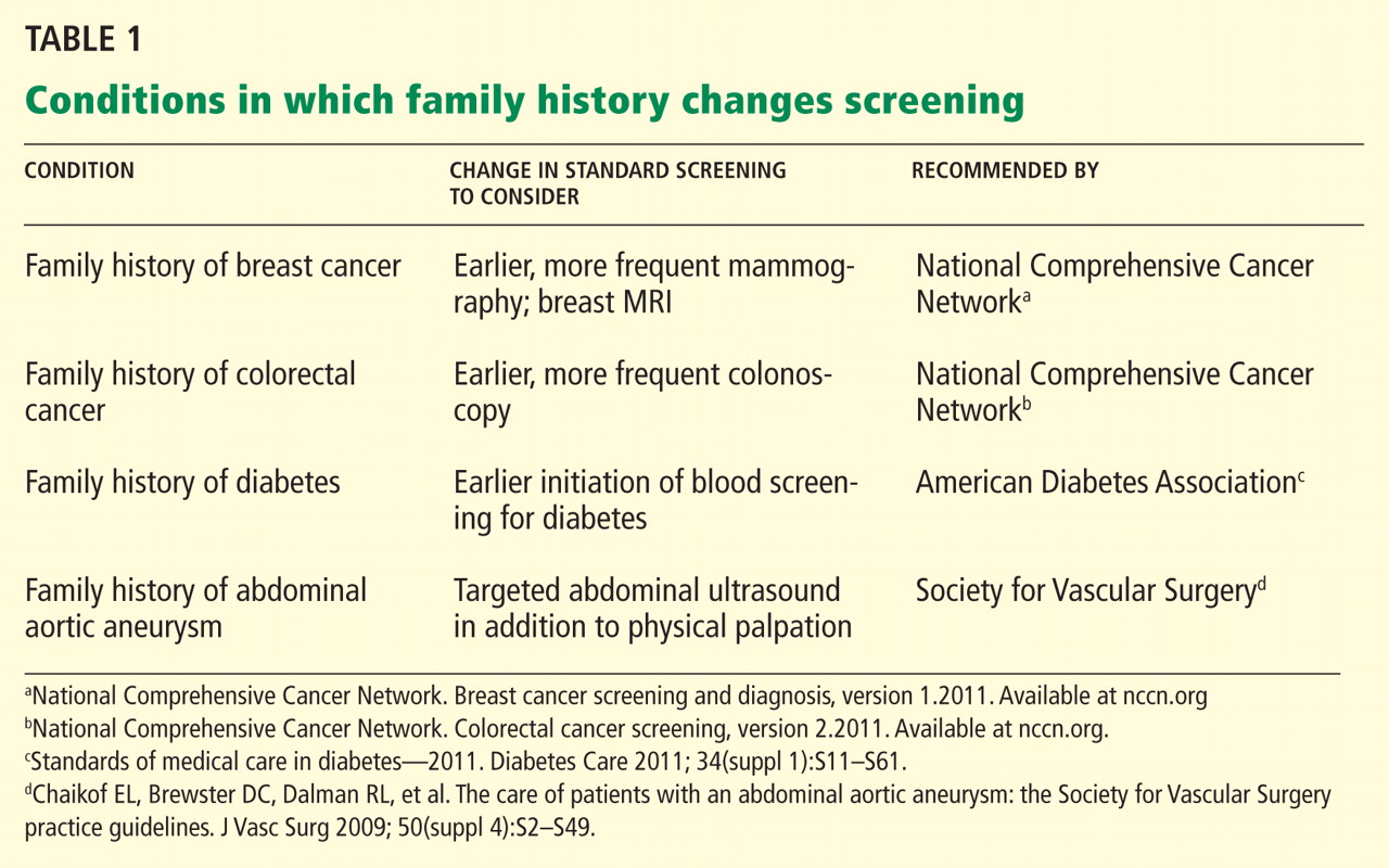
Furthermore, knowing they are at risk might empower people and encourage them to engage with the medical system. For example, counseling people at risk of diabetes as reflected in the family history has been shown to increase their understanding of the risk and of preventive behaviors. Further study is needed to determine if such messages can engender lasting changes in behavior across many diseases.42–46
TOWARD PERSONALIZED CARE
Especially now that caregivers are striving to provide value-based health care with emphasis on preventive care, the family history remains an important tool for detecting risk of disease. The evidence clearly indicates that medical providers have room for improvement in taking a family history and in using it.
We hope that asking patients about family history and recognizing patterns of disease will help us create personalized preventive-care plans, providing greater opportunity to educate and motivate our patients to work with us towards better health. Future solutions need to focus on time-effective ways to collect and analyze family history and on innovative methods of teaching medical providers at all levels to apply the family history to clinical practice.
- Guttmacher AE, Collins FS, Carmona RH. The family history—more important than ever. N Engl J Med 2004; 351:2333–2336.
- American College of Obstetricians and Gynecologists Committee on Genetics. Committee Opinion No. 478: Family history as a risk assessment tool. Obstet Gynecol 2011; 117:747–750.
- Rich EC, Burke W, Heaton CJ, et al. Reconsidering the family history in primary care. J Gen Intern Med 2004; 19:273–280.
- Green RF. Summary of workgroup meeting on use of family history information in pediatric primary care and public health. Pediatrics 2007; 120(suppl 2):S87–S100.
- American College of Obstetricians and Gynecologists. ACOG Practice Bulletin No. 103: Hereditary breast and ovarian cancer syndrome. Obstet Gynecol 2009; 113:957–966.
- Scheuner MT, Setodji CM, Pankow JS, Blumenthal RS, Keeler E. General Cardiovascular Risk Profile identifies advanced coronary artery calcium and is improved by family history: the multiethnic study of atherosclerosis. Circ Cardiovasc Genet 2010; 3:97–105.
- Yang Q, Liu T, Valdez R, Moonesinghe R, Khoury MJ. Improvements in ability to detect undiagnosed diabetes by using information on family history among adults in the United States. Am J Epidemiol 2010; 171:1079–1089.
- Kones R. Primary prevention of coronary heart disease: integration of new data, evolving views, revised goals, and role of rosuvastatin in management. A comprehensive survey. Drug Des Devel Ther 2011; 5:325–380.
- Rex DK, Johnson DA, Anderson JC, Schoenfeld PS, Burke CA, Inadomi JM; American College of Gastroenterology. American College of Gastroenterology guidelines for colorectal cancer screening 2009 (corrected). Am J Gastroenterol 2009; 104:739–750.
- American Diabetes Association. Standards of medical care in diabetes—2011. Diabetes Care 2011; 34(suppl 1):S11–S61.
- Kanis JA, Johansson H, Oden A, McCloskey EV. Assessment of fracture risk. Eur J Radiol 2009; 71:392–397.
- US Preventive Services Task Force. Genetic risk assessment and BRCA mutation testing for breast and ovarian cancer susceptibility: recommendation statement. Ann Intern Med 2005; 143:355–361.
- Williams SB, Salami S, Regan MM, et al. Selective detection of histologically aggressive prostate cancer: An Early Detection Research Network Prediction model to reduce unnecessary prostate biopsies with validation in the Prostate Cancer Prevention Trial. Cancer 2011; Oct 17(Epub ahead of print.)
- Dinh TA, Rosner BI, Atwood JC, et al. Health benefits and cost-effectiveness of primary genetic screening for Lynch syndrome in the general population. Cancer Prev Res (Phila) 2011; 4:9–22.
- Kwon JS, Scott JL, Gilks CB, Daniels MS, Sun CC, Lu KH. Testing women with endometrial cancer to detect Lynch syndrome. J Clin Oncol 2011; 29:2247–2252.
- Fuller M, Myers M, Webb T, Tabangin M, Prows C. Primary care providers’ responses to patient-generated family history. J Genet Couns 2010; 19:84–96.
- Acheson LS, Wiesner GL, Zyzanski SJ, Goodwin MA, Stange KC. Family history-taking in community family practice: implications for genetic screening. Genet Med 2000; 2:180–185.
- Yoon PW, Scheuner MT, Jorgensen C, Khoury MJ. Developing Family Healthware, a family history screening tool to prevent common chronic diseases. Prev Chronic Dis 2009; 6:A33.
- Valdez R, Yoon PW, Qureshi N, Green RF, Khoury MJ. Family history in public health practice: a genomic tool for disease prevention and health promotion. Annu Rev Public Health 2010; 31:69–87.
- Ziogas A, Anton-Culver H. Validation of My Family Health Portrait for six common heritable conditions. Am J Prev Med 2003; 24:190–198.
- Wideroff L, Garceau AO, Greene MH, et al. Coherence and completeness of population-based family cancer reports. Cancer Epidemiol Biomarkers Prev 2010; 19:799–810.
- Facio FM, Feero WG, Linn A, Oden N, Manickam K, Biesecker LG. Validation of My Family Health Portrait for six common heritable conditions. Genet Med 2010; 12:370–375.
- Owens KM, Marvin ML, Gelehrter TD, Ruffin MT, Uhlmann WR. Clinical use of the Surgeon General’s “My Family Health Portrait” (MFHP) tool: opinions of future health care providers. J Genet Couns 2011; 20:510–525.
- Tyler CV, Snyder CW. Cancer risk assessment: examining the family physician’s role. J Am Board Fam Med 2006; 19:468–477.
- Rubenstein WS. The genetics of breast cancer. In:Vogel VG, editor. Management of Patients at High Risk for Breast Cancer. Malden, MA: Blackwell Science; 2001:19–55.
- Antoniou A, Pharoah PD, Narod S, et al. Average risks of breast and ovarian cancer associated with BRCA1 or BRCA2 mutations detected in case series unselected for family history: a combined analysis of 22 studies. Am J Hum Genet 2003; 72:1117–1130.
- Korde LA, Zujewski JA, Kamin L, et al. Multidisciplinary meeting on male breast cancer: summary and research recommendations. J Clin Oncol 2010; 28:2114–2122.
- Myriad Genetic Laboratories, Inc. Mutation prevalence tables. http://www.myriad.com/lib/brac/brca-prevalence-tables.pdf. Accessed April 2, 2012.
- Schousboe JT, Kerlikowske K, Loh A, Cummings SR. Personalizing mammography by breast density and other risk factors for breast cancer: analysis of health benefits and cost-effectiveness. Ann Intern Med 2011; 155:10–20.
- National Cancer Institute. http://www.cancer.gov. Accessed January 20, 2012.
- Saslow D, Boetes C, Burke W, et al; American Cancer Society Breast Cancer Advisory Group. American Cancer Society guidelines for breast screening with MRI as an adjunct to mammography. CA Cancer J Clin 2007; 57:75–89.
- Agency for Healthcare Research and Quality; John M. Medications to reduce the risk of primary breast cancer in women: clinician’s guide. http://www.effectivehealthcare.ahrq.gov/index.cfm/searchfor-guides-reviews-and-reports/?productid=390&pageaction=displayproduct. Accessed April 2, 2012.
- Domchek SM, Friebel TM, Singer CF, et al. Association of risk-reducing surgery in BRCA1 or BRCA2 mutation carriers with cancer risk and mortality. JAMA 2010; 304:967–975.
- Anderson K, Jacobson JS, Heitjan DF, et al. Cost-effectiveness of preventive strategies for women with a BRCA1 or a BRCA2 mutation. Ann Intern Med 2006; 144:397–406.
- Kwon JS, Daniels MS, Sun CC, Lu KH. Preventing future cancers by testing women with ovarian cancer for BRCA mutations. J Clin Oncol 2009; 28:675–682.
- Wang G, Beattie MS, Ponce NA, Phillips KA. Eligibility criteria in private and public coverage policies for BRCA genetic testing and genetic counseling. Genet Med 2011; 13:1045–1050.
- Hinton RB. The family history: reemergence of an established tool. Crit Care Nurs Clin North Am 2008; 20:149–158.
- Murff HJ, Greevy RA, Syngal S. The comprehensiveness of family cancer history assessments in primary care. Community Genet 2007; 10:174–180.
- Wallace E, Hinds A, Campbell H, Mackay J, Cetnarskyj R, Porteous ME. A cross-sectional survey to estimate the prevalence of family history of colorectal, breast and ovarian cancer in a Scottish general practice population. Br J Cancer 2004; 91:1575–1579.
- Schroy PC, Barrison AF, Ling BS, Wilson S, Geller AC. Family history and colorectal cancer screening: a survey of physician knowledge and practice patterns. Am J Gastroenterol 2002; 97:1031–1036.
- Department of Health and Human Services. Genetics education and training: report of the Secretary’s Advisory Committee on Genetics, Health, and Society; 2011. http://oba.od.nih.gov/oba/SACGHS/reports/SACGHS_education_report_2011.pdf. Accessed April 2, 2012.
- Qureshi N, Kai J. Informing patients of familial diabetes mellitus risk: How do they respond? A cross-sectional survey. BMC Health Serv Res 2008; 8:37.
- Zlot AI, Bland MP, Silvey K, Epstein B, Mielke B, Leman RF. Influence of family history of diabetes on health care provider practice and patient behavior among nondiabetic Oregonians. Prev Chronic Dis 2009; 6:A27.
- Pijl M, Timmermans DR, Claassen L, et al. Impact of communicating familial risk of diabetes on illness perceptions and self-reported behavioral outcomes: a randomized controlled trial. Diabetes Care 2009; 32:597–599.
- Ruffin MT, Nease DE, Sen A, et al; Family History Impact Trial (FHITr) Group. Effect of preventive messages tailored to family history on health behaviors: the Family Healthware Impact Trial. Ann Fam Med 2011; 9:3–11.
- Claassen L, Henneman L, Janssens AC, et al. Using family history information to promote healthy lifestyles and prevent diseases; a discussion of the evidence. BMC Public Health 2010; 10:248.
At the dawn of the genomics era, is the family history still relevant? The answer is a resounding yes.1,2
The family history is clinically useful because it is a proxy for genetic, environmental, and behavioral risks to health. It can be used to inform risk stratification, allowing for judicious use of screening and opening the door to early and even prophylactic treatment.3–8 As people live longer, we will need to detect common chronic conditions early in their course so that we can continue to improve health outcomes. Family history can help physicians personalize preventive care for conditions such as diabetes, osteoporosis, and cancers of the breast, colon, and prostate.2,9–15
However, there is ample evidence that the family history is underused. Most practitioners ask about it infrequently and inconsistently.16,17 Why is this, and how can we encourage the use of this powerful tool to enhance our daily clinical practice and improve care?
We will discuss here some of the challenges that make it difficult for physicians to collect and use the family history in clinical practice, and review strategies for collecting and using the family history in a more consistent manner. We anticipate that this discussion will be helpful to clinicians, as the family history is an essential input to personalized, preventive care plans.
CHALLENGE 1: ARE PATIENTS’ REPORTS RELIABLE?
A question that often arises when discussing the utility of the family history is the reliability of patients’ reports. Can we trust that patients can accurately report their family history? For many conditions, the answer is yes.18,19
Ziogas and Anton-Culverl20 asked 1,111 cancer patients whether their relatives had ever had cancer and verified their answers. In more than 95% of cases, if the patient said that a first-degree or second-degree relative did not have cancer of any type, that relative truly did not have cancer. Overall, over-reporting of cancer was rare, occurring in 2.4% of cases.
If the patient said that a relative did have cancer, that statement was usually true as well. The reliability of a report of cancer in first-degree relatives was greater than 75% for most types of cancer (female breast, ovarian, esophageal, colorectal, pancreas, lung, melanoma, brain, thyroid, lymphoma, leukemia). For several of these types of cancer (female breast, colorectal, and brain), the reliability was 90% or higher. For second-degree relatives, the reliability of a reported positive history was moderate (50% to 80%) for the same types of cancer, and for third-degree relatives, the reliability dropped further for all types of cancer except female breast, brain, pancreas, and leukemia, for which the reliability of a positive report remained at 70%.
Wideroff et al21 had similar findings in a study of more than 1,000 patients and more than 20,000 of their relatives.
Yoon et al,18 at the US Centers for Disease Control and Prevention, developed a Web-based risk-assessment tool called Family Healthware, currently undergoing validation trials. They found that patients’ reports were highly reliable for coronary heart disease, stroke, diabetes, and breast, ovarian, and colorectal cancers. They also calculated the degree of risk associated with a positive family history and the prevalence of a family history of each of these diseases.
For the primary care physician, these studies support the reliability of patients’ reports and provide guidance for targeting specific conditions when obtaining a family history.
CHALLENGE 2: NO TIME OR REIMBURSEMENT
Perhaps the most obvious barriers to collecting a family history are lack of time and reimbursement.
Acheson et al,17 in an observational study of 138 primary care physicians and 4,454 patient visits, found that family history was discussed during 51% of new patient visits and 22% of established patient visits. The rate at which the family history was taken varied from 0% (some physicians never asked) to 81% of all patient visits. On average, physicians spent less than 2.5 minutes collecting the family history.
Not surprisingly, the family history was discussed more often at well-care visits than at illness visits, as the former type of visit tends to be longer and, by definition, to be spent partly on preventive care. A difficulty with this strategy is that, given the shortage of primary care physicians, limited access, and patient preference, most preventive-care visits are combined with problem-focused visits, further decreasing the time available to collect and discuss a family history. While some argue that the family history should routinely be obtained and discussed during preventive-care visits regardless of reimbursement and time, the reality is that it may simply drop on the list of priorities for each visit.
Rich et al3 estimated that taking a family history would increase reimbursement for only one new patient evaluation and management code (99202) and one return-visit code (99213) in Current Procedural Terminology. This action would increase reimbursement enough to support about 10 minutes of physician effort for collecting, documenting, and analyzing the family history. While this is certainly better than the average of less than 2.5 minutes observed by Acheson et al,17 doctors would probably do it more if they were paid more for it.
Electronic solutions
Given that a lack of time is a barrier, what are some ways to minimize the time it takes to collect a family history?
With more physicians using electronic health records and with increasing use of Internet-based tools in the population at large, information-technology systems have been developed to help obtain the family history. One of the most widely used is the US surgeon general’s My Family Health Portrait, available free at https://familyhistory.hhs.gov. It is one of the broadest electronic family-history collection tools and has been validated for use in risk assessment for diabetes and cancer of the colon, breast, and ovaries.22
However, electronic solutions have their own challenges. Not all patients have access to the Internet, many need help using these programs, and these tools may not work well with existing electronic medical records systems.23 Ideally, these programs would also provide built-in decision support for the provider, thereby maximizing data use for final patient risk assessment.23 Furthermore, electronic solutions are not a one-time-only risk assessment— periodic re-review of family history and reassessment of familial risk are required.24
Does taking a family history improve outcomes? Lessons from breast cancer
One of the reasons physicians don’t get reimbursed for collecting a family history is that it has been difficult to measure any improvement in outcomes associated with risk prediction through family history.
The best examples of improvement in outcomes associated with family history-based risk prediction come from studies of breast cancer. From 5% to 10% of cases of breast cancer are part of hereditary cancer syndromes, many of which have a known genetic cause. The most prevalent of these genetic syndromes is the hereditary breast and ovarian cancer (HBOC) syndrome, caused by mutations in the breast cancer 1 (BRCA1) and breast cancer 2 (BRCA2) genes. Clinical testing for BRCA mutations has been available since 1998.25 Women with a BRCA mutation have up to a 65% lifetime risk of developing breast cancer and up to a 40% lifetime risk of developing ovarian cancer.26 Men with a BRCA mutation are at 10 to 100 times the risk of the general population (1% to 10% vs 0.1%) for developing breast cancer, and are also at higher risk of prostate and other cancers.27
People who have a relative who developed breast cancer at a young age are more likely to harbor one of these mutations. For example, based on genetic testing in more than 185,000 people, the prevalence of BRCA mutations among people without cancer, not of Ashkenazi Jewish ancestry (a risk factor for breast cancer), and with no family history of early breast cancer or of ovarian cancer in any relative is 1.5%.28 In contrast, people with no personal history of cancer who have a family history of breast cancer before age 50 have a 5.6% prevalence of BRCA mutation, and if they are of Ashkenazi Jewish ancestry, this number is 16.4%.28
Medical and surgical interventions are available to reduce the risk of cancer in people with hereditary cancer syndromes such as HBOC. Options include screening more often, using advanced screening tests,29 giving preventive drugs such as tamoxifen (Nolvadex), and prophylactic surgery.30–32 What is the evidence that early screening and intervention in these people improve outcomes?
Domcheck et al33 prospectively followed more than 2,400 women who had BRCA mutations to assess the effect of prophylactic mastectomy or salpingo-oophorectomy on cancer outcomes. Mastectomy was indeed associated with a lower risk of breast cancer: 0 cases of breast cancer were diagnosed in 3 years of prospective follow-up in the 247 women who elected to undergo mastectomy, compared with 98 cases diagnosed in the 1,372 women who did not elect it over a similar period.
Women who elected to undergo salpingo-oophorectomy had a similarly lower rate of ovarian cancer compared with those who did not elect surgery (1% vs 6%). Additionally, fewer women who elected prophylactic salpingo-oophorectomy died of any cause (3% vs 10%), died of breast cancer (2% vs 6%), or died of ovarian cancer (0.4% vs 3%) compared with women who did not elect surgery.
Taking a family history reduces costs
What is the evidence that appropriate use of the family history decreases health care costs? Let us continue with the example of HBOC syndrome due to BRCA mutations.
Given that germline mutations account for 5% to 10% of cases of breast cancer in the United States and that the women who develop cancer associated with such mutations do so at a relatively young age, these mutations account for a disproportionate share of life-years lost due to cancer.34 Through taking a family history, these women at high risk can be identified and referred for genetic testing. Genetic testing, though costly, is more cost-effective than diagnosing and treating cancer.
Anderson et al,34 in 2006, estimated that cost-effective policies on testing and preventive treatment for persons at high risk of breast cancer could save up to $800 million of the more than $8 billion spent each year on breast cancer diagnosis, prevention, and treatment.
Kwon et al,35 in a simulation model (not a study in real patients), compared four different criteria for BRCA testing in women with ovarian cancer to see which strategy would be most cost-effective in preventing breast and ovarian cancers in their first-degree relatives. The best strategy, according to this analysis, is to test women with ovarian cancer for BRCA mutations if they also have a personal history of breast cancer, have a family history of breast or ovarian cancer, or are of Ashkenazi Jewish ancestry. The estimated cost per life-year gained with this strategy was $32,018, much lower than the widely accepted threshold for cost-effectiveness of $50,000 per life-year gained.
Although many professional organizations, including the US Preventive Services Task Force, have endorsed family-history-based eligibility criteria for genetic counseling and BRCA testing, awareness of the value of genetic testing in people who have been prescreened by family history has been relatively slow in seeping out to insurance carriers, especially Medicaid.12,36 As evidence continues to accumulate showing that this approach can improve outcomes for at-risk family members, reimbursement and time allotted for obtaining and using the family history should be adjusted.
CHALLENGE 3: A KNOWLEDGE GAP IN CLINICIANS
Another challenge often cited as a cause of the underuse of the family history as a predictor of disease risk is that clinicians may not know enough about the topic. Several studies indicated that even when physicians had obtained some components of the family history, they did not document risk appropriately or recognize the significance of the pattern of inheritance observed.37–39
In a study comparing primary care physicians and gastroenterologists in their use of the family history to predict the risk of hereditary colon cancer, gastroenterologists were more likely to elicit a family history of colorectal cancer and implement appropriate screening strategies, but overall compliance with screening guidelines was suboptimal in both groups.40
A 2011 report by an advisory committee to the secretary of the US Department of Health and Human Services concluded that lack of genetics education in medical school limits the integration of genetics into clinical care.41
How can we close this knowledge gap?
Recognizing a need, the National Coalition for Health Professional Education in Genetics was established in 1996 by the American Medical Association, the American Nurses Association, and the National Human Genome Research Institute (www.nchpeg.org). Its mission is to promote the education of health professionals and access to information about advances in human genetics to improve the health care of the nation. It offers educational materials, including a newly updated “Core Principles in Family History” program, which can be used to educate medical providers and their patients about various concepts related to genetics and family history.
In addition, physicians can use many risk assessment tools based on family history in patient care. Two of the best known are:
- The Gail breast cancer risk assessment model, hosted by the National Cancer Institute (www.cancer.gov/bcrisktool/)
- The FRAX osteoporosis risk assessment model, developed by the World Health Organization (www.shef.ac.uk/FRAX).
As we continue to educate the medical community about the value of the family history in predicting disease, it will be important to increase efforts in medical schools and residency programs and to find new, more interactive ways of teaching these concepts.
A possible strategy is to highlight the use of pedigree drawing to recognize patterns of inheritance.2 In a study of physician attitudes toward using patient-generated pedigrees in practice, such as those produced by the US surgeon general’s My Family Health Portrait, 73% of physicians stated that the patient-generated pedigree would improve their ability to assess the risk of disease, and the majority also agreed that it would not extend the time of the assessment.16
Is this information clinically useful?
Furthermore, knowing they are at risk might empower people and encourage them to engage with the medical system. For example, counseling people at risk of diabetes as reflected in the family history has been shown to increase their understanding of the risk and of preventive behaviors. Further study is needed to determine if such messages can engender lasting changes in behavior across many diseases.42–46
TOWARD PERSONALIZED CARE
Especially now that caregivers are striving to provide value-based health care with emphasis on preventive care, the family history remains an important tool for detecting risk of disease. The evidence clearly indicates that medical providers have room for improvement in taking a family history and in using it.
We hope that asking patients about family history and recognizing patterns of disease will help us create personalized preventive-care plans, providing greater opportunity to educate and motivate our patients to work with us towards better health. Future solutions need to focus on time-effective ways to collect and analyze family history and on innovative methods of teaching medical providers at all levels to apply the family history to clinical practice.
- Guttmacher AE, Collins FS, Carmona RH. The family history—more important than ever. N Engl J Med 2004; 351:2333–2336.
- American College of Obstetricians and Gynecologists Committee on Genetics. Committee Opinion No. 478: Family history as a risk assessment tool. Obstet Gynecol 2011; 117:747–750.
- Rich EC, Burke W, Heaton CJ, et al. Reconsidering the family history in primary care. J Gen Intern Med 2004; 19:273–280.
- Green RF. Summary of workgroup meeting on use of family history information in pediatric primary care and public health. Pediatrics 2007; 120(suppl 2):S87–S100.
- American College of Obstetricians and Gynecologists. ACOG Practice Bulletin No. 103: Hereditary breast and ovarian cancer syndrome. Obstet Gynecol 2009; 113:957–966.
- Scheuner MT, Setodji CM, Pankow JS, Blumenthal RS, Keeler E. General Cardiovascular Risk Profile identifies advanced coronary artery calcium and is improved by family history: the multiethnic study of atherosclerosis. Circ Cardiovasc Genet 2010; 3:97–105.
- Yang Q, Liu T, Valdez R, Moonesinghe R, Khoury MJ. Improvements in ability to detect undiagnosed diabetes by using information on family history among adults in the United States. Am J Epidemiol 2010; 171:1079–1089.
- Kones R. Primary prevention of coronary heart disease: integration of new data, evolving views, revised goals, and role of rosuvastatin in management. A comprehensive survey. Drug Des Devel Ther 2011; 5:325–380.
- Rex DK, Johnson DA, Anderson JC, Schoenfeld PS, Burke CA, Inadomi JM; American College of Gastroenterology. American College of Gastroenterology guidelines for colorectal cancer screening 2009 (corrected). Am J Gastroenterol 2009; 104:739–750.
- American Diabetes Association. Standards of medical care in diabetes—2011. Diabetes Care 2011; 34(suppl 1):S11–S61.
- Kanis JA, Johansson H, Oden A, McCloskey EV. Assessment of fracture risk. Eur J Radiol 2009; 71:392–397.
- US Preventive Services Task Force. Genetic risk assessment and BRCA mutation testing for breast and ovarian cancer susceptibility: recommendation statement. Ann Intern Med 2005; 143:355–361.
- Williams SB, Salami S, Regan MM, et al. Selective detection of histologically aggressive prostate cancer: An Early Detection Research Network Prediction model to reduce unnecessary prostate biopsies with validation in the Prostate Cancer Prevention Trial. Cancer 2011; Oct 17(Epub ahead of print.)
- Dinh TA, Rosner BI, Atwood JC, et al. Health benefits and cost-effectiveness of primary genetic screening for Lynch syndrome in the general population. Cancer Prev Res (Phila) 2011; 4:9–22.
- Kwon JS, Scott JL, Gilks CB, Daniels MS, Sun CC, Lu KH. Testing women with endometrial cancer to detect Lynch syndrome. J Clin Oncol 2011; 29:2247–2252.
- Fuller M, Myers M, Webb T, Tabangin M, Prows C. Primary care providers’ responses to patient-generated family history. J Genet Couns 2010; 19:84–96.
- Acheson LS, Wiesner GL, Zyzanski SJ, Goodwin MA, Stange KC. Family history-taking in community family practice: implications for genetic screening. Genet Med 2000; 2:180–185.
- Yoon PW, Scheuner MT, Jorgensen C, Khoury MJ. Developing Family Healthware, a family history screening tool to prevent common chronic diseases. Prev Chronic Dis 2009; 6:A33.
- Valdez R, Yoon PW, Qureshi N, Green RF, Khoury MJ. Family history in public health practice: a genomic tool for disease prevention and health promotion. Annu Rev Public Health 2010; 31:69–87.
- Ziogas A, Anton-Culver H. Validation of My Family Health Portrait for six common heritable conditions. Am J Prev Med 2003; 24:190–198.
- Wideroff L, Garceau AO, Greene MH, et al. Coherence and completeness of population-based family cancer reports. Cancer Epidemiol Biomarkers Prev 2010; 19:799–810.
- Facio FM, Feero WG, Linn A, Oden N, Manickam K, Biesecker LG. Validation of My Family Health Portrait for six common heritable conditions. Genet Med 2010; 12:370–375.
- Owens KM, Marvin ML, Gelehrter TD, Ruffin MT, Uhlmann WR. Clinical use of the Surgeon General’s “My Family Health Portrait” (MFHP) tool: opinions of future health care providers. J Genet Couns 2011; 20:510–525.
- Tyler CV, Snyder CW. Cancer risk assessment: examining the family physician’s role. J Am Board Fam Med 2006; 19:468–477.
- Rubenstein WS. The genetics of breast cancer. In:Vogel VG, editor. Management of Patients at High Risk for Breast Cancer. Malden, MA: Blackwell Science; 2001:19–55.
- Antoniou A, Pharoah PD, Narod S, et al. Average risks of breast and ovarian cancer associated with BRCA1 or BRCA2 mutations detected in case series unselected for family history: a combined analysis of 22 studies. Am J Hum Genet 2003; 72:1117–1130.
- Korde LA, Zujewski JA, Kamin L, et al. Multidisciplinary meeting on male breast cancer: summary and research recommendations. J Clin Oncol 2010; 28:2114–2122.
- Myriad Genetic Laboratories, Inc. Mutation prevalence tables. http://www.myriad.com/lib/brac/brca-prevalence-tables.pdf. Accessed April 2, 2012.
- Schousboe JT, Kerlikowske K, Loh A, Cummings SR. Personalizing mammography by breast density and other risk factors for breast cancer: analysis of health benefits and cost-effectiveness. Ann Intern Med 2011; 155:10–20.
- National Cancer Institute. http://www.cancer.gov. Accessed January 20, 2012.
- Saslow D, Boetes C, Burke W, et al; American Cancer Society Breast Cancer Advisory Group. American Cancer Society guidelines for breast screening with MRI as an adjunct to mammography. CA Cancer J Clin 2007; 57:75–89.
- Agency for Healthcare Research and Quality; John M. Medications to reduce the risk of primary breast cancer in women: clinician’s guide. http://www.effectivehealthcare.ahrq.gov/index.cfm/searchfor-guides-reviews-and-reports/?productid=390&pageaction=displayproduct. Accessed April 2, 2012.
- Domchek SM, Friebel TM, Singer CF, et al. Association of risk-reducing surgery in BRCA1 or BRCA2 mutation carriers with cancer risk and mortality. JAMA 2010; 304:967–975.
- Anderson K, Jacobson JS, Heitjan DF, et al. Cost-effectiveness of preventive strategies for women with a BRCA1 or a BRCA2 mutation. Ann Intern Med 2006; 144:397–406.
- Kwon JS, Daniels MS, Sun CC, Lu KH. Preventing future cancers by testing women with ovarian cancer for BRCA mutations. J Clin Oncol 2009; 28:675–682.
- Wang G, Beattie MS, Ponce NA, Phillips KA. Eligibility criteria in private and public coverage policies for BRCA genetic testing and genetic counseling. Genet Med 2011; 13:1045–1050.
- Hinton RB. The family history: reemergence of an established tool. Crit Care Nurs Clin North Am 2008; 20:149–158.
- Murff HJ, Greevy RA, Syngal S. The comprehensiveness of family cancer history assessments in primary care. Community Genet 2007; 10:174–180.
- Wallace E, Hinds A, Campbell H, Mackay J, Cetnarskyj R, Porteous ME. A cross-sectional survey to estimate the prevalence of family history of colorectal, breast and ovarian cancer in a Scottish general practice population. Br J Cancer 2004; 91:1575–1579.
- Schroy PC, Barrison AF, Ling BS, Wilson S, Geller AC. Family history and colorectal cancer screening: a survey of physician knowledge and practice patterns. Am J Gastroenterol 2002; 97:1031–1036.
- Department of Health and Human Services. Genetics education and training: report of the Secretary’s Advisory Committee on Genetics, Health, and Society; 2011. http://oba.od.nih.gov/oba/SACGHS/reports/SACGHS_education_report_2011.pdf. Accessed April 2, 2012.
- Qureshi N, Kai J. Informing patients of familial diabetes mellitus risk: How do they respond? A cross-sectional survey. BMC Health Serv Res 2008; 8:37.
- Zlot AI, Bland MP, Silvey K, Epstein B, Mielke B, Leman RF. Influence of family history of diabetes on health care provider practice and patient behavior among nondiabetic Oregonians. Prev Chronic Dis 2009; 6:A27.
- Pijl M, Timmermans DR, Claassen L, et al. Impact of communicating familial risk of diabetes on illness perceptions and self-reported behavioral outcomes: a randomized controlled trial. Diabetes Care 2009; 32:597–599.
- Ruffin MT, Nease DE, Sen A, et al; Family History Impact Trial (FHITr) Group. Effect of preventive messages tailored to family history on health behaviors: the Family Healthware Impact Trial. Ann Fam Med 2011; 9:3–11.
- Claassen L, Henneman L, Janssens AC, et al. Using family history information to promote healthy lifestyles and prevent diseases; a discussion of the evidence. BMC Public Health 2010; 10:248.
KEY POINTS
- The family history is an underused tool for predicting the risk of disease and for personalizing preventive care.
- Barriers to the appropriate collection and use of the family history include concerns over the reliability of patient reporting, a lack of time and reimbursement, and provider knowledge gaps.
- Use of family history to inform genetic testing for hereditary cancer syndromes has been shown to improve outcomes and may reduce overall health care costs.
- Future solutions need to focus on creating time-effective ways to collect and analyze the family history, and on developing innovative methods of educating medical providers at all levels of training as to how to apply the family history in clinical practice.
Personalizing patient care
The concept and promise of personalized health care have been anticipated for decades. Yet in its breadth and in the way we would like it practiced, it is in its infancy.
Personalized health care aims to individualize care by integrating a person’s unique clinical, molecular (ie, genetic, genomic), and environmental information. Applied not only when the patient is sick but also when he or she is well, it builds on and enhances our current standards of care.
As Sir William Osler recognized more than a century ago, “Variability is the law of life, and as no two faces are the same, so no two bodies are alike, and no two individuals react alike and behave alike under the abnormal conditions which we know as disease.”1
PREDICTING THE RISK OF DISEASE
For years, we have attempted to predict and stratify the risk of disease, and the Human Genome Project has given us a new set of tools to help understand the complexity of disease and its variability.
We know and have known for decades—and in some cultures, for centuries—that family history is the most clinically validated tool for predicting the risk of disease.
Nevertheless, evidence suggests that we physicians are not collecting adequate family histories, and because of this, we are missing opportunities to intervene and prevent diseases predicted by the family history. The current standard of care is to use family medical histories to hone genetic differential diagnoses, and based on the differential diagnoses, to target specific genes to test in the setting of genetic counseling. Current genetic testing is used for molecular diagnoses and predictive testing so that gene-specific clinical management can be subsequently tailored.
PREDICTING RESPONSE TO TREATMENT
The use of personalized health care to predict response to treatment is a novel and constantly evolving practice.
ABO blood typing is a form of genetics-based personalization of safe transfusion that dates back to World War II.
A prominent, recent success story is in cancer treatment. For example, the American Society of Clinical Oncology now recommends that tumors from patients with node-negative, estrogen-receptor-positive breast cancer be evaluated with the Oncotype DX assay.2 This test measures the expression of 21 genes, and the score obtained identifies patients most likely to benefit from adjuvant chemotherapy. A similar 12-gene expression signature has been developed for colon cancer, and others have been developed for hematologic cancers.2,3 As with other new but apparently valid tests, the risk scores derived are sensitive at the extremes but ambiguous in the mid-ranges. We anticipate many more developments in this field.
In the field of pharmacogenomics, there is evidence to suggest that prior knowledge of CYP2C9 and VKORC1 genotypes enhances outcomes for patients starting treatment with warfarin. The US Food and Drug Administration revised the label on warfarin in February 2010, suggesting that genotypes be taken into consideration when the drug is prescribed.4
However, clinicians have been slow to adopt genotype testing when prescribing warfarin. Some cite the paucity of large, randomized, controlled trials demonstrating clinical utility of genotype-informed prescribing. Others cite concern that warfarin will soon become obsolete with the arrival of newer anticoagulants (such as factor X inhibitors) that do not carry warfarin’s adverse effects, and these genotypes will therefore become moot. Perhaps, as we move forward and new drugs are developed, companion genotype tests could be developed at the same time to be used with them.5
IMPROVING CARE, SAVING MONEY, AND EMPOWERING PATIENTS
The goal of personalized health care, by customizing treatments (medication types and dosages) and preventive strategies, is to optimize medical care and improve outcomes for each patient. It could improve the quality of care by targeting interventions and reducing adverse events, topics that are important to all of us in the current environment of health care reform.
A personalized approach might also, in the long run, decrease the cost of health care by driving appropriate utilization of resources.
Lastly, the true value of personalized health care may be in its potential to improve patient satisfaction and to empower our patients to work with us towards better health.
WE LAUNCH A NEW SERIES
To keep physicians up-to-date on progress in personalized health care, the Cleveland Clinic Journal of Medicine will present a series of articles on the topic. The series, to run once a quarter, begins in this issue, on page 331, with an article on the importance of the family history as a piece of genetic information that can help to predict the risk of disease and inform preventive care plans. Future topics will include the role of genetics and genomics in personalized care of patients with breast and colorectal cancers; the genetic counselor as a part of the health care team; pharmacogenomics; and ethical, legal, and societal considerations.
Our goal in this series is to provide practical information to help our readers incorporate personalized approaches into daily practice. In addition, as patients become more interested in and informed about personalized health care, we hope this information will help clinicians to effectively coach them about its potential benefits and risks. We also hope this information will enable our readers to ask the right questions so that patient and health care provider can work together to help the patient grow old gracefully.
As the series unfolds, we ask you to send us feedback and to suggest other topics in personalized health care you would like us to cover in this series.
- Osler W. Aequanimitas, With Other Addresses to Medical Students, Nurses and Practitioners of Medicine. 2nd edition. Philadelphia, PA: P. Blakiston’s Sone & Co, 1906:348.
- McDermott U, Downing JR, Stratton MR. Genomics and the continuum of cancer care. N Engl J Med 2011; 364:340–350.
- Eng C. Microenvironmental protection in diffuse large-B-cell lymphoma. N Engl J Med 2008; 359:2379–2381.
- Wang L, McLeod HL, Weinshilboum RM. Genomics and drug response. N Engl J Med 2011; 364:1144–1153.
- Hamburg MA, Collins FS. The path to personalized medicine. N Engl J Med 2010; 363:301–304.
The concept and promise of personalized health care have been anticipated for decades. Yet in its breadth and in the way we would like it practiced, it is in its infancy.
Personalized health care aims to individualize care by integrating a person’s unique clinical, molecular (ie, genetic, genomic), and environmental information. Applied not only when the patient is sick but also when he or she is well, it builds on and enhances our current standards of care.
As Sir William Osler recognized more than a century ago, “Variability is the law of life, and as no two faces are the same, so no two bodies are alike, and no two individuals react alike and behave alike under the abnormal conditions which we know as disease.”1
PREDICTING THE RISK OF DISEASE
For years, we have attempted to predict and stratify the risk of disease, and the Human Genome Project has given us a new set of tools to help understand the complexity of disease and its variability.
We know and have known for decades—and in some cultures, for centuries—that family history is the most clinically validated tool for predicting the risk of disease.
Nevertheless, evidence suggests that we physicians are not collecting adequate family histories, and because of this, we are missing opportunities to intervene and prevent diseases predicted by the family history. The current standard of care is to use family medical histories to hone genetic differential diagnoses, and based on the differential diagnoses, to target specific genes to test in the setting of genetic counseling. Current genetic testing is used for molecular diagnoses and predictive testing so that gene-specific clinical management can be subsequently tailored.
PREDICTING RESPONSE TO TREATMENT
The use of personalized health care to predict response to treatment is a novel and constantly evolving practice.
ABO blood typing is a form of genetics-based personalization of safe transfusion that dates back to World War II.
A prominent, recent success story is in cancer treatment. For example, the American Society of Clinical Oncology now recommends that tumors from patients with node-negative, estrogen-receptor-positive breast cancer be evaluated with the Oncotype DX assay.2 This test measures the expression of 21 genes, and the score obtained identifies patients most likely to benefit from adjuvant chemotherapy. A similar 12-gene expression signature has been developed for colon cancer, and others have been developed for hematologic cancers.2,3 As with other new but apparently valid tests, the risk scores derived are sensitive at the extremes but ambiguous in the mid-ranges. We anticipate many more developments in this field.
In the field of pharmacogenomics, there is evidence to suggest that prior knowledge of CYP2C9 and VKORC1 genotypes enhances outcomes for patients starting treatment with warfarin. The US Food and Drug Administration revised the label on warfarin in February 2010, suggesting that genotypes be taken into consideration when the drug is prescribed.4
However, clinicians have been slow to adopt genotype testing when prescribing warfarin. Some cite the paucity of large, randomized, controlled trials demonstrating clinical utility of genotype-informed prescribing. Others cite concern that warfarin will soon become obsolete with the arrival of newer anticoagulants (such as factor X inhibitors) that do not carry warfarin’s adverse effects, and these genotypes will therefore become moot. Perhaps, as we move forward and new drugs are developed, companion genotype tests could be developed at the same time to be used with them.5
IMPROVING CARE, SAVING MONEY, AND EMPOWERING PATIENTS
The goal of personalized health care, by customizing treatments (medication types and dosages) and preventive strategies, is to optimize medical care and improve outcomes for each patient. It could improve the quality of care by targeting interventions and reducing adverse events, topics that are important to all of us in the current environment of health care reform.
A personalized approach might also, in the long run, decrease the cost of health care by driving appropriate utilization of resources.
Lastly, the true value of personalized health care may be in its potential to improve patient satisfaction and to empower our patients to work with us towards better health.
WE LAUNCH A NEW SERIES
To keep physicians up-to-date on progress in personalized health care, the Cleveland Clinic Journal of Medicine will present a series of articles on the topic. The series, to run once a quarter, begins in this issue, on page 331, with an article on the importance of the family history as a piece of genetic information that can help to predict the risk of disease and inform preventive care plans. Future topics will include the role of genetics and genomics in personalized care of patients with breast and colorectal cancers; the genetic counselor as a part of the health care team; pharmacogenomics; and ethical, legal, and societal considerations.
Our goal in this series is to provide practical information to help our readers incorporate personalized approaches into daily practice. In addition, as patients become more interested in and informed about personalized health care, we hope this information will help clinicians to effectively coach them about its potential benefits and risks. We also hope this information will enable our readers to ask the right questions so that patient and health care provider can work together to help the patient grow old gracefully.
As the series unfolds, we ask you to send us feedback and to suggest other topics in personalized health care you would like us to cover in this series.
- Osler W. Aequanimitas, With Other Addresses to Medical Students, Nurses and Practitioners of Medicine. 2nd edition. Philadelphia, PA: P. Blakiston’s Sone & Co, 1906:348.
- McDermott U, Downing JR, Stratton MR. Genomics and the continuum of cancer care. N Engl J Med 2011; 364:340–350.
- Eng C. Microenvironmental protection in diffuse large-B-cell lymphoma. N Engl J Med 2008; 359:2379–2381.
- Wang L, McLeod HL, Weinshilboum RM. Genomics and drug response. N Engl J Med 2011; 364:1144–1153.
- Hamburg MA, Collins FS. The path to personalized medicine. N Engl J Med 2010; 363:301–304.
Disparities in prostate cancer in African American men: What primary care physicians can do
Prostate cancer is the most common cancer affecting American men. In 2010, an estimated 217,730 men were diagnosed with it and 32,050 died of it.1 African American men are disproportionately affected, with a prostate cancer incidence two-thirds higher than whites and a mortality rate twice as high.1 Owing to such disparities, the life expectancy of African Americans is several years shorter than that of non-Hispanic whites.2
For the primary care provider, who is often the first access point for health care in the United States, it is important to understand what mechanisms may underlie these differences and what can be done to narrow the gap.3
WHAT IS THE CAUSE OF THESE DIFFERENCES?
Many studies have looked into the causes of the higher incidence of prostate cancer in African American men and their higher mortality rate from it. The disparity may be due to a variety of factors, some socioeconomic and some biologic.
Poorer access to care, or lower-quality care?
A study of US servicemen who had equal access to care showed that African American men had a higher rate of prostate cancer regardless of access to care and socioeconomic status.4
However, the 2002 Institute of Medicine report, Unequal Treatment: Confronting Racial and Ethnic Disparities in Health Care, found evidence that racial and ethnic minorities tend to receive lower-quality health care than whites, “even when access-related factors, such as patients’ insurance status and income, are controlled.”5
Genetic predisposition?
Some have proposed that the disparity may be a function of genetic predisposition.
Evidence of a genetic component to the high incidence and mortality rate in African American men comes from epidemiologic studies of men with similar genetic backgrounds. For example, men in Nigeria and Ghana also have a high incidence of prostate cancer, as do men of African descent in the Caribbean islands and in the United Kingdom.6
Chromosome 8q24 variants have been shown in several studies to be associated with prostate cancer risk and are more common in African American men.7–10 Some studies have also shown a higher rate of variations in cell apoptosis genes such as BCL211 and tumor-suppression genes such as EphB2 in African American men.12
These findings suggest that genetic differences may contribute to the higher prostate cancer incidence and mortality rate seen in African American men.
More-aggressive cancer, or later detection?
Not only do African American men tend to have a higher incidence of prostate cancer, they also tend to have more-aggressive disease (ie, a higher pathologic grade) at the time of diagnosis, which may contribute to the disparity in mortality rates.13–19
Initially, there was some controversy as to whether this observation is a result of genetic and biologic factors that may predispose African American men to more-aggressive disease, or if it is due to inadequate screening and delayed presentation. However, a body of evidence supports the contention that prostate cancer is more aggressive in African American men.
For example, a study of autopsy data from men who died of prostate cancer at ages 20 to 49 showed that the age of onset of prostate cancer was similar between African American and white men.20 The Surveillance Epidemiology and End Results (SEER) database showed that African American men had a higher incidence of metastatic disease across all age groups.20 A similar study conducted 10 years later confirmed that rates of subclinical prostate cancer in African American and white men do not differ by race at the early ages, but that advanced or metastatic disease occurred nearly four times as frequently in African American men.21
Another study examined prostate biopsies from African American men and found that their tumors expressed higher levels of biomarkers, suggesting they had more-aggressive disease.22
SCREENING FOR PROSTATE CANCER
Serum prostate-specific antigen (PSA) testing has become the method of choice for prostate cancer screening. However, PSA screening in asymptomatic men is under debate, because it can lead to overdetection and subsequent overtreatment of indolent disease.23
Several recent studies showed differing results from prostate cancer screening.
The US Prostate, Lung, Colorectal, and Ovarian Cancer Screening Trial found that the mortality rate was no lower with combined PSA screening and digital rectal examination during a median follow-up of 11 years than in a control group that had a lower rate of screening.24 However, further analysis of these data, with stratifying by comorbidities, showed that PSA screening in young and healthy men reduces the risk of death from prostate cancer, with minimal overtreatment.25
The European Randomized Study of Screening for Prostate Cancer found a statistically significant 20% reduction in deaths from prostate cancer with PSA screening, but that it was necessary to treat 48 men in order to save one life.26
Another study, published in 2010, showed that regular PSA screening reduced the rate of prostate cancer mortality by half over 14 years.27
African American men generally present with disease that is more advanced than in white men.28 This historically has been attributed to the fact that African Americans have been less likely to be screened for prostate cancer, though recent data indicate the gap is lessening.29–31 A cross-sectional study from the Texas Medical Center showed that 54.4% of African American men had received PSA screening, compared with 63.2% of white men.32
Another study showed that African Americans were more likely to have had a longer interval between PSA screenings before diagnosis, and that a longer PSA screening interval was associated with greater odds of having advanced disease at diagnosis.33 However, when the researchers controlled for the PSA screening interval, they found that African Americans had the same odds of being diagnosed with advanced prostate cancer as white patients did. They concluded that more frequent or systematic PSA screening may reduce the racial differences in cancer stage at diagnosis and in deaths.
Reasons for the disparities in screening
Many reasons have been proposed to explain why African Americans receive less screening, including poor communication between physicians and minority patients due to lack of cultural competency among physicians, lack of health insurance (and poor access to quality care as a result), and deficiency of knowledge about screening. Though awareness is rising, many African Americans are unaware of early detection methods for prostate cancer (eg, PSA testing), and other barriers such as cost and transportation exist that may prevent African American men from being screened.34,35
As gatekeepers, primary care physicians are in a position to address these shortcomings in patient education and to enhance the physician-patient relationship.36
Black men have higher PSA levels, with or without cancer
Physicians must also be aware of racial differences in PSA levels and realize that the predictive value of PSA in the diagnosis of prostate cancer may differ between African Americans and whites.
Black men, with or without prostate cancer, have been found to have higher PSA levels. Kyle and colleagues37 found that African American men without prostate cancer had significantly higher mean PSA levels than white men across all age groups. Furthermore, Vijayakumar et al38 found that African Americans with newly diagnosed localized prostate cancer had higher serum PSA levels than whites at diagnosis.
Although PSA cutoff levels have not been officially modified according to race, primary care physicians should have a lower threshold for referring African American men who have a suspiciously high PSA level for further urologic evaluation. Close partnership between the internist, family practitioner, and urologist will aid in the optimal use of PSA testing for the early detection of prostate cancer.
When to start PSA screening? How often to screen?
The age at which African American men should begin to have their PSA levels checked (with or without a digital rectal examination) continues to debated. However, the American Cancer Society39 recommends that African American men who have a father or brother who had prostate cancer before age 65 should begin having discussions with their physician on this topic and, with their informed consent, screening at age 45.
The frequency of PSA screening depends on the individual’s PSA level. The National Comprehensive Cancer Network40 recommends that men at high risk be offered a baseline PSA measurement and digital rectal examination at age 40 and, if the PSA level is higher than 1 ng/mL, that they be offered annual follow-ups. If the PSA level is less than 1 ng/mL, they recommend screening again at age 45. Risk factors for prostate cancer include family history as well as African American race.41
How should PSA levels be interpreted?
Interpreting PSA results is important in detecting prostate cancer at early stages.
At first, we believed the normal range of PSA for all men was 4.0 ng/mL or less. However, the American Urological Association now recognizes that the normal PSA range, in addition to varying along racial lines, also is age-dependent.42 The Cleveland Clinic Minority Men's Health Center's suggested normal ranges of PSA in African American men are:
- Age 40–49: ≤ 2.5 ng/mL
- Age 50–59: ≤ 3.0 ng/mL
- Age 60–69: ≤ 3.5 ng/mL
- Age 70–79: ≤ 4.5 ng/mL
- Age > 80: ≤ 5.0 ng/mL.
Remember that an elevated PSA does not necessarily signify prostate cancer, and that these are reference ranges only and may vary in individual men.
SURVIVAL AFTER DIAGNOSIS
African American men with prostate cancer have significantly higher mortality rates than white men. The possible causes of worse outcomes are many, and there have been many studies that attempted to address this disparity. The question of a more biologically aggressive cancer was previously discussed, but additional factors such as socioeconomic factors, comorbidities, and treatment received have also been studied, and data are mixed.43–45
In a large SEER database review, once confounding variables of socioeconomic status, cancer stage, and treatment received were eliminated, African Americans had similar stage-for-stage survival from prostate cancer.46 Another study found, in 2,046 men, that differences in socioeconomic status explained the difference in mortality rates between white and black patients.47
However, other studies that adjusted for socioeconomic status as well as patient and tumor characteristics found that African American and Hispanic men were more likely to die of prostate cancer than white men.48
Do African American men receive less-aggressive care?
Studies have also determined that there may be differences in treatments offered to patients, which in turn negatively affect survival.28,49–53 Potentially curative local therapies (including radical surgery or radiation) may be recommended less often to black men because of major comorbidities or socioeconomic considerations.49–52
Additionally, potential metastatic disease may be identified in a less timely and accurate manner, as African American men are less likely to undergo pelvic lymph node dissection. This was associated with worse survival in men with poorly differentiated prostate cancer.53
However, returning to the possibility that prostate cancer is biologically more aggressive in African American men, some studies have shown that even after adjusting for treatment, African Americans continue to have worse survival rates.54,55 One study in men with stage T1 to T3 prostate cancer who chose brachytherapy for treatment reported that after adjusting for PSA, clinical stage, socioeconomic status, and comorbidities, African American and Hispanic race were associated with higher all-cause mortality rates.55
Equal care, equal outcomes?
In total, these results suggest that factors unrelated to tumor biology may be additional reasons for the poorer survival rates in African American men with prostate cancer. More favorable survival outcomes for African Americans with localized disease may be achieved with uniform assignment of treatment.
Fowler and Terrell56 reviewed the outcomes of 148 black and 209 white men with localized prostate cancer treated with surgery or radiation therapy over an 11-year period at a Veterans Administration hospital. Not surprisingly, the black men presented more often with advanced disease. However, survival outcomes were equivalent between whites and blacks when treatment was assigned in a uniform manner without regard to race. After a median follow-up of 96 months, there were no significant differences in all-cause, cause-specific, metastasis-free, clinical disease-free, or PSA recurrence-free survival rates in 109 black and 167 white men with low-stage cancer treated with surgery or radiation therapy or in 39 black and 42 white men with high-stage disease treated with radiotherapy.56
Similarly, Tewari et al57 studied a cohort of 402 African American and 642 white men, all of whom underwent radical prostatectomy for clinically localized prostate cancer. They were followed for PSA recurrence to determine if race-specific differences in PSA doubling time or histopathologic variables might account for the higher mortality rate in black men. While there were race-specific differences in baseline serum PSA and incidence of high-grade prostatic intraepithelial neoplasia, race was not an independent risk factor for biochemical recurrence. Instead, other variables such as the Gleason pathology score, bilateral cancers, and margin positivity were independently associated with biochemical recurrence.
Furthermore, researchers at Louisiana State University58 retrospectively analyzed data from 205 men of different races with early-stage prostate cancer. The African American men had a higher serum PSA level, suggesting more advanced disease or greater tumor burden at presentation, but no statistically significant differences were found among the pretreatment biopsy variables, including prostate volume (measured by ultrasonography), Gleason score, millimeters of cancer within the biopsy specimen, and percentage of cancer within the biopsy specimen. After treatment, there were no significant differences in survival outcomes along racial lines, leading the authors to conclude that early detection and treatment of prostate cancer in African Americans would be the best approach to lowering mortality rates.
Taken together, these data suggest that if localized prostate cancer is treated adequately and appropriately, African American patients may have improved survival rates.
DIETARY AND LIFESTYLE FACTORS
The incidence of prostate cancer is increasing in other countries where Western diets and lifestyles have been adopted,59,60 suggesting that nutritional factors may also contribute partly to prostate carcinogenesis. Culture- and race-specific differences in diet may play an important role in prostate cancer risk in certain racial minorities. Many aspects of diet and nutrition have been studied for their impact on prostate cancer.
Dietary risk factors
Too much red meat and processed meat? Although some have suggested that diets high in red and processed meats may lead to a higher risk of prostate cancer, a meta-analysis showed no association.61,62
Too much calcium? The European Prospective Investigation Into Cancer and Nutrition study found that high dietary intake of dairy protein and calcium from dairy products was associated with a higher risk of prostate cancer.63 A cohort study in the United States had similar findings with regard to calcium.64 However, the higher risk of prostate cancer was associated with consumption of 2,000 mg or more of calcium per day, which was consumed by only 2% of the study’s cohort and, as the study’s authors reported, fewer than 1% of US men. As such, only a small population of American men seem to be exposing themselves to a higher risk of prostate cancer by high calcium consumption.
High fat intake? Certain fatty acids have been implicated in general tumor genesis, and that risk has been extrapolated to prostate cancer.65 For example, high fat intake and obesity are associated with increased levels of insulin-like growth factor 1, which in turn has been shown to correlate with a significantly elevated risk of prostate cancer.63,65
Obesity has been shown to increase the risk of more-aggressive prostate cancer, but not of less-aggressive tumors.66 Moreover, men who lost weight had a lower risk of prostate cancer than those who maintained their weight over 10 years.66 Obesity may be particularly risky for African American men, in whom it was found to be associated with shorter biochemical relapse-free survival, whereas it was not an independent risk factor in white men.67
Preventive dietary agents have been elusive
Unfortunately, despite attempts to identify preventive dietary agents, none has yet been confirmed.
No benefit from selenium or vitamin E. The Selenium and Vitamin E Cancer Prevention Trial was discontinued, as there was no evidence that either agent prevented prostate cancer in relatively healthy men.68
Vitamin D? It has been suggested that lower levels of vitamin D could contribute to the higher rates of prostate cancer in African Americans, as vitamin D deficiency is more common in African Americans.69 However, several meta-analyses have shown no association between vitamin D and prostate cancer.70–72
Soy? Attempts at correlating the relatively low incidence of prostate cancer in Asians have revealed that high soy intake may be protective. Asians consume more soy than Americans do (100 vs 3 mg/day), and soy isoflavones such as genistein, glycitein, and daidzein lower the incidence of prostate cancer in laboratory mice.73
Other lifestyle factors
Other lifestyle factors have also been analyzed to see if they contribute to prostate cancer.
Pollution. Some studies have suggested that the etiology of prostate cancer may lie in environmental exposures to pesticides,74 metal industrial facilities,75 and urban living.76
Smoking. Watters et al77 found that current and former cigarette smokers were actually at a lower risk of being diagnosed with non-advanced prostate cancer, but current smokers were at higher risk of dying from prostate cancer.
Physical activity. A prospective study of lifetime physical activity of more than 45,000 men found that men who were not sedentary during work and who walked or bicycled more than 30 minutes per day during adult life had an approximately 20% lower incidence of prostate cancer.78
In sum, primary care providers who are generally promoting healthy lifestyles can point to a reduction in risk for prostate cancer as yet another benefit to a low-fat diet, a healthy body mass index, and daily exercise.
HOW PRIMARY CARE PHYSICIANS CAN HELP CLOSE THE GAP
Primary care physicians serve as the first point of health access for many in the United States today.
The diagnosis of prostate cancer is made more frequently in African American men than in other American men, often at a higher pathological grade, and with a worse mortality rate. Primary care physicians can help improve these statistics. Interventions targeting overall health, such as promotion of a healthy diet, could be established at primary care visits and could also reduce the incidence of prostate cancer in African American men. Patient education regarding prostate cancer screening, the impact of family history, and the rate of PSA screening could be improved.
Primary care physicians serve a vital role in health education and prostate cancer screening, and therefore they begin the process in potentially reducing the impact of prostate cancer in African American men. The racial disparity seen in prostate cancer may begin to be minimized with primary care physicians and specialists working together to ensure that all men receive appropriate treatment.
- Altekruse SF, Kosary CL, Krapcho M, et al. SEER Cancer Statistics Review, 1975–2007, National Cancer Institute. Bethesda, MD. http://seer.cancer.gov/csr/1975_2007/, based on November 2009 SEER data submission, posted to the SEER web site, 2010. Accessed April 2, 2011.
- Arias E. United States life tables, 2007. National vital statistics reports; vol 59 no 9. Hyattsville, MD: National Center for Health Statistics. 2011.
- Klein JB, Nguyen CT, Saffore L, Modlin C, Modlin CS. Racial disparities in urologic health care. J Natl Med Assoc 2010; 102:108–117.
- Wells TS, Bukowinski AT, Smith TC, et al. Racial differences in prostate cancer risk remain among US servicemen with equal access to care. Prostate 2010; 70:727–734.
- Smedley BD, Stith AY, Nelson AR, editors. Unequal Treatment: Confronting Racial and Ethnic Disparities in Health Care. Institute of Medicine. National Academy Press; 2002.
- Odedina FT, Akinremi TO, Chinegwundoh F, et al. Prostate cancer disparities in black men of African descent: a comparative literature review of prostate cancer burden among black men in the United States, Caribbean, United Kingdom, and West Africa. Infect Agent Cancer 2009; 4(suppl 1):S2.
- Okobia MN, Zmuda JM, Ferrell RE, Patrick AL, Bunker CH. Chromosome 8q24 variants are associated with prostate cancer risk in a high risk population of African ancestry. Prostate 2011; 71:1054–1063.
- Haiman CA, Chen GK, Blot WJ, et al. Characterizing genetic risk at known prostate cancer susceptibility loci in African Americans. PLoS Genet 2011; 7:e1001387.
- Freedman ML, Haiman CA, Patterson N, et al. Admixture mapping identifies 8q24 as a prostate cancer risk locus in African-American men. Proc Natl Acad Sci U S A 2006; 103:14068–14073.
- Chang BL, Isaacs SD, Wiley KE, et al. Genome-wide screen for prostate cancer susceptibility genes in men with clinically significant disease. Prostate 2005; 64:356–361.
- Hatcher D, Daniels G, Osman I, Lee P. Molecular mechanisms involving prostate cancer racial disparity. Am J Transl Res 2009; 1:235–248.
- Robbins CM, Hooker S, Kittles RA, Carpten JD. EphB2 SNPs and sporadic prostate cancer risk in African American men. PLoS One 2011; 6:e19494.
- American Cancer Society. Cancer Facts & Figures for African Americans 2009–2010. http://www.cancer.org/acs/groups/content/@nho/documents/document/cffaa20092010pdf.pdf. Accessed April 2, 2012.
- Ayanian JZ, Udvarhelyi IS, Gatsonis CA, Pashos CL, Epstein AM. Racial differences in the use of revascularization procedures after coronary angiography. JAMA 1993; 269:2642–2646.
- Fine MJ, Ibrahim SA, Thomas SB. The role of race and genetics in health disparities research. Am J Public Health 2005; 95:2125–2128.
- Horner RD, Oddone EZ, Matchar DB. Theories explaining racial differences in the utilization of diagnostic and therapeutic procedures for cerebrovascular disease. Milbank Q 1995; 73:443–462.
- Juckett G. Cross-cultural medicine. Am Fam Physician 2005; 72:2267–2274.
- Ndubuisi SC, Kofie VY, Andoh JY, Schwartz EM. Black-white differences in the stage at presentation of prostate cancer in the District of Columbia. Urology 1995; 46:71–77.
- Misra-Hebert AD. Physician cultural competence: cross-cultural communication improves care. Cleve Clin J Med 2003; 70:289,293,296–298.
- Powell I, Sakr W, Weiss L, et al. Prostate cancer is biologically more aggressive among African Americans than Caucasian men under age 70: hypothesis supported by autopsy and SEER data. Program and abstracts from the American Urological Association 95th Annual Meeting; April 29–May 4, 2000: Atlanta, GA.
- Powell IJ, Bock CH, Ruterbusch JJ, Sakr W. Evidence supports a faster growth rate and/or earlier transformation to clinically significant prostate cancer in black than in white American men, and influences racial progression and mortality disparity. J Urol 2010; 183:1792–1796.
- Kim HS, Moreira DM, Jayachandran J, et al. Prostate biopsies from black men express higher levels of aggressive disease biomarkers than prostate biopsies from white men. Prostate Cancer Prostatic Dis 2011; 14:262–265.
- Duffy MJ. Prostate-specific antigen: does the current evidence support its use in prostate cancer screening? Ann Clin Biochem 2011; 48:310–316.
- Andriole GL, Crawford ED, Grubb RL, et al; PLCO Project Team. Mortality results from a randomized prostate-cancer screening trial. N Engl J Med 2009; 360:1310–1319.
- Crawford ED, Grubb R, Black A, et al. Comorbidity and mortality results from a randomized prostate cancer screening trial. J Clin Oncol 2011; 29:355–361.
- Schröder FH, Hugosson J, Roobol MJ, et al; ERSPC Investigators. Screening and prostate-cancer mortality in a randomized European study. N Engl J Med 2009; 360:1320–1328.
- Hugosson J, Carlsson S, Aus G, et al. Mortality results from the Göteborg randomised population-based prostate-cancer screening trial. Lancet Oncol 2010; 11:725–732.
- Chornokur G, Dalton K, Borysova ME, Kumar NB. Disparities at presentation, diagnosis, treatment, and survival in African American men, affected by prostate cancer. Prostate 2011; 71:985–997.
- Boyd MD, Weinrich SP, Weinrich M, Norton A. Obstacles to prostate cancer screening in African-American men. J Natl Black Nurses Assoc 2001; 12:1–5.
- Freedland SJ, Isaacs WB. Explaining racial differences in prostate cancer in the United States: sociology or biology? Prostate 2005; 62:243–252.
- Ross LE, Berkowitz Z, Ekwueme DU. Use of the prostate-specific antigen test among U.S. men: findings from the 2005 National Health Interview Survey. Cancer Epidemiol Biomarkers Prev 2008; 17:636–644.
- Hosain GM, Sanderson M, Du XL, Chan W, Strom SS. Racial/ethnic differences in predictors of PSA screening in a tri-ethnic population. Cent Eur J Public Health 2011; 19:30–34.
- Carpenter WR, Howard DL, Taylor YJ, Ross LE, Wobker SE, Godley PA. Racial differences in PSA screening interval and stage at diagnosis. Cancer Causes Control 2010; 21:1071–1080.
- Betancourt JR, Maina AW. The Institute of Medicine report “Unequal Treatment”: implications for academic health centers. Mt Sinai J Med 2004; 71:314–321.
- Patel K, Kenerson D, Wang H, et al. Factors influencing prostate cancer screening in low-income African Americans in Tennessee. J Health Care Poor Underserved 2010; 21(suppl 1):114–126.
- Modlin CS. Culture, race, and disparities in health care. Cleve Clin J Med 2003; 70:283–288.
- Kyle C, Ewing T, Wu XC, et al. Statewide analysis of serum prostate specific antigen levels in Louisiana men without prostate cancer. J La State Med Soc 2004; 156:319–323.
- Vijayakumar S, Winter K, Sause W, et al. Prostate-specific antigen levels are higher in African-American than in white patients in a multicenter registration study: results of RTOG 94-12. Int J Radiat Oncol Biol Phys 1998; 40:17–25.
- Chang BL, Spangler E, Gallagher S, et al. Validation of genome-wide prostate cancer associations in men of African descent. Cancer Epidemiol Biomarkers Prev 2011; 20:23–32.
- National Comprehensive Cancer Network (NCCN). NCCN Stresses Importance of PSA Testing in High-Risk Men. http://www.nccn.org/about/news/newsinfo.asp?NewsID=218. Accessed April 2, 2012.
- National Cancer Institute. Prostate-Specific Antigen (PSA) Test. http://www.cancer.gov/cancertopics/factsheet/detection/PSA. Accessed April 2, 2012.
- Duggan D, Zheng SL, Knowlton M, et al. Two genome-wide association studies of aggressive prostate cancer implicate putative prostate tumor suppressor gene DAB2IP. Natl Cancer Inst 2007; 99:1836–1844.
- Grossfeld GD, Latini DM, Downs T, Lubeck DP, Mehta SS, Carroll PR. Is ethnicity an independent predictor of prostate cancer recurrence after radical prostatectomy? J Urol 2002; 168:2510–2515.
- Hoffman RM, Harlan LC, Klabunde CN, et al. Racial differences in initial treatment for clinically localized prostate cancer. Results from the prostate cancer outcomes study. J Gen Intern Med 2003; 18:845–853.
- Polednak AP. Prostate cancer treatment in black and white men: the need to consider both stage at diagnosis and socioeconomic status. J Natl Med Assoc 1998; 90:101–104.
- Merrill RM, Lyon JL. Explaining the difference in prostate cancer mortality rates between white and black men in the United States. Urology 2000; 55:730–735.
- Tewari AK, Gold HT, Demers RY, et al. Effect of socioeconomic factors on long-term mortality in men with clinically localized prostate cancer. Urology 2009; 73:624–630.
- White A, Coker AL, Du XL, Eggleston KS, Williams M. Racial/ethnic disparities in survival among men diagnosed with prostate cancer in Texas. Cancer 2011; 117:1080–1088.
- Moses KA, Paciorek AT, Penson DF, Carroll PR, Master VA. Impact of ethnicity on primary treatment choice and mortality in men with prostate cancer: data from CaPSURE. J Clin Oncol 2010; 28:1069–1074.
- Demers RY, Tiwari A, Wei J, Weiss LK, Severson RK, Montie J. Trends in the utilization of androgen-deprivation therapy for patients with prostate carcinoma suggest an effect on mortality. Cancer 2001; 92:2309–2317.
- Hsing AW, Chokkalingam AP. Prostate cancer epidemiology. Front Biosci 2006; 11:1388–1413.
- Schwartz K, Powell IJ, Underwood W, George J, Yee C, Banerjee M. Interplay of race, socioeconomic status, and treatment on survival of patients with prostate cancer. Urology 2009; 74:1296–1302.
- Hayn MH, Orom H, Shavers VL, et al. Racial/ethnic differences in receipt of pelvic lymph node dissection among men with localized/regional prostate cancer. Cancer 2011. [Epub ahead of print]
- Du XL, Lin CC, Johnson NJ, Altekruse S. Effects of individual-level socioeconomic factors on racial disparities in cancer treatment and survival: findings from the National Longitudinal Mortality Study, 1979–2003. Cancer 2011; 117:3242–3251.
- Winkfield KM, Chen MH, Dosoretz DE, et al. Race and survival following brachytherapy-based treatment for men with localized or locally advanced adenocarcinoma of the prostate. Int J Radiat Oncol Biol Phys 20115; 81:e345–e350.
- Fowler JE, Terrell F. Survival in blacks and whites after treatment for localized prostate cancer. J Urol 1996; 156:133–136.
- Tewari A, Horninger W, Badani KK, et al. Racial differences in serum prostate-specific antigen (PSA) doubling time, histopathological variables and long-term PSA recurrence between African-American and white American men undergoing radical prostatectomy for clinically localized prostate cancer. BJU Int 2005; 96:29–33.
- Bozeman C, Williams BJ, Whatley T, Crow A, Eastham J. Clinical and biopsy specimen features in black and white men with clinically localized prostate cancer. South Med J 2000; 93:400–402.
- Delongchamps NB, Singh A, Haas GP. Epidemiology of prostate cancer in Africa: another step in the understanding of the disease? Curr Probl Cancer 2007; 31:226–236.
- Quinn M, Babb P. Patterns and trends in prostate cancer incidence, survival, prevalence and mortality. Part I: international comparisons. BJU Int 2002; 90:162–173.
- Muller DC, Severi G, Baglietto L, et al. Dietary patterns and prostate cancer risk. Cancer Epidemiol Biomarkers Prev 2009; 18:3126–3129.
- Alexander DD, Mink PJ, Cushing CA, Sceurman B. A review and meta-analysis of prospective studies of red and processed meat intake and prostate cancer. Nutr J 2010; 9:50.
- Gonzalez CA, Riboli E. Diet and cancer prevention: contributions from the European Prospective Investigation into Cancer and Nutrition (EPIC) study. Eur J Cancer 2010; 46:2555–2562.
- Rodriguez C, McCullough ML, Mondul AM, et al. Calcium, dairy products, and risk of prostate cancer in a prospective cohort of United States men. Cancer Epidemiol Biomarkers Prev 2003; 12:597–603.
- McCarty MF. Mortality from Western cancers rose dramatically among African-Americans during the 20th century: are dietary animal products to blame? Med Hypotheses 2001; 57:169–174.
- Rodriguez C, Freedland SJ, Deka A, et al. Body mass index, weight change, and risk of prostate cancer in the Cancer Prevention Study II Nutrition Cohort. Cancer Epidemiol Biomarkers Prev 2007; 16:63–69.
- Spangler E, Zeigler-Johnson CM, Coomes M, Malkowicz SB, Wein A, Rebbeck TR. Association of obesity with tumor characteristics and treatment failure of prostate cancer in African-American and European American men. J Urol 2007; 178:1939–1944.
- Lippman SM, Klein EA, Goodman PJ, et al. Effect of selenium and vitamin E on risk of prostate cancer and other cancers: the Selenium and Vitamin E Cancer Prevention Trial (SELECT). JAMA 2009; 301:39–51.
- Oakley-Girvan I, Feldman D, Eccleshall TR, et al. Risk of early-onset prostate cancer in relation to germ line polymorphisms of the vitamin D receptor. Cancer Epidemiol Biomarkers Prev 2004; 13:1325–1330.
- Gilbert R, Martin RM, Beynon R, et al. Associations of circulating and dietary vitamin D with prostate cancer risk: a systematic review and dose-response meta-analysis. Cancer Causes Control 2011; 22:319–340.
- Gandini S, Boniol M, Haukka J, et al. Meta-analysis of observational studies of serum 25-hydroxyvitamin D levels and colorectal, breast and prostate cancer and colorectal adenoma. Int J Cancer 2011; 128:1414–1424.
- Yin L, Raum E, Haug U, Arndt V, Brenner H. Meta-analysis of longitudinal studies: serum vitamin D and prostate cancer risk. Cancer Epidemiol 2009; 33:435–445.
- McCormick DL, Johnson WD, Bosland MC, Lubet RA, Steele VE. Chemoprevention of rat prostate carcinogenesis by soy isoflavones and by Bowman-Birk inhibitor. Nutr Cancer 2007; 57:184–193.
- Belpomme D, Irigaray P, Ossondo M, Vacque D, Martin M. Prostate cancer as an environmental disease: an ecological study in the French Caribbean islands, Martinique and Guadeloupe. Int J Oncol 2009; 34:1037–1044.
- Ramis R, Diggle P, Cambra K, López-Abente G. Prostate cancer and industrial pollution. Risk around putative focus in a multi-source scenario. Environ Int 2011; 37:577–585.
- Dey S, Zhang Z, Hablas A, et al. Geographic patterns of cancer in the population-based registry of Egypt: possible links to environmental exposures. Cancer Epidemiol 2011; 35:254–264.
- Watters JL, Park Y, Hollenbeck A, Schatzkin A, Albanes D. Cigarette smoking and prostate cancer in a prospective US cohort study. Cancer Epidemiol Biomarkers Prev 2009; 18:2427–2435.
- Orsini N, Bellocco R, Bottai M, et al. A prospective study of lifetime physical activity and prostate cancer incidence and mortality. Br J Cancer 2009; 101:1932–1938.
Prostate cancer is the most common cancer affecting American men. In 2010, an estimated 217,730 men were diagnosed with it and 32,050 died of it.1 African American men are disproportionately affected, with a prostate cancer incidence two-thirds higher than whites and a mortality rate twice as high.1 Owing to such disparities, the life expectancy of African Americans is several years shorter than that of non-Hispanic whites.2
For the primary care provider, who is often the first access point for health care in the United States, it is important to understand what mechanisms may underlie these differences and what can be done to narrow the gap.3
WHAT IS THE CAUSE OF THESE DIFFERENCES?
Many studies have looked into the causes of the higher incidence of prostate cancer in African American men and their higher mortality rate from it. The disparity may be due to a variety of factors, some socioeconomic and some biologic.
Poorer access to care, or lower-quality care?
A study of US servicemen who had equal access to care showed that African American men had a higher rate of prostate cancer regardless of access to care and socioeconomic status.4
However, the 2002 Institute of Medicine report, Unequal Treatment: Confronting Racial and Ethnic Disparities in Health Care, found evidence that racial and ethnic minorities tend to receive lower-quality health care than whites, “even when access-related factors, such as patients’ insurance status and income, are controlled.”5
Genetic predisposition?
Some have proposed that the disparity may be a function of genetic predisposition.
Evidence of a genetic component to the high incidence and mortality rate in African American men comes from epidemiologic studies of men with similar genetic backgrounds. For example, men in Nigeria and Ghana also have a high incidence of prostate cancer, as do men of African descent in the Caribbean islands and in the United Kingdom.6
Chromosome 8q24 variants have been shown in several studies to be associated with prostate cancer risk and are more common in African American men.7–10 Some studies have also shown a higher rate of variations in cell apoptosis genes such as BCL211 and tumor-suppression genes such as EphB2 in African American men.12
These findings suggest that genetic differences may contribute to the higher prostate cancer incidence and mortality rate seen in African American men.
More-aggressive cancer, or later detection?
Not only do African American men tend to have a higher incidence of prostate cancer, they also tend to have more-aggressive disease (ie, a higher pathologic grade) at the time of diagnosis, which may contribute to the disparity in mortality rates.13–19
Initially, there was some controversy as to whether this observation is a result of genetic and biologic factors that may predispose African American men to more-aggressive disease, or if it is due to inadequate screening and delayed presentation. However, a body of evidence supports the contention that prostate cancer is more aggressive in African American men.
For example, a study of autopsy data from men who died of prostate cancer at ages 20 to 49 showed that the age of onset of prostate cancer was similar between African American and white men.20 The Surveillance Epidemiology and End Results (SEER) database showed that African American men had a higher incidence of metastatic disease across all age groups.20 A similar study conducted 10 years later confirmed that rates of subclinical prostate cancer in African American and white men do not differ by race at the early ages, but that advanced or metastatic disease occurred nearly four times as frequently in African American men.21
Another study examined prostate biopsies from African American men and found that their tumors expressed higher levels of biomarkers, suggesting they had more-aggressive disease.22
SCREENING FOR PROSTATE CANCER
Serum prostate-specific antigen (PSA) testing has become the method of choice for prostate cancer screening. However, PSA screening in asymptomatic men is under debate, because it can lead to overdetection and subsequent overtreatment of indolent disease.23
Several recent studies showed differing results from prostate cancer screening.
The US Prostate, Lung, Colorectal, and Ovarian Cancer Screening Trial found that the mortality rate was no lower with combined PSA screening and digital rectal examination during a median follow-up of 11 years than in a control group that had a lower rate of screening.24 However, further analysis of these data, with stratifying by comorbidities, showed that PSA screening in young and healthy men reduces the risk of death from prostate cancer, with minimal overtreatment.25
The European Randomized Study of Screening for Prostate Cancer found a statistically significant 20% reduction in deaths from prostate cancer with PSA screening, but that it was necessary to treat 48 men in order to save one life.26
Another study, published in 2010, showed that regular PSA screening reduced the rate of prostate cancer mortality by half over 14 years.27
African American men generally present with disease that is more advanced than in white men.28 This historically has been attributed to the fact that African Americans have been less likely to be screened for prostate cancer, though recent data indicate the gap is lessening.29–31 A cross-sectional study from the Texas Medical Center showed that 54.4% of African American men had received PSA screening, compared with 63.2% of white men.32
Another study showed that African Americans were more likely to have had a longer interval between PSA screenings before diagnosis, and that a longer PSA screening interval was associated with greater odds of having advanced disease at diagnosis.33 However, when the researchers controlled for the PSA screening interval, they found that African Americans had the same odds of being diagnosed with advanced prostate cancer as white patients did. They concluded that more frequent or systematic PSA screening may reduce the racial differences in cancer stage at diagnosis and in deaths.
Reasons for the disparities in screening
Many reasons have been proposed to explain why African Americans receive less screening, including poor communication between physicians and minority patients due to lack of cultural competency among physicians, lack of health insurance (and poor access to quality care as a result), and deficiency of knowledge about screening. Though awareness is rising, many African Americans are unaware of early detection methods for prostate cancer (eg, PSA testing), and other barriers such as cost and transportation exist that may prevent African American men from being screened.34,35
As gatekeepers, primary care physicians are in a position to address these shortcomings in patient education and to enhance the physician-patient relationship.36
Black men have higher PSA levels, with or without cancer
Physicians must also be aware of racial differences in PSA levels and realize that the predictive value of PSA in the diagnosis of prostate cancer may differ between African Americans and whites.
Black men, with or without prostate cancer, have been found to have higher PSA levels. Kyle and colleagues37 found that African American men without prostate cancer had significantly higher mean PSA levels than white men across all age groups. Furthermore, Vijayakumar et al38 found that African Americans with newly diagnosed localized prostate cancer had higher serum PSA levels than whites at diagnosis.
Although PSA cutoff levels have not been officially modified according to race, primary care physicians should have a lower threshold for referring African American men who have a suspiciously high PSA level for further urologic evaluation. Close partnership between the internist, family practitioner, and urologist will aid in the optimal use of PSA testing for the early detection of prostate cancer.
When to start PSA screening? How often to screen?
The age at which African American men should begin to have their PSA levels checked (with or without a digital rectal examination) continues to debated. However, the American Cancer Society39 recommends that African American men who have a father or brother who had prostate cancer before age 65 should begin having discussions with their physician on this topic and, with their informed consent, screening at age 45.
The frequency of PSA screening depends on the individual’s PSA level. The National Comprehensive Cancer Network40 recommends that men at high risk be offered a baseline PSA measurement and digital rectal examination at age 40 and, if the PSA level is higher than 1 ng/mL, that they be offered annual follow-ups. If the PSA level is less than 1 ng/mL, they recommend screening again at age 45. Risk factors for prostate cancer include family history as well as African American race.41
How should PSA levels be interpreted?
Interpreting PSA results is important in detecting prostate cancer at early stages.
At first, we believed the normal range of PSA for all men was 4.0 ng/mL or less. However, the American Urological Association now recognizes that the normal PSA range, in addition to varying along racial lines, also is age-dependent.42 The Cleveland Clinic Minority Men's Health Center's suggested normal ranges of PSA in African American men are:
- Age 40–49: ≤ 2.5 ng/mL
- Age 50–59: ≤ 3.0 ng/mL
- Age 60–69: ≤ 3.5 ng/mL
- Age 70–79: ≤ 4.5 ng/mL
- Age > 80: ≤ 5.0 ng/mL.
Remember that an elevated PSA does not necessarily signify prostate cancer, and that these are reference ranges only and may vary in individual men.
SURVIVAL AFTER DIAGNOSIS
African American men with prostate cancer have significantly higher mortality rates than white men. The possible causes of worse outcomes are many, and there have been many studies that attempted to address this disparity. The question of a more biologically aggressive cancer was previously discussed, but additional factors such as socioeconomic factors, comorbidities, and treatment received have also been studied, and data are mixed.43–45
In a large SEER database review, once confounding variables of socioeconomic status, cancer stage, and treatment received were eliminated, African Americans had similar stage-for-stage survival from prostate cancer.46 Another study found, in 2,046 men, that differences in socioeconomic status explained the difference in mortality rates between white and black patients.47
However, other studies that adjusted for socioeconomic status as well as patient and tumor characteristics found that African American and Hispanic men were more likely to die of prostate cancer than white men.48
Do African American men receive less-aggressive care?
Studies have also determined that there may be differences in treatments offered to patients, which in turn negatively affect survival.28,49–53 Potentially curative local therapies (including radical surgery or radiation) may be recommended less often to black men because of major comorbidities or socioeconomic considerations.49–52
Additionally, potential metastatic disease may be identified in a less timely and accurate manner, as African American men are less likely to undergo pelvic lymph node dissection. This was associated with worse survival in men with poorly differentiated prostate cancer.53
However, returning to the possibility that prostate cancer is biologically more aggressive in African American men, some studies have shown that even after adjusting for treatment, African Americans continue to have worse survival rates.54,55 One study in men with stage T1 to T3 prostate cancer who chose brachytherapy for treatment reported that after adjusting for PSA, clinical stage, socioeconomic status, and comorbidities, African American and Hispanic race were associated with higher all-cause mortality rates.55
Equal care, equal outcomes?
In total, these results suggest that factors unrelated to tumor biology may be additional reasons for the poorer survival rates in African American men with prostate cancer. More favorable survival outcomes for African Americans with localized disease may be achieved with uniform assignment of treatment.
Fowler and Terrell56 reviewed the outcomes of 148 black and 209 white men with localized prostate cancer treated with surgery or radiation therapy over an 11-year period at a Veterans Administration hospital. Not surprisingly, the black men presented more often with advanced disease. However, survival outcomes were equivalent between whites and blacks when treatment was assigned in a uniform manner without regard to race. After a median follow-up of 96 months, there were no significant differences in all-cause, cause-specific, metastasis-free, clinical disease-free, or PSA recurrence-free survival rates in 109 black and 167 white men with low-stage cancer treated with surgery or radiation therapy or in 39 black and 42 white men with high-stage disease treated with radiotherapy.56
Similarly, Tewari et al57 studied a cohort of 402 African American and 642 white men, all of whom underwent radical prostatectomy for clinically localized prostate cancer. They were followed for PSA recurrence to determine if race-specific differences in PSA doubling time or histopathologic variables might account for the higher mortality rate in black men. While there were race-specific differences in baseline serum PSA and incidence of high-grade prostatic intraepithelial neoplasia, race was not an independent risk factor for biochemical recurrence. Instead, other variables such as the Gleason pathology score, bilateral cancers, and margin positivity were independently associated with biochemical recurrence.
Furthermore, researchers at Louisiana State University58 retrospectively analyzed data from 205 men of different races with early-stage prostate cancer. The African American men had a higher serum PSA level, suggesting more advanced disease or greater tumor burden at presentation, but no statistically significant differences were found among the pretreatment biopsy variables, including prostate volume (measured by ultrasonography), Gleason score, millimeters of cancer within the biopsy specimen, and percentage of cancer within the biopsy specimen. After treatment, there were no significant differences in survival outcomes along racial lines, leading the authors to conclude that early detection and treatment of prostate cancer in African Americans would be the best approach to lowering mortality rates.
Taken together, these data suggest that if localized prostate cancer is treated adequately and appropriately, African American patients may have improved survival rates.
DIETARY AND LIFESTYLE FACTORS
The incidence of prostate cancer is increasing in other countries where Western diets and lifestyles have been adopted,59,60 suggesting that nutritional factors may also contribute partly to prostate carcinogenesis. Culture- and race-specific differences in diet may play an important role in prostate cancer risk in certain racial minorities. Many aspects of diet and nutrition have been studied for their impact on prostate cancer.
Dietary risk factors
Too much red meat and processed meat? Although some have suggested that diets high in red and processed meats may lead to a higher risk of prostate cancer, a meta-analysis showed no association.61,62
Too much calcium? The European Prospective Investigation Into Cancer and Nutrition study found that high dietary intake of dairy protein and calcium from dairy products was associated with a higher risk of prostate cancer.63 A cohort study in the United States had similar findings with regard to calcium.64 However, the higher risk of prostate cancer was associated with consumption of 2,000 mg or more of calcium per day, which was consumed by only 2% of the study’s cohort and, as the study’s authors reported, fewer than 1% of US men. As such, only a small population of American men seem to be exposing themselves to a higher risk of prostate cancer by high calcium consumption.
High fat intake? Certain fatty acids have been implicated in general tumor genesis, and that risk has been extrapolated to prostate cancer.65 For example, high fat intake and obesity are associated with increased levels of insulin-like growth factor 1, which in turn has been shown to correlate with a significantly elevated risk of prostate cancer.63,65
Obesity has been shown to increase the risk of more-aggressive prostate cancer, but not of less-aggressive tumors.66 Moreover, men who lost weight had a lower risk of prostate cancer than those who maintained their weight over 10 years.66 Obesity may be particularly risky for African American men, in whom it was found to be associated with shorter biochemical relapse-free survival, whereas it was not an independent risk factor in white men.67
Preventive dietary agents have been elusive
Unfortunately, despite attempts to identify preventive dietary agents, none has yet been confirmed.
No benefit from selenium or vitamin E. The Selenium and Vitamin E Cancer Prevention Trial was discontinued, as there was no evidence that either agent prevented prostate cancer in relatively healthy men.68
Vitamin D? It has been suggested that lower levels of vitamin D could contribute to the higher rates of prostate cancer in African Americans, as vitamin D deficiency is more common in African Americans.69 However, several meta-analyses have shown no association between vitamin D and prostate cancer.70–72
Soy? Attempts at correlating the relatively low incidence of prostate cancer in Asians have revealed that high soy intake may be protective. Asians consume more soy than Americans do (100 vs 3 mg/day), and soy isoflavones such as genistein, glycitein, and daidzein lower the incidence of prostate cancer in laboratory mice.73
Other lifestyle factors
Other lifestyle factors have also been analyzed to see if they contribute to prostate cancer.
Pollution. Some studies have suggested that the etiology of prostate cancer may lie in environmental exposures to pesticides,74 metal industrial facilities,75 and urban living.76
Smoking. Watters et al77 found that current and former cigarette smokers were actually at a lower risk of being diagnosed with non-advanced prostate cancer, but current smokers were at higher risk of dying from prostate cancer.
Physical activity. A prospective study of lifetime physical activity of more than 45,000 men found that men who were not sedentary during work and who walked or bicycled more than 30 minutes per day during adult life had an approximately 20% lower incidence of prostate cancer.78
In sum, primary care providers who are generally promoting healthy lifestyles can point to a reduction in risk for prostate cancer as yet another benefit to a low-fat diet, a healthy body mass index, and daily exercise.
HOW PRIMARY CARE PHYSICIANS CAN HELP CLOSE THE GAP
Primary care physicians serve as the first point of health access for many in the United States today.
The diagnosis of prostate cancer is made more frequently in African American men than in other American men, often at a higher pathological grade, and with a worse mortality rate. Primary care physicians can help improve these statistics. Interventions targeting overall health, such as promotion of a healthy diet, could be established at primary care visits and could also reduce the incidence of prostate cancer in African American men. Patient education regarding prostate cancer screening, the impact of family history, and the rate of PSA screening could be improved.
Primary care physicians serve a vital role in health education and prostate cancer screening, and therefore they begin the process in potentially reducing the impact of prostate cancer in African American men. The racial disparity seen in prostate cancer may begin to be minimized with primary care physicians and specialists working together to ensure that all men receive appropriate treatment.
- Altekruse SF, Kosary CL, Krapcho M, et al. SEER Cancer Statistics Review, 1975–2007, National Cancer Institute. Bethesda, MD. http://seer.cancer.gov/csr/1975_2007/, based on November 2009 SEER data submission, posted to the SEER web site, 2010. Accessed April 2, 2011.
- Arias E. United States life tables, 2007. National vital statistics reports; vol 59 no 9. Hyattsville, MD: National Center for Health Statistics. 2011.
- Klein JB, Nguyen CT, Saffore L, Modlin C, Modlin CS. Racial disparities in urologic health care. J Natl Med Assoc 2010; 102:108–117.
- Wells TS, Bukowinski AT, Smith TC, et al. Racial differences in prostate cancer risk remain among US servicemen with equal access to care. Prostate 2010; 70:727–734.
- Smedley BD, Stith AY, Nelson AR, editors. Unequal Treatment: Confronting Racial and Ethnic Disparities in Health Care. Institute of Medicine. National Academy Press; 2002.
- Odedina FT, Akinremi TO, Chinegwundoh F, et al. Prostate cancer disparities in black men of African descent: a comparative literature review of prostate cancer burden among black men in the United States, Caribbean, United Kingdom, and West Africa. Infect Agent Cancer 2009; 4(suppl 1):S2.
- Okobia MN, Zmuda JM, Ferrell RE, Patrick AL, Bunker CH. Chromosome 8q24 variants are associated with prostate cancer risk in a high risk population of African ancestry. Prostate 2011; 71:1054–1063.
- Haiman CA, Chen GK, Blot WJ, et al. Characterizing genetic risk at known prostate cancer susceptibility loci in African Americans. PLoS Genet 2011; 7:e1001387.
- Freedman ML, Haiman CA, Patterson N, et al. Admixture mapping identifies 8q24 as a prostate cancer risk locus in African-American men. Proc Natl Acad Sci U S A 2006; 103:14068–14073.
- Chang BL, Isaacs SD, Wiley KE, et al. Genome-wide screen for prostate cancer susceptibility genes in men with clinically significant disease. Prostate 2005; 64:356–361.
- Hatcher D, Daniels G, Osman I, Lee P. Molecular mechanisms involving prostate cancer racial disparity. Am J Transl Res 2009; 1:235–248.
- Robbins CM, Hooker S, Kittles RA, Carpten JD. EphB2 SNPs and sporadic prostate cancer risk in African American men. PLoS One 2011; 6:e19494.
- American Cancer Society. Cancer Facts & Figures for African Americans 2009–2010. http://www.cancer.org/acs/groups/content/@nho/documents/document/cffaa20092010pdf.pdf. Accessed April 2, 2012.
- Ayanian JZ, Udvarhelyi IS, Gatsonis CA, Pashos CL, Epstein AM. Racial differences in the use of revascularization procedures after coronary angiography. JAMA 1993; 269:2642–2646.
- Fine MJ, Ibrahim SA, Thomas SB. The role of race and genetics in health disparities research. Am J Public Health 2005; 95:2125–2128.
- Horner RD, Oddone EZ, Matchar DB. Theories explaining racial differences in the utilization of diagnostic and therapeutic procedures for cerebrovascular disease. Milbank Q 1995; 73:443–462.
- Juckett G. Cross-cultural medicine. Am Fam Physician 2005; 72:2267–2274.
- Ndubuisi SC, Kofie VY, Andoh JY, Schwartz EM. Black-white differences in the stage at presentation of prostate cancer in the District of Columbia. Urology 1995; 46:71–77.
- Misra-Hebert AD. Physician cultural competence: cross-cultural communication improves care. Cleve Clin J Med 2003; 70:289,293,296–298.
- Powell I, Sakr W, Weiss L, et al. Prostate cancer is biologically more aggressive among African Americans than Caucasian men under age 70: hypothesis supported by autopsy and SEER data. Program and abstracts from the American Urological Association 95th Annual Meeting; April 29–May 4, 2000: Atlanta, GA.
- Powell IJ, Bock CH, Ruterbusch JJ, Sakr W. Evidence supports a faster growth rate and/or earlier transformation to clinically significant prostate cancer in black than in white American men, and influences racial progression and mortality disparity. J Urol 2010; 183:1792–1796.
- Kim HS, Moreira DM, Jayachandran J, et al. Prostate biopsies from black men express higher levels of aggressive disease biomarkers than prostate biopsies from white men. Prostate Cancer Prostatic Dis 2011; 14:262–265.
- Duffy MJ. Prostate-specific antigen: does the current evidence support its use in prostate cancer screening? Ann Clin Biochem 2011; 48:310–316.
- Andriole GL, Crawford ED, Grubb RL, et al; PLCO Project Team. Mortality results from a randomized prostate-cancer screening trial. N Engl J Med 2009; 360:1310–1319.
- Crawford ED, Grubb R, Black A, et al. Comorbidity and mortality results from a randomized prostate cancer screening trial. J Clin Oncol 2011; 29:355–361.
- Schröder FH, Hugosson J, Roobol MJ, et al; ERSPC Investigators. Screening and prostate-cancer mortality in a randomized European study. N Engl J Med 2009; 360:1320–1328.
- Hugosson J, Carlsson S, Aus G, et al. Mortality results from the Göteborg randomised population-based prostate-cancer screening trial. Lancet Oncol 2010; 11:725–732.
- Chornokur G, Dalton K, Borysova ME, Kumar NB. Disparities at presentation, diagnosis, treatment, and survival in African American men, affected by prostate cancer. Prostate 2011; 71:985–997.
- Boyd MD, Weinrich SP, Weinrich M, Norton A. Obstacles to prostate cancer screening in African-American men. J Natl Black Nurses Assoc 2001; 12:1–5.
- Freedland SJ, Isaacs WB. Explaining racial differences in prostate cancer in the United States: sociology or biology? Prostate 2005; 62:243–252.
- Ross LE, Berkowitz Z, Ekwueme DU. Use of the prostate-specific antigen test among U.S. men: findings from the 2005 National Health Interview Survey. Cancer Epidemiol Biomarkers Prev 2008; 17:636–644.
- Hosain GM, Sanderson M, Du XL, Chan W, Strom SS. Racial/ethnic differences in predictors of PSA screening in a tri-ethnic population. Cent Eur J Public Health 2011; 19:30–34.
- Carpenter WR, Howard DL, Taylor YJ, Ross LE, Wobker SE, Godley PA. Racial differences in PSA screening interval and stage at diagnosis. Cancer Causes Control 2010; 21:1071–1080.
- Betancourt JR, Maina AW. The Institute of Medicine report “Unequal Treatment”: implications for academic health centers. Mt Sinai J Med 2004; 71:314–321.
- Patel K, Kenerson D, Wang H, et al. Factors influencing prostate cancer screening in low-income African Americans in Tennessee. J Health Care Poor Underserved 2010; 21(suppl 1):114–126.
- Modlin CS. Culture, race, and disparities in health care. Cleve Clin J Med 2003; 70:283–288.
- Kyle C, Ewing T, Wu XC, et al. Statewide analysis of serum prostate specific antigen levels in Louisiana men without prostate cancer. J La State Med Soc 2004; 156:319–323.
- Vijayakumar S, Winter K, Sause W, et al. Prostate-specific antigen levels are higher in African-American than in white patients in a multicenter registration study: results of RTOG 94-12. Int J Radiat Oncol Biol Phys 1998; 40:17–25.
- Chang BL, Spangler E, Gallagher S, et al. Validation of genome-wide prostate cancer associations in men of African descent. Cancer Epidemiol Biomarkers Prev 2011; 20:23–32.
- National Comprehensive Cancer Network (NCCN). NCCN Stresses Importance of PSA Testing in High-Risk Men. http://www.nccn.org/about/news/newsinfo.asp?NewsID=218. Accessed April 2, 2012.
- National Cancer Institute. Prostate-Specific Antigen (PSA) Test. http://www.cancer.gov/cancertopics/factsheet/detection/PSA. Accessed April 2, 2012.
- Duggan D, Zheng SL, Knowlton M, et al. Two genome-wide association studies of aggressive prostate cancer implicate putative prostate tumor suppressor gene DAB2IP. Natl Cancer Inst 2007; 99:1836–1844.
- Grossfeld GD, Latini DM, Downs T, Lubeck DP, Mehta SS, Carroll PR. Is ethnicity an independent predictor of prostate cancer recurrence after radical prostatectomy? J Urol 2002; 168:2510–2515.
- Hoffman RM, Harlan LC, Klabunde CN, et al. Racial differences in initial treatment for clinically localized prostate cancer. Results from the prostate cancer outcomes study. J Gen Intern Med 2003; 18:845–853.
- Polednak AP. Prostate cancer treatment in black and white men: the need to consider both stage at diagnosis and socioeconomic status. J Natl Med Assoc 1998; 90:101–104.
- Merrill RM, Lyon JL. Explaining the difference in prostate cancer mortality rates between white and black men in the United States. Urology 2000; 55:730–735.
- Tewari AK, Gold HT, Demers RY, et al. Effect of socioeconomic factors on long-term mortality in men with clinically localized prostate cancer. Urology 2009; 73:624–630.
- White A, Coker AL, Du XL, Eggleston KS, Williams M. Racial/ethnic disparities in survival among men diagnosed with prostate cancer in Texas. Cancer 2011; 117:1080–1088.
- Moses KA, Paciorek AT, Penson DF, Carroll PR, Master VA. Impact of ethnicity on primary treatment choice and mortality in men with prostate cancer: data from CaPSURE. J Clin Oncol 2010; 28:1069–1074.
- Demers RY, Tiwari A, Wei J, Weiss LK, Severson RK, Montie J. Trends in the utilization of androgen-deprivation therapy for patients with prostate carcinoma suggest an effect on mortality. Cancer 2001; 92:2309–2317.
- Hsing AW, Chokkalingam AP. Prostate cancer epidemiology. Front Biosci 2006; 11:1388–1413.
- Schwartz K, Powell IJ, Underwood W, George J, Yee C, Banerjee M. Interplay of race, socioeconomic status, and treatment on survival of patients with prostate cancer. Urology 2009; 74:1296–1302.
- Hayn MH, Orom H, Shavers VL, et al. Racial/ethnic differences in receipt of pelvic lymph node dissection among men with localized/regional prostate cancer. Cancer 2011. [Epub ahead of print]
- Du XL, Lin CC, Johnson NJ, Altekruse S. Effects of individual-level socioeconomic factors on racial disparities in cancer treatment and survival: findings from the National Longitudinal Mortality Study, 1979–2003. Cancer 2011; 117:3242–3251.
- Winkfield KM, Chen MH, Dosoretz DE, et al. Race and survival following brachytherapy-based treatment for men with localized or locally advanced adenocarcinoma of the prostate. Int J Radiat Oncol Biol Phys 20115; 81:e345–e350.
- Fowler JE, Terrell F. Survival in blacks and whites after treatment for localized prostate cancer. J Urol 1996; 156:133–136.
- Tewari A, Horninger W, Badani KK, et al. Racial differences in serum prostate-specific antigen (PSA) doubling time, histopathological variables and long-term PSA recurrence between African-American and white American men undergoing radical prostatectomy for clinically localized prostate cancer. BJU Int 2005; 96:29–33.
- Bozeman C, Williams BJ, Whatley T, Crow A, Eastham J. Clinical and biopsy specimen features in black and white men with clinically localized prostate cancer. South Med J 2000; 93:400–402.
- Delongchamps NB, Singh A, Haas GP. Epidemiology of prostate cancer in Africa: another step in the understanding of the disease? Curr Probl Cancer 2007; 31:226–236.
- Quinn M, Babb P. Patterns and trends in prostate cancer incidence, survival, prevalence and mortality. Part I: international comparisons. BJU Int 2002; 90:162–173.
- Muller DC, Severi G, Baglietto L, et al. Dietary patterns and prostate cancer risk. Cancer Epidemiol Biomarkers Prev 2009; 18:3126–3129.
- Alexander DD, Mink PJ, Cushing CA, Sceurman B. A review and meta-analysis of prospective studies of red and processed meat intake and prostate cancer. Nutr J 2010; 9:50.
- Gonzalez CA, Riboli E. Diet and cancer prevention: contributions from the European Prospective Investigation into Cancer and Nutrition (EPIC) study. Eur J Cancer 2010; 46:2555–2562.
- Rodriguez C, McCullough ML, Mondul AM, et al. Calcium, dairy products, and risk of prostate cancer in a prospective cohort of United States men. Cancer Epidemiol Biomarkers Prev 2003; 12:597–603.
- McCarty MF. Mortality from Western cancers rose dramatically among African-Americans during the 20th century: are dietary animal products to blame? Med Hypotheses 2001; 57:169–174.
- Rodriguez C, Freedland SJ, Deka A, et al. Body mass index, weight change, and risk of prostate cancer in the Cancer Prevention Study II Nutrition Cohort. Cancer Epidemiol Biomarkers Prev 2007; 16:63–69.
- Spangler E, Zeigler-Johnson CM, Coomes M, Malkowicz SB, Wein A, Rebbeck TR. Association of obesity with tumor characteristics and treatment failure of prostate cancer in African-American and European American men. J Urol 2007; 178:1939–1944.
- Lippman SM, Klein EA, Goodman PJ, et al. Effect of selenium and vitamin E on risk of prostate cancer and other cancers: the Selenium and Vitamin E Cancer Prevention Trial (SELECT). JAMA 2009; 301:39–51.
- Oakley-Girvan I, Feldman D, Eccleshall TR, et al. Risk of early-onset prostate cancer in relation to germ line polymorphisms of the vitamin D receptor. Cancer Epidemiol Biomarkers Prev 2004; 13:1325–1330.
- Gilbert R, Martin RM, Beynon R, et al. Associations of circulating and dietary vitamin D with prostate cancer risk: a systematic review and dose-response meta-analysis. Cancer Causes Control 2011; 22:319–340.
- Gandini S, Boniol M, Haukka J, et al. Meta-analysis of observational studies of serum 25-hydroxyvitamin D levels and colorectal, breast and prostate cancer and colorectal adenoma. Int J Cancer 2011; 128:1414–1424.
- Yin L, Raum E, Haug U, Arndt V, Brenner H. Meta-analysis of longitudinal studies: serum vitamin D and prostate cancer risk. Cancer Epidemiol 2009; 33:435–445.
- McCormick DL, Johnson WD, Bosland MC, Lubet RA, Steele VE. Chemoprevention of rat prostate carcinogenesis by soy isoflavones and by Bowman-Birk inhibitor. Nutr Cancer 2007; 57:184–193.
- Belpomme D, Irigaray P, Ossondo M, Vacque D, Martin M. Prostate cancer as an environmental disease: an ecological study in the French Caribbean islands, Martinique and Guadeloupe. Int J Oncol 2009; 34:1037–1044.
- Ramis R, Diggle P, Cambra K, López-Abente G. Prostate cancer and industrial pollution. Risk around putative focus in a multi-source scenario. Environ Int 2011; 37:577–585.
- Dey S, Zhang Z, Hablas A, et al. Geographic patterns of cancer in the population-based registry of Egypt: possible links to environmental exposures. Cancer Epidemiol 2011; 35:254–264.
- Watters JL, Park Y, Hollenbeck A, Schatzkin A, Albanes D. Cigarette smoking and prostate cancer in a prospective US cohort study. Cancer Epidemiol Biomarkers Prev 2009; 18:2427–2435.
- Orsini N, Bellocco R, Bottai M, et al. A prospective study of lifetime physical activity and prostate cancer incidence and mortality. Br J Cancer 2009; 101:1932–1938.
KEY POINTS
- African American men have the dual disadvantages of being less likely to receive adequate care and also, possibly, of having biological differences that make them more prone to prostate cancer and more-aggressive cancer.
- Prostate-specific antigen (PSA) cutoff levels have not been officially modified according to race, but we believe primary care physicians should have a lower threshold for referring African American men who have a suspiciously high PSA level for further urologic evaluation.
- A healthy lifestyle, with a low-fat diet, healthy body mass index, and daily exercise, may decrease the risk of prostate cancer, among other benefits.
- Primary care physicians, who are often the gatekeepers to care, play a key role in educating and screening their patients.