User login
AYA cancer survivors struggle with social functioning
New research suggests young cancer survivors struggle to get their social lives “back to normal” within the first 2 years of their diagnosis.
The study showed that adolescents and young adults (AYAs) with cancer had significantly worse social functioning than the general population around the time of cancer diagnosis as well as 1 year and 2 years later.
These findings were published in Cancer.
“The research is important to help these young survivors better reintegrate into society,” said study author Brad Zebrack, PhD, of the University of Michigan in Ann Arbor.
He and his colleagues collected data from 141 AYA cancer patients (ages 14 to 39) who visited 1 of 5 US medical facilities between March 2008 and April 2010.
The patients completed a self-report measure of social functioning within the first 4 months of diagnosis, then again at 12 months and 24 months.
Compared to the general population, the AYA cancer patients had significantly worse social functioning scores at all time points:
- Around the time of diagnosis—52.0 vs 85.1 (P<0.001)
- At 12 months—73.1 vs 85.1 (P<0.001)
- At 24 months—69.2 vs 85.1 (P<0.001).
Overall, the patients did experience improvements in social functioning from baseline to the 12-month time point, but their scores remained stable after that.
The researchers noted that 9% of patients had consistently high/normal social functioning, 47% had improvements in social functioning over time, 13% had worsening social functioning over time, and 32% had consistently low social functioning.
“This finding highlights the need to screen, identify, and respond to the needs of high-risk young adult-adolescent patients at the time of diagnosis and then monitor them over time,” Dr Zebrack said.
“They are likely the ones most in need of help in managing work, school, and potentially problematic relationships with family members and friends.”![]()
FDA grants orphan designation to gilteritinib in AML
The US Food and Drug Administration (FDA) has granted orphan drug designation to gilteritinib for the treatment of patients with acute myeloid leukemia (AML).
Gilteritinib, an inhibitor of FLT3 and AXL, has demonstrated activity against FLT3 internal tandem duplication (ITD) as well as tyrosine kinase domain, 2 mutations that are seen in up to a third of patients with AML.
Astellas Pharma Inc. is currently investigating gilteritinib in phase 3 trials of AML patients.
Results from a phase 1/2 study of gilteritinib in AML were presented at the 2017 ASCO Annual Meeting.
The goal of the study was to determine the tolerability and antileukemic activity of once-daily gilteritinib in a FLT3-ITD-enriched, relapsed/refractory AML population.
The drug exhibited “potent” FLT3 inhibition at doses greater than 80 mg/day. In patients who received such doses, the greatest overall response rate was 52%, and the longest median overall survival was 31 weeks.
The maximum tolerated dose of gilteritinib was 300 mg/day. Dose-limiting toxicities included diarrhea and liver function abnormalities.
About orphan designation
The FDA grants orphan designation to products intended to treat, diagnose, or prevent diseases/disorders that affect fewer than 200,000 people in the US.
The designation provides incentives for sponsors to develop products for rare diseases. This may include tax credits toward the cost of clinical trials, prescription drug user fee waivers, and 7 years of market exclusivity if the product is approved. ![]()
FDA approves generic tranexamic acid
Zydus Cadila has received approval from the US Food and Drug Administration (FDA) to market a tranexamic acid product for use in patients with hemophilia.
The company’s product—Tranexamic Acid Injection, 1000 mg/10 mL (100 mg/mL) Single Dose Vial—can be used to prevent or reduce bleeding in hemophilia patients undergoing tooth extraction.
Zydus Cadila’s tranexamic acid will be produced at a manufacturing facility in Moraiya, Gujarat, India. ![]()
Predicting response to azacitidine in MDS
Research published in Cell Reports helps explain why some patients with myelodysplastic syndrome (MDS) do not respond to treatment with azacitidine.
The study showed that patients who were resistant to the drug had relatively quiescent hematopoietic progenitor cells (HPCs).
A smaller proportion of their HPCs were undergoing active cell-cycle progression when compared to the HPCs of patients who responded to azacitidine.
This discovery could provide the first method to identify non-responders to azacitidine early, according to study author Ashwin Unnikrishnan, PhD, of the University of New South Wales in Sydney, Australia.
“These are early days, but this could avoid what has really been a ‘wait and see’ approach with patients that sometimes results in them receiving futile treatment for 6 months,” Dr Unnikrishnan said.
“By that stage, the patient’s disease has progressed, and there’s no alternative for them.”
Dr Unnikrishnan and his colleagues also found the HPC quiescence in non-responders was mediated by integrin α5 (ITGA5) signaling, and the cells’ hematopoietic potential improved when the team combined azacitidine treatment with an ITGA5 inhibitor.
This suggests a potential avenue for future combination therapies that would improve azacitidine responsiveness.
Lastly, the researchers made discoveries that could explain why some patients who initially respond to azacitidine eventually relapse.
“All of the pernicious mutations that we associate with MDS never disappear in these patients, even after years of treatment,” Dr Unnikrishnan said. “From a clinical perspective, blood cell production is restored in patients, but they are a ticking time bomb, waiting to relapse.”
“[Azacitidine] is not a cure, and we are starting to understand why it does what it does. We need to find better treatments than azacitidine if we want a more durable therapy for MDS, and that’s the basis for our future work.” ![]()
Product can reduce, prevent bleeding in kids with FXD
BERLIN—Results of a phase 3 study suggest prophylaxis with plasma-derived factor X (pdFX, Coagadex®) prevents or reduces bleeding episodes in children with moderate to severe hereditary factor X deficiency (FXD).
pdFX was well tolerated in this study as well, with no treatment-related adverse events (AEs) reported.
“For the first time, we have data providing physicians with important evidence of the efficacy and safety of Coagadex as a prophylactic regimen for the reduction and prevention of bleeding episodes, as well as potential guidance on dosing for children with hereditary factor X deficiency,” said Michael Gattens, MBChB, a consultant pediatric hematologist at Cambridge University Hospitals in the UK and an investigator involved in this trial.
Data from the trial, known as TEN02, were presented in a poster (PB 818) at the International Society on Thrombosis and Haemostasis (ISTH) 2017 Congress. The trial was sponsored by Bio Products Laboratory Limited.
TEN02 was a prospective study conducted in children younger than 12 years with moderate or severe congenital FXD (basal plasma factor X activity <5 IU/dL at diagnosis) and either a history of severe bleeding or an F10 gene mutation causing a documented severe bleeding type.
The study enrolled 9 patients. Eight had severe FXD (FX:C <1 IU/dL), and 1 had moderate FXD (FX:C 1–5 IU/dL).
The patients’ mean age was 7.3 (range, 2.6 years to 11.9 years). Four patients were in the 0-5 age group, and 5 were in the 6-11 age group. Fifty-six percent of patients were female, 78% were Asian, and 22% were white.
The patients had a total of 21 prior bleeds, including overt (n=10), covert (n=9), and menorrhagic (n=2). The past bleeds were spontaneous (n=16) or caused by injury (n=2), menorrhagia (n=2), or surgery (n=2).
Treatment
Patients received 50 IU/kg of pdFX at visit 1 (baseline), followed 72 hours later by a second dose (visit 2). At the investigator’s discretion, visit 2 may have been conducted 48 hours after the first dose for subjects ages 0 to 5.
Following visit 2, patients used pdFX at 40-50 IU/kg as routine prophylaxis every 72 ± 2 hours or every 48 ± 2 hours for those age 0 to 5, at the investigator’s discretion. Dose and frequency were adjusted over the initial 6 weeks to maintain FX:C levels ≥5 IU/dL (with peak levels ≤120 IU/dL).
Treatment was continued for at least 26 weeks, with a minimum of 50 treatment days.
Following visit 2, patients returned to the study site for 3 additional visits (after 9-28, 29-42, and 50 treatment days/26 weeks; visits 3-5), during which time blood samples were collected to assess FX:C trough levels and vital signs and AEs were checked.
A final bolus dose of 50 IU/kg was administered during visit 5 to assess post-dose incremental recovery.
A dose of 25 IU/kg was recommended to treat minor bleeds, and 50 IU/kg was recommended for major bleeds. Treatment was to be repeated as often as necessary based on FX:C recovery levels and clinical need.
Nine patients completed 11 treatment cycles, including 2 patients who had 50 exposure days but less than 26 study weeks. These patients were re-screened and completed a second, per-protocol treatment cycle.
Results
A total of 537 prophylactic infusions were administered, with a mean dose of 38.6 IU/kg per patient.
Investigators rated the prophylactic efficacy of pdFX as “excellent.”
There were 10 bleeds in 3 patients. Four bleeds in 2 patients (both in the 6-11 age group) required treatment with a single infusion of pdFX (mean dose, 31.7±10.1 IU/kg).
The overall mean 30-minute incremental recovery was 1.66 IU/dL per IU/kg for patients’ baseline visit, 1.82 IU/dL per IU/kg for the end-of-study visit, and 1.74 IU/dL per IU/kg for both visits combined.
Incremental recovery was significantly lower in the 0-5 age group than the 6-11 age group:
- 1.45 vs 1.83 at baseline (P=0.027)
- 1.62 vs 1.99 at end of study (P=0.025)
- 1.53 vs 1.91 combined (P=0.001).
All patients had FX:C trough levels greater than 5% after visit 4 (days 29-42).
There were 28 AEs reported in 8 patients, but none of these events were considered related to treatment.
One patient did have 2 serious treatment-emergent AEs—lower respiratory tract infection and influenza.
There were no other serious AEs, no evidence of factor X inhibitor development, and no deaths. ![]()
Long-term maintenance deemed feasible in PV
MADRID—Long-term maintenance with ropeginterferon alfa-2b is feasible, effective, and well-tolerated in patients with polycythemia vera (PV), according to researchers.
In the ongoing phase 1/2 PEGINVERA study, patients have received ropeginterferon alfa-2b for a median of 4 years.
After the first 2 years, patients switched from bi-weekly dosing to receiving ropeginterferon alfa-2b once every 4 weeks.
None of the patients discontinued treatment after the switch, and many were able to maintain their best response.
Most adverse events (AEs) were mild, although there were several severe treatment-related AEs.
These results were presented in a poster (abstract P707) at the 22nd Congress of the European Hematology Association (EHA). The research was funded by AOP Orphan Pharmaceuticals AG.
The trial enrolled 51 patients, but the researchers reported results in the 29 patients who had completed 2 years of treatment and switched from bi-weekly dosing to receiving treatment once every 4 weeks.
All 29 patients remained on the 4-week schedule with a median observation period of roughly 2 years. The median monthly dose was 308 μg before the switch and 165 μg after.
At study entry, the patients’ median age was 58 (range, 40-80), and 76% were male. Their median spleen length was 12.8 cm (range, 8.0-22.0), and 34% of patients had prior treatment with hydroxyurea.
Patients’ median hematocrit was 45.40% (range, 36.9-53.8), their median platelet count was 431 G/L (range, 225-1016), their median leukocyte count was 11.1 G/L (range, 4.7-30.9), and their median JAKV617F allelic burden was 78% (range, 2-91.5).
Results
More than 80% of patients achieved a hematologic response, with more than 50% achieving a complete hematologic response. The same percentage of patients maintained their best hematologic response before and 6 months after switching to the 4-week schedule—51.7%.
More than 80% of patients achieved a molecular response, with nearly 20% achieving a complete molecular response. The percentage of patients maintaining their best molecular response was 62.1% before switching to the 4-week schedule and 58.6% 6 months after the switch.
The researchers said changes in hematocrit, platelet count, leukocyte count, and spleen size after the switch were “minimal and without clinical relevance.”
The median hematocrit changed from 42.3% to 42.6%, the median platelet count changed from 201.0 x 10⁹/L to 211.9 x 10⁹/L, the median leukocyte count changed from 5.0 x 10⁹/L to 5.6 x 10⁹/L, and the median spleen size changed from 12.8 cm to 12.4 cm.
The need for phlebotomy did not change, with 24.1% of patients requiring phlebotomy both before and 6 months after the switch.
The researchers also noted that ropeginterferon alfa-2b decreased mutant JAK2 allele burden in all of the patients over time, with the strongest effect observed in the second year of treatment.
After 2 years, most patients had a burden below 10%, and this was not affected by the change in dosing.
There were no cases of progression to myelofibrosis or leukemic transformation.
Seventy-one percent of AEs were mild, and 40.4% were considered likely related to ropeginterferon alfa-2b. The most frequent treatment-related AEs were arthralgia (29.4%) and fatigue (21.6%).
There were 34 severe AEs, 11 of which were related to ropeginterferon alfa-2b. ![]()
CAR T-cell therapy ‘highly effective’ in high-risk CLL
A chimeric antigen receptor (CAR) T-cell therapy is “highly effective” in high-risk patients with chronic lymphocytic leukemia (CLL), according to researchers.
The CD19 CAR T-cell therapy, JCAR014, produced an overall response rate of 71% and a complete response (CR) rate of 17% in a phase 1/2 trial of patients with relapsed/refractory CLL.
Eighty-three percent of patients experienced cytokine release syndrome (CRS), and 33% developed neurotoxicity. One patient died of CRS/neurotoxicity.
Researchers reported these results in The Journal of Clinical Oncology.
The study was supported by Juno Therapeutics, Life Science Discovery Fund, the Bezos Family, the University of British Columbia Clinical Investigator Program, and grants from the National Cancer Institute and National Institute of Diabetes and Digestive and Kidney Diseases.
The trial included 24 patients with relapsed or refractory CLL. The patients’ median age was 61 (range, 40-73), and they had received a median of 5 prior therapies (range, 3-9). Nineteen patients had progressed while on ibrutinib, and 3 could not tolerate the drug.
“It was not known whether CAR T cells could be used to treat these high-risk CLL patients,” said study author Cameron Turtle, MBBS, PhD, of Fred Hutchinson Cancer Research Center in Seattle, Washington.
“Our study shows that CD19 CAR T cells are a highly promising treatment for CLL patients who have failed ibrutinib.”
Treatment and safety
All 24 patients received lymphodepletion prior to JCAR014. Lymphodepleting regimens consisted of cyclophosphamide, fludarabine, or both.
Patients received JCAR014 at 1 of 3 dose levels—2 x 10⁵, 2 x 10⁶, or 2 x 10⁷ CAR T cells/kg.
Twenty patients (83%) developed CRS—18 with grade 1/2, 1 with grade 4, and 1 with grade 5 CRS.
Eight patients (33%) developed neurotoxicity, all of whom also had CRS. Five patients had grade 3 neurotoxicity, and 1 had grade 5 (same patient with grade 5 CRS).
Initial response
The overall response rate, according to IWCLL criteria, was 71%. Seventeen patients responded, 4 with CRs and 13 with partial responses (PRs).
One of the patients who achieved a CR had received a second dose of JCAR014, without lymphodepletion, 14 days after the first dose.
One of the patients with a PR initially had stable disease (SD) at the 4-week assessment but was in PR 8 weeks later.
Twenty-one patients had a bone marrow evaluation 4 weeks after treatment with JCAR014. Seventeen of these patients (81%) had no residual disease according to high-resolution flow cytometry.
The researchers also performed IGH sequencing of bone marrow in 12 patients with no residual disease by flow cytometry. Seven of these patients (58%) had no detectable malignant IGH sequences 4 weeks after receiving JCAR014.
Second dose
Six patients who had persistent disease or who relapsed after receiving JCAR014 received a second cycle of lymphodepletion and JCAR014 at the same dose (n=1) or a 10-fold higher dose (n=5).
Four of these patients developed CRS (2 grade 3 or higher), and 1 developed reversible neurotoxicity (grade 3).
Two patients achieved a CR and had no residual disease by flow cytometry or IGH sequencing.
Survival
Responders had significantly superior progression-free survival (PFS) and overall survival (OS) compared to non-responders.
The median PFS was 9.8 months in patients who achieved a CR, was not reached in those with a PR, and was 1.1 months in patients with progressive disease (PD) or SD (P=0.0068 for CR/PR vs SD/PD).
The median OS was not reached in patients who achieved a CR or a PR, but it was 11.2 months in patients with SD or PD (P=0.0011 for CR/PR vs SD/PD).
The researchers also found that IGH sequencing revealed patients with durable PFS.
The median PFS was not reached in patients with no malignant IGH sequences, but it was 8.5 months in IGH-positive patients (P=0.253). The median OS was not reached in either group (P=0.25). ![]()
A chimeric antigen receptor (CAR) T-cell therapy is “highly effective” in high-risk patients with chronic lymphocytic leukemia (CLL), according to researchers.
The CD19 CAR T-cell therapy, JCAR014, produced an overall response rate of 71% and a complete response (CR) rate of 17% in a phase 1/2 trial of patients with relapsed/refractory CLL.
Eighty-three percent of patients experienced cytokine release syndrome (CRS), and 33% developed neurotoxicity. One patient died of CRS/neurotoxicity.
Researchers reported these results in The Journal of Clinical Oncology.
The study was supported by Juno Therapeutics, Life Science Discovery Fund, the Bezos Family, the University of British Columbia Clinical Investigator Program, and grants from the National Cancer Institute and National Institute of Diabetes and Digestive and Kidney Diseases.
The trial included 24 patients with relapsed or refractory CLL. The patients’ median age was 61 (range, 40-73), and they had received a median of 5 prior therapies (range, 3-9). Nineteen patients had progressed while on ibrutinib, and 3 could not tolerate the drug.
“It was not known whether CAR T cells could be used to treat these high-risk CLL patients,” said study author Cameron Turtle, MBBS, PhD, of Fred Hutchinson Cancer Research Center in Seattle, Washington.
“Our study shows that CD19 CAR T cells are a highly promising treatment for CLL patients who have failed ibrutinib.”
Treatment and safety
All 24 patients received lymphodepletion prior to JCAR014. Lymphodepleting regimens consisted of cyclophosphamide, fludarabine, or both.
Patients received JCAR014 at 1 of 3 dose levels—2 x 10⁵, 2 x 10⁶, or 2 x 10⁷ CAR T cells/kg.
Twenty patients (83%) developed CRS—18 with grade 1/2, 1 with grade 4, and 1 with grade 5 CRS.
Eight patients (33%) developed neurotoxicity, all of whom also had CRS. Five patients had grade 3 neurotoxicity, and 1 had grade 5 (same patient with grade 5 CRS).
Initial response
The overall response rate, according to IWCLL criteria, was 71%. Seventeen patients responded, 4 with CRs and 13 with partial responses (PRs).
One of the patients who achieved a CR had received a second dose of JCAR014, without lymphodepletion, 14 days after the first dose.
One of the patients with a PR initially had stable disease (SD) at the 4-week assessment but was in PR 8 weeks later.
Twenty-one patients had a bone marrow evaluation 4 weeks after treatment with JCAR014. Seventeen of these patients (81%) had no residual disease according to high-resolution flow cytometry.
The researchers also performed IGH sequencing of bone marrow in 12 patients with no residual disease by flow cytometry. Seven of these patients (58%) had no detectable malignant IGH sequences 4 weeks after receiving JCAR014.
Second dose
Six patients who had persistent disease or who relapsed after receiving JCAR014 received a second cycle of lymphodepletion and JCAR014 at the same dose (n=1) or a 10-fold higher dose (n=5).
Four of these patients developed CRS (2 grade 3 or higher), and 1 developed reversible neurotoxicity (grade 3).
Two patients achieved a CR and had no residual disease by flow cytometry or IGH sequencing.
Survival
Responders had significantly superior progression-free survival (PFS) and overall survival (OS) compared to non-responders.
The median PFS was 9.8 months in patients who achieved a CR, was not reached in those with a PR, and was 1.1 months in patients with progressive disease (PD) or SD (P=0.0068 for CR/PR vs SD/PD).
The median OS was not reached in patients who achieved a CR or a PR, but it was 11.2 months in patients with SD or PD (P=0.0011 for CR/PR vs SD/PD).
The researchers also found that IGH sequencing revealed patients with durable PFS.
The median PFS was not reached in patients with no malignant IGH sequences, but it was 8.5 months in IGH-positive patients (P=0.253). The median OS was not reached in either group (P=0.25).
Death is most frequent major adverse outcome after VTE
BERLIN—Data from the GARFIELD-VTE registry showed that, in the first 6 months after a patient was diagnosed with venous thromboembolism (VTE), death was the most frequent major adverse outcome.
More than half of the deaths were related to cancer, with small percentages of patients dying from VTE complications and bleeding events.
GARFIELD-VTE is a prospective registry designed to provide insight into the management of VTE in everyday clinical practice.
Six-month results from the registry were presented in a poster at the International Society on Thrombosis and Haemostasis (ISTH) 2017 Congress (PB 1196).
The GARFIELD-VTE registry has enrolled more than 10,000 patients with acute VTE—including deep vein thrombosis (DVT) and pulmonary embolism (PE)—from across 415 sites in 28 countries.
Alexander G. G. Turpie, MD, of McMaster University in Hamilton, Ontario, Canada, and his colleagues presented data on 10,315 patients with at least 6 months of follow-up.
Baseline characteristics
The patients’ median age was 60.2 (range, 46.2-71.7), and 49.9% were female. Most patients were white (69.3%), 19.6% were Asian, 4.4% were black, 0.6% were multiracial, 4.3% were classified as “other,” and 1.9% were of unknown race.
Six percent of patients had a family history of VTE (first-degree relatives), 15.1% had a prior episode of DVT and/or PE themselves, and 37.5% had at least 1 provoking factor for VTE.
Most patients (61.8%) had DVT alone, but 38.3% had PE with or without DVT.
The registry included patients with active cancer (9.1%), a history of cancer (10.7%), thrombophilia (2.9%), chronic immobilization (5.6%), heart failure (3.2%), and renal insufficiency (3.5%).
Outcomes
Over 6 months of follow-up, the following events were reported:
- Any bleeding—622 total bleeds or 13.6 per 100 person-years
- All-cause mortality—460 total deaths or 9.7 per 100 person-years
- Recurrent VTE—169 events or 3.6 per 100 person-years
- Major bleeding—106 events or 2.2 per 100 person-years
- Myocardial infarction—42 events or 0.9 per 100 person-years
- Stroke/transient ischemic attack—38 events or 0.8 per 100 person-years.
Nearly 5% of patients died (4.5%, n=460). More than half (54.3%, n=250) of these deaths were cancer-related.
Other causes of death included:
- Cardiac death—7.0% (n=32)
- PE—3.5% (n=16)
- Bleeding—3.3% (n=15)
- VTE complications—1.3% (n=6)
- Stroke—1.1% (n=5)
- Other cause—17.8% (n=82)
- Unknown cause—11.7% (n=54).
Additional data from the GARFIELD-VTE registry were presented at ISTH 2017 as posters (PB 460 and PB 1188) and in an oral presentation (ASY 35.4). The next set of data from the registry is slated to be presented at the 2017 ASH Annual Meeting.
GARFIELD-VTE is supported by an unrestricted educational grant from Bayer AG. For further information on the registry, visit http://www.garfieldregistry.org.
Combo may be option for elderly patients with untreated AML
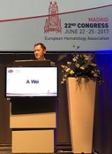
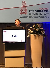
MADRID—The combination of venetoclax and low-dose cytarabine (VEN+LDAC) appears to be a feasible treatment option for elderly patients with untreated acute myeloid leukemia (AML) who are ineligible for intensive chemotherapy.
In a phase 1/2 study of such patients, VEN+LDAC was considered well-tolerated, conferring moderate myelosuppression and largely low-grade non-hematologic toxicities.
In addition, the combination produced “rapid and durable” responses, and early death rates were low, according to Andrew H. Wei, MBBS, PhD, of Monash University in Melbourne, Victoria, Australia.
However, nearly three-quarters of patients ultimately discontinued the treatment, many due to disease progression.
Dr Wei presented these results at the 22nd Congress of the European Hematology Association (EHA) as abstract S473. AbbVie and Genentech, the companies developing and marketing venetoclax, provided financial support for this study.
“Expression of pro-survival proteins is an established mechanism of chemoresistance in diverse cancers,” Dr Wei noted. “BCL-2 is 1 of 5 pro-survival molecules which functions to sequester pro-apoptotic molecules and tip the balance in favor of cell survival.”
“Venetoclax is a potent and specific inhibitor of BCL-2 which releases these pro-apoptotic molecules, tipping the balance in favor of cell death. Cytotoxic drugs are well-known to increase the burden of BH3-only proteins, and so it was surmised that the combination of chemotherapy, such as cytarabine, with venetoclax could augment the clinical response.”
Patients
Dr Wei presented data on 61 AML patients treated with VEN+LDAC. He noted that this was a poor-risk population, with nearly half of patients over the age of 75 at baseline.
The patients’ median age was 74 (range, 66-87), and 64% were male. Nearly half of patients had an ECOG performance status of 1 (49%), 30% had a status of 0, and 21% had a status of 2.
Forty-four percent of patients had secondary AML, and 28% had prior treatment with a hypomethylating agent (HMA). Sixty-one percent of patients had intermediate-risk cytogenetics, and 31% had poor-risk cytogenetics.
Treatment
The patients received oral venetoclax at 600 mg daily on days 1 to 28 and subcutaneous cytarabine at 20 mg/m2 daily on days 1 to 10 of each 28-day cycle.
In the first cycle, the dose of venetoclax was ramped up gradually—no dose on day 1, 50 mg on day 2, 100 mg on day 3, 200 mg on day 4, 400 mg on day 5, and 600 mg thereafter.
Patients received prophylaxis for tumor lysis syndrome prior to starting cycle 1, and they were hospitalized to enable observation.
The median time on study treatment was 6 months (range, <1 to 19 months). Seventy-two percent of patients discontinued treatment.
Reasons for discontinuation included:
- Progressive disease without death—26%
- Progressive disease with death—10%
- Adverse event (AE) related to progression—10%
- AE not related to progression—8%
- Withdrawn consent—8%
- Other reasons—18%.
Safety
The most common AEs of any grade (occurring in at least 30% of patients) were nausea (74%), hypokalemia (46%), diarrhea (46%), fatigue (44%), decreased appetite (41%), constipation (34%), hypomagnesemia (34%), vomiting (31%), thrombocytopenia (44%), febrile neutropenia (38%), and neutropenia (33%).
Grade 3/4 hematologic AEs (occurring in at least 10% of patients) included thrombocytopenia (44%), febrile neutropenia (36%), neutropenia (33%), and anemia (28%).
Grade 3/4 non-hematologic AEs (occurring in at least 10% of patients) included hypokalemia (16%), hypophosphatemia (13%), and hypertension (12%).
Response and survival
The overall response rate was 65%, with 25% of patients achieving a complete response (CR), 38% having a CR with incomplete blood count recovery (CRi), and 2% experiencing a partial response.
Dr Wei noted that VEN+LDAC was active across subgroups.
The CR/CRi rate was 76% among patients with intermediate-risk cytogenetics and 47% among patients with poor-risk cytogenetics.
The CR/CRi rate was 70% among patients older than 75, 52% among patients with secondary AML, 66% among patients with no prior HMA exposure, and 53% in patients with prior HMA exposure.
“Although responses were slightly lower in patients with poor cytogenetic risk, prior HMA exposure, and secondary AML . . ., these responses are far in excess of what we would expect with [LDAC] alone,” Dr Wei said.
“Furthermore, the median time to response was very rapid, and this is extremely important to get patients into remission and avoid the medium-term consequences of active AML.”
The median time to response was 1 month (range, <1 to 9 months).
The 30-day death rate was 3%, the 60-day death rate was 15%, and the median overall survival was approximately 12 months.
Based on these results, AbbVie has initiated a phase 3 trial comparing VEN+LDAC to LDAC alone in elderly patients with untreated AML who are ineligible for intensive chemotherapy.
RNAi therapeutic reduces ABR in hemophilia A and B
BERLIN—Researchers have reported positive results from an ongoing phase 2 trial of fitusiran in patients with hemophilia A or B, with or without inhibitors.
Once-monthly treatment with fitusiran reduced the median annualized bleeding rate (ABR) from 20 to 1 in all patients. In patients with inhibitors, the median ABR fell from 38 to 0.
Most adverse events (AEs) were mild or moderate in severity, with the most common AEs being alanine aminotransferase (ALT) increases and injection-site reactions.
These results were presented* at the International Society on Thrombosis and Haemostasis (ISTH) 2017 Congress.
The research was sponsored by Alnylam Pharmaceuticals, Inc., the company developing fitusiran in collaboration with Sanofi Genzyme.
Fitusiran is an RNAi therapeutic targeting antithrombin for the treatment of patients with hemophilia A and B. Fitusiran is designed to lower levels of antithrombin and promote sufficient thrombin generation upon activation of the clotting cascade to restore hemostasis and prevent bleeding.
John Pasi, PhD, of Barts and the London School of Medicine and Dentistry in London, UK, and his colleagues tested fitusiran in 33 patients, ages 19 to 61, with hemophilia A (n=27) or hemophilia B (n=6), with inhibitors (n=14) or without (n=19).
Fitusiran was administered as a monthly, subcutaneous, fixed dose of 50 mg (n=13) or 80 mg (n=20).
Patients were treated for up to 20 months, with a median of 11 months on study. Five patients discontinued treatment—4 due to withdrawn consent and 1 due to an AE.
Safety
The incidence of AEs was 70%. Most were mild or moderate in severity and unrelated to fitusiran.
Non-laboratory AEs included injection site reactions (18%, n=6), abdominal pain (9%, n=3), diarrhea (9%, n=3), and headache (9%, n=3).
Eleven patients had asymptomatic ALT increases greater than 3 times the upper limit of normal, without concurrent elevations in bilirubin greater than 2 times the upper limit of normal. All of these patients were hepatitis C antibody-positive.
At last follow-up, all ALT elevations were resolved (n=10) or resolving (n=1).
There were serious AEs in 6 patients, and 2 of these events were considered possibly related to fitusiran. One event was seizure with confusion in a patient with a prior history of seizure disorder.
The other serious AE was an asymptomatic ALT elevation in a patient with chronic hepatitis C virus infection. This patient discontinued treatment due to the event.
There were no thromboembolic events, no laboratory evidence for pathological clot formation, and no instances of anti-drug antibody formation.
Efficacy
Fitusiran resulted in approximately 80% lowering of antithrombin, with corresponding increases in thrombin generation.
In all patients, fitusiran reduced the median ABR from 20 (interquartile range [IQR]: 4-36) to 1 (IQR: 0-3).
In patients with inhibitors, fitusiran reduced the median ABR from 38 (IQR: 20-48) to 0 (IQR: 0-3).
Forty-eight percent of all patients (n=16) remained bleed-free during the observation period, and 67% (n=22) did not experience any spontaneous bleeds.
All breakthrough bleeds were successfully managed with replacement factor (recombinant factor VIII or recombinant factor IX) or bypassing agents (recombinant factor VIIa or activated prothrombin complex concentrate).
Based on these results, Sanofi and Alnylam initiated the ATLAS phase 3 program for fitusiran in patients with hemophilia A and B, with or without inhibitors.
*Data in the abstract differ from data presented at the meeting.


