User login
Analysis reveals common sources of bias in scientific research
A recent study suggested that bias in research varies across scientific disciplines, but there are some common factors the different fields share.
The data consistently showed that small studies, early studies, and highly cited studies overestimated effect size.
In addition, a scientist’s early career status, isolation from other researchers, and involvement in misconduct appeared to be risk factors for unreliable results.
John P. A. Ioannidis, MD, of Stanford University School of Medicine in Stanford, California, and his colleagues reported these findings in PNAS.
The team reviewed more than 3000 meta-analyses that included nearly 50,000 individual studies across 22 scientific fields.
“I think that this is a mapping exercise,” Dr Ioannidis said. “It maps all the main biases that have been proposed across all 22 scientific disciplines. Now, we have a map for each scientific discipline, which biases are important, and which have a bigger impact, and, therefore, scientists can think about where do they want to go next with their field.”
Types of bias
The researchers examined several hypothesized kinds of scientific bias, including:
- Small-study effect: When studies with small sample sizes report large effect sizes.
- Gray literature bias: The tendency of smaller or statistically insignificant effects to be reported in PhD theses, conference proceedings, or personal communications rather than in peer-reviewed literature.
- Early extremes effect: When extreme or controversial findings are published early just because they are astonishing.
- Decline effect: When reports of extreme effects are followed by subsequent reports of reduced effects.
- Citation bias: The larger the effect size, the more likely the study will be cited.
- United States effect: When US researchers overestimate effect sizes.
- Industry bias: When industry sponsorship and affiliation affect the direction and size of reported effects.
Dr Ioannidis and his colleagues also looked at other factors that might potentially affect the risk of bias, such as size and types of collaborations, the gender of the researchers, and pressure to publish.
Results
Small studies, highly cited studies, and those published in peer-reviewed journals seemed more likely to overestimate effects. US studies and early studies seemed to report more extreme effects.
Early career researchers and researchers working in small or long-distance collaborations were more likely to overestimate effect sizes. And researchers with a history of misconduct tended to overestimate effect sizes.
On the other hand, studies by highly cited authors who published frequently were not more affected by bias than average. And there was no difference in bias according to gender.
In addition, scientists in countries with strong incentives to publish were not more affected by bias than scientists from countries where there was less pressure to publish.
Dr Ioannidis said that, in the data he and his colleagues examined, the influence of different kinds of bias changed over time and seemed to depend on the individual scientist.
“We show that some of the patterns and risk factors seem to be getting worse in intensity over time,” he said. “This is particularly driven by the social sciences . . . . It seems that the social sciences are seeing the more prominent worsening of these biases over time.”
Another finding of this study is that the kinds and amounts of bias were irregularly distributed across the literature.
“Although bias may be worryingly high in specific research areas, it is nonexistent in many others,” said study author Daniele Fanelli, PhD, of the Meta-Research Innovation Center at Stanford (METRICS), Stanford University in Palo Alto. “So bias does not undermine the scientific enterprise as a whole.”
Yet another finding is that the relative magnitude of biases closely reflects the level of attention they receive in the literature. That is, the kinds of biases researchers are most concerned about are, in fact, the ones they should be concerned about.
“Our understanding of bias is improving, and our priorities are set on the right targets,” Dr Fanelli said, though he noted that researchers should not become complacent when it comes to bias.
“We perhaps understand bias better, but we are far from having rid science of it. Indeed, our results suggest that the challenge might be greater than many think because interventions might need to be tailored to the needs and problems of individual disciplines of fields. One-size-fits all solutions are unlikely to work.” ![]()
Nanoparticles allow for creation of CAR T cells in vivo
Researchers say they have developed biodegradable nanoparticles that can be used to genetically reprogram T cells while they are still in the body.
The nanoparticles were able to program T cells with genes encoding leukemia-specific chimeric antigen receptors (CARs).
The resulting CAR T cells were able to eliminate leukemia or slow the progression of disease in a mouse model of B-cell acute lymphoblastic leukemia (B-ALL).
Researchers reported these results in Nature Nanotechnology.
“Our technology is the first that we know of to quickly program tumor-recognizing capabilities into T cells without extracting them for laboratory manipulation,” said Matthias Stephan, MD, PhD, of Fred Hutchinson Cancer Research Center in Seattle, Washington.
“The reprogrammed cells begin to work within 24 to 48 hours and continue to produce these receptors for weeks. This suggests that our technology has the potential to allow the immune system to quickly mount a strong enough response to destroy cancerous cells before the disease becomes fatal.”
Dr Stephan and his colleagues designed their nanoparticles to carry genes that encode for CARs intended to target and eliminate B-ALL. The nanoparticles are coated with ligands that make them seek out and bind to T cells.
When a nanoparticle binds to a T cell, the cell engulfs the particle. The nanoparticle then travels to the cell’s nucleus and dissolves.
The CAR genes integrate into chromosomes housed in the nucleus, making it possible for the T cells to begin decoding the new genes and producing CARs within 1 or 2 days.
Once they determined their CAR-carrying nanoparticles reprogrammed a noticeable percentage of T cells, the researchers tested the T cells’ efficacy in a mouse model of B-ALL.
The team infused the nanoparticles into 10 mice and found the treatment eradicated tumors in 7 of the animals. The other 3 mice “showed substantial regression” of leukemia, the researchers said.
On average, mice that received CAR-carrying nanoparticles had a 58-day improvement in survival compared to control mice.
Mice that received the nanoparticles also had “dramatically reduced” B-cell numbers in their spleens. The researchers noted that this is consistent with the reversible B-cell aplasia observed in patients who receive conventional CD19 CAR T-cell therapy.
Dr Stephan and his colleagues also tested conventional CAR T-cell therapy in the B-ALL mouse model. The mice received cyclophosphamide followed by CAR T cells created ex vivo.
These mice had significantly better survival than controls, but their survival was comparable to that of the mice that received the CAR-carrying nanoparticles.
Although these nanoparticles are several steps away from the clinic, Dr Stephan said he imagines a future in which nanoparticles transform cell-based immunotherapies into easily administered, off-the-shelf treatments that are available anywhere.
“I’ve never had cancer, but if I did get a cancer diagnosis, I would want to start treatment right away,” Dr Stephan said. “I want to make cellular immunotherapy a treatment option the day of diagnosis and have it able to be done in an outpatient setting near where people live.” ![]()
Health Canada expands approval of daratumumab in MM
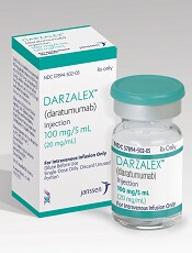
The drug is now approved for use in combination with lenalidomide and dexamethasone or bortezomib and dexamethasone to treat MM patients who have received at least 1 prior therapy.
Health Canada granted daratumumab priority review for this indication due to a high unmet medical need in patients with MM.
When a drug is granted priority review, Health Canada aims to complete its review of the drug within 180 days from the time an application is submitted.
Health Canada grants priority review to products intended for the treatment, prevention, or diagnosis of serious, life-threatening, or severely debilitating diseases or conditions.
About daratumumab
Daratumumab is a human IgG1k monoclonal antibody that binds to CD38, which is highly expressed on the surface of MM cells.
The drug is being developed by Janssen Biotech, Inc. under an exclusive worldwide license from Genmab.
Daratumumab received conditional approval in Canada last year.
In June 2016, Health Canada issued a Notice of Compliance with Conditions approving daratumumab for MM patients who had received at least 3 prior lines of therapy, including a proteasome inhibitor and an immunomodulatory agent, or patients who are refractory to both a proteasome inhibitor and an immunomodulatory agent.
Phase 3 trial data
Health Canada’s expanded approval for daratumumab is based on data from the phase 3 POLLUX and CASTOR trials.
In the POLLUX trial, researchers compared treatment with lenalidomide and dexamethasone to treatment with daratumumab, lenalidomide, and dexamethasone in patients with relapsed or refractory MM.
Patients who received daratumumab in combination had a significantly higher response rate and longer progression-free survival than patients who received the 2-drug combination.
However, treatment with daratumumab was associated with infusion-related reactions and a higher incidence of neutropenia.
Results from this trial were published in NEJM in October 2016.
In the CASTOR trial, researchers compared treatment with bortezomib and dexamethasone to treatment with daratumumab, bortezomib, and dexamethasone in patients with previously treated MM.
Patients who received the 3-drug combination had a higher response rate, longer progression-free survival, and a higher incidence of grade 3/4 adverse events than those who received the 2-drug combination.
Results from this trial were published in NEJM in August 2016. ![]()
Inhibitor exhibits activity against hematologic malignancies
A dual kinase inhibitor is active against a range of hematologic malignancies, according to preclinical research.
Investigators found that ASN002, a SYK/JAK inhibitor, exhibited “potent” antiproliferative activity in leukemia, lymphoma, and myeloma cell lines.
ASN002 also inhibited tumor growth in mouse models of these malignancies and proved active against ibrutinib-resistant diffuse large B-cell lymphoma (DLBCL).
The investigators presented these results at the AACR Annual Meeting 2017 (abstract 4204).
The work was conducted by employees of Asana BioSciences, the company developing ASN002.
The investigators tested ASN002 in 178 cell lines and found the drug exhibited “strong antiproliferative activity” in a range of hematologic cancer cell lines, including:
- Leukemia—HEL31, HL60, Jurkat, MOLM-13, and MOLM-16
- Lymphoma—KARPAS-299, OCI-LY10, OCI-LY-19, Pfeiffer, Raji, Ramos, SU-DHL-1, SU-DHL-6, and SU-DHL-10
- Myeloma—H929, JJN3, OPM2, and U266.
In addition, ASN002 was active against ibrutinib-resistant clones derived from the DLBCL cell line SU-DHL-6.
In a SU-DHL-6 xenograft model, the combination of ASN002 and ibrutinib was more effective than either compound alone.
The investigators also found that ASN002 alone demonstrated “strong tumor growth inhibition” in mouse models of DLBCL (Pfeiffer and SU-DHL-6), myeloma (H929), and erythroleukemia (HEL).
The team pointed out that ASN002 is currently under investigation in a phase 1/2 study of patients with B-cell lymphomas (DLBCL, mantle cell lymphoma, and follicular lymphoma) as well as solid tumors.
The investigators said that, to date, ASN002 has been well tolerated and has shown encouraging early evidence of clinical activity and symptomatic benefit in the lymphoma patients. ![]()
Early prophylaxis may preserve joint health in hemophilia patients
New research suggests that starting prophylactic factor VIII (FVIII) therapy early in life can preserve joint health in patients with hemophilia A.
In studying data on more than 6000 patients, researchers found the use of FVIII prophylaxis increased from 1999 to 2010.
And the rate of bleeding events, including joint bleeds, decreased over this time period.
The study also indicated that starting FVIII prophylaxis before age 4 decreases a patient’s risk of losing normal range of motion (ROM) in his joints.
Marilyn J. Manco-Johnson, MD, of the University of Colorado Denver in Aurora, Colorado, and her colleagues reported these findings in Blood.
The team studied information collected from 6196 males between 2 and 69 years of age with severe hemophilia A. Altogether, these patients had 26,614 visits to US hemophilia treatment centers between 1999 and 2010.
The data collected from the clinic visits were used to examine trends in FVIII prophylaxis use and changes in the patients’ health over the study period.
Prophylaxis and bleeds
Overall, prophylaxis use increased from 31% in 1999 to 59% in 2010. Three-quarters of patients younger than 20 years of age were on prophylaxis by 2010.
The rate of total bleeds fell 17% from 1999 to 2010 for patients receiving prophylaxis—from a mean of 4.91 bleeds every 6 months to a mean of 4.07 bleeds every 6 months. For patients not on prophylaxis, the rate of total bleeds fell 30%, from 14.2 to 9.87 bleeds.
For patients on prophylaxis, the rate of joint bleeding fell 22%, from a mean of 3.03 bleeds every 6 months in 1999 to a mean of 2.36 bleeds every 6 months in 2010. Among patients not on prophylaxis, the rate of joint bleeding fell 23%, from 9.42 to 7.25 bleeds.
The researchers noted that rates of joint bleeding and total bleeding events in patients not on prophylaxis were roughly twice the rates for patients who were using prophylaxis.
Joint ROM
The researchers also conducted longitudinal analyses to assess joint ROM on 3078 patients who had 14,130 visits to hemophilia treatment centers during the study period.
The team found a few factors that were significantly associated with a decrease in overall joint ROM at the patients’ initial visit. This included advancing age (P<0.001), non-white race (P<0.001), and obesity (P=0.003).
Obesity was associated with a significant increase in loss of joint ROM over time as well (P<0.001).
On the other hand, starting prophylaxis before age 4 was associated with a significant decrease in loss of joint ROM over time (P=0.03).
Questions and next steps
The researchers said it isn’t clear why prophylaxis is best able to protect joint ROM when treatment is started at a very young age. And it’s not clear why hemophilia patients who might benefit from prophylaxis aren’t using it.
The team said studies are needed to understand why some hemophilia patients don’t use prophylaxis and to develop strategies that are successful in changing this behavior.
In addition, more work is needed to understand the factors that may lead to joint bleeds and to develop treatment strategies for hemophilia patients who have a higher risk for joint disease. ![]()
Imbalance drives development of B-ALL, team says
Researchers say they have discovered an imbalance that drives the development of B-cell acute lymphoblastic leukemia (B-ALL).
The group’s study suggests that activation of STAT5 causes competition among other transcription factors that leads to B-ALL.
Therefore, the researchers believe that inhibiting the activation of STAT5 and restoring the natural balance of proteins could mean more effective treatment for B-ALL.
Seth Frietze, PhD, of the University of Vermont in Burlington, Vermont, and his colleagues conducted this research and reported the results in Nature Immunology.
The researchers first studied the role of STAT5 in B-ALL using mouse models.
The experiments revealed that STAT5 activation and defects in a signaling pathway worked together to promote B-ALL. The defects were in signaling components of the B-cell antigen receptor precursor—IKAROS, NF-κB, BLNK, BTK, and PKCβ.
With further investigation, the researchers found that STAT5 “antagonized NF-κB and IKAROS by opposing the regulation of shared target genes.”
The team also studied samples from patients with B-ALL and found that patients with a high ratio of active STAT5 to NF-κB or IKAROS had more aggressive disease.
Specifically, the ratio of active STAT5 to IKAROS was negatively correlated with patient survival and the duration of remission. The ratio of active STAT5 to the NF-κB subunit RELA correlated with remission duration but not survival.
“The major outcome of this story is that a signature emerged from looking at the level of activated proteins compared to other proteins that’s very predictive of how a patient will respond to therapy,” Dr Frietze said.
“That’s a novel finding. If we could find drugs to target that activation, that could be an incredibly effective way to treat leukemia.” ![]()
TKI shows promise in preclinical study of AML
WASHINGTON, DC—Preclinical data suggest a novel tyrosine kinase inhibitor (TKI) may be an effective treatment for patients with NTRK-rearranged acute myeloid leukemia (AML).
Entrectinib is a TKI targeting tumors that harbor TRK, ROS1, or ALK fusions.
Researchers found that entrectinib inhibited cell proliferation in NTRK-rearranged AML cell lines.
In mouse models of NTRK-rearranged AML, entrectinib induced tumor regression and eliminated residual AML cells from the bone marrow.
These results were presented at the AACR Annual Meeting 2017 (abstract 5158). The research was conducted by employees of Ignyta, Inc., the company developing entrectinib.
The researchers first tested entrectinib in AML cell lines. They observed “potent” anti-proliferative activity in a pair of NTRK-fusion-positive AML cell lines, IMS-M2 and M0-91.
Entrectinib inhibited TRK signaling and induced cell-cycle arrest in these cell lines. The TKI also induced both caspase 3-dependent apoptosis and PARP cleavage in a dose- and time-dependent manner.
However, entrectinib showed minimal activity against an NTRK-fusion-negative AML cell line, Kasumi-1.
The researchers also tested entrectinib in mouse models of NTRK-fusion-driven AML.
The TKI induced tumor regression in both IMS-M2 and M0-91 models, and the drug eliminated leukemic cells in the bone marrow.
The researchers said these results provide rationale for the clinical development of entrectinib in molecularly defined hematologic malignancies.
Entrectinib is currently being studied in a phase 2 trial of solid tumor malignancies. ![]()
WASHINGTON, DC—Preclinical data suggest a novel tyrosine kinase inhibitor (TKI) may be an effective treatment for patients with NTRK-rearranged acute myeloid leukemia (AML).
Entrectinib is a TKI targeting tumors that harbor TRK, ROS1, or ALK fusions.
Researchers found that entrectinib inhibited cell proliferation in NTRK-rearranged AML cell lines.
In mouse models of NTRK-rearranged AML, entrectinib induced tumor regression and eliminated residual AML cells from the bone marrow.
These results were presented at the AACR Annual Meeting 2017 (abstract 5158). The research was conducted by employees of Ignyta, Inc., the company developing entrectinib.
The researchers first tested entrectinib in AML cell lines. They observed “potent” anti-proliferative activity in a pair of NTRK-fusion-positive AML cell lines, IMS-M2 and M0-91.
Entrectinib inhibited TRK signaling and induced cell-cycle arrest in these cell lines. The TKI also induced both caspase 3-dependent apoptosis and PARP cleavage in a dose- and time-dependent manner.
However, entrectinib showed minimal activity against an NTRK-fusion-negative AML cell line, Kasumi-1.
The researchers also tested entrectinib in mouse models of NTRK-fusion-driven AML.
The TKI induced tumor regression in both IMS-M2 and M0-91 models, and the drug eliminated leukemic cells in the bone marrow.
The researchers said these results provide rationale for the clinical development of entrectinib in molecularly defined hematologic malignancies.
Entrectinib is currently being studied in a phase 2 trial of solid tumor malignancies.
Leukemia, NHL more common in African-born blacks than US-born blacks
The cancer profile of black US immigrants born in sub-Saharan Africa (ABs) differs from that of US-born black individuals (USBs) and varies by region of birth, according to a new study.
For instance, the data showed that ABs who immigrated to the US had a higher incidence of leukemia and non-Hodgkin lymphoma (NHL) than USBs.
And there was a higher incidence of NHL among ABs born in Eastern Africa than among those born in Western Africa.
“Typically, cancer occurrence among blacks in the United States is presented as one homogeneous group, with no breakdown by country or region of birth,” said Ahemdin Jemal, DVM, PhD, of the American Cancer Society in Atlanta, Georgia.
“Our study shows that approach masks important potential differences that may be key to guiding cancer prevention programs for African-born black immigrants.”
Dr Jemal and his colleagues described this study in the journal Cancer.
The team reviewed cancer incidence data from the Surveillance, Epidemiology, and End Results (SEER 17) program covering the period from 2000 through 2012.
They calculated age-standardized proportional incidence ratios (PIRs), comparing the frequency of the top 15 cancers in ABs with that of USBs, by sex and region of birth.
ABs had a significantly higher proportion of hematologic malignancies (leukemia and NHL), infection-related cancers (liver, stomach, and Kaposi sarcoma), prostate cancer, and thyroid cancers (in females only), when compared to USBs.
In females, the incidence of NHL was 3.4% in ABs and 2.5% in USBs, with a PIR of 1.19. The incidence of leukemia was 3.0% in ABs and 1.9% in USBs, with a PIR of 1.62.
In males, the incidence of NHL was 5.9% in ABs and 3.0% in USBs, with a PIR of 1.34. The incidence of leukemia was 3.0% in ABs and 2.2% in USBs, with a PIR of 1.40.
The investigators also calculated the PIRs of the 5 most frequent cancers in ABs compared to USBs by region of origin (Western Africa vs Eastern Africa).
For both sexes, the incidence of NHL was 4.0% in ABs born in Western Africa (PIR=1.17) and 5.5% in ABs born in Eastern Africa (PIR=1.44).
Leukemia was not included in this analysis because it was not among the top 5 cancers.
Therapy increases factor IX activity in hemophilia B
SCOTTSDALE, ARIZONA—The investigational therapy SPK-9001 delivers “consistent and sustained” increases in factor IX (FIX) activity in patients with hemophilia B, according to researchers.
All 10 subjects who received SPK-9001 in a phase 1/2 trial have consistently achieved the targeted therapeutic range of FIX activity.
There have been no confirmed bleeds, and 9 of the subjects have not received any infusions of FIX therapy since receiving SPK-9001.
SPK-9001 reduced the patients’ annualized bleeding rate by 96% and the annualized infusion rate by 99%.
Adam Cuker, MD, of the Perelman School of Medicine at the University of Pennsylvania in Philadelphia, and his colleagues presented these results at the Hemostasis and Thrombosis Research Society 2017 Scientific Symposium.
The trial is sponsored by Spark Therapeutics and Pfizer, the companies developing SPK-9001.
SPK-9001 is a bio-engineered adeno-associated virus capsid expressing a codon-optimized, high-activity human FIX variant enabling endogenous production of FIX.
In the phase 1/2 trial, 10 hemophilia B patients received a single administration of SPK-9001 (5 x 10¹¹ vector genomes/kg body weight).
As of the data cutoff (March 24, 2017), 9 of the 10 patients have not taken FIX concentrates to prevent or control bleeding events since they received SPK-9001.
One patient with severe joint disease has self-administered infusions. The patient self-infused FIX therapy for a suspected ankle bleed on day 2 after receiving SPK-9001 and self-administered precautionary infusions another 9 times between December 2, 2016, and January 2, 2017, for persistent knee pain. The patient has not used additional FIX therapy since January 2.
For all 10 patients, the mean steady-state FIX activity level 12 weeks after SPK-9001 administration was a sustained 33% (range as of the data cutoff: 14% to 81%).
Based on individual patient history prior to the study, the annualized bleeding rate was reduced by 96% to a mean of 0.39 annual bleeds, compared with 9.2 bleeds before SPK-9001 administration.
The annualized infusion rate was reduced 99% to a mean of 0.98 annual infusions, compared with 68.5 infusions before SPK-9001 administration.
To date, no serious adverse events have been reported, including no FIX inhibitors and no thrombotic events.
Two of the 10 patients experienced an asymptomatic, transient elevation in liver enzymes, or decline in FIX activity, potentially indicative of an immune response to the Spark100 vector capsid, that occurred several weeks post-infusion.
Both patients received a tapering dose of oral corticosteroids, after which their alanine aminotransferase levels returned to baseline.
The FIX activity level of one of these patients has stabilized at approximately 15% for more than 9 weeks post-corticosteroid use. The other patient had a FIX activity level between 70% and 80% upon completion of steroid use.
CAR T-cell therapy demonstrates efficacy in mice with MM
WASHINGTON, DC—A chimeric antigen receptor (CAR) T-cell therapy is active against multiple myeloma (MM), according to preclinical research.
The therapy, known as P-BCMA-101, demonstrated persistent anti-tumor activity in a mouse model of MM.
Treatment with P-BCMA-101 eliminated tumors after relapse and prolonged survival in the mice, when compared to other CAR-T cell therapies.
These results were presented in a poster at the AACR Annual Meeting 2017 (abstract 3759).
The research was conducted by employees of Poseida Therapeutics Inc., the company developing P-BCMA-101, as well as others.
About P-BCMA-101
The researchers explained that P-BCMA-101 employs a B-cell maturation antigen (BCMA)-specific Centyrin™ rather than a single-chain variable fragment (scFv) for antigen detection.
Centyrins™ are fully human and have similar binding affinities as scFvs. However, Centyrins are smaller, more thermostable, and predicted to be less immunogenic.
P-BCMA-101 is engineered using the PiggyBac™ DNA modification system. PiggyBac eliminates the need to use lentivirus or gamma-retrovirus as a gene delivery mechanism, resulting in improved manufacturing and cost savings.
In addition, the increased cargo capacity of PiggyBac allows for the incorporation of a safety switch and a selectable gene. The safety switch can be “flipped” to enable depletion in case adverse events occur. And the selectable gene allows for enrichment of CARTyrin+ cells using a non-genotoxic drug.
Findings
The researchers found that more than 70% of P-BCMA-101 cells possessed a stem cell memory phenotype, creating a significant population of self-renewing, multipotent progenitors capable of reconstituting the entire spectrum of memory and effector T-cell subsets required to prevent cancer relapse. Similar competitor products typically report 0% to 20% stem cell memory phenotype.
In addition, P-BCMA-101 was enriched with more than 95% of T cells successfully modified, which compares favorably to the roughly 30% to 50% commonly expected with clinical manufacture using lentivirus.
P-BCMA-101 did not exhibit effects of CAR-mediated tonic signaling, a common cause of T-cell exhaustion that leads to poor durability. Tonic signaling is caused by oligomerization of unstable binding domains commonly seen with traditional scFv CARs.
The researchers tested P-BCMA-101 in NSG mice bearing luciferase+ MM.1S cells. The mice received a single administration of either 4 x 10⁶ or 12 x 106 P-BCMA-101 cells.
P-BCMA-101 treatment reduced tumor burden to the limit of detection within 7 days. Conversely, all untreated control mice succumbed to MM within 4 weeks.
P-BCMA-101 expanded and persisted in the treated mice, eliminated tumors following relapse, and prolonged survival.
Most treated mice survived 110 days, and none of them died from tumor burden during the study. This compares favorably to lentivirus-based products that have shown roughly 50-day survival in the same model.
Based on these results, Poseida plans to initiate a phase 1 trial of P-BCMA-101 in patients with relapsed or refractory MM.


