User login
Fractional laser offers new hope for old burn scars
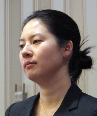
BOSTON – The appearance of mature burn scars significantly improved after treatment with a nonablative fractional laser, based on data from a randomized controlled trial.
In a split-lesion study, laser-treated skin appeared smoother than did adjacent untreated control sites, and within 3 months both patients and clinicians rated laser-treated areas as significantly improved, said Dr. Merete Haedersdal of the University of Copenhagen, Denmark.

"We consider this as a safe treatment, we now have long-term clinical and histological efficacy, and we do have documentation that the efficacy improves over time. We are operating with an intact skin barrier, which may give us a good potential for when we are going to treat patients with large areas of burn scars," she said at the annual meeting of the American Society for Laser Medicine and Surgery.
Dr. Haedersdal and her colleagues in Denmark, Germany, and Belgium examined long-term outcomes from the treatment of burn scars using a 1,540-nm fractional nonablative laser with compression handpieces to deliver energy to deep and superficial tissues.
Twenty patients (median age, 38 years) were enrolled, and 17 completed the study. The patients, all Fitzpatrick skin types II or III, had mature burn scars from fires (75%) or scalding (25%), and 75% had previously received skin grafts. The mature scars, with a median duration of 7 years, involved the trunk and/or extremities.
Side-by-side areas of each lesion were randomly assigned to receive three monthly deep and superficial treatments with Palomar Medical Technologies’ 1,540-nm nonablative fractional laser or to serve as untreated controls.
With the deep (XD) handpiece, the energy was applied with 15-ms pulses at 70 mJ per microbeam in three stacks for 10 passes. The compression tip of the handpiece squeezes moisture away from the applicator end, allowing delivery of energy into deep tissues.
The superficial (XF) handpiece was then used to deliver 50 mJ per microbeam in a 15-ms pulse for one stack with two passes.
"By ultrasound, we saw that we were able to deliver the energy into the deep layers of the skin," Dr. Haedersdal said. She cited the example of one patient whose scar was 1.08 mm thick before treatment, and immediately after treatment it was 3.52 mm thick from edema in the mid and deep dermal layers.
On-site clinical evaluations at 6 months performed by blinded observers rated 15 of 18 treated areas as improved, 3 as showing no response, and none as worsening. In contrast, all 18 control sites were rated as having no response.
The investigators also used the Patient and Observer Scar Assessment Scale (POSAS), which rates vascularity, pigmentation, thickness, relief, pliability, and surface area, as well as overall impression. The scale ranges from 1 for "normal skin" to 10 for "worst scar imaginable." The baseline median score was 7 (range, 3-8) for both groups.
At 1 month after treatment, there was no significant difference in POSAS score between the treated and untreated sides of the scars, but by 3 months the untreated sides were rated as a median of 7, compared with 5 for the treated sides (P = .0185). At 6 months the difference had increased slightly, with a median rating of 7 for the untreated sides and 4 for the treated sides (P = .0008).
The researchers also observed significant improvements in laser-treated (but not control) scars from baseline to 3 months (P = .0185), baseline to 6 months (P = .0008), and from 3 to 6 months (P = .0092), "which actually supports for the first time that when giving these nonablative fractional treatments, there is a continued improvement over time," Dr. Haedersdal said.
The researchers found that meshed (transplanted) skin tended to respond better than nontransplanted skin. Preliminary histology showed treatment-induced remodeling of the stratum corneum, frequently with a thicker epidermal compartment. In addition, post-treatment collagen deposition appeared closer to that found in normal skin, Dr. Haedersdal noted.
The immediate post-treatment responses included edema in 17 of 20 patients, erythema in 18, and purpura in 15, but there was no blistering of skin, and the skin barrier remained intact.
During the study period, 6 of 20 patients had hyperpigmentation, which resolved gradually over time.
"We also saw a grid pattern in three of the patients using these parameters, but we softened up the treatments afterward by giving following treatments with the XF handpiece," Dr. Haedersdal said.
The study was supported by an equipment loan and research grant to Dr. Haedersdal from Palomar Medical Technologies.
BOSTON – The appearance of mature burn scars significantly improved after treatment with a nonablative fractional laser, based on data from a randomized controlled trial.
In a split-lesion study, laser-treated skin appeared smoother than did adjacent untreated control sites, and within 3 months both patients and clinicians rated laser-treated areas as significantly improved, said Dr. Merete Haedersdal of the University of Copenhagen, Denmark.
"We consider this as a safe treatment, we now have long-term clinical and histological efficacy, and we do have documentation that the efficacy improves over time. We are operating with an intact skin barrier, which may give us a good potential for when we are going to treat patients with large areas of burn scars," she said at the annual meeting of the American Society for Laser Medicine and Surgery.
Dr. Haedersdal and her colleagues in Denmark, Germany, and Belgium examined long-term outcomes from the treatment of burn scars using a 1,540-nm fractional nonablative laser with compression handpieces to deliver energy to deep and superficial tissues.
Twenty patients (median age, 38 years) were enrolled, and 17 completed the study. The patients, all Fitzpatrick skin types II or III, had mature burn scars from fires (75%) or scalding (25%), and 75% had previously received skin grafts. The mature scars, with a median duration of 7 years, involved the trunk and/or extremities.
Side-by-side areas of each lesion were randomly assigned to receive three monthly deep and superficial treatments with Palomar Medical Technologies’ 1,540-nm nonablative fractional laser or to serve as untreated controls.
With the deep (XD) handpiece, the energy was applied with 15-ms pulses at 70 mJ per microbeam in three stacks for 10 passes. The compression tip of the handpiece squeezes moisture away from the applicator end, allowing delivery of energy into deep tissues.
The superficial (XF) handpiece was then used to deliver 50 mJ per microbeam in a 15-ms pulse for one stack with two passes.
"By ultrasound, we saw that we were able to deliver the energy into the deep layers of the skin," Dr. Haedersdal said. She cited the example of one patient whose scar was 1.08 mm thick before treatment, and immediately after treatment it was 3.52 mm thick from edema in the mid and deep dermal layers.
On-site clinical evaluations at 6 months performed by blinded observers rated 15 of 18 treated areas as improved, 3 as showing no response, and none as worsening. In contrast, all 18 control sites were rated as having no response.
The investigators also used the Patient and Observer Scar Assessment Scale (POSAS), which rates vascularity, pigmentation, thickness, relief, pliability, and surface area, as well as overall impression. The scale ranges from 1 for "normal skin" to 10 for "worst scar imaginable." The baseline median score was 7 (range, 3-8) for both groups.
At 1 month after treatment, there was no significant difference in POSAS score between the treated and untreated sides of the scars, but by 3 months the untreated sides were rated as a median of 7, compared with 5 for the treated sides (P = .0185). At 6 months the difference had increased slightly, with a median rating of 7 for the untreated sides and 4 for the treated sides (P = .0008).
The researchers also observed significant improvements in laser-treated (but not control) scars from baseline to 3 months (P = .0185), baseline to 6 months (P = .0008), and from 3 to 6 months (P = .0092), "which actually supports for the first time that when giving these nonablative fractional treatments, there is a continued improvement over time," Dr. Haedersdal said.
The researchers found that meshed (transplanted) skin tended to respond better than nontransplanted skin. Preliminary histology showed treatment-induced remodeling of the stratum corneum, frequently with a thicker epidermal compartment. In addition, post-treatment collagen deposition appeared closer to that found in normal skin, Dr. Haedersdal noted.
The immediate post-treatment responses included edema in 17 of 20 patients, erythema in 18, and purpura in 15, but there was no blistering of skin, and the skin barrier remained intact.
During the study period, 6 of 20 patients had hyperpigmentation, which resolved gradually over time.
"We also saw a grid pattern in three of the patients using these parameters, but we softened up the treatments afterward by giving following treatments with the XF handpiece," Dr. Haedersdal said.
The study was supported by an equipment loan and research grant to Dr. Haedersdal from Palomar Medical Technologies.
AT LASER 2013
Major finding: At 3 months, untreated areas were rated 7 on the POSAS scale, compared with 5 for laser-treated areas (P = .0185).
Data source: A randomized controlled trial in 20 patients, comparing side-by-side areas of untreated and laser-treated mature burn scars.
Disclosures: The study was supported by an equipment loan and research grant to Dr. Haedersdal from Palomar Medical Technologies.

Safety First: Fractional Nonablative Laser Resurfacing in Fitzpatrick Skin Types IV to VI

In the April 2013 issue of the Journal of Drugs in Dermatology (2013;12:428-431), Clark et al retrospectively reviewed 115 laser sessions with the 1550-nm erbium-doped fractional nonablative laser (Fraxel Re:Store SR 1550, Solta Medical) in 45 patients with Fitzpatrick skin types IV to VI to assess the rate of postinflammatory hyperpigmentation and the associated laser parameters. The fluence, treatment level, and number of passes were all reviewed, as well as any posttreatment complications (ie, erythema, blistering, edema, bruising, pain) and long-term (1 month) complications (ie, hypopigmentation, hyperpigmentation). All patients were pretreated with hydroquinone cream 4% 2 weeks before, stopping 7 days before treatment and then continuing 4 weeks thereafter. Also, continuous forced-air cooling was used during treatment as well as posttreatment ice packs. Fifty-eight percent (26/45) of treatments were performed in patients with Fitzpatrick skin type IV, 24% (11/45) with type V, and 18% (8/45) with type VI. Laser parameters ranged from 4 to 70 mJ, treatment level 2 to 9, and 4 to 8 passes. Of 115 sessions, 5 (4%) were associated with postinflammatory hyperpigmentation; 2 of these instances occurred in 1 patient. The occurrence of postinflammatory hyperpigmentation was found to be statistically significant (P=.05), correlating with higher mean energy levels compared to those without hyperpigmentation (60.8 vs 44.7 mJ). Only 1 episode of postinflammatory hyperpigmentation lasted longer than 1 month, and 2 of 5 cases had only transient (<7 days) hyperpigmentation. All 5 cases resolved.
What’s the issue?
The 1550-nm erbium-doped fractional nonablative laser is being used for many skin conditions and has a low incidence of adverse effects when appropriate laser parameters are chosen. When treating darker skin phototypes with this technology, the concern for postinflammatory pigmentary alteration is more concerning. Higher treatment densities used in darker phototypes have been associated with a greater risk for postinflammatory hyperpigmentation. In their review, the authors showed that higher energy levels were associated with their cases of postinflammatory hyperpigmentation, with the caveat that they were careful not to use higher density or treatment levels than they would have used in lighter phototypes. Importantly, all 5 cases of hyperpigmentation did resolve and only 1 lasted longer than 1 month (2 months in total). This analysis reinforces that the 1550-nm erbium-doped fractional nonablative laser is quite safe in Fitzpatrick skin types IV to VI when appropriate parameters are utilized, as well as methodical pretreatment and posttreatment with hydroquinone, concomitant cooling, and strict posttreatment sun protection. With the right parameters, the treatment is quite safe; however, what are the optimal treatment parameters to provide efficacious and lasting results?

Ipilimumab plus surgery boosted advanced melanoma survival
NATIONAL HARBOR, MD – Patients with stage IV melanoma treated with a combination of ipilimumab and surgical resection had a high rate of melanoma-specific and overall survival, a retrospective study of a single-center case series has shown.
"To our knowledge, this is the first report of 5-year melanoma-specific survival data on patients who have undergone surgical resection and ipilimumab treatment, and the data suggests that surgical resection and ipilimumab treatment may result in long-term survival in select metastatic melanoma patients," Dr. Junko Ozao-Choy said at the annual Society of Surgical Oncology Cancer Symposium.

Among 44 patients treated with the CTLA-4 (cytotoxic T-lymphocyte antigen 4) inhibitor ipilimumab (Yervoy) and surgical resection, the 5-year melanoma-specific survival (MSS) rate was 51% and the median overall survival duration was 60 months, reported Dr. Ozao-Choy of the John Wayne Cancer Institute at Saint John’s Health Center in Santa Monica, Calif.
For 24 patients who received ipilimumab before resection, the 5-year MSS was 61% at a median of 60 months, and for 18 of 20 patients treated with ipilimumab after surgery, the 5-year MSS was 42% at a median of 47 months, but this difference was not significant (data were incomplete for 2 patients in the latter group), she noted.
In a recent study of retrospective data on patients with metastatic melanoma treated at her center, the 4-year survival of patients who underwent resection of metastatic lesions with or without systemic medical therapy was 20.8%, compared with 7% for those who underwent systemic medical therapy alone. The study investigators concluded that more than half of patients with metastatic melanoma were eligible for metastasectomy (Ann. Surg. Oncol. 2012;19:2547-55).
Dr. Ozao-Choy and her colleagues reviewed the center’s records on patients with metastatic melanoma who underwent resection and had received ipilimumab, looking at disease-specific survival from the date of diagnosis of stage IV disease.
The groups were well balanced in terms of age, sex, mean Breslow thickness scores, and nodal status. However, significantly more patients who received ipilimumab before surgery had brain metastases (13 of 24 vs. 3 of 18, P = .001). In a univariate analysis, patients with brain metastases had a significantly worse 5-year MSS (31% vs. 60%, P = .049).
The only other significant variables associated in the univariate analysis with better survival were prior immunotherapy, with 69% of patients who had received any immunotherapy having a 5-year MSS of 69%, compared with 29% for those with no immunotherapy (P = .01), and previous number of resections, with more resections being associated with better survival (P = .01).
Neither previous treatment with Bacillus Calmette-Guérin vaccine, previous chemotherapy, T stage, N stage, or timing of ipilimumab were significantly associated with MSS.
In a multivariate analysis (which controlled for demographic and disease factors), only the previous number of resections remained a significant predictor of MSS (P = .01).
In the audience response segment following the presentation, Dr. Daniel G. Coit, a surgical oncologist at Memorial Sloan-Kettering Cancer Center in New York City, pointed out that the previous number of resections is a not an adequate independent predictor for survival. "One of the inescapable truths is that if you have to have more than one operation, you have to still be alive. ... Of necessity, older people live longer than younger people; people who die at an older age live longer than people who die at a younger age," he said.
The study was internally funded. Dr. Ozao-Choy and Dr. Coit reported having no relevant financial disclosures.
NATIONAL HARBOR, MD – Patients with stage IV melanoma treated with a combination of ipilimumab and surgical resection had a high rate of melanoma-specific and overall survival, a retrospective study of a single-center case series has shown.
"To our knowledge, this is the first report of 5-year melanoma-specific survival data on patients who have undergone surgical resection and ipilimumab treatment, and the data suggests that surgical resection and ipilimumab treatment may result in long-term survival in select metastatic melanoma patients," Dr. Junko Ozao-Choy said at the annual Society of Surgical Oncology Cancer Symposium.
Among 44 patients treated with the CTLA-4 (cytotoxic T-lymphocyte antigen 4) inhibitor ipilimumab (Yervoy) and surgical resection, the 5-year melanoma-specific survival (MSS) rate was 51% and the median overall survival duration was 60 months, reported Dr. Ozao-Choy of the John Wayne Cancer Institute at Saint John’s Health Center in Santa Monica, Calif.
For 24 patients who received ipilimumab before resection, the 5-year MSS was 61% at a median of 60 months, and for 18 of 20 patients treated with ipilimumab after surgery, the 5-year MSS was 42% at a median of 47 months, but this difference was not significant (data were incomplete for 2 patients in the latter group), she noted.
In a recent study of retrospective data on patients with metastatic melanoma treated at her center, the 4-year survival of patients who underwent resection of metastatic lesions with or without systemic medical therapy was 20.8%, compared with 7% for those who underwent systemic medical therapy alone. The study investigators concluded that more than half of patients with metastatic melanoma were eligible for metastasectomy (Ann. Surg. Oncol. 2012;19:2547-55).
Dr. Ozao-Choy and her colleagues reviewed the center’s records on patients with metastatic melanoma who underwent resection and had received ipilimumab, looking at disease-specific survival from the date of diagnosis of stage IV disease.
The groups were well balanced in terms of age, sex, mean Breslow thickness scores, and nodal status. However, significantly more patients who received ipilimumab before surgery had brain metastases (13 of 24 vs. 3 of 18, P = .001). In a univariate analysis, patients with brain metastases had a significantly worse 5-year MSS (31% vs. 60%, P = .049).
The only other significant variables associated in the univariate analysis with better survival were prior immunotherapy, with 69% of patients who had received any immunotherapy having a 5-year MSS of 69%, compared with 29% for those with no immunotherapy (P = .01), and previous number of resections, with more resections being associated with better survival (P = .01).
Neither previous treatment with Bacillus Calmette-Guérin vaccine, previous chemotherapy, T stage, N stage, or timing of ipilimumab were significantly associated with MSS.
In a multivariate analysis (which controlled for demographic and disease factors), only the previous number of resections remained a significant predictor of MSS (P = .01).
In the audience response segment following the presentation, Dr. Daniel G. Coit, a surgical oncologist at Memorial Sloan-Kettering Cancer Center in New York City, pointed out that the previous number of resections is a not an adequate independent predictor for survival. "One of the inescapable truths is that if you have to have more than one operation, you have to still be alive. ... Of necessity, older people live longer than younger people; people who die at an older age live longer than people who die at a younger age," he said.
The study was internally funded. Dr. Ozao-Choy and Dr. Coit reported having no relevant financial disclosures.
AT SSO 2013
Major finding: The 5-year melanoma-specific survival rate was 51%, and the median overall survival duration was 60 months for patients with advanced melanoma treated with ipilimumab and resection.
Data source: A retrospective study of a single-center case series of 44 patients.
Disclosures: The study was internally funded. Dr. Ozao-Choy and Dr. Coit reported having no relevant financial disclosures.
Agencies continue push for indoor tanning regulations
MIAMI BEACH – Calls for a ban on the use of tanning beds by minors in the United States have thus far gone unheeded, but medical organizations are increasingly supporting such a ban – and with good reason, according to Alan Geller of the Harvard School of Public Health, Boston.
The data linking tanning bed use and melanoma are consistent and convincing. A 2010 University of Minnesota case-control study, for example, demonstrated that melanoma risk was significantly increased among users, compared with nonusers, of UVB-enhanced tanning devices (adjusted odds ratio, 2.86) and primarily UVA-emitting devices (AOR, 4.44), Mr. Geller said at the annual meeting of the American Academy of Dermatology.

The risk increased as tanning bed use increased (Cancer Epidemiol. Biomarkers Prev. 2010;19:1557-68).
A more recent study demonstrated that with every visit to a tanning bed, the risk of melanoma increased by 1.8% – and the risk was even greater among those who started tanning at a younger age (BMJ 2012;345:e4757).
"We are clearly in the throes of a modern-day epidemic, particularly among teenage white girls and young women between the ages of 18 and 25," Mr. Geller said, noting that study after study shows that about a third of white teenage girls and about 20% of all teenage girls use a tanning bed by the age of 17.
And yet only five states restrict the use of tanning beds by those under age 18. Others have parental consent restrictions, but these have been shown to have no effect on tanning bed use by minors. That means that in 45 states, children aged 15 years and younger are free to visit tanning salons with no restrictions, he said, noting that a Washington University in St. Louis survey released in February showed that 65% of Missouri tanning salon owners would allow preteens aged 10-12 years to use their tanning beds – and that 43% of tanning salon employees believe indoor tanning poses no health risks.
Data show that 7% of girls use tanning beds by age 14 years. This doubles from age 14 to 15, and doubles again from age 15 to 17, he said, noting that girls are about five to six times more likely than boys to use tanning beds.
Of particular concern, not only are girls using tanning beds early, but they are using them more often.
A Centers for Disease Control and Prevention survey showed that while the rate of use (20% among all girls) has remained constant in recent years, the "prom phenomenon" – the occasional use of tanning beds before a special event – is no longer the norm; the average yearly number of uses of tanning beds among those surveyed was 28.
"We’re way past the prom phenomenon," Mr. Geller said, noting that one reason for this is that tanning salons "do a wonderful job of selling giant packages of use for very little money."
"When people are beginning to think of some kind of restrictions on the tanning bed industry, that would be one we could surely consider," he said, noting that based on the data showing a 1.8% increase in melanoma risk with each tanning bed use, the risk would be 54%-90% in a teen who starts tanning at age 18 and quits at age 19.
That’s a conservative estimate, because most teens start before age 18 and don’t stop at age 19, he said.
Surveillance, Epidemiology, and End Results (SEER) data from the National Cancer Institute show that the risk of melanoma has doubled among women aged 20-24 years since the 1980s, while the risk in men has declined in some age groups, and remained the same in others.
"You have to ask what’s happened during that time," Mr. Geller said, adding that there is concern about the late effects of tanning bed use, especially given that sun exposure time hasn’t changed in that age group over time.
As for what can be done from a public health perspective to reduce tanning bed use, Mr. Geller said a number of research, legislative, and public health campaigns are underway.
"We know from doing qualitative work, that indoor tanning is largely socially driven. "When [girls] are not tanning, they talk about tanning, they blog about tanning," he said, explaining that "the tanning culture involves some kind of socially driven bonds."
The key is to figure out how to break up those bonds.
"If one girl in a social group quits tanning, will this have an effect on the others? We don’t know," he said, adding that this is among the areas that require further research.
Researchers are also studying the effects of antitanning campaigns and legislation in other countries, a number of which have restricted access to tanning beds for minors. A recent web-based advertising campaign in Denmark targeted teens, and, along with legislation restricting access, resulted in a substantial drop in tanning bed use there, he said.
The results of campaigns and legislative efforts like these are being closely monitored so that the lessons learned about if and how they work can be incorporated into efforts here.
Lessons from the campaign against smoking launched three decades ago also are being incorporated into the current effort to reduce tanning, he said.
Although the link between tanning and melanoma isn’t quite as strong as the link between smoking and lung cancer, the seven key principles that made the antismoking campaign a success can be adapted for this purpose. These are surveillance, taxation, legal strategies, public health advertising campaigns, educational programs, legislation, and "some move to mandate enforcement," he said.
Some progress has been made with respect to these principles. For example, state-by-state surveillance and scoring of states’ level of compliance with existing regulations are underway, a 10% tax has been imposed on tanning salons, cost-efficacy studies are being planned, and lawsuits have been filed in multiple states. However, most of these efforts are in their infancy, Mr. Geller said.
For now, what exists across the United States is a "patchwork quilt of pretty crummy regulations," he said.
While intense pressure is on the Food and Drug Administration to ban tanning bed use by those under age 18 – including pressure from the American Academy of Dermatology – and while the agency is cognizant of the risks and has acknowledged a need for more regulations, "politics have prevailed, and at this point we don’t have the ban," he said.
The FDA website does, however, indicate plans for revising regulations and strengthening warning labels to make consumers more aware of the risks, he noted.
"This is good, but I think it’s a really faulty response to everything that we know about the link between tanning beds and melanoma," he said.
Despite the slow progress toward a ban for those under age 18, there have been some successes in the antitanning campaign. For one, numerous organizations have taken up the cause, including the World Health Organization, the American Academy of Pediatrics, the American Medical Association, the Society of Surgical Oncology, and the Canadian Pediatric Society.
Also, thanks to a Federal Trade Commission crackdown in 2010, the tanning industry is no longer allowed to claim that tanning has certain health benefits, such as reducing the risks of some types of cancers. And in 2012, the U.S. Preventive Services Task Force issued its first guidelines on tanning, stating that the evidence is strong enough to recommend that women aged 10-24 years who have fair skin should avoid prime-time sun exposure and tanning beds.
Additionally, a wellness provision of the Patient Protection and Affordable Care Act that will go into effect in May provides for full reimbursement to health care providers for counseling about skin cancer prevention and tanning bed reduction.
"We want to study this because we think this will have a huge effect on increasing the rate of counseling," he said.
Mr. Geller reported having no disclosures.
MIAMI BEACH – Calls for a ban on the use of tanning beds by minors in the United States have thus far gone unheeded, but medical organizations are increasingly supporting such a ban – and with good reason, according to Alan Geller of the Harvard School of Public Health, Boston.
The data linking tanning bed use and melanoma are consistent and convincing. A 2010 University of Minnesota case-control study, for example, demonstrated that melanoma risk was significantly increased among users, compared with nonusers, of UVB-enhanced tanning devices (adjusted odds ratio, 2.86) and primarily UVA-emitting devices (AOR, 4.44), Mr. Geller said at the annual meeting of the American Academy of Dermatology.
The risk increased as tanning bed use increased (Cancer Epidemiol. Biomarkers Prev. 2010;19:1557-68).
A more recent study demonstrated that with every visit to a tanning bed, the risk of melanoma increased by 1.8% – and the risk was even greater among those who started tanning at a younger age (BMJ 2012;345:e4757).
"We are clearly in the throes of a modern-day epidemic, particularly among teenage white girls and young women between the ages of 18 and 25," Mr. Geller said, noting that study after study shows that about a third of white teenage girls and about 20% of all teenage girls use a tanning bed by the age of 17.
And yet only five states restrict the use of tanning beds by those under age 18. Others have parental consent restrictions, but these have been shown to have no effect on tanning bed use by minors. That means that in 45 states, children aged 15 years and younger are free to visit tanning salons with no restrictions, he said, noting that a Washington University in St. Louis survey released in February showed that 65% of Missouri tanning salon owners would allow preteens aged 10-12 years to use their tanning beds – and that 43% of tanning salon employees believe indoor tanning poses no health risks.
Data show that 7% of girls use tanning beds by age 14 years. This doubles from age 14 to 15, and doubles again from age 15 to 17, he said, noting that girls are about five to six times more likely than boys to use tanning beds.
Of particular concern, not only are girls using tanning beds early, but they are using them more often.
A Centers for Disease Control and Prevention survey showed that while the rate of use (20% among all girls) has remained constant in recent years, the "prom phenomenon" – the occasional use of tanning beds before a special event – is no longer the norm; the average yearly number of uses of tanning beds among those surveyed was 28.
"We’re way past the prom phenomenon," Mr. Geller said, noting that one reason for this is that tanning salons "do a wonderful job of selling giant packages of use for very little money."
"When people are beginning to think of some kind of restrictions on the tanning bed industry, that would be one we could surely consider," he said, noting that based on the data showing a 1.8% increase in melanoma risk with each tanning bed use, the risk would be 54%-90% in a teen who starts tanning at age 18 and quits at age 19.
That’s a conservative estimate, because most teens start before age 18 and don’t stop at age 19, he said.
Surveillance, Epidemiology, and End Results (SEER) data from the National Cancer Institute show that the risk of melanoma has doubled among women aged 20-24 years since the 1980s, while the risk in men has declined in some age groups, and remained the same in others.
"You have to ask what’s happened during that time," Mr. Geller said, adding that there is concern about the late effects of tanning bed use, especially given that sun exposure time hasn’t changed in that age group over time.
As for what can be done from a public health perspective to reduce tanning bed use, Mr. Geller said a number of research, legislative, and public health campaigns are underway.
"We know from doing qualitative work, that indoor tanning is largely socially driven. "When [girls] are not tanning, they talk about tanning, they blog about tanning," he said, explaining that "the tanning culture involves some kind of socially driven bonds."
The key is to figure out how to break up those bonds.
"If one girl in a social group quits tanning, will this have an effect on the others? We don’t know," he said, adding that this is among the areas that require further research.
Researchers are also studying the effects of antitanning campaigns and legislation in other countries, a number of which have restricted access to tanning beds for minors. A recent web-based advertising campaign in Denmark targeted teens, and, along with legislation restricting access, resulted in a substantial drop in tanning bed use there, he said.
The results of campaigns and legislative efforts like these are being closely monitored so that the lessons learned about if and how they work can be incorporated into efforts here.
Lessons from the campaign against smoking launched three decades ago also are being incorporated into the current effort to reduce tanning, he said.
Although the link between tanning and melanoma isn’t quite as strong as the link between smoking and lung cancer, the seven key principles that made the antismoking campaign a success can be adapted for this purpose. These are surveillance, taxation, legal strategies, public health advertising campaigns, educational programs, legislation, and "some move to mandate enforcement," he said.
Some progress has been made with respect to these principles. For example, state-by-state surveillance and scoring of states’ level of compliance with existing regulations are underway, a 10% tax has been imposed on tanning salons, cost-efficacy studies are being planned, and lawsuits have been filed in multiple states. However, most of these efforts are in their infancy, Mr. Geller said.
For now, what exists across the United States is a "patchwork quilt of pretty crummy regulations," he said.
While intense pressure is on the Food and Drug Administration to ban tanning bed use by those under age 18 – including pressure from the American Academy of Dermatology – and while the agency is cognizant of the risks and has acknowledged a need for more regulations, "politics have prevailed, and at this point we don’t have the ban," he said.
The FDA website does, however, indicate plans for revising regulations and strengthening warning labels to make consumers more aware of the risks, he noted.
"This is good, but I think it’s a really faulty response to everything that we know about the link between tanning beds and melanoma," he said.
Despite the slow progress toward a ban for those under age 18, there have been some successes in the antitanning campaign. For one, numerous organizations have taken up the cause, including the World Health Organization, the American Academy of Pediatrics, the American Medical Association, the Society of Surgical Oncology, and the Canadian Pediatric Society.
Also, thanks to a Federal Trade Commission crackdown in 2010, the tanning industry is no longer allowed to claim that tanning has certain health benefits, such as reducing the risks of some types of cancers. And in 2012, the U.S. Preventive Services Task Force issued its first guidelines on tanning, stating that the evidence is strong enough to recommend that women aged 10-24 years who have fair skin should avoid prime-time sun exposure and tanning beds.
Additionally, a wellness provision of the Patient Protection and Affordable Care Act that will go into effect in May provides for full reimbursement to health care providers for counseling about skin cancer prevention and tanning bed reduction.
"We want to study this because we think this will have a huge effect on increasing the rate of counseling," he said.
Mr. Geller reported having no disclosures.
EXPERT ANALYSIS FROM THE AAD ANNUAL MEETING

Alstonia scholaris
Alstonia scholaris, a tree that grows 50-80 feet high and belongs to the Apocynaceae family, has a long history of use in traditional and homeopathic medicine, including Ayurvedic medicine in India, where it is known as sapthaparna (Integr. Cancer Ther. 2009;8:273-9), in traditional Chinese medicine (J. Ethnopharmacol. 2010;129:293-8; J. Ethnopharmacol. 2010;129:174-81), and in traditional medicine in Africa and Australia (Integr. Cancer Ther. 2010;9:261-9). The bark contains the alkaloids ditamine, echitamine (or ditaine), and echitanines; and decoctions or other preparations of the bark have been used to treat gastrointestinal conditions (Grieve M. A Modern Herbal (Vol. 1). New York, Dover Publications, 1971, p. 29). Often called the devil’s tree, the bark of A. scholaris also has been used to treat malaria, cutaneous diseases, tumors, ulcers, chronic respiratory conditions (such as asthma and bronchitis), helminthiasis, and agalactia (Chin. J. Integr. Med. 2012 Mar 28 [Epub ahead of print]).
In the study of A. scholaris most directly pertinent to potential dermatologic treatment, Lee et al. found that ethanolic bark extracts of A. scholaris significantly suppressed retinoid-induced skin irritation in vitro and in vivo, in human HaCat keratinocytes. The investigators identified echitamine and loganin as the primary components likely responsible for the anti-inflammatory effects.

Data showed that A. scholaris dose-dependently inhibited the all-trans retinoic acid–induced releases of the pro-inflammatory cytokines monocyte chemoattractant protein-1 (MCP-1) and interleukin-8 (IL-8) in vitro. Also in vitro, A. scholaris extract potently suppressed radiation-induced increases in matrix metalloproteinase-1 (MMP-1). Importantly, in a cumulative irritation patch test, the botanical extract diminished retinol-induced skin irritation while enhancing retinoid activity in blocking MMP-1 expression, which is linked closely to cutaneous aging. The authors concluded that A. scholaris appears to have the dual benefits of decreasing irritation associated with retinoids while augmenting their antiaging impact (Evid. Based Complement. Alternat. Med. 2012;2012:190370).
The leaf extract of A. scholaris has been used to treat cold symptoms and tracheitis, and it has been prescribed in hospitals and approved for commercial over-the-counter sale by the State Food and Drugs Administration of China (SFDA) (J. Ethnopharmacol. 2010;129:293-8; J. Ethnopharmacol. 2010;129:174-81). The broad range of biological properties associated with A. scholaris has been ascribed to particular constituent categories, including alkaloids, flavonoids, and terpenoids (specifically, phenolic acids) (Chin. J. Integr. Med. 2012 Mar 28 [Epub ahead of print]). These properties include, but are reportedly not limited to, antioxidant, anticancer, anti-inflammatory, antistress, analgesic, antimutagenic, hepatoprotective, immunomodulatory, and chemopreventive activity (Integr. Cancer Ther. 2010;9:261-9; Chin. J. Integr. Med. 2012 Mar 28 [Epub ahead of print]). Antineoplastic effects have been linked directly to phytochemical constituents including echitamine, alstonine, pleiocarpamine, O-methylmacralstonine, macralstonine, and lupeol (Integr. Cancer Ther. 2010;9:261-9).
In 2006, Jagetia and Baliga investigated the anticancer activity of A. scholaris alkaloid fractions in vitro in cultured human neoplastic cell lines. They also conducted in vivo studies in tumor-bearing mice. The in vitro data in HeLa cells revealed a time-dependent rise in antineoplastic activity after 24 hours of exposure (25 mcg/mL). Further, once-daily administration of A. scholaris (240 mg/kg) to tumor-bearing mice yielded dose-dependent remissions, although there were toxic presentations at this dosage. The next-lower dose of 210 mg/kg was found to be most effective, with 20% of the mice surviving for as long as 120 days after tumor cell inoculation, compared with none of the control animals treated with saline (Phytother. Res. 2006;20:103-9).
Using an acute-restraint stress model in mice in 2009, Kulkarni and Juvekar evaluated the effects of stress and the impact of a methanolic extract of A. scholaris bark. Pretreatments with the extract of 100, 250, and 500 mg/kg for 7 days were found to exert significant antistress effects. In addition, nootropic activities were observed, with memory functions clearly enhanced in learning tasks. A. scholaris also was associated with significant antioxidant properties. The extract at 200 mcg/mL exhibited maximum scavenging of the stable radical 1,1-diphenyl-2-picrylhydrazyl at 90.11% and the nitric oxide radical at 62.77% (Indian J. Exp. Biol. 2009;47:47-52).
Later in 2009, Jahan et al. reported on their investigation of potential antioxidant and chemopreventive activity displayed by A. scholaris in a two-stage murine model. Skin carcinogenesis development was initiated in Swiss albino mice through one application of 7, 12-dimethyabenz(a)anthrecene (DMBA) and then promoted two weeks later by repeated application of croton oil three times per week through 16 weeks. The investigators found a lower incidence of tumors, tumor yield, tumor burden, and number of papillomas in mice treated with A. scholaris extract as compared to untreated controls (Integr. Cancer Ther. 2009;8:273-9).
In 2010, Shang et al. conducted multiple studies using A. scholaris. In the first published report, they assessed the anti-inflammatory and analgesic properties of the ethanolic leaf extract to validate its use in traditional Chinese medicine and modern clinical medicine. The investigators first determined that analgesic activity was conferred as the ethyl acetate and alkaloid fractions significantly diminished acetic acid-induced reactions in mice and, along with the ethanolic extract, reduced xylene-induced ear edema.
The researchers also performed in vivo and in vitro assessments of anti-inflammatory activity again on xylene-induced ear edema and carrageenan-induced air pouch formation in mice, as well as cyclooxygenase (COX)-1, -2 and 5-LOX inhibition.
In the air pouch model, A. scholaris alkaloids were found to have significantly spurred superoxide dismutase activity while lowering nitric oxide, prostaglandin E2, and malondialdehyde levels. In vitro tests, supporting evidence from animal models, showed that the three primary alkaloids isolated from A. scholaris leaves (picrinine, vallesamine, and scholaricine) inhibited the inflammatory mediators COX-1, COX-2, and 5-LOX. The researchers also noted that the in vitro anti-inflammatory assay results reinforced the notion of these alkaloids as the bioactive fraction of the plant (J. Ethnopharmacol. 2010;129:174-81).
In their second published report that year, Shang et al. investigated the antitussive and anti-asthmatic activities of the ethanolic extract, fractions, and chief alkaloids of A. scholaris leaf.
The researchers tested for antitussive effects using ammonia-induced or sulfur dioxide-induced coughing in mice and citric acid-induced coughing in guinea pigs. They evaluated anti-asthmatic activity via histamine-induced bronchoconstriction in guinea pigs. They also measured phenol red volume in murine tracheas to assess expectorant activity.
The data indicated antitussive activity, with significant alkaloid suppression of ammonia-induced coughing frequency in mice. Latency periods of sulfur dioxide-induced cough in mice and citric acid-induced cough in guinea pigs increased, and cough frequency in guinea pigs decreased.
Anti-asthmatic effects, such as suppression of convulsion, were observed in guinea pigs. In the expectorant assessment, tracheal phenol red production was increased. The researchers identified picrinine as the primary alkaloid responsible for these activities (J. Ethnopharmacol. 2010;129:293-8).
In addition, Jahan and Goyal showed that pretreatment with A. scholaris bark extract protected the bone marrow of mice against radiation-induced chromosomal damage and micronuclei induction (J Environ. Pathol. Toxicol. Oncol. 2010;29:101-11).
Conclusion
Despite the dearth of research on A. scholaris, the existing data are intriguing, particularly the findings that A. scholaris may have the capacity to amplify the anti-aging activity of retinoids while blunting their irritating effects. Although more research is needed to determine the dermatologic value of A. scholaris, the pursuit may potentially prove fruitful.
Dr. Baumann is in private practice in Miami Beach. She did not disclose any conflicts of interest. To respond to this column, or to suggest topics for future columns, write to her at sknews@elsevier.com.
Alstonia scholaris, a tree that grows 50-80 feet high and belongs to the Apocynaceae family, has a long history of use in traditional and homeopathic medicine, including Ayurvedic medicine in India, where it is known as sapthaparna (Integr. Cancer Ther. 2009;8:273-9), in traditional Chinese medicine (J. Ethnopharmacol. 2010;129:293-8; J. Ethnopharmacol. 2010;129:174-81), and in traditional medicine in Africa and Australia (Integr. Cancer Ther. 2010;9:261-9). The bark contains the alkaloids ditamine, echitamine (or ditaine), and echitanines; and decoctions or other preparations of the bark have been used to treat gastrointestinal conditions (Grieve M. A Modern Herbal (Vol. 1). New York, Dover Publications, 1971, p. 29). Often called the devil’s tree, the bark of A. scholaris also has been used to treat malaria, cutaneous diseases, tumors, ulcers, chronic respiratory conditions (such as asthma and bronchitis), helminthiasis, and agalactia (Chin. J. Integr. Med. 2012 Mar 28 [Epub ahead of print]).
In the study of A. scholaris most directly pertinent to potential dermatologic treatment, Lee et al. found that ethanolic bark extracts of A. scholaris significantly suppressed retinoid-induced skin irritation in vitro and in vivo, in human HaCat keratinocytes. The investigators identified echitamine and loganin as the primary components likely responsible for the anti-inflammatory effects.
Data showed that A. scholaris dose-dependently inhibited the all-trans retinoic acid–induced releases of the pro-inflammatory cytokines monocyte chemoattractant protein-1 (MCP-1) and interleukin-8 (IL-8) in vitro. Also in vitro, A. scholaris extract potently suppressed radiation-induced increases in matrix metalloproteinase-1 (MMP-1). Importantly, in a cumulative irritation patch test, the botanical extract diminished retinol-induced skin irritation while enhancing retinoid activity in blocking MMP-1 expression, which is linked closely to cutaneous aging. The authors concluded that A. scholaris appears to have the dual benefits of decreasing irritation associated with retinoids while augmenting their antiaging impact (Evid. Based Complement. Alternat. Med. 2012;2012:190370).
The leaf extract of A. scholaris has been used to treat cold symptoms and tracheitis, and it has been prescribed in hospitals and approved for commercial over-the-counter sale by the State Food and Drugs Administration of China (SFDA) (J. Ethnopharmacol. 2010;129:293-8; J. Ethnopharmacol. 2010;129:174-81). The broad range of biological properties associated with A. scholaris has been ascribed to particular constituent categories, including alkaloids, flavonoids, and terpenoids (specifically, phenolic acids) (Chin. J. Integr. Med. 2012 Mar 28 [Epub ahead of print]). These properties include, but are reportedly not limited to, antioxidant, anticancer, anti-inflammatory, antistress, analgesic, antimutagenic, hepatoprotective, immunomodulatory, and chemopreventive activity (Integr. Cancer Ther. 2010;9:261-9; Chin. J. Integr. Med. 2012 Mar 28 [Epub ahead of print]). Antineoplastic effects have been linked directly to phytochemical constituents including echitamine, alstonine, pleiocarpamine, O-methylmacralstonine, macralstonine, and lupeol (Integr. Cancer Ther. 2010;9:261-9).
In 2006, Jagetia and Baliga investigated the anticancer activity of A. scholaris alkaloid fractions in vitro in cultured human neoplastic cell lines. They also conducted in vivo studies in tumor-bearing mice. The in vitro data in HeLa cells revealed a time-dependent rise in antineoplastic activity after 24 hours of exposure (25 mcg/mL). Further, once-daily administration of A. scholaris (240 mg/kg) to tumor-bearing mice yielded dose-dependent remissions, although there were toxic presentations at this dosage. The next-lower dose of 210 mg/kg was found to be most effective, with 20% of the mice surviving for as long as 120 days after tumor cell inoculation, compared with none of the control animals treated with saline (Phytother. Res. 2006;20:103-9).
Using an acute-restraint stress model in mice in 2009, Kulkarni and Juvekar evaluated the effects of stress and the impact of a methanolic extract of A. scholaris bark. Pretreatments with the extract of 100, 250, and 500 mg/kg for 7 days were found to exert significant antistress effects. In addition, nootropic activities were observed, with memory functions clearly enhanced in learning tasks. A. scholaris also was associated with significant antioxidant properties. The extract at 200 mcg/mL exhibited maximum scavenging of the stable radical 1,1-diphenyl-2-picrylhydrazyl at 90.11% and the nitric oxide radical at 62.77% (Indian J. Exp. Biol. 2009;47:47-52).
Later in 2009, Jahan et al. reported on their investigation of potential antioxidant and chemopreventive activity displayed by A. scholaris in a two-stage murine model. Skin carcinogenesis development was initiated in Swiss albino mice through one application of 7, 12-dimethyabenz(a)anthrecene (DMBA) and then promoted two weeks later by repeated application of croton oil three times per week through 16 weeks. The investigators found a lower incidence of tumors, tumor yield, tumor burden, and number of papillomas in mice treated with A. scholaris extract as compared to untreated controls (Integr. Cancer Ther. 2009;8:273-9).
In 2010, Shang et al. conducted multiple studies using A. scholaris. In the first published report, they assessed the anti-inflammatory and analgesic properties of the ethanolic leaf extract to validate its use in traditional Chinese medicine and modern clinical medicine. The investigators first determined that analgesic activity was conferred as the ethyl acetate and alkaloid fractions significantly diminished acetic acid-induced reactions in mice and, along with the ethanolic extract, reduced xylene-induced ear edema.
The researchers also performed in vivo and in vitro assessments of anti-inflammatory activity again on xylene-induced ear edema and carrageenan-induced air pouch formation in mice, as well as cyclooxygenase (COX)-1, -2 and 5-LOX inhibition.
In the air pouch model, A. scholaris alkaloids were found to have significantly spurred superoxide dismutase activity while lowering nitric oxide, prostaglandin E2, and malondialdehyde levels. In vitro tests, supporting evidence from animal models, showed that the three primary alkaloids isolated from A. scholaris leaves (picrinine, vallesamine, and scholaricine) inhibited the inflammatory mediators COX-1, COX-2, and 5-LOX. The researchers also noted that the in vitro anti-inflammatory assay results reinforced the notion of these alkaloids as the bioactive fraction of the plant (J. Ethnopharmacol. 2010;129:174-81).
In their second published report that year, Shang et al. investigated the antitussive and anti-asthmatic activities of the ethanolic extract, fractions, and chief alkaloids of A. scholaris leaf.
The researchers tested for antitussive effects using ammonia-induced or sulfur dioxide-induced coughing in mice and citric acid-induced coughing in guinea pigs. They evaluated anti-asthmatic activity via histamine-induced bronchoconstriction in guinea pigs. They also measured phenol red volume in murine tracheas to assess expectorant activity.
The data indicated antitussive activity, with significant alkaloid suppression of ammonia-induced coughing frequency in mice. Latency periods of sulfur dioxide-induced cough in mice and citric acid-induced cough in guinea pigs increased, and cough frequency in guinea pigs decreased.
Anti-asthmatic effects, such as suppression of convulsion, were observed in guinea pigs. In the expectorant assessment, tracheal phenol red production was increased. The researchers identified picrinine as the primary alkaloid responsible for these activities (J. Ethnopharmacol. 2010;129:293-8).
In addition, Jahan and Goyal showed that pretreatment with A. scholaris bark extract protected the bone marrow of mice against radiation-induced chromosomal damage and micronuclei induction (J Environ. Pathol. Toxicol. Oncol. 2010;29:101-11).
Conclusion
Despite the dearth of research on A. scholaris, the existing data are intriguing, particularly the findings that A. scholaris may have the capacity to amplify the anti-aging activity of retinoids while blunting their irritating effects. Although more research is needed to determine the dermatologic value of A. scholaris, the pursuit may potentially prove fruitful.
Dr. Baumann is in private practice in Miami Beach. She did not disclose any conflicts of interest. To respond to this column, or to suggest topics for future columns, write to her at sknews@elsevier.com.
Turn your laser into a Swiss Army knife!
WAILEA, HAWAII – Laser manufacturers want to convince dermatologists to buy a different device for every application. Nothing doing: With an understanding of laser physics and the laser/tissue interaction, a dermatologist can coax a laser to perform a variety of tasks, according to Dr. E. Victor Ross.
"Making the lasers you already have smarter can really save you money. The critical thing, especially if you’re in a private practice, is to take three or four lasers, or maybe just one or two, and make those lasers do as many things as they can. Play to their strengths. Take the tool you have and make it like a Swiss Army knife. You want a multiple-trick pony, not a one-trick pony," he said at the Hawaii Dermatology Seminar sponsored by the Global Academy for Medical Education/Skin Disease Education Foundation.

It’s all about knowing the laser energy’s relative absorption by blood, melanin, and water, according to Dr. Ross, director of the laser and cosmetic dermatology center at the Scripps Clinic, San Diego.
Armed with this understanding, here is some of the multitasking that popular dermatologic lasers are capable of:
• 810-nm diode laser: A popular device for hair removal, it’s also effective in treating deeper venous lakes and reticular facial veins with the handpiece placed against the skin and cooling turned on. With the cooling off, the 810-nm diode laser is also useful in treating various epidermal pigmented lesions.
• Nonfractional CO2 laser: An old dog capable of many new tricks, according to Dr. Ross. Among them are treatment of seborrheic keratoses, skin tags, nevi, hidrocystomas, and lyomas and lasering and curettage of basal cell carcinomas. With several treatment sessions, roughly 90% of the ink in red lip-liner tattoos can be eliminated.
"I use this laser every day. I’m still a big fan," the dermatologist said.
• Pulsed-dye laser: This laser have been typecast for years as a tool for removal of port wine stains, but it can be applied to anything that’s red and inflammatory. It works particularly well for red striae on fair-skinned patients, scars, warts, sebaceous hyperplasia, lentigo, and red fibrous papules. Dr. Ross said he’s had mixed results with pulsed dye laser therapy for granuloma annulare. He has even used the laser to treat superficial basal cell carcinomas.
"You have to hit them hard, with stacked pulses at about 14 Joules/cm2, no cooling, at a duration of 1.5-3.0 millisec. But I have to say, I think excision and curettage works about as well," the dermatologist continued.
• KTP laser: The potassium titanyl phosphate laser is among the devices Dr. Ross turns to most often. It’s the best laser out there for vascular lesions. But it’s well suited for any red or brown lesions, including seborrheic keratoses, red fibrous papules, leg veins, poikiloderma, warts, and dermatosis papulosa nigra.
• Q-switched YAG: Often employed for tattoo removal. But it can also be utilized for treatment of scars, Hori’s nevus, melasma, and for laser skin toning. In addition, it works very well for compound nevi.
"Hit them multiple times with 10 Hz at about 12 Joules/cm2. After two or three treatments, they’re gone," according to Dr. Ross.
• Long-pulsed alexandrite laser: It’s normally used for hair removal. But it’s also excellent for epidermal pigmented lesions. In addition, it can provide good single-treatment results for venous lakes, telangiectasias, vascular lesions, seborrheic keratoses, and port wine stains.
"Put numbing cream on about 45 minutes beforehand, turn the cooling off, and blast away," the dermatologist advised.
• Nonfractional erbium YAG: A good laser in addressing stucco keratoses, flesh-colored fibrous papules on the nose, large sebaceous hyperplasia lesions, and seborrheic keratoses.
• Intense pulsed light devices: Lots of applications for these, including activation of photodynamic therapy, removal of port wine stains, and eradication of warts. Templates can be used to mask the beam, allowing high-energy treatment of focal lesions while sparing normal skin.
The SDEF and this news organization are owned by the same parent company.
Dr. Ross reported financial relationships with Alma, Lumenis, Miramar Laboratories, Palomar, and Synernon.
WAILEA, HAWAII – Laser manufacturers want to convince dermatologists to buy a different device for every application. Nothing doing: With an understanding of laser physics and the laser/tissue interaction, a dermatologist can coax a laser to perform a variety of tasks, according to Dr. E. Victor Ross.
"Making the lasers you already have smarter can really save you money. The critical thing, especially if you’re in a private practice, is to take three or four lasers, or maybe just one or two, and make those lasers do as many things as they can. Play to their strengths. Take the tool you have and make it like a Swiss Army knife. You want a multiple-trick pony, not a one-trick pony," he said at the Hawaii Dermatology Seminar sponsored by the Global Academy for Medical Education/Skin Disease Education Foundation.
It’s all about knowing the laser energy’s relative absorption by blood, melanin, and water, according to Dr. Ross, director of the laser and cosmetic dermatology center at the Scripps Clinic, San Diego.
Armed with this understanding, here is some of the multitasking that popular dermatologic lasers are capable of:
• 810-nm diode laser: A popular device for hair removal, it’s also effective in treating deeper venous lakes and reticular facial veins with the handpiece placed against the skin and cooling turned on. With the cooling off, the 810-nm diode laser is also useful in treating various epidermal pigmented lesions.
• Nonfractional CO2 laser: An old dog capable of many new tricks, according to Dr. Ross. Among them are treatment of seborrheic keratoses, skin tags, nevi, hidrocystomas, and lyomas and lasering and curettage of basal cell carcinomas. With several treatment sessions, roughly 90% of the ink in red lip-liner tattoos can be eliminated.
"I use this laser every day. I’m still a big fan," the dermatologist said.
• Pulsed-dye laser: This laser have been typecast for years as a tool for removal of port wine stains, but it can be applied to anything that’s red and inflammatory. It works particularly well for red striae on fair-skinned patients, scars, warts, sebaceous hyperplasia, lentigo, and red fibrous papules. Dr. Ross said he’s had mixed results with pulsed dye laser therapy for granuloma annulare. He has even used the laser to treat superficial basal cell carcinomas.
"You have to hit them hard, with stacked pulses at about 14 Joules/cm2, no cooling, at a duration of 1.5-3.0 millisec. But I have to say, I think excision and curettage works about as well," the dermatologist continued.
• KTP laser: The potassium titanyl phosphate laser is among the devices Dr. Ross turns to most often. It’s the best laser out there for vascular lesions. But it’s well suited for any red or brown lesions, including seborrheic keratoses, red fibrous papules, leg veins, poikiloderma, warts, and dermatosis papulosa nigra.
• Q-switched YAG: Often employed for tattoo removal. But it can also be utilized for treatment of scars, Hori’s nevus, melasma, and for laser skin toning. In addition, it works very well for compound nevi.
"Hit them multiple times with 10 Hz at about 12 Joules/cm2. After two or three treatments, they’re gone," according to Dr. Ross.
• Long-pulsed alexandrite laser: It’s normally used for hair removal. But it’s also excellent for epidermal pigmented lesions. In addition, it can provide good single-treatment results for venous lakes, telangiectasias, vascular lesions, seborrheic keratoses, and port wine stains.
"Put numbing cream on about 45 minutes beforehand, turn the cooling off, and blast away," the dermatologist advised.
• Nonfractional erbium YAG: A good laser in addressing stucco keratoses, flesh-colored fibrous papules on the nose, large sebaceous hyperplasia lesions, and seborrheic keratoses.
• Intense pulsed light devices: Lots of applications for these, including activation of photodynamic therapy, removal of port wine stains, and eradication of warts. Templates can be used to mask the beam, allowing high-energy treatment of focal lesions while sparing normal skin.
The SDEF and this news organization are owned by the same parent company.
Dr. Ross reported financial relationships with Alma, Lumenis, Miramar Laboratories, Palomar, and Synernon.
EXPERT OPINION FROM SDEF HAWAII DERMATOLOGY SEMINAR
Maximize your resources for treating rosacea
MAUI, HAWAII – Within the next year or two, two promising new topical medications may join the roster of products for managing rosacea, according to Dr. Joseph F. Fowler Jr.
The two coming attractions are brimonidine tartrate 0.5% gel and oxymetazoline cream. Brimonidine is further along in development; Galderma has submitted an application for Food and Drug Administration marketing approval of the product. Phase II studies of oxymetazoline cream are ongoing.
"Having seen both of these drugs in studies, I think both are going to be effective," said Dr. Fowler, of the University of Louisville (Ky.). "I have no idea if one will be more effective than the other, but I can tell you that both of them are probably going to be a lot better than anything else we have now for the erythema of rosacea," he noted.
"It usually takes around a year after that for a drug to reach the market, assuming no problems arise," Dr. Fowler said at the Hawaii Dermatology Seminar sponsored by Global Academy for Medical Education/Skin Disease Education Foundation.

Both drugs are vasoconstrictors; they are already marketed in other formulations for indications other than rosacea. Oxymetazoline is used as a decongestant in some versions of Afrin nasal spray. Brimonidine is an alpha-2 agonist formulated as a prescription eye drop for the treatment of glaucoma, said Dr. Fowler, who was codirector of the seminar.
The only two topical therapies currently approved for treatment of rosacea – metronidazole and azelaic acid – don’t do much at all to improve the erythematous component of rosacea, in Dr. Fowler’s view. They do reduce inflammatory lesion counts, but not the background redness, he said.
In a separate presentation during the seminar, Dr. Guy W. Webster described his off-label experience in treating rosacea using oxymetazoline and brimonidine in their current formulations.
"These are two off-label products that really work," he said. "I have rosacea patients who are such spectacular flushers that they can’t go outside in the wintertime, but many of them do great with one of these two off-label medicines. It’s something to think about" when other efforts to improve erythema and flushing fail, said Dr. Webster of Thomas Jefferson University, Philadelphia.
Of the two products, the brimonidine eye drops work better when applied to the skin, said Dr. Webster. In fact, the eye drops are so effective that patients require careful instruction in off-label use or they will end up with white streaking on a background of untreated redness that may last for 4-8 hours, he said. Dr. Webster also recommends a preemptive phone call to a patient’s pharmacist to confirm that the "apply to cheeks" instruction on the prescription for the glaucoma medication is in fact correct.
Alternatively, the version of Afrin that contains oxymetazoline can be sprayed on the cheeks for temporary relief of rosacea. However, the investigational cream formulation works better, Dr. Webster said.
Dr. Webster also discussed the use of the two approved topical agents for rosacea and several other drugs with well-established off-label use.
Topical metronidazole 0.75% was the first the original concentration approved for rosacea, but the more recently approved 1% concentration is "vastly superior," in Dr. Webster’s view.
"Unfortunately, a lot of our insurers make patients get the old generic form, which I find is like a placebo," he noted.
Dr. Webster said that some of his patients respond to azelaic acid – the other FDA-approved topical drug – but not to metronidazole, and vice versa.
Topical benzoyl peroxide/clindamycin products often improve papular inflammatory rosacea, although the mechanism of action is unclear, he added.
Dr. Webster said he is unimpressed with the efficacy of sodium sulfacetamide/sulfur for rosacea. "For the amount of activity it gives, it’s almost not worth the expense," he said.
In Dr. Webster’s experience, tacrolimus and pimecrolimus are not useful in uncomplicated rosacea, but he said he finds the topical calcineurin inhibitors invaluable in patients whose rosacea is exacerbated by comorbid atopic dermatitis or seborrheic dermatitis.
"I find I can’t get the rosacea better when it’s being tweaked by a coexisting inflammatory disease unless I get the atopic dermatitis or seborrheic dermatitis better. These two drugs, off label, are critical to getting the rosacea to be able to respond because rosacea is provoked by other inflammation," he explained.
Dr. Webster serves as a consultant to half a dozen pharmaceutical companies, including Galderma and Allergan, which are developing brimonidine gel and oxymetazoline cream, respectively, as rosacea drugs.
Dr. Fowler is a consultant to multiple pharmaceutical companies, including Galderma, and is a research investigator for multiple companies including Galderma and Allergan.
SDEF and this news organization are owned by the same parent company.
MAUI, HAWAII – Within the next year or two, two promising new topical medications may join the roster of products for managing rosacea, according to Dr. Joseph F. Fowler Jr.
The two coming attractions are brimonidine tartrate 0.5% gel and oxymetazoline cream. Brimonidine is further along in development; Galderma has submitted an application for Food and Drug Administration marketing approval of the product. Phase II studies of oxymetazoline cream are ongoing.
"Having seen both of these drugs in studies, I think both are going to be effective," said Dr. Fowler, of the University of Louisville (Ky.). "I have no idea if one will be more effective than the other, but I can tell you that both of them are probably going to be a lot better than anything else we have now for the erythema of rosacea," he noted.
"It usually takes around a year after that for a drug to reach the market, assuming no problems arise," Dr. Fowler said at the Hawaii Dermatology Seminar sponsored by Global Academy for Medical Education/Skin Disease Education Foundation.
Both drugs are vasoconstrictors; they are already marketed in other formulations for indications other than rosacea. Oxymetazoline is used as a decongestant in some versions of Afrin nasal spray. Brimonidine is an alpha-2 agonist formulated as a prescription eye drop for the treatment of glaucoma, said Dr. Fowler, who was codirector of the seminar.
The only two topical therapies currently approved for treatment of rosacea – metronidazole and azelaic acid – don’t do much at all to improve the erythematous component of rosacea, in Dr. Fowler’s view. They do reduce inflammatory lesion counts, but not the background redness, he said.
In a separate presentation during the seminar, Dr. Guy W. Webster described his off-label experience in treating rosacea using oxymetazoline and brimonidine in their current formulations.
"These are two off-label products that really work," he said. "I have rosacea patients who are such spectacular flushers that they can’t go outside in the wintertime, but many of them do great with one of these two off-label medicines. It’s something to think about" when other efforts to improve erythema and flushing fail, said Dr. Webster of Thomas Jefferson University, Philadelphia.
Of the two products, the brimonidine eye drops work better when applied to the skin, said Dr. Webster. In fact, the eye drops are so effective that patients require careful instruction in off-label use or they will end up with white streaking on a background of untreated redness that may last for 4-8 hours, he said. Dr. Webster also recommends a preemptive phone call to a patient’s pharmacist to confirm that the "apply to cheeks" instruction on the prescription for the glaucoma medication is in fact correct.
Alternatively, the version of Afrin that contains oxymetazoline can be sprayed on the cheeks for temporary relief of rosacea. However, the investigational cream formulation works better, Dr. Webster said.
Dr. Webster also discussed the use of the two approved topical agents for rosacea and several other drugs with well-established off-label use.
Topical metronidazole 0.75% was the first the original concentration approved for rosacea, but the more recently approved 1% concentration is "vastly superior," in Dr. Webster’s view.
"Unfortunately, a lot of our insurers make patients get the old generic form, which I find is like a placebo," he noted.
Dr. Webster said that some of his patients respond to azelaic acid – the other FDA-approved topical drug – but not to metronidazole, and vice versa.
Topical benzoyl peroxide/clindamycin products often improve papular inflammatory rosacea, although the mechanism of action is unclear, he added.
Dr. Webster said he is unimpressed with the efficacy of sodium sulfacetamide/sulfur for rosacea. "For the amount of activity it gives, it’s almost not worth the expense," he said.
In Dr. Webster’s experience, tacrolimus and pimecrolimus are not useful in uncomplicated rosacea, but he said he finds the topical calcineurin inhibitors invaluable in patients whose rosacea is exacerbated by comorbid atopic dermatitis or seborrheic dermatitis.
"I find I can’t get the rosacea better when it’s being tweaked by a coexisting inflammatory disease unless I get the atopic dermatitis or seborrheic dermatitis better. These two drugs, off label, are critical to getting the rosacea to be able to respond because rosacea is provoked by other inflammation," he explained.
Dr. Webster serves as a consultant to half a dozen pharmaceutical companies, including Galderma and Allergan, which are developing brimonidine gel and oxymetazoline cream, respectively, as rosacea drugs.
Dr. Fowler is a consultant to multiple pharmaceutical companies, including Galderma, and is a research investigator for multiple companies including Galderma and Allergan.
SDEF and this news organization are owned by the same parent company.
EXPERT ANALYSIS FROM SDEF HAWAII DERMATOLOGY SEMINAR
Laser choice enhances hair removal for darker skin
MIAMI BEACH – Choosing the right laser and the correct parameters, along with proper patient selection and counseling, can reduce the risk of complications and promote safe and effective hair removal in skin of color patients, according to Dr. H. Ray Jalian of the University of California, Los Angeles.
Patient selection and counseling come first, Dr. Jalian said at the annual meeting of the American Academy of Dermatology. Explain to darker-skinned patients that they may need more treatments than patients with lighter skin (Fitzpatrick types I to III), he said.

Both the 800-810-nm long-pulsed diode laser and the long-pulsed 1064 Nd:YAG laser have proven safe and effective in laser hair removal in darker skin types, but Dr. Jalian said he prefers the long-pulsed 1064 Nd:YAG.
To improve safety, pay attention to the laser parameters, Dr. Jalian advised. Use longer wavelengths to ensure less melanin absorption, he said. In addition, the pulse duration should to be longer than the thermal relaxation time (TRT) of the epidermal melanosomes. For example, the TRT for a melanosome is 250 ns, and a typical laser pulse duration is 10-100 ns; the TRT for a terminal hair follicle is 100 ms, and a typical pulse duration is 3-100 ms, he said.
Before a procedure, Dr. Jalian advises his patients to use sun protection and to shave the area, because the burning hair can act like a "hot coal." He also puts patients on oral antibiotics if they report a history of pseudofolliculitis barbae flares.
In addition, "perform the procedure on a test spot that’s representative of the area for hair removal, and reevaluate it in 4 weeks before treating the entire area," said Dr. Jalian.
During the procedure, look for desired endpoints, including perifollicular erythema and singed hairs. But also look for undesired endpoints, including epidermal graying, blisters, and excessive pain, Dr. Jalian said.
He also recommended epidermal cooling to minimize epidermal damage caused by the absorption of light by melanin. Cooling strategies include using passive cooling methods such as cold gel, and using the cooling tools available on many lasers, such as the sapphire tip, cryogen spray, and forced chilled air, he added.
"And remember that there can be too much of a good thing," in terms of cooling, said Dr. Jalian. "There should be a balance between heating and cooling of the skin to achieve best results."
After a laser hair removal procedure, he recommends a single application of a midpotency topical steroid, and sun protection.
Common complications of laser hair removal in darker skin types include hyperpigmentation and hypopigmentation, infections and folliculitis, scarring, and eye injury. Dr. Jalian advised against using an Nd:YAG laser close to the orbit of the eye to reduce the odds of such an injury.
Paradoxical hypertrichosis after laser hair removal has been reported, mostly in darker skin types, and with all light sources. Some risk factors include Mediterranean, Middle Eastern, and Indian ethnicities, a low-set frontal hair line, and fine or intermediate hair. Subtherapeutic fluence also may cause induction of hair cycle at the edge of a laser spot, he said.
Dr. Jalian had no financial conflicts to disclose.
On Twitter @naseemsmiller
MIAMI BEACH – Choosing the right laser and the correct parameters, along with proper patient selection and counseling, can reduce the risk of complications and promote safe and effective hair removal in skin of color patients, according to Dr. H. Ray Jalian of the University of California, Los Angeles.
Patient selection and counseling come first, Dr. Jalian said at the annual meeting of the American Academy of Dermatology. Explain to darker-skinned patients that they may need more treatments than patients with lighter skin (Fitzpatrick types I to III), he said.
Both the 800-810-nm long-pulsed diode laser and the long-pulsed 1064 Nd:YAG laser have proven safe and effective in laser hair removal in darker skin types, but Dr. Jalian said he prefers the long-pulsed 1064 Nd:YAG.
To improve safety, pay attention to the laser parameters, Dr. Jalian advised. Use longer wavelengths to ensure less melanin absorption, he said. In addition, the pulse duration should to be longer than the thermal relaxation time (TRT) of the epidermal melanosomes. For example, the TRT for a melanosome is 250 ns, and a typical laser pulse duration is 10-100 ns; the TRT for a terminal hair follicle is 100 ms, and a typical pulse duration is 3-100 ms, he said.
Before a procedure, Dr. Jalian advises his patients to use sun protection and to shave the area, because the burning hair can act like a "hot coal." He also puts patients on oral antibiotics if they report a history of pseudofolliculitis barbae flares.
In addition, "perform the procedure on a test spot that’s representative of the area for hair removal, and reevaluate it in 4 weeks before treating the entire area," said Dr. Jalian.
During the procedure, look for desired endpoints, including perifollicular erythema and singed hairs. But also look for undesired endpoints, including epidermal graying, blisters, and excessive pain, Dr. Jalian said.
He also recommended epidermal cooling to minimize epidermal damage caused by the absorption of light by melanin. Cooling strategies include using passive cooling methods such as cold gel, and using the cooling tools available on many lasers, such as the sapphire tip, cryogen spray, and forced chilled air, he added.
"And remember that there can be too much of a good thing," in terms of cooling, said Dr. Jalian. "There should be a balance between heating and cooling of the skin to achieve best results."
After a laser hair removal procedure, he recommends a single application of a midpotency topical steroid, and sun protection.
Common complications of laser hair removal in darker skin types include hyperpigmentation and hypopigmentation, infections and folliculitis, scarring, and eye injury. Dr. Jalian advised against using an Nd:YAG laser close to the orbit of the eye to reduce the odds of such an injury.
Paradoxical hypertrichosis after laser hair removal has been reported, mostly in darker skin types, and with all light sources. Some risk factors include Mediterranean, Middle Eastern, and Indian ethnicities, a low-set frontal hair line, and fine or intermediate hair. Subtherapeutic fluence also may cause induction of hair cycle at the edge of a laser spot, he said.
Dr. Jalian had no financial conflicts to disclose.
On Twitter @naseemsmiller
EXPERT ANALYSIS FROM THE AAD ANNUAL MEETING

Prevent pigment problems in skin of color
MIAMI BEACH – When it comes to procedures such as chemical peels, microdermabrasion, and laser therapies, one size doesn’t fit all, and dermatologists should take special precautions when treating patients with darker skin.
Dr. Marta Rendon, a dermatologist in Boca Raton, Florida, said she sees at least two patients a week who are seeking treatment for pigmentary complications that have been caused by prior cosmetic procedures performed by other physicians.
Some she can treat, and some are beyond repair.
"If I were to sum up my presentation, I would tell you that above all, be conservative and don’t be aggressive," especially in patients with ethnic skin, she told her audience at the annual meeting of the American Academy of Dermatology.
Dr. Rendon, who is also the president of the Skin of Color Society, urged physicians to take precautions because with the increasing diversity of the United States, their patient population is only going to get more diverse.
"Do the patient history. Take your time," advised Dr. Rendon. "Find out what their ethnic background is. Ask them about their grandmother, and where they are from."
The second most important part of history, she said, is asking about reaction to prior procedures or surgeries and prior history of postinflammatory hyperpigmentation (PIH).
"Ask them about what they do, what their hobbies are, or if they play a sport," she said. And take into account the season. During warmer temperatures, consider superficial peels and be careful with lasers. Medium peels and laser resurfacing are more appropriate during cooler and cloudier seasons, she said.
Take caution
Dr. Rendon had the following advice for various procedures in ethnic skin:
• Don’t perform chemical peels on patients on tretinoin. Don’t start with a high concentration. Don’t do excessive layers. And don’t combine surgical procedures with peels in the same visit.
• When performing microdermabrasion, don’t be too aggressive to avoid PIH, streaking, and scratch marks. Don\'t perform the procedure too close to the eyes. Avoid extremely sensitive skin and pressure urticaria. And always start with the lowest strength and time interval.
• With fillers, be careful with superficial placement since the colored material can be seen through the skin. Hyaluronic acid is safer, because it’s colorless and less risky to use. Be mindful that severe bruising can lead to hemosiderin. And be sure that all your patients are using sunscreen.
• For laser and light therapies, always do a test spot. Have a solid understanding of laser-tissue interaction. And be prepared if pigmentation problems develop, so that you can treat them early.
Treatment
Aggressive and early intervention is crucial in treating side effects from cosmetic procedures.
One of the keys to prevent hyperpigmentation is UV protection, whether it’s with sunscreens, cosmetics, antioxidants such as topical vitamin C and E, or systemic agents such as chloroquine, fish oil, or green-tea extract, said Dr. Rendon.
Several topical agents can be used to treat hyperpigmentation. They include hydroquinone, hydroquinone combination, glycolic/retinoid/steroid combination, or antioxidants.
Dr. Rendon also listed several combination bleaching agents including hydroquinone 4%, tretinoin 0.05%, and fluocinolone 0.01%; hydroquinone 4% and retinol 0.3%; hydroquinone microentrapped 4% and retinol 0.15%; hydroquinone and glycolic acid 10%; hydroquinone, glycolic acid 10% + and hyaluronic acid; mequinol and tretinoin 0.01%; retinaldehyde and glycolic acid; and compounded hydroquinone 6%-8%.
Niacinamide is one of the cosmeceutical skin-lightening agents that act as a vitamin exfoliant, reducing melanosome transfer. Soy-protease inhibitors and glutathione also have skin-lightening effects. Dr. Rendon also listed several skin lightening products including Melanozyme, Melaplex, Lumixyl (oligopeptide), retinaldehyde, lactic acid, ferrulic acid, and sunscreen.
She said her favorite method of treating pigmentary complications is to combine treatments. For instance, she combines peels with microdermabrasion; peels with laser; microdermabrasion with IPL; and fractional resurfacing with topical regimens. She added that she maximizes the procedures with topical regimens.
The bottom line is aggressive and early intervention for side effects, Dr. Rendon said.
As a result of growing ethnic population, the treatment options for pigmented skin is expanding, but in the meantime, dermatologists should ensure that their procedures are specific and individualized, Dr. Rendon advised.
"There’s no way of predicting who will hyperpigment, unless you take a good history," she said. And remember two pearls to stay out of trouble: Be conservative, and don’t use aggressive techniques.
Dr. Rendon has performed clinical research for and/or served as a consultant for several companies, including Amgen, Aveeno, Galderma, J&J, Neutrogena, and Sanofi-Aventis. She is a global spokesperson for the H&S brand.
On Twitter @naseemsmiller
EXPERT ANALYSIS FROM THE AAD ANNUAL MEETING
Cosmetic Corner: Dermatologists Weigh in on Antiwrinkle Treatments
To improve patient care and outcomes, leading dermatologists offered their recommendations on the top antiwrinkle treatments. Consideration must be given to:
• eCO2
Lutronic
"For antiwrinkle treatments, nothing beats a resurfacing procedure. At this time I believe that fractional CO2 laser resurfacing gives the best results with the least risk. You give up some efficacy compared to fully ablative CO2 laser resurfacing, but there is a markedly lower incidence of significant adverse events with the CO2 laser. We have been using the Lutronic eCO2 laser for the last few years and have been very happy with it."—Mark G. Rubin, MD, Beverly Hills, California
• Eucerin Q10 Anti-Wrinkle Sensitive Skin Creme
Beiersdorf, Inc
Recommended by Deborah S. Sarnoff, MD, New York, New York
• Kinerase Pro+ Therapy Tretinoin Gel 0.05%
Valeant Pharmaceuticals North America LLC
"Kinerase tretinoin has superior tolerability to generic tretinoin. The vehicle is elegant, and our patients complain less about peeling and irritation."—Amy J. Derick, MD, Barrington, Illinois
• Lifeline Skin Care
International Stem Cell Corporation
"Lifeline Skin Care is the first antiaging product derived from nonembryonic human stem cells. The serums lead to increased hydration and stimulate collagen to improve the appearance of wrinkles. I have noticed a definite improvement in my skin."—Elizabeth K. Hale, MD, New York, New York
• Rapid Wrinkle Repair Night Moisturizer in conjunction with Olay Pro-X Wrinkle Smoothing Cream
Neutrogena Corporation and Procter & Gamble, respectively
Recommended by Marian Northington, MD, Birmingham, Alabama
Cutis invites readers to send us their recommendations. Sunscreens, shampoos, and facial moisturizers will be featured in upcoming editions of Cosmetic Corner. Please e-mail your recommendation(s) to msteiger@frontlinemedcom.com.
Disclaimer: Opinions expressed herein do not necessarily reflect those of Cutis or Frontline Medical Communications Inc and shall not be used for product endorsement purposes. Any reference made to a specific commercial product does not indicate or imply that Cutis or Frontline Medical Communications Inc endorses, recommends, or favors the product mentioned. No guarantee is given to the effects of recommended products.