User login
Put Your Best Hand Forward
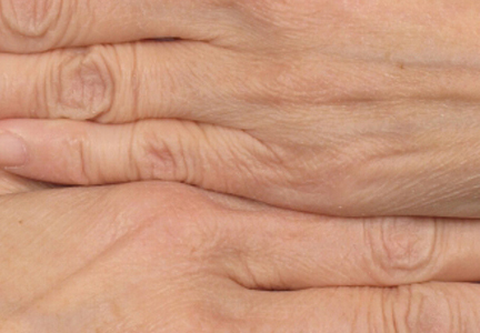
Understanding Elastic Recoil Key to Aging Skin
DANA POINT, CALIF. – Finding ways to restore elasticity of the skin is a better goal than is developing and refining ways to tighten the skin, according to Dr. R. Rox Anderson.
"The loss of natural skin tension with aging is due to a loss of its elasticity," Dr. Anderson said at the Summit in Aesthetic Medicine sponsored by Skin Disease Education Foundation (SDEF). "It’s the impressive loss of cutaneous elastic recoil with aging that accounts for the effectiveness of Botox, for example. A simple hypothesis is that restoring skin elasticity is a better goal than tightening the skin."

The "players" in cutaneous elastic recoil, he said, are numerous, but it remains unknown which are pivotal. Is it the extracellular matrix content and cross-linking, the fibril microstructure, or the active cytoskeleton?
"When you lose elastic recoil, we don’t know which ones of these are most important," said Dr. Anderson, professor of dermatology at Harvard Medical School, Boston. "If we knew that, we could design treatments that actually treat the cause as opposed to [treating] flabby skin."
He said that "powerful, verified" research tools are needed to improve understanding of cutaneous elastic recoil, including "composite, dynamic structural models" to expand understanding of the difference between microscopic and macroscopic skin. "Those exist; we don’t have to invent them," he said. "I’m an adjunct professor at Massachusetts Institute of Technology, [which has] an entire department that does nothing but material science and modeling. They’re really good at composite materials. That’s what we [humans] are: we are complex composite material."
Stress-strain component analysis is another key tool to improve understanding of elastic recoil. "Each of those components has its own structural and dynamic behavior," said Dr. Anderson, who also directs the Wellman Center for Photomedicine. "I think it’s important to recognize that the skin is a dynamic system that responds to stress. So Langer’s lines are due to particular strain receptors and fibroblasts. The genes that are strain inducible have been at least partially noted, but they have not been studied in the context of skin tightening and rejuvenation. I would love to see that: a dose response analysis for strain-mediated gene expression."
Studies involving in vivo microscopy will also be important. "Elastin, for example, is easy as pie to image in vivo in human skin," he said. "We should be able to map mechanical properties of skin."
Dr. Anderson said that he had no relevant financial conflicts to disclose.
DANA POINT, CALIF. – Finding ways to restore elasticity of the skin is a better goal than is developing and refining ways to tighten the skin, according to Dr. R. Rox Anderson.
"The loss of natural skin tension with aging is due to a loss of its elasticity," Dr. Anderson said at the Summit in Aesthetic Medicine sponsored by Skin Disease Education Foundation (SDEF). "It’s the impressive loss of cutaneous elastic recoil with aging that accounts for the effectiveness of Botox, for example. A simple hypothesis is that restoring skin elasticity is a better goal than tightening the skin."
The "players" in cutaneous elastic recoil, he said, are numerous, but it remains unknown which are pivotal. Is it the extracellular matrix content and cross-linking, the fibril microstructure, or the active cytoskeleton?
"When you lose elastic recoil, we don’t know which ones of these are most important," said Dr. Anderson, professor of dermatology at Harvard Medical School, Boston. "If we knew that, we could design treatments that actually treat the cause as opposed to [treating] flabby skin."
He said that "powerful, verified" research tools are needed to improve understanding of cutaneous elastic recoil, including "composite, dynamic structural models" to expand understanding of the difference between microscopic and macroscopic skin. "Those exist; we don’t have to invent them," he said. "I’m an adjunct professor at Massachusetts Institute of Technology, [which has] an entire department that does nothing but material science and modeling. They’re really good at composite materials. That’s what we [humans] are: we are complex composite material."
Stress-strain component analysis is another key tool to improve understanding of elastic recoil. "Each of those components has its own structural and dynamic behavior," said Dr. Anderson, who also directs the Wellman Center for Photomedicine. "I think it’s important to recognize that the skin is a dynamic system that responds to stress. So Langer’s lines are due to particular strain receptors and fibroblasts. The genes that are strain inducible have been at least partially noted, but they have not been studied in the context of skin tightening and rejuvenation. I would love to see that: a dose response analysis for strain-mediated gene expression."
Studies involving in vivo microscopy will also be important. "Elastin, for example, is easy as pie to image in vivo in human skin," he said. "We should be able to map mechanical properties of skin."
Dr. Anderson said that he had no relevant financial conflicts to disclose.
EXPERT ANALYSIS AT THE SDEF SUMMIT IN AESTHETIC MEDICINE

Skin Flaps Remedy Defects of the Ear
SAN DIEGO – In the clinical experience of Dr. Michael A. Keefe, 70%-80% of ear defects from auricular cancer treatment can be easily remedied with skin flaps.
The most common locations of auricular cancer are the helix, the posterior auricle skin, and the antihelix, Dr. Keefe said at a meeting on superficial anatomy and cutaneous surgery.

"More than 70% of lesions are smaller than 3 cm in size, and auricular lesions make up an estimated 8% of all skin cancers," said Dr. Keefe, a plastic surgeon with the division of head and neck surgery at Sharp Rees-Stealy Medical Group in San Diego. "The defects are unique, and the underlying cartilage structure makes it all the more interesting."
And challenging – defects may be located on the skin of the ear only, on the lateral side, or on the posterior side, or they may involve a combination of skin and cartilage. Healing by secondary intention is effective for concave defects, but the size of the defect drives the reconstruction options. "If there is no perichondrium, punch holes through cartilage with a 2-3 mm punch to allow granulation tissue to grow through, and then use a skin graft or allow it to heal with secondary intention," he said. "Keep the area moist with antibiotic ointment."
Options for reconstruction of defects in the middle one-third of the ear include primary closure, full-thickness skin grafts (FTSGs), the helical advancement flap, and the retroauricular composite advancement flap, while options for defects in the lower one-third of the ear include primary closure and the preauricular tubed flap. Options for reconstruction of defects in the upper one-third of the ear include primary closure, FTSGs, the helical advancement flap, the retroauricular and preauricular tubed flaps, and constructing an autogenous cartilage framework with FTSGs.
Dr. Keefe said that most small helical rim defects limited to the skin can be closed primarily. "There might be slight rim asymmetry [after closure]," he said at the meeting, which was sponsored by the University of California, San Diego, School of Medicine and the Scripps Clinic. "Some patients might not care [about this], but you have to advise them of that," he added.
A bilobed advancement flap is another option for helical rim defects limited to the skin. This flap "works well for cutaneous defects 2 cm or smaller in the helical rim or the posterior auricle," he said. "The other thing you can do with these bilobed flaps is advance them over the edge to correct helical rim defects."
The banner flap is another effective flap for helical rim defects, especially those located on the superior helix. It does not replace cartilage, but it conceals the incision well. For small composite helix and anterior defects, Dr. Keefe favors the chondrocutaneous advancement flap.
He said that he favors using FTSGs on the anterior surface of the helix for skin defects whenever possible. "You can use a composite skin graft as well, especially to replace cartilage or skin defects that are smaller than 1 cm in size," he said. "A FTSG is easy to harvest and has minimal contraction. Common donor sites include the preauricular, postauricular, supraclavicular, and clavicular regions. Make sure you trim off the fat." For posterior surface defects, the bilobe or advancement flaps work well.
Grafts must be placed on tissue with an adequate blood supply. Effective grafts establish imbibition in the first 24 hours, inosculation within 48-72 hours, and restoration of circulation within 4-7 days.
Dr. Keefe said that he had no relevant financial conflicts to disclose.
SAN DIEGO – In the clinical experience of Dr. Michael A. Keefe, 70%-80% of ear defects from auricular cancer treatment can be easily remedied with skin flaps.
The most common locations of auricular cancer are the helix, the posterior auricle skin, and the antihelix, Dr. Keefe said at a meeting on superficial anatomy and cutaneous surgery.
"More than 70% of lesions are smaller than 3 cm in size, and auricular lesions make up an estimated 8% of all skin cancers," said Dr. Keefe, a plastic surgeon with the division of head and neck surgery at Sharp Rees-Stealy Medical Group in San Diego. "The defects are unique, and the underlying cartilage structure makes it all the more interesting."
And challenging – defects may be located on the skin of the ear only, on the lateral side, or on the posterior side, or they may involve a combination of skin and cartilage. Healing by secondary intention is effective for concave defects, but the size of the defect drives the reconstruction options. "If there is no perichondrium, punch holes through cartilage with a 2-3 mm punch to allow granulation tissue to grow through, and then use a skin graft or allow it to heal with secondary intention," he said. "Keep the area moist with antibiotic ointment."
Options for reconstruction of defects in the middle one-third of the ear include primary closure, full-thickness skin grafts (FTSGs), the helical advancement flap, and the retroauricular composite advancement flap, while options for defects in the lower one-third of the ear include primary closure and the preauricular tubed flap. Options for reconstruction of defects in the upper one-third of the ear include primary closure, FTSGs, the helical advancement flap, the retroauricular and preauricular tubed flaps, and constructing an autogenous cartilage framework with FTSGs.
Dr. Keefe said that most small helical rim defects limited to the skin can be closed primarily. "There might be slight rim asymmetry [after closure]," he said at the meeting, which was sponsored by the University of California, San Diego, School of Medicine and the Scripps Clinic. "Some patients might not care [about this], but you have to advise them of that," he added.
A bilobed advancement flap is another option for helical rim defects limited to the skin. This flap "works well for cutaneous defects 2 cm or smaller in the helical rim or the posterior auricle," he said. "The other thing you can do with these bilobed flaps is advance them over the edge to correct helical rim defects."
The banner flap is another effective flap for helical rim defects, especially those located on the superior helix. It does not replace cartilage, but it conceals the incision well. For small composite helix and anterior defects, Dr. Keefe favors the chondrocutaneous advancement flap.
He said that he favors using FTSGs on the anterior surface of the helix for skin defects whenever possible. "You can use a composite skin graft as well, especially to replace cartilage or skin defects that are smaller than 1 cm in size," he said. "A FTSG is easy to harvest and has minimal contraction. Common donor sites include the preauricular, postauricular, supraclavicular, and clavicular regions. Make sure you trim off the fat." For posterior surface defects, the bilobe or advancement flaps work well.
Grafts must be placed on tissue with an adequate blood supply. Effective grafts establish imbibition in the first 24 hours, inosculation within 48-72 hours, and restoration of circulation within 4-7 days.
Dr. Keefe said that he had no relevant financial conflicts to disclose.
EXPERT ANALYSIS FROM A MEETING ON SUPERFICIAL ANATOMY AND CUTANEOUS SURGERY

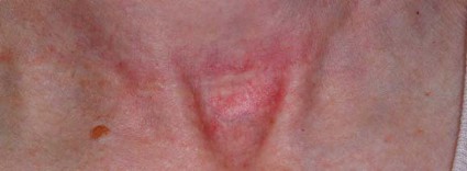
Early Scar Treatment Is 'Critical'
SAN DIEGO – The future of treating hypertrophic and keloidal scars will involve earlier intervention with new and existing technologies – even at the genesis of scar formation, said Dr. E. Victor Ross.
"I think you’re going to see a lot more in the future about scars, not just in the laser area, but also in the biologic arena, because we’re learning more about the way scars behave," Dr. Ross said at a meeting on superficial anatomy and cutaneous surgery. "Some physicians are treating scars as early as the time of Mohs surgery, for example, by applying the PDL [pulsed-dye laser] at the time of suture placement. That’s perhaps a bit extreme, but I think you are going to see newer technologies and drugs used synergistically to give us a better fighting chance to prevent and treat scars."
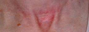
Dr. Ross of Scripps Clinic Laser and Cosmetic Dermatology Center in Carmel Valley, Calif., said that there is a lack of consensus regarding how the two main types of scars hypertrophic and keloidal – are defined. Historically, "we’ve said that hypertrophic scars don’t go beyond the boundary of where the scar tissue was, and keloidal scars go around the perimeter of where the scar boundaries were," he noted. "If the scar is red, even if it’s longstanding, I tend to call it a hypertrophic scar. If it tends to be more flesh colored, and aged like a fine wine, I tend to call it a keloidal scar. The critical thing with these scars is how long it takes the wound to heal. If an open wound takes more than 3-4 weeks to heal, often it will be hypertrophic."
Existing therapies that are commonly used to treat scars include intralesional steroids, intralesional 5-fluorouracil, oral antihistamines, cyclooxygenase-2 inhibitors, lasers, hydrogel sheeting, and compression. "The critical thing is to treat relatively early; you have to use all the weapons that are available to you," Dr. Ross said at the meeting, which was sponsored by the University of California San Diego School of Medicine and the Scripps Clinic.
He said that when treating scars, a modifiable approach should be taken. "You want to modify the scar. After it’s formed, you want to rehabilitate the scar and make it more like the skin around it."
When using intralesional steroids, Dr. Ross prefers to use very low volumes with a very high concentration of Kenalog, "typically 40 mg/mL in tiny amounts with a 3-gauge, half-inch needle," he said. "You want to keep the needle tip relatively superficial. If the steroid floats into the scar too easily you’re probably too deep or under the scar."
He favors using fractional lasers for scars whenever possible. These devices "create microscopic wounds in the skin," he said. "It turns out that if you fractionate a wound, the reservoirs of normal, undamaged skin act as ‘seeds’ to make the wounds heal quickly. I like to use purpuric settings with the pulsed-dye laser. They tend to give you better results than other settings."
For scars that form after thyroid surgery, Dr. Ross likes to use a PDL or IPL (intense pulsed light) to reduce the redness, followed by a nonablative fractional laser. With that tandem approach "you can almost make the scar go away, which is a complete rehabilitation of the scar," he said.
Innovative scar therapies include topical mitomycin C, which has worked well for postoperative keloids; oral and topical tamoxifen, which helps in the formation of fibroblasts; and oral methotrexate, which has demonstrated efficacy in the treatment and prevention of keloids. Imiquimod has also been used, "but I’m not a believer in it," Dr. Ross said. "We’ve tried it several times and we found that it irritated the skin most of the time. Retinoids are good and bad. They decrease fibroblast activity but also decrease collagenase."
Dr. Ross disclosed that he is a consultant for Cutera, Palomar Medical Technologies, and Lumenis. He has also received research support from Palomar, Sciton, and Syneron Medical.
SAN DIEGO – The future of treating hypertrophic and keloidal scars will involve earlier intervention with new and existing technologies – even at the genesis of scar formation, said Dr. E. Victor Ross.
"I think you’re going to see a lot more in the future about scars, not just in the laser area, but also in the biologic arena, because we’re learning more about the way scars behave," Dr. Ross said at a meeting on superficial anatomy and cutaneous surgery. "Some physicians are treating scars as early as the time of Mohs surgery, for example, by applying the PDL [pulsed-dye laser] at the time of suture placement. That’s perhaps a bit extreme, but I think you are going to see newer technologies and drugs used synergistically to give us a better fighting chance to prevent and treat scars."
Dr. Ross of Scripps Clinic Laser and Cosmetic Dermatology Center in Carmel Valley, Calif., said that there is a lack of consensus regarding how the two main types of scars hypertrophic and keloidal – are defined. Historically, "we’ve said that hypertrophic scars don’t go beyond the boundary of where the scar tissue was, and keloidal scars go around the perimeter of where the scar boundaries were," he noted. "If the scar is red, even if it’s longstanding, I tend to call it a hypertrophic scar. If it tends to be more flesh colored, and aged like a fine wine, I tend to call it a keloidal scar. The critical thing with these scars is how long it takes the wound to heal. If an open wound takes more than 3-4 weeks to heal, often it will be hypertrophic."
Existing therapies that are commonly used to treat scars include intralesional steroids, intralesional 5-fluorouracil, oral antihistamines, cyclooxygenase-2 inhibitors, lasers, hydrogel sheeting, and compression. "The critical thing is to treat relatively early; you have to use all the weapons that are available to you," Dr. Ross said at the meeting, which was sponsored by the University of California San Diego School of Medicine and the Scripps Clinic.
He said that when treating scars, a modifiable approach should be taken. "You want to modify the scar. After it’s formed, you want to rehabilitate the scar and make it more like the skin around it."
When using intralesional steroids, Dr. Ross prefers to use very low volumes with a very high concentration of Kenalog, "typically 40 mg/mL in tiny amounts with a 3-gauge, half-inch needle," he said. "You want to keep the needle tip relatively superficial. If the steroid floats into the scar too easily you’re probably too deep or under the scar."
He favors using fractional lasers for scars whenever possible. These devices "create microscopic wounds in the skin," he said. "It turns out that if you fractionate a wound, the reservoirs of normal, undamaged skin act as ‘seeds’ to make the wounds heal quickly. I like to use purpuric settings with the pulsed-dye laser. They tend to give you better results than other settings."
For scars that form after thyroid surgery, Dr. Ross likes to use a PDL or IPL (intense pulsed light) to reduce the redness, followed by a nonablative fractional laser. With that tandem approach "you can almost make the scar go away, which is a complete rehabilitation of the scar," he said.
Innovative scar therapies include topical mitomycin C, which has worked well for postoperative keloids; oral and topical tamoxifen, which helps in the formation of fibroblasts; and oral methotrexate, which has demonstrated efficacy in the treatment and prevention of keloids. Imiquimod has also been used, "but I’m not a believer in it," Dr. Ross said. "We’ve tried it several times and we found that it irritated the skin most of the time. Retinoids are good and bad. They decrease fibroblast activity but also decrease collagenase."
Dr. Ross disclosed that he is a consultant for Cutera, Palomar Medical Technologies, and Lumenis. He has also received research support from Palomar, Sciton, and Syneron Medical.
EXPERT ANALYSIS FROM A MEETING ON SUPERFICIAL ANATOMY AND CUTANEOUS SURGERY
Skin of Color: Barriers to Melanoma Detection
Melanoma accounts for 75% of all skin cancer deaths. Ultraviolet exposure is still targeted as the major etiologic factor, but for black patients, skin erythema has been estimated to occur at a UV radiation dose 6-to-33 times greater than that experienced by white patients. Many have concluded that this is why black patients experience lower rates of melanoma than white patients.
Black patients, however, present at a later stage and have a higher melanoma-specific mortality, both of which have been linked to time of diagnosis and the ability to seek care at the onset of localized disease. Thus, there still exists a barrier to the detection and treatment of melanoma in black patients.
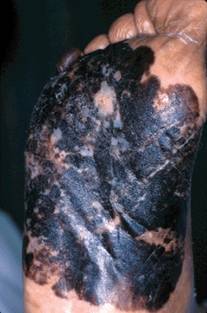
In the July issue of Archives of Dermatology (2012;148:797-801), an article highlighted the anatomic distribution of malignant melanoma in the non-Hispanic black patient in an effort to explore how distribution of melanoma relates to UV exposure.
Data from 46 population-based cancer registries were analyzed. The most frequent site of melanoma in non-Hispanic black patients – both male and female, between the years 1998 and 2007 – was found to be the lower limbs and hip (58.9%). Of those, 27% were of the acral lentiginous type, which is not associated with exposure to UV rays.
The second most common location was the trunk (16.5%), which affected patients at a younger age; 46% of females and 31% of males were less than 44 years of age. The median age was 56 years for males and 48 years for females at presentation.
This study reiterates the burden of melanoma in the black community. It also highlights gaps in the detection of melanoma, which may be because of site of diagnosis – such as those of the acral lentiginous types – and unclear risk factors, the general underestimation of risk, and access to care.
- Lily Talakoub, M.D.
Do you have questions about treating patients with darker skin? If so, send them to sknews@elsevier.com.
Melanoma accounts for 75% of all skin cancer deaths. Ultraviolet exposure is still targeted as the major etiologic factor, but for black patients, skin erythema has been estimated to occur at a UV radiation dose 6-to-33 times greater than that experienced by white patients. Many have concluded that this is why black patients experience lower rates of melanoma than white patients.
Black patients, however, present at a later stage and have a higher melanoma-specific mortality, both of which have been linked to time of diagnosis and the ability to seek care at the onset of localized disease. Thus, there still exists a barrier to the detection and treatment of melanoma in black patients.
In the July issue of Archives of Dermatology (2012;148:797-801), an article highlighted the anatomic distribution of malignant melanoma in the non-Hispanic black patient in an effort to explore how distribution of melanoma relates to UV exposure.
Data from 46 population-based cancer registries were analyzed. The most frequent site of melanoma in non-Hispanic black patients – both male and female, between the years 1998 and 2007 – was found to be the lower limbs and hip (58.9%). Of those, 27% were of the acral lentiginous type, which is not associated with exposure to UV rays.
The second most common location was the trunk (16.5%), which affected patients at a younger age; 46% of females and 31% of males were less than 44 years of age. The median age was 56 years for males and 48 years for females at presentation.
This study reiterates the burden of melanoma in the black community. It also highlights gaps in the detection of melanoma, which may be because of site of diagnosis – such as those of the acral lentiginous types – and unclear risk factors, the general underestimation of risk, and access to care.
- Lily Talakoub, M.D.
Do you have questions about treating patients with darker skin? If so, send them to sknews@elsevier.com.
Frozen Fat Plumps Up Aging Face: The Skinny Vodcast
Skin & Allergy News Managing Editor Amy Pfeiffer and Senior Editor Terry Rudd review hot news in dermatology with the experts in this month's Skinny Vodcast.
Highlights include an interview with Dr. Axel Hauschild about two new promising treatments for BRAF-mutant melanoma. Doug Brunk talks to Dr. Christopher Zachary about a new light source that may someday replace lasers.
And, Dr. Lisa M. Donofrio discusses the advantages of fat transfer for volumizing the face.
Lastly, Dr. Lily Talakoub recommends a classification system for defining under eye circles.
Cosmeceutical Critique: Safflower Oil
Safflower (Carthamus tinctorius), a thistlelike annual, is one of the oldest cultivated crops, its use dating back to ancient Egypt. It was traditionally grown for its seeds, which were used in foods and folk medicine. Safflower is now primarily cultivated for its vegetable oil, which is extracted from its seeds. Safflower oil has been found to exert notable health benefits when consumed through the diet and also when used in topical formulations.
Linoleic acid is a primary constituent of safflower seeds, and is the component to which the oil’s cutaneous benefits are typically ascribed. In fact, safflower oil is one of the richest sources of linoleic acid, which is necessary for the endogenous production of ceramides, key components of the epidermal layer that play a crucial role in barrier function and help the skin retain water.

In skin care products, safflower oil is incorporated in moisturizing agents for its occlusive properties. Occlusive agents coat the stratum corneum to slow transepidermal water loss. Typically, such substances have the capacity to dissolve fats and are therefore used in many skin care cosmetics.
Research on Topical Applications
In an early study on the antiproliferative potential of C. tinctorius extracts, Yasukawa et al., in 1996, isolated erythro-alkane-6,8-diols from the flowers of C. tinctorius and applied the tumor-promoting agent 12-O-tetradecanoylphorbol-13-acetate (TPA) to the ears of mice (1 mcg/ear) to induce inflammation. The investigators reported that five of the eight alkane-6,8-diols assayed suppressed inflammation, and the mixture of erythro-alkane-6,8-diols significantly inhibited TPA-induced skin tumor formation in mice after initiation with 7,12-dimethylbenz[a]anthracene (Oncology 1996;53:133-6).
In 2004, Roh et al. investigated the melanogenesis-suppressing activity of safflower seeds to develop a novel skin-whitening agent. They reported that an 80% aqueous methanol extract and ethyl acetate fraction from the seeds significantly inhibited mushroom tyrosinase, and the researchers identified three active constituents [N-feruloylserotonin, N-(p-coumaroyl)serotonin, and acacetin]. Of these, N-feruloylserotonin and N-(p-coumaroyl)serotonin were found to more potently suppress the melanin synthesis of Streptomyces bikiniensis and B16 melanoma cells than arbutin, a well-known inhibitor of melanogenesis (Biol. Pharm. Bull. 2004;27:1976-8).
In 2005, Solanki et al. conducted a short, randomized controlled study in a tertiary-care neonatal intensive care unit (NICU) of a large teaching hospital, to assess the transcutaneous absorption of oil traditionally used in massage of newborns, and to compare the effects of safflower oil and coconut oil on fatty acid profiles of massaged babies. The investigators randomly assigned 120 babies to three groups – safflower oil, coconut oil, or no-oil controls (40 in each group). The babies were massaged with 5 mL of oil four times daily for 5 days. Blood triglyceride levels were significantly elevated in all groups, though much more so in the noncontrol groups. Significant increases in essential fatty acids (linolenic and arachidonic) were seen in the safflower oil group, and similar increases in saturated fats were seen in the coconut oil group, with changes more evident in term babies. The researchers concluded that topically applied oil is absorbed in neonates and is likely available nutritionally. Consequently, they deemed the fatty acid constituents of the massage oils significant in potentially impacting the fatty acid profiles of patients (Indian Pediatr. 2005;42:998-1005). Safflower oil is rich in essential fatty acids, and coconut oil is rife with saturated fat.
Potential Dietary Benefits
Safflower oil has also been found, as has olive oil, to confer dietary benefits on diabetic pregnant rats and their embryos, preventing diabetes-induced developmental harm during early organogenesis (Mol. Hum. Reprod. 2010;16:286-95). Supplementation with either oil has also been demonstrated to prevent excessive activity by matrix metalloproteinases (specifically MMP-2 and MMP-9) in the placenta of diabetic rats, with salubrious effects manifesting in the sera (Placenta 2012;33:8-16). In addition, in a recent study, safflower oil and folic acid supplementations were shown to interact, protecting rat embryos from diabetes-induced harm through reductions in proinflammatory mediators (Mol. Hum. Reprod. 2012;18:253-64).
As mentioned above, safflower oil is available in several topical products, but it is more likely beneficial through diet. Topically, safflower oil, as found in a Neutrogena bath oil, for example, contains linoleic acid and may be useful when added to bathwater or applied to wet skin. Of course, oils in general are not suitable for all skin types. Safflower oil is indicated for individuals with dry or damaged skin.
Conclusion
Safflower oil, rich in the essential omega-6 fatty acid linoleic acid, is known to confer health benefits via diet. It is also included in skin care products, such as bath oils, which anecdotally appear to be effective. Nevertheless, there is a dearth of data on the use of safflower oil for dermatologic purposes. Much more research is necessary, including randomized controlled clinical trials in humans, to establish the potential for more extensive uses of safflower oil for skin health.
Dr. Baumann is in private practice in Miami Beach. She did not disclose any conflicts of interest. To respond to this column, or to suggest topics for future columns, write to her at sknews@elsevier.com.
Safflower (Carthamus tinctorius), a thistlelike annual, is one of the oldest cultivated crops, its use dating back to ancient Egypt. It was traditionally grown for its seeds, which were used in foods and folk medicine. Safflower is now primarily cultivated for its vegetable oil, which is extracted from its seeds. Safflower oil has been found to exert notable health benefits when consumed through the diet and also when used in topical formulations.
Linoleic acid is a primary constituent of safflower seeds, and is the component to which the oil’s cutaneous benefits are typically ascribed. In fact, safflower oil is one of the richest sources of linoleic acid, which is necessary for the endogenous production of ceramides, key components of the epidermal layer that play a crucial role in barrier function and help the skin retain water.
In skin care products, safflower oil is incorporated in moisturizing agents for its occlusive properties. Occlusive agents coat the stratum corneum to slow transepidermal water loss. Typically, such substances have the capacity to dissolve fats and are therefore used in many skin care cosmetics.
Research on Topical Applications
In an early study on the antiproliferative potential of C. tinctorius extracts, Yasukawa et al., in 1996, isolated erythro-alkane-6,8-diols from the flowers of C. tinctorius and applied the tumor-promoting agent 12-O-tetradecanoylphorbol-13-acetate (TPA) to the ears of mice (1 mcg/ear) to induce inflammation. The investigators reported that five of the eight alkane-6,8-diols assayed suppressed inflammation, and the mixture of erythro-alkane-6,8-diols significantly inhibited TPA-induced skin tumor formation in mice after initiation with 7,12-dimethylbenz[a]anthracene (Oncology 1996;53:133-6).
In 2004, Roh et al. investigated the melanogenesis-suppressing activity of safflower seeds to develop a novel skin-whitening agent. They reported that an 80% aqueous methanol extract and ethyl acetate fraction from the seeds significantly inhibited mushroom tyrosinase, and the researchers identified three active constituents [N-feruloylserotonin, N-(p-coumaroyl)serotonin, and acacetin]. Of these, N-feruloylserotonin and N-(p-coumaroyl)serotonin were found to more potently suppress the melanin synthesis of Streptomyces bikiniensis and B16 melanoma cells than arbutin, a well-known inhibitor of melanogenesis (Biol. Pharm. Bull. 2004;27:1976-8).
In 2005, Solanki et al. conducted a short, randomized controlled study in a tertiary-care neonatal intensive care unit (NICU) of a large teaching hospital, to assess the transcutaneous absorption of oil traditionally used in massage of newborns, and to compare the effects of safflower oil and coconut oil on fatty acid profiles of massaged babies. The investigators randomly assigned 120 babies to three groups – safflower oil, coconut oil, or no-oil controls (40 in each group). The babies were massaged with 5 mL of oil four times daily for 5 days. Blood triglyceride levels were significantly elevated in all groups, though much more so in the noncontrol groups. Significant increases in essential fatty acids (linolenic and arachidonic) were seen in the safflower oil group, and similar increases in saturated fats were seen in the coconut oil group, with changes more evident in term babies. The researchers concluded that topically applied oil is absorbed in neonates and is likely available nutritionally. Consequently, they deemed the fatty acid constituents of the massage oils significant in potentially impacting the fatty acid profiles of patients (Indian Pediatr. 2005;42:998-1005). Safflower oil is rich in essential fatty acids, and coconut oil is rife with saturated fat.
Potential Dietary Benefits
Safflower oil has also been found, as has olive oil, to confer dietary benefits on diabetic pregnant rats and their embryos, preventing diabetes-induced developmental harm during early organogenesis (Mol. Hum. Reprod. 2010;16:286-95). Supplementation with either oil has also been demonstrated to prevent excessive activity by matrix metalloproteinases (specifically MMP-2 and MMP-9) in the placenta of diabetic rats, with salubrious effects manifesting in the sera (Placenta 2012;33:8-16). In addition, in a recent study, safflower oil and folic acid supplementations were shown to interact, protecting rat embryos from diabetes-induced harm through reductions in proinflammatory mediators (Mol. Hum. Reprod. 2012;18:253-64).
As mentioned above, safflower oil is available in several topical products, but it is more likely beneficial through diet. Topically, safflower oil, as found in a Neutrogena bath oil, for example, contains linoleic acid and may be useful when added to bathwater or applied to wet skin. Of course, oils in general are not suitable for all skin types. Safflower oil is indicated for individuals with dry or damaged skin.
Conclusion
Safflower oil, rich in the essential omega-6 fatty acid linoleic acid, is known to confer health benefits via diet. It is also included in skin care products, such as bath oils, which anecdotally appear to be effective. Nevertheless, there is a dearth of data on the use of safflower oil for dermatologic purposes. Much more research is necessary, including randomized controlled clinical trials in humans, to establish the potential for more extensive uses of safflower oil for skin health.
Dr. Baumann is in private practice in Miami Beach. She did not disclose any conflicts of interest. To respond to this column, or to suggest topics for future columns, write to her at sknews@elsevier.com.

BMI Drives Body Contouring Results
DANA POINT, CALIF. – Three factors affect the results of body contouring outcomes after bariatric surgery: body mass index at presentation, fat deposition pattern, and quality of the skin-fat envelope, according to Dr. Al S. Aly.
"The fat deposition pattern is genetically controlled; we cannot alter that," Dr. Aly, professor and vice-chair of plastic surgery at the University of California, Irvine, said at the Summit in Aesthetic Medicine sponsored by Skin Disease Education Foundation (SDEF). "The quality of the skin-fat envelope is very important. If you have a very loose skin-fat envelope – meaning that they’ve had a tremendous drop in weight – you can do a lot more for them than with other patients."

The two main body contouring treatments for the lower trunk are abdominoplasty and circumferential belt lipectomy, said Dr. Aly. "Abdominoplasties are ideally suited for problems limited to the belly, or those located between the two anterior superior iliac spines. For larger patients you need to do something else."
The rehabilitation of larger patients involves a circumferential procedure commonly referred to as a body lift or belt lipectomy, which involves removing a circumferential wedge of tissue from around the trunk. "There are many reasons why we need circumferential excisions for these large patients, but the main one is that you need circumferential improvement above and below the area of resection as opposed to a regular tummy tuck," he explained.
Markings for the belt lipectomy "are the essence of the procedure," Dr. Aly said, who noted that about 10% of his patients are male. "The markings are adjustable guidelines, and they vary according to anatomy and desires. It’s a fairly complex set of issues but essentially you have to understand how tissues drape as the patient loses weight, and there are zones of adherence or areas of the body where the skin and the soft tissue envelope is stuck to the underlying musculoskeletal anatomy. It’s important to understand those before you can mark these patients. Surgical improvement is related to the amount of tension that you create above and below an area of excision."
Dr. Aly said that he routinely performs belt lipectomies with another surgeon and strives for surgical times under 6 hours. He uses a general anesthetic and an epidural for postoperative pain management. Moving patients into multiple positions during surgery is required, "otherwise you can’t accomplish the excisions," he said. "There is a variety of different approaches. Mine happens to be one of supine first then lateral/lateral."
Results of body contouring procedures generally correlate with the patient’s body mass index (BMI) at the time of surgery. "So people who present to us above a BMI of 35 kg/m2 ... their results are okay," he said. "Results are better for patients with a BMI of 30-35 kg/m2. If you’re lucky enough to get a patient at an ideal BMI – 26 kg/m2 and lower – then you can create almost normal anatomy."
He noted that, compared with abdominoplasties, results of belt lipectomies take longer than most procedures to mature. "However, their results are also far superior to those attainable with a tummy tuck," Dr. Aly said. Complications, including an increased risk for seroma and formation of hematoma, "correlate with BMI," he said. "If you’re not ready to deal with complications, massive weight loss patients are a tough group to deal with. It’s part of the nature of these patients."
Other areas that often need contouring include upper arm reductions, upper back roll reductions, reconstruction of the breasts (in women and men), and thigh reductions.
Dr. Aly said that he had no relevant conflicts to disclose. SDEF and this news organization are owned by Elsevier.
DANA POINT, CALIF. – Three factors affect the results of body contouring outcomes after bariatric surgery: body mass index at presentation, fat deposition pattern, and quality of the skin-fat envelope, according to Dr. Al S. Aly.
"The fat deposition pattern is genetically controlled; we cannot alter that," Dr. Aly, professor and vice-chair of plastic surgery at the University of California, Irvine, said at the Summit in Aesthetic Medicine sponsored by Skin Disease Education Foundation (SDEF). "The quality of the skin-fat envelope is very important. If you have a very loose skin-fat envelope – meaning that they’ve had a tremendous drop in weight – you can do a lot more for them than with other patients."
The two main body contouring treatments for the lower trunk are abdominoplasty and circumferential belt lipectomy, said Dr. Aly. "Abdominoplasties are ideally suited for problems limited to the belly, or those located between the two anterior superior iliac spines. For larger patients you need to do something else."
The rehabilitation of larger patients involves a circumferential procedure commonly referred to as a body lift or belt lipectomy, which involves removing a circumferential wedge of tissue from around the trunk. "There are many reasons why we need circumferential excisions for these large patients, but the main one is that you need circumferential improvement above and below the area of resection as opposed to a regular tummy tuck," he explained.
Markings for the belt lipectomy "are the essence of the procedure," Dr. Aly said, who noted that about 10% of his patients are male. "The markings are adjustable guidelines, and they vary according to anatomy and desires. It’s a fairly complex set of issues but essentially you have to understand how tissues drape as the patient loses weight, and there are zones of adherence or areas of the body where the skin and the soft tissue envelope is stuck to the underlying musculoskeletal anatomy. It’s important to understand those before you can mark these patients. Surgical improvement is related to the amount of tension that you create above and below an area of excision."
Dr. Aly said that he routinely performs belt lipectomies with another surgeon and strives for surgical times under 6 hours. He uses a general anesthetic and an epidural for postoperative pain management. Moving patients into multiple positions during surgery is required, "otherwise you can’t accomplish the excisions," he said. "There is a variety of different approaches. Mine happens to be one of supine first then lateral/lateral."
Results of body contouring procedures generally correlate with the patient’s body mass index (BMI) at the time of surgery. "So people who present to us above a BMI of 35 kg/m2 ... their results are okay," he said. "Results are better for patients with a BMI of 30-35 kg/m2. If you’re lucky enough to get a patient at an ideal BMI – 26 kg/m2 and lower – then you can create almost normal anatomy."
He noted that, compared with abdominoplasties, results of belt lipectomies take longer than most procedures to mature. "However, their results are also far superior to those attainable with a tummy tuck," Dr. Aly said. Complications, including an increased risk for seroma and formation of hematoma, "correlate with BMI," he said. "If you’re not ready to deal with complications, massive weight loss patients are a tough group to deal with. It’s part of the nature of these patients."
Other areas that often need contouring include upper arm reductions, upper back roll reductions, reconstruction of the breasts (in women and men), and thigh reductions.
Dr. Aly said that he had no relevant conflicts to disclose. SDEF and this news organization are owned by Elsevier.
AT THE SDEF SUMMIT IN AESTHETIC MEDICINE
ASCO, SSO Back Sentinel Lymph Node Biopsy in Melanoma
All newly diagnosed melanoma patients with tumors of intermediate thickness – defined as those with a Breslow thickness of between 1 and 4 mm – should undergo sentinel lymph node biopsy, according to a new guideline from two professional societies.
The guideline, issued jointly July 9 by the American Society of Clinical Oncology and the Society of Surgical Oncology, also advises that while the use of sentinel lymph node biopsy (SLNB) in people with thicker or thinner melanomas remains contentious, both categories of patients could benefit from SLNB in some circumstances.
All melanoma patients with a positive SLNB, the guideline says, should be treated with completion lymph node dissection (CLND), the current standard of care, although, the authors noted, it is not yet known whether CLND after a positive SLN biopsy improves 10-year overall survival. This is the subject of a large, ongoing randomized trial, the Multicenter Selective Lymphadenectomy Trial II (MSLT-11).
The guideline’s authors, led by Dr. Sandra L. Wong of the University of Michigan, Ann Arbor, and Dr. Gary H. Lyman of Duke University, Durham, N.C., aimed to use the most current evidence to clarify both the indications for sentinel node biopsy and the role of completion biopsy in people with melanomas.
"It is critically important to identify those patients for whom the expected benefits of resecting regional lymph nodes outweigh the risks of surgical morbidity," they wrote in their analysis.
To this end, Dr. Wong, Dr. Lyman, and 12 other expert panel members undertook a broad literature search and identified 73 studies (most of them observational in design) that met the criteria for inclusion in their meta-analysis. Some 25,000 patients were enrolled in the included studies.
The authors found that while robust evidence supports routine use of sentinel lymph node biopsy (SLNB) in intermediate melanomas, there is also some evidence to support the procedure in patients with thin melanomas (less than 1 mm) when certain other risk factors are present, and in patients with thick melanomas (greater than 4 mm).
For people with melanomas of less than 1 mm Breslow thickness and one or more risk factors such as tumor ulceration or a mitotic rate of 1/mm2 or greater, they wrote, "the benefits of pathologic staging may outweigh the potential risks of the procedure," particularly in the subgroup of patients with melanomas ranging from 0.75 mm to 0.99 mm.
Patients with melanomas of 4 mm or greater may also benefit, the guideline says. "Conventional wisdom asserts that patients with thick melanomas have a high risk of systemic disease at the time of diagnosis and that no survival benefit can be derived from removal of regional lymph nodes," the authors wrote.
"However, among patients without distant disease, it can be argued that those with thick melanomas have indications for SLN biopsy similar to those of patients with intermediate-thickness melanomas and derive the same benefits from SLN biopsy as a pathologic staging procedure. One of the main advantages of SLN biopsy in patients with thick melanomas is better regional disease control."
The guideline reiterates that CLND should remain the standard of care for patients with tumor-positive SLNs, even absent survival data from the ongoing MSLT-II trial. The authors cited in support of this evidence from studies that saw nodal recurrence after CLND of between 4.2% and 4.9%. By contrast, as Dr. Wong and colleagues reported in an earlier study, patients in whom CLND was not performed saw a 15% rate of regional nodal recurrence as a site of first metastasis and 41% overall regional nodal recurrence rate (Ann Surg Oncol 2006 13:302-309).
"Until final results of MSLT-II are available, we will not be able to determine, with higher-level evidence, the impact of CLND on regional disease control. Until that time, the best available evidence suggests that CLND is effective at achieving regional disease control in the majority of patients with positive SLNs," the authors wrote.
The guideline was commissioned by ASCO and SSO. The authors disclosed no conflicts of interest.
This guideline is important in that it provides further strong support from two of the world’s premier oncologic societies for the use of sentinel node biopsy in appropriate patients with clinically localized melanoma. Most melanoma patients today present with clinically localized disease and hence may be potential candidates for this procedure.
Importantly, the ASCO/SSO guideline panel included medical oncologists, pathologists, and at least one melanoma survivor, so the objectivity of this panel should help reassure patients facing a decision about whether to undergo sentinel node biopsy as part of their melanoma treatment.
But it is also important to note that there are limitations imposed by the specific guideline development process that ASCO uses to define allowable studies for inclusion in the analysis. Because of those limitations, the panel appropriately concluded that sentinel node biopsy should not be used for ALL thin melanoma patients. However, they were not able to specifically define what "high risk features" surgeons and patients should consider, and that is probably the single greatest deficiency of these guidelines.
 |
|
In particular, both surgeons and patients have unanswered questions about whether patients with stage I melanoma automatically need a sentinel node biopsy if they are called stage IB because the pathologist finds at least one mitosis in the dermal component of the tumor. In my opinion, based on my years of experience and also a recent review of our experience with 271 stage I melanoma patients undergoing sentinel node biopsy at Moffitt Cancer Center over the past 5 years (Ann. Surg. Oncol. [doi: 10.1245/s10434-012-2469-1]), sentinel node biopsy is NOT necessary or appropriate for the overwhelming majority of patients whose melanoma is less than 0.76 mm in thickness, whether or not it is stage IB. However, for patients with melanomas between 0.76 and 1.00 mm in thickness, I believe that many will be appropriate candidates for the procedure, even if they are stage IA.
It’s also worth emphasizing the importance of the pathology report in making a final recommendation about whether to undergo a sentinel node biopsy. Patients should be sure to discuss their pathology report in detail with their doctor, and ask whether they should be evaluated by a surgeon or surgical oncologist for consideration of a sentinel node biopsy. If all the pathologic features required to make an informed decision have not been reported, and sometimes even if they are, we have found a second pathology opinion by a dermatopathologist who specializes in melanoma to be extremely helpful, and have sometimes dramatically changed our recommendations as a result.
But again, the guideline should reassure patients that sentinel node biopsy is often but by no means always appropriate – and a thoughtful discussion of the pros and cons of the procedure is important in every case.
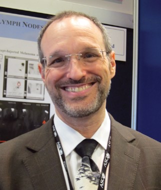
Vernon K. Sondak, MD, chair, department of cutaneous oncology, Moffitt Cancer Center, Tampa, Fla. Dr. Sondak is a paid consultant to Navidea, a company that is developing an agent for use in sentinel node biopsy for melanoma and breast cancer, and to Merck.
This guideline is important in that it provides further strong support from two of the world’s premier oncologic societies for the use of sentinel node biopsy in appropriate patients with clinically localized melanoma. Most melanoma patients today present with clinically localized disease and hence may be potential candidates for this procedure.
Importantly, the ASCO/SSO guideline panel included medical oncologists, pathologists, and at least one melanoma survivor, so the objectivity of this panel should help reassure patients facing a decision about whether to undergo sentinel node biopsy as part of their melanoma treatment.
But it is also important to note that there are limitations imposed by the specific guideline development process that ASCO uses to define allowable studies for inclusion in the analysis. Because of those limitations, the panel appropriately concluded that sentinel node biopsy should not be used for ALL thin melanoma patients. However, they were not able to specifically define what "high risk features" surgeons and patients should consider, and that is probably the single greatest deficiency of these guidelines.
|
|
In particular, both surgeons and patients have unanswered questions about whether patients with stage I melanoma automatically need a sentinel node biopsy if they are called stage IB because the pathologist finds at least one mitosis in the dermal component of the tumor. In my opinion, based on my years of experience and also a recent review of our experience with 271 stage I melanoma patients undergoing sentinel node biopsy at Moffitt Cancer Center over the past 5 years (Ann. Surg. Oncol. [doi: 10.1245/s10434-012-2469-1]), sentinel node biopsy is NOT necessary or appropriate for the overwhelming majority of patients whose melanoma is less than 0.76 mm in thickness, whether or not it is stage IB. However, for patients with melanomas between 0.76 and 1.00 mm in thickness, I believe that many will be appropriate candidates for the procedure, even if they are stage IA.
It’s also worth emphasizing the importance of the pathology report in making a final recommendation about whether to undergo a sentinel node biopsy. Patients should be sure to discuss their pathology report in detail with their doctor, and ask whether they should be evaluated by a surgeon or surgical oncologist for consideration of a sentinel node biopsy. If all the pathologic features required to make an informed decision have not been reported, and sometimes even if they are, we have found a second pathology opinion by a dermatopathologist who specializes in melanoma to be extremely helpful, and have sometimes dramatically changed our recommendations as a result.
But again, the guideline should reassure patients that sentinel node biopsy is often but by no means always appropriate – and a thoughtful discussion of the pros and cons of the procedure is important in every case.
Vernon K. Sondak, MD, chair, department of cutaneous oncology, Moffitt Cancer Center, Tampa, Fla. Dr. Sondak is a paid consultant to Navidea, a company that is developing an agent for use in sentinel node biopsy for melanoma and breast cancer, and to Merck.
All newly diagnosed melanoma patients with tumors of intermediate thickness – defined as those with a Breslow thickness of between 1 and 4 mm – should undergo sentinel lymph node biopsy, according to a new guideline from two professional societies.
The guideline, issued jointly July 9 by the American Society of Clinical Oncology and the Society of Surgical Oncology, also advises that while the use of sentinel lymph node biopsy (SLNB) in people with thicker or thinner melanomas remains contentious, both categories of patients could benefit from SLNB in some circumstances.
All melanoma patients with a positive SLNB, the guideline says, should be treated with completion lymph node dissection (CLND), the current standard of care, although, the authors noted, it is not yet known whether CLND after a positive SLN biopsy improves 10-year overall survival. This is the subject of a large, ongoing randomized trial, the Multicenter Selective Lymphadenectomy Trial II (MSLT-11).
The guideline’s authors, led by Dr. Sandra L. Wong of the University of Michigan, Ann Arbor, and Dr. Gary H. Lyman of Duke University, Durham, N.C., aimed to use the most current evidence to clarify both the indications for sentinel node biopsy and the role of completion biopsy in people with melanomas.
"It is critically important to identify those patients for whom the expected benefits of resecting regional lymph nodes outweigh the risks of surgical morbidity," they wrote in their analysis.
To this end, Dr. Wong, Dr. Lyman, and 12 other expert panel members undertook a broad literature search and identified 73 studies (most of them observational in design) that met the criteria for inclusion in their meta-analysis. Some 25,000 patients were enrolled in the included studies.
The authors found that while robust evidence supports routine use of sentinel lymph node biopsy (SLNB) in intermediate melanomas, there is also some evidence to support the procedure in patients with thin melanomas (less than 1 mm) when certain other risk factors are present, and in patients with thick melanomas (greater than 4 mm).
For people with melanomas of less than 1 mm Breslow thickness and one or more risk factors such as tumor ulceration or a mitotic rate of 1/mm2 or greater, they wrote, "the benefits of pathologic staging may outweigh the potential risks of the procedure," particularly in the subgroup of patients with melanomas ranging from 0.75 mm to 0.99 mm.
Patients with melanomas of 4 mm or greater may also benefit, the guideline says. "Conventional wisdom asserts that patients with thick melanomas have a high risk of systemic disease at the time of diagnosis and that no survival benefit can be derived from removal of regional lymph nodes," the authors wrote.
"However, among patients without distant disease, it can be argued that those with thick melanomas have indications for SLN biopsy similar to those of patients with intermediate-thickness melanomas and derive the same benefits from SLN biopsy as a pathologic staging procedure. One of the main advantages of SLN biopsy in patients with thick melanomas is better regional disease control."
The guideline reiterates that CLND should remain the standard of care for patients with tumor-positive SLNs, even absent survival data from the ongoing MSLT-II trial. The authors cited in support of this evidence from studies that saw nodal recurrence after CLND of between 4.2% and 4.9%. By contrast, as Dr. Wong and colleagues reported in an earlier study, patients in whom CLND was not performed saw a 15% rate of regional nodal recurrence as a site of first metastasis and 41% overall regional nodal recurrence rate (Ann Surg Oncol 2006 13:302-309).
"Until final results of MSLT-II are available, we will not be able to determine, with higher-level evidence, the impact of CLND on regional disease control. Until that time, the best available evidence suggests that CLND is effective at achieving regional disease control in the majority of patients with positive SLNs," the authors wrote.
The guideline was commissioned by ASCO and SSO. The authors disclosed no conflicts of interest.
A Mending Ozone: The Skinny Podcast
In this month's program, Dr. Axel Hauschild discusses the results of a study that showed a 70% improvement in progression-free survival in patients with melanoma randomized to dabrafenib in the open-label break-3 trial.
Then, our reporter Heidi Splete reviews a study showing that an at-home hair removal device may work no better than standard shaving.

The Environment Protection Agency has reported that the ozone layer is improving, thanks to the worldwide ban of chlorofluorocarbons in 1989. We interviewed Drusilla Hufford at the EPA to find out more.
Cosmetic Counter host, Dr. Lily Talakoub, offers tips for educating patients about retinoids and anti-wrinkle creams. And lastly, Dr. Alan Rockoff tells a story that may induce contractions.
Don’t miss another episode of The Skinny Podcast; subscribe for free on iTunes!
In this month's program, Dr. Axel Hauschild discusses the results of a study that showed a 70% improvement in progression-free survival in patients with melanoma randomized to dabrafenib in the open-label break-3 trial.
Then, our reporter Heidi Splete reviews a study showing that an at-home hair removal device may work no better than standard shaving.
The Environment Protection Agency has reported that the ozone layer is improving, thanks to the worldwide ban of chlorofluorocarbons in 1989. We interviewed Drusilla Hufford at the EPA to find out more.
Cosmetic Counter host, Dr. Lily Talakoub, offers tips for educating patients about retinoids and anti-wrinkle creams. And lastly, Dr. Alan Rockoff tells a story that may induce contractions.
Don’t miss another episode of The Skinny Podcast; subscribe for free on iTunes!