User login
Preserving normal hematopoietic function in AML
Preclinical research has revealed a treatment that might preserve normal hematopoietic function in patients with acute myeloid leukemia (AML).
Researchers found that AML suppresses adipocytes in the bone marrow, which leads to imbalanced regulation of endogenous hematopoietic stem and progenitor cells and impaired myelo-erythroid maturation.
However, a PPARγ agonist can induce adipogenesis, which suppresses AML cells and stimulates the regeneration of healthy blood cells.
Researchers reported these findings in Nature Cell Biology.
The team found that adipocytes in the bone marrow support myelo-erythroid maturation of hematopoietic stem and progenitor cells. But AML has a negative effect on the maturation of adipocytes, which explains why deficient myelo-erythropoiesis is “a consistent feature” of AML.
The researchers also found that pro-adipogenesis therapy—treatment with the PPARγ agonist GW1929—protects healthy myelo-erythropoiesis and limits leukemic self-renewal.
“Our approach represents a different way of looking at leukemia and considers the entire bone marrow as an ecosystem, rather than the traditional approach of studying and trying to directly kill the diseased cells themselves,” said study author Allison Boyd, PhD, of McMaster University in Hamilton, Ontario, Canada.
“These traditional approaches have not delivered enough new therapeutic options for patients. The standard of care for this disease hasn’t changed in several decades.”
“The focus of chemotherapy and existing standard of care is on killing cancer cells, but, instead, we took a completely different approach, which changes the environment the cancer cells live in,” said study author Mick Bhatia, PhD, of McMaster University.
“This not only suppressed the ‘bad’ cancer cells but also bolstered the ‘good’ healthy cells, allowing them to regenerate in the new drug-induced environment. The fact that we can target one cell type in one tissue using an existing drug makes us excited about the possibilities of testing this in patients.”
“We can envision this becoming a potential new therapeutic approach that can either be added to existing treatments or even replace others in the near future. The fact that this drug activates blood regeneration may provide benefits for those waiting for bone marrow transplants by activating their own healthy cells.” ![]()
Flu vaccine appears ineffective in young leukemia patients
Vaccination may fail to protect young leukemia patients from developing influenza during cancer treatment, according to research published in the Journal of Pediatrics.
Researchers found that young patients with acute leukemia who received flu shots were just as likely as their unvaccinated peers to develop the flu.
The team said these results are preliminary, but they suggest a need for more research and additional efforts to prevent flu in young patients with leukemia.
“The annual flu shot, whose side effects are generally mild and short-lived, is still recommended for patients with acute leukemia who are being treated for their disease,” said study author Elisabeth Adderson, MD, of St. Jude Children’s Research Hospital in Memphis, Tennessee.
“However, the results do highlight the need for additional research in this area and for us to redouble our efforts to protect our patients through other means.”
In this retrospective study, Dr Adderson and her colleagues looked at rates of flu infection during 3 successive flu seasons (2010-2013) in 498 patients treated for acute leukemia at St. Jude.
The patients’ median age was 6 years (range, 1-21). Most patients had acute lymphoblastic leukemia (ALL, 94%), though some had acute myeloid leukemia (4.8%) or mixed-lineage leukemia (1.2%).
Most patients (n=354) received flu shots, including 98 patients who received booster doses. The remaining 144 patients were not vaccinated.
The vaccinated patients received the trivalent vaccine, which is designed to protect against 3 flu strains predicted to be in wide circulation during a particular flu season. The vaccine was a fairly good match for circulating flu viruses during the flu seasons included in this analysis.
Demographic characteristic were largely similar between vaccinated and unvaccinated patients. The exceptions were that more vaccinated patients had ALL (95.5% vs 90.3%; P=0.034) and vaccinated patients were more likely to be in a low-intensity phase of cancer therapy (90.7% vs 73.6%, P<0.0001).
Results
There were no significant differences between vaccinated and unvaccinated patients when it came to flu rates or rates of flu-like illnesses.
There were 37 episodes of flu in vaccinated patients and 16 episodes in unvaccinated patients. The rates (per 1000 patient days) were 0.73 and 0.70, respectively (P=0.874).
There were 123 cases of flu-like illnesses in vaccinated patients and 55 cases in unvaccinated patients. The rates were 2.44 and 2.41, respectively (P=0.932).
Likewise, there was no significant difference in the rates of flu or flu-like illnesses between patients who received 1 dose of flu vaccine and those who received 2 doses.
The flu rates were 0.60 and 1.02, respectively (P=0.107). And the rates of flu-like illnesses were 2.42 and 2.73, respectively (P=0.529).
Dr Adderson said additional research is needed to determine if a subset of young leukemia patients may benefit from vaccination.
She added that patients at risk of flu should practice good hand hygiene and avoid crowds during the flu season. Patients may also benefit from “cocooning,” a process that focuses on getting family members, healthcare providers, and others in close contact with at-risk patients vaccinated. ![]()
Team devises new method to analyze cells
Biophysicists have developed a new method to determine a cell’s mechanical properties, and they believe this method could provide insights regarding cancers, sickle cell anemia, and other diseases.
The method allows researchers to make standardized measurements of single cells, determine each cell’s stiffness, and assign it a number, generally between 10 and 20,000, in pascals.
“Measuring cells with our calibrated instrument is like measuring time with a standardized clock,” said Amy Rowat, PhD, of the University of California Los Angeles.
“Our method can be used to obtain stiffness measurements of hundreds of cells per second.”
Dr Rowat and her colleagues described their method in Biophysical Journal.
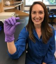
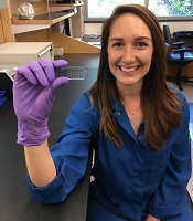
The method is called quantitative deformability cytometry (q-DC). It involves a small device (about 1 inch by 2 inches) made of a soft, flexible rubber that has integrated circuit chips like those in computers.
The researchers use gel particles containing molecules derived from seaweed to force cells through tiny pores in the device. As the cells flow through the device, the researchers take videos at thousands of frames per second—more than 100 times faster than standard video.
Dr Rowat and her colleagues used the device to analyze promyelocytic leukemia cells (HL-60) and breast cancer cells.
The researchers believe this work will provide scientists with a more precise, standardized method to distinguish cancer cells from normal cells.
The team thinks that, in the future, their method could be used to track a cancer patient over time to see how a drug is affecting the patient’s cancer cells.
“By using q-DC, we can very rapidly assess how specific drug treatments affect physical properties of single cells—such as shape, size, and stiffness—and achieve calibrated, quantitative measurements,” Dr Rowat said.
She and her colleagues believe q-DC might also help predict how invasive a cancer cell could be and which drugs might be most effective in fighting the cancer, as well as revealing which proteins are important in regulating the invasion of a cancer cell.
The researchers are now applying q-DC to other types of cancer cells. The team would like to better understand the relationship between a cancer cell’s physical properties and how easily cancer cells can spread through the body.
Dr Rowat’s hypothesis is that properties such as stiffness, size, and a cell’s ability to change shape are important in enabling cancer cells to maneuver.
The researchers said they can also use q-DC to measure other types of cells, such as normal and sickled red blood cells. ![]()
Compound induces selective apoptosis in AML
Researchers say they have discovered a compound that can overcome resistance to apoptosis in acute myeloid leukemia (AML).
The compound, BTSA1, works by activating the BCL-2 family protein BAX.
BTSA1 prompted apoptosis in leukemia cells while sparing healthy cells. It also suppressed AML in mice without producing side effects.
Evripidis Gavathiotis, PhD, of Albert Einstein College of Medicine in Bronx, New York, and his colleagues described these results in Cancer Cell.
The team knew that apoptosis occurs when BAX is activated by pro-apoptotic proteins. However, cancer cells can avoid apoptosis by producing anti-apoptotic proteins that suppress BAX and the proteins that activate it.
“Our novel compound revives suppressed BAX molecules in cancer cells by binding with high affinity to BAX’s activation site,” Dr Gavathiotis said. “BAX can then swing into action, killing cancer cells while leaving healthy cells unscathed.”
Dr Gavathiotis was the lead author of a paper published in Nature in 2008 that first described the structure and shape of BAX’s activation site. He has since looked for small molecules that can activate BAX strongly enough to overcome cancer cells’ resistance to apoptosis.
His team initially screened more than 1 million compounds to reveal those with BAX-binding potential. The most promising 500 compounds were then evaluated in the lab.
“A compound dubbed BTSA1 (short for BAX Trigger Site Activator 1) proved to be the most potent BAX activator, causing rapid and extensive apoptosis when added to several different human AML cell lines,” said Denis Reyna, a doctoral student in Dr Gavathiotis’s lab.
The researchers also tested BTSA1 in blood samples from patients with high-risk AML. BTSA1 induced apoptosis in the patients’ AML cells but did not affect healthy hematopoietic stem cells.
Finally, the researchers generated mouse models of AML. BTSA1 was given to half the mice, while the other half served as controls.
On average, the BTSA1-treated mice survived significantly longer than the control mice—55 days and 40 days, respectively (P=0.0009). In fact, 43% of BTSA1-treated mice were still alive after 60 days and showing no signs of AML.
In addition, the mice treated with BTSA1 showed no evidence of toxicity.
“BTSA1 activates BAX and causes apoptosis in AML cells while sparing healthy cells and tissues, probably because the cancer cells are primed for apoptosis,” Dr Gavathiotis said.
He and his colleagues found that AML cells contained significantly higher BAX levels than normal blood cells from healthy subjects.
“With more BAX available in AML cells, even low BTSA1 doses will trigger enough BAX activation to cause apoptotic death, while sparing healthy cells that contain low levels of BAX or none at all,” Dr Gavathiotis said.
He and his team plan to determine if BTSA1 will elicit similar results in other cancer types. ![]()
Natural selection opportunities tied to cancer rates
Countries with the lowest opportunities for natural selection have higher cancer rates than countries with the highest opportunities for natural selection, according to a study published in Evolutionary Applications.
Researchers said this is because modern medicine is enabling people to survive cancers, and their genetic backgrounds are passing from one generation to the next.
The team said the rate of some cancers has doubled and even quadrupled over the past 100 to 150 years, and human evolution has moved away from “survival of the fittest.”
“Modern medicine has enabled the human species to live much longer than would otherwise be expected in the natural world,” said study author Maciej Henneberg, PhD, DSc, of the University of Adelaide in South Australia.
“Besides the obvious benefits that modern medicine gives, it also brings with it an unexpected side-effect—allowing genetic material to be passed from one generation to the next that predisposes people to have poor health, such as type 1 diabetes or cancer.”
“Because of the quality of our healthcare in western society, we have almost removed natural selection as the ‘janitor of the gene pool.’ Unfortunately, the accumulation of genetic mutations over time and across multiple generations is like a delayed death sentence.”
Country comparison
The researchers studied global cancer data from the World Health Organization as well as other health and socioeconomic data from the United Nations and the World Bank of 173 countries. The team compared the top 10 countries with the highest opportunities for natural selection to the 10 countries with the lowest opportunities for natural selection.
“We looked at countries that offered the greatest opportunity to survive cancer compared with those that didn’t,” said study author Wenpeng You, a PhD student at the University of Adelaide. “This does not only take into account factors such as socioeconomic status, urbanization, and quality of medical services but also low mortality and fertility rates, which are the 2 distinguishing features in the ‘better’ world.”
“Countries with low mortality rates may allow more people with cancer genetic background to reproduce and pass cancer genes/mutations to the next generation. Meanwhile, low fertility rates in these countries may not be able to have diverse biological variations to provide the opportunity for selecting a naturally fit population—for example, people without or with less cancer genetic background. Low mortality rate and low fertility rate in the ‘better’ world may have formed a self-reinforcing cycle which has accumulated cancer genetic background at a greater rate than previously thought.”
Based on the researchers’ analysis, the 20 countries are:
| Lowest opportunities for natural selection | Highest opportunities for natural selection |
| Iceland | Burkina Faso |
| Singapore | Chad |
| Japan | Central African Republic |
| Switzerland | Afghanistan |
| Sweden | Somalia |
| Luxembourg | Sierra Leone |
| Germany | Democratic Republic of the Congo |
| Italy | Guinea-Bissau |
| Cyprus | Burundi |
| Andorra | Cameroon |
Cancer incidence
The researchers found the rates of most cancers were higher in the 10 countries with the lowest opportunities for natural selection. The incidence of all cancers was 2.326 times higher in the low-opportunity countries than the high-opportunity ones.
The increased incidences of hematologic malignancies were as follows:
- Non-Hodgkin lymphoma—2.019 times higher in the low-opportunity countries
- Hodgkin lymphoma—3.314 times higher in the low-opportunity countries
- Leukemia—3.574 times higher in the low-opportunity countries
- Multiple myeloma—4.257 times higher in the low-opportunity countries .
Dr Henneberg said that, having removed natural selection as the “janitor of the gene pool,” our modern society is faced with a controversial issue.
“It may be that the only way humankind can be rid of cancer once and for all is through genetic engineering—to repair our genes and take cancer out of the equation,” he said. ![]()
NCCN completes resource on radiation therapy
The National Comprehensive Cancer Network® (NCCN) has announced the release of the newly completed NCCN Radiation Therapy Compendium™.
This resource includes information designed to support clinical decision-making regarding the use of radiation therapy in cancer patients.
The content is based on the NCCN Clinical Practice Guidelines in Oncology and includes information from the 41 guidelines that reference radiation therapy.
“By compiling every recommendation for radiation therapy in one place, we’ve made it significantly easier for specialists . . . to stay up-to-date on the very latest recommendations, regardless of how many different cancer types they treat,” said Robert W. Carlson, MD, chief executive officer of NCCN.
“This targeted content provides radiation oncologists with the specific, cutting-edge information they need, without forcing them to sift through any extraneous information. It’s part of our ongoing effort to always provide the most pertinent data on emerging treatment practices in the clearest, most efficient way possible.”
The NCCN Radiation Therapy Compendium includes a full complement of radiation therapy recommendations found in the current NCCN guidelines, including specific treatment modalities such as 2D/3D conformal external beam radiation therapy, intensity modulated radiation therapy, intra-operative radiation therapy, stereotactic radiosurgery/stereotactic body radiotherapy/stereotactic ablative body radiotherapy, image-guided radiation therapy, low dose-rate/high dose-rate brachytherapy, radioisotope, and particle therapy.
NCCN first announced the launch of the Radiation Therapy Compendium in March at the NCCN Annual Conference: Improving the Quality, Effectiveness, and Efficiency of Cancer Care.
At the time, the NCCN released a preliminary version of the compendium featuring 24 cancer types. The newly completed version now contains all 41 disease sites that are currently being treated using radiation therapy.
The compendium will be updated on a continual basis in conjunction with the library of clinical guidelines.
For more information and to access the NCCN Radiation Therapy Compendium, visit NCCN.org/RTCompendium. The compendium is available free-of-charge through March 2018. ![]()
AML capsules
Detecting hereditary MDS/AML
Unexplained cytopenias or failure to mobilize stem cells in related donors of patients with myelodysplastic syndrome (MDS) or acute myeloid leukemia (AML) – important clues for the detection of a hereditary MDS/AML syndrome – should prompt efforts to obtain germ line tissue sources for collaborative research, Jane E. Churpek, MD, of the Hereditary Hematologic Malignancies Program at The University of Chicago Medicine, wrote.
Partnering with families to obtain germ line tissue sources uncontaminated by tumor cells from large numbers of patients with MDS/AML, and international collaboration to determine the mechanisms and multi-step processes from the carrier state to overt disease, have enormous potential to improve the outcomes of patients with both familial and sporadic forms of MDS/AML. Incorporating collection of ideal germ line tissue along with family histories to the already robust international MDS/AML registries and data sharing are all essential to future progress in this field, she wrote in a recent article published onlne in Best Practice & Research Clinical Haematology.
Prophylactic granulocyte transfusions
Giving prophylactic granulocyte transfusions to AML patients during the induction phase of therapy is feasible and safe, but assuring an adequate donor pool and patient continuation of the transfusions are another matter.
In a phase 2 trial of 45 neutropenic patients with AML or high-risk myelodysplastic syndrome undergoing induction or first-salvage therapy, non-irradiated allogeneic granulocyte transfusions were to be given every 3-4 days until sustained ANC recovery, initiation of new therapy, or completion of 6 weeks on study. But logistical problems limited the success of the protocol: 5 patients never received a granulocyte transfusion due to donor screening failure or donor unavailability and the other 40 received a median of 3 (range 1-9) granulocyte transfusions.
“We anticipated approximately 8 GTs per patient ... only 11 received 6 or more GTs,” Fleur M. Aung, MD, and colleagues at The MD Anderson Cancer Center, Houston, wrote in the Journal of Blood Disorders and Transfusion (DOI 10.4172/2155-9864.1000376).
The authors concluded that while the process is feasible, ex vivo expanded neutrophils, produced from multiple donors and cryopreserved to provide an “off the shelf” myeloid progenitor product, will likely prove more reliable for treating patients with prolonged neutropenia.
From ONC201 to ONC212
Oncoceutics has expanded its research collaboration agreement with The University of Texas MD Anderson Cancer Center, Houston, to include the clinical development of a second novel imipridone called ONC212.
Oncoceutics and MD Anderson are already collaborating on the clinical development of another imipridone, ONC201. Both drugs have the same chemical core that interacts with G-Protein Coupled Receptors (GPCRs). ONC212 targets GPR132, which influences the growth of acute leukemias and has not previously been successfully targeted by a small molecule.
This alliance between MD Anderson and Oncoceutics will support Phase I and Phase II clinical trials of ONC212 in patients with refractory acute myeloid leukemia (AML), according to Oncoceutics. The alliance provides for a sharing of risk and rewards from potential commercialization of ONC212.
Top risk factor miRNA
The miRNA hsa-mir-425 was identified as the top risk factor miRNA of AML survival and CD44 was identified as one of the top three risk factor target-genes associated with AML survival, based on a miRNA-mRNA interaction network.
Chunmei Zhang, MD, of the department of hematology at Taian City Central Hospital, Taian, Shandong, China, and colleagues used The Cancer Genome Atlas database to obtain miRNA and mRNA expression profiles from AML patients. Of 14 miRNAs associated with AML survival, 3 were associated with risk – hsa-mir-425, hsa-mir-1201, and hsa-mir-1978. GTSF1, RTN4R, and CD44 were the top risk factor target-genes associated with AML survival. Med Sci Monit 2017; 23:4705-14 (DOI: 10.12659/MSM.903989)
Minimal residual disease measures not yet impactful for AML patients
SAN FRANCISCO – Routine testing for minimal residual disease is probably not of value in acute myeloid leukemia, as there is no evidence that changing treatment based on MRD status currently makes a difference in patient outcomes, experts said at the annual congress on Hematologic Malignancies held by the National Comprehensive Cancer Network.
“If we find minimal residual disease, we don’t always have a better therapy to offer our patients,” Jessica Altman, MD, associate professor of hematology and oncology at the Northwestern University Feinberg School of Medicine, said.
Beyond that therapeutic reality, there are no clear guidelines and standards for MRD testing. The optimal timing for MRD testing and a standard threshold for an MRD classification are not yet established, she said.
“Having MRD is bad, not having it is better,” Richard Stone, MD, PhD, clinical director of the adult leukemia program at the Dana-Farber Cancer Institute, said. The problem in AML, he said, is, “So?” There is no reliable “MRD eraser” in AML, he said. Until then, there is not much point in knowing whether a patient is MRD positive or not.
A recent survey conducted by researchers at Moffitt Cancer Center, Tampa, addressed MRD testing at 13 major cancer centers. While most centers reported that they test for MRD, many physicians said that they are unsure about what to do with the results.
A 2013 study by the HOVON group found that patients who were in complete remission but MRD positive after their first course of therapy, subsequently became MRD negative after their second course of therapy. But the second regimen would not have been different based on knowledge of MRD status, according to the HOVON/SAKK AML 42A study (J Clin Oncol. 2013; 31:3889-97).
The AML community is awaiting guidelines on MRD use from the NCCN and other groups, Dr. Altman said. An option for using NPM1 mutations to assess MRD should be available soon, and could be an improvement on existing options (N Engl J Med 2016; 374:422-33).
Given the treatment limitations, knowing about MRD status can have a negative mental toll on patients, Dr. Stone said. “I would not underplay the psychological burden.” Nevertheless, MRD should be measured in clinical trials, and it could be a valuable surrogate marker by which to compare drug efficacy.
One of the biggest hopes is that MRD status could eventually be useful in determining the need for allogeneic stem cell transplant in patients deemed intermediate risk, Dr. Altman said. “I think we are finally on the brink of this being actionable.”
Dr. Altman reports financial relationships with Astellas, Bristol-Myers Squibb, Celgene, Janssen, Novartis, and Syros. Dr. Stone reports financial relationships with AbbVie, Actinium, Agios, Amgen and many other companies.
EXPERT ANALYSIS AT NCCN HEMATOLOGIC MALIGNANCIES CONGRESS
FDA grants drug fast track designation for rel/ref AML
The US Food and Drug Administration (FDA) has granted fast track designation to gilteritinib for the treatment of adults with FLT3 mutation-positive relapsed or refractory acute myeloid leukemia (AML).
Gilteritinib is a compound that has demonstrated inhibitory activity against FLT3 internal tandem duplication (ITD) and FLT3 tyrosine kinase domain, 2 mutations that are seen in approximately one-third of patients with AML.
Gilteritinib has also demonstrated inhibition of AXL, which is reported to be associated with therapeutic resistance.
Astellas Pharma Inc. is currently investigating gilteritinib in phase 3 trials of AML patients.
Results from a phase 1/2 study of gilteritinib in AML were presented at the 2017 ASCO Annual Meeting.
The goal of the study was to determine the tolerability and antileukemic activity of once-daily gilteritinib in a FLT3-ITD-enriched, relapsed/refractory AML population.
Researchers said the drug exhibited potent FLT3 inhibition at doses greater than 80 mg/day. In patients who received such doses, the greatest overall response rate was 52%, and the longest median overall survival was 31 weeks.
The maximum tolerated dose of gilteritinib was 300 mg/day. Dose-limiting toxicities included diarrhea and liver function abnormalities.
About fast track designation
The FDA’s fast track program is designed to facilitate the development and expedite the review of products intended to treat or prevent serious or life-threatening conditions and address unmet medical need.
Through the fast track program, a product may be eligible for priority review. In addition, the company developing the product may be allowed to submit sections of the new drug application or biologics license application on a rolling basis as data become available.
Fast track designation also provides the company with opportunities for more frequent meetings and written communications with the FDA.
About orphan designation
Gilteritinib also has orphan drug designation for the treatment of AML.
The FDA grants orphan designation to products intended to treat, diagnose, or prevent diseases/disorders that affect fewer than 200,000 people in the US.
The designation provides incentives for sponsors to develop products for rare diseases. This may include tax credits toward the cost of clinical trials, prescription drug user fee waivers, and 7 years of market exclusivity if the product is approved. ![]()
The US Food and Drug Administration (FDA) has granted fast track designation to gilteritinib for the treatment of adults with FLT3 mutation-positive relapsed or refractory acute myeloid leukemia (AML).
Gilteritinib is a compound that has demonstrated inhibitory activity against FLT3 internal tandem duplication (ITD) and FLT3 tyrosine kinase domain, 2 mutations that are seen in approximately one-third of patients with AML.
Gilteritinib has also demonstrated inhibition of AXL, which is reported to be associated with therapeutic resistance.
Astellas Pharma Inc. is currently investigating gilteritinib in phase 3 trials of AML patients.
Results from a phase 1/2 study of gilteritinib in AML were presented at the 2017 ASCO Annual Meeting.
The goal of the study was to determine the tolerability and antileukemic activity of once-daily gilteritinib in a FLT3-ITD-enriched, relapsed/refractory AML population.
Researchers said the drug exhibited potent FLT3 inhibition at doses greater than 80 mg/day. In patients who received such doses, the greatest overall response rate was 52%, and the longest median overall survival was 31 weeks.
The maximum tolerated dose of gilteritinib was 300 mg/day. Dose-limiting toxicities included diarrhea and liver function abnormalities.
About fast track designation
The FDA’s fast track program is designed to facilitate the development and expedite the review of products intended to treat or prevent serious or life-threatening conditions and address unmet medical need.
Through the fast track program, a product may be eligible for priority review. In addition, the company developing the product may be allowed to submit sections of the new drug application or biologics license application on a rolling basis as data become available.
Fast track designation also provides the company with opportunities for more frequent meetings and written communications with the FDA.
About orphan designation
Gilteritinib also has orphan drug designation for the treatment of AML.
The FDA grants orphan designation to products intended to treat, diagnose, or prevent diseases/disorders that affect fewer than 200,000 people in the US.
The designation provides incentives for sponsors to develop products for rare diseases. This may include tax credits toward the cost of clinical trials, prescription drug user fee waivers, and 7 years of market exclusivity if the product is approved.
Clinical trials summary: AGILE
Study of AG-120 (Ivosidenib) vs. Placebo in Combination With Azacitidine in Patients With Previously Untreated Acute Myeloid Leukemia With an IDH1 Mutation (AGILE)
AG120-C-009 (NCT03173248) will evaluate the efficacy and safety of AG-120 (ivosidenib) plus azacitidine vs. placebo plus azacitidine in adult subjects with previously untreated IDH1m AML who are considered appropriate candidates for non-intensive therapy.
The primary endpoint of this global, phase 3, multicenter, double-blind, randomized, placebo-controlled clinical trial is overall survival. Key secondary efficacy endpoints are event-free survival, rate of complete remission, rate of complete remission with partial hematologic recovery, and overall response rate. Subjects will be randomized 1:1 to receive either oral AG-120 or matched placebo, both administered in combination with subcutaneous or intravenous azacitidine. An estimated 392 subjects will participate in the study, which is sponsored by Agios Pharmaceuticals. Estimated completion date is June 2022.
Contact: Medical Affairs Agios Pharmaceuticals, Inc.; (844) 633-2332, e-mail: medinfo@agios.com
