User login
Haplo-HSCT regimen can cure SCD, team says
A haploidentical transplant regimen has led to long-term engraftment in adults with sickle cell disease (SCD), according to researchers.
Seven of 8 patients treated with this regimen were still alive at last follow-up, and 6 of them maintained engraftment.
Two patients developed graft-versus-host disease (GVHD). One of them had acute and chronic GVHD and died about 400 days after transplant. The other had acute GVHD that resolved with treatment.
Damiano Rondelli, MD, of the University of Illinois at Chicago, and his colleagues reported these results in Biology of Blood and Marrow Transplantation.
The researchers initially screened 50 adult SCD patients as candidates for haploidentical hematopoietic stem cell transplant (haplo-HSCT) between January 2014 and March 2017. Most patients were ineligible or declined the procedure.
Ultimately, 10 patients received a haplo-HSCT. Unfortunately, the first 2 patients failed to engraft. These patients had received conditioning with alemtuzumab and 3 Gy total body irradiation (TBI) as well as post-transplant cyclophosphamide.
Because of this failure, the researchers used the following regimen for the remaining 8 patients. It’s a modified version of the regimen described in Blood in 2012.
“We modified the transplant protocol by increasing the dose of radiation used before the transplant and by infusing growth factor-mobilized peripheral blood stem cells instead of bone marrow cells,” Dr Rondelli said. “These two modifications helped ensure the patient’s body could accept the healthy donor cells.”
Modified regimen
Patients received growth-factor-mobilized peripheral blood stem cells after conditioning with rabbit antithymocyte globulin (0.5 mg/kg on day -9, 2 mg/kg on day -8 and -7), cyclophosphamide (14.5 mg/kg on day -6 and -5), fludarabine (30 mg/m2 on day -6 to -2), and single-dose TBI (3 Gy on day -1).
For GVHD prophylaxis, patients received intravenous cyclophosphamide (50 mg/kg on day 3 and 4), oral mycophenolate mofetil (15 mg/kg 3 times daily from day 5 to 35), and sirolimus (from day 5 dosed for a target trough of 5 to 15 ng/mL). In patients who had T-cell chimerism greater than 50% at 1 year after HSCT and did not have signs of GVHD, sirolimus was tapered off over 3 months.
Patients stopped taking hydroxyurea on day -9. They received red blood cell exchange transfusion on day -10 (with the goal of getting hemoglobin S below 30%) and received platelet transfusions to maintain platelet counts greater than 50 x 109 cells/L.
Patients also received penicillin V (250 mg twice daily) in addition to standard antimicrobial prophylaxis.
Results
All 8 patients on the modified regimen engrafted. The median time to neutrophil engraftment was 22 days (range, 18 to 23 days). One patient experienced secondary graft failure on day 90.
Seven neutropenic patients with hemoglobin S less than 30% received G-CSF after transplant. They received a median of 7 doses (range, 3 to 14) at 5 μg/kg, starting at day 12 post-HSCT. One of these patients experienced mild bone pain in the lower extremities.
Two patients developed GVHD. At day 83, one patient developed acute or chronic GVHD involving the skin, liver, and eyes. Steroids and sirolimus improved eye and liver symptoms, but the patient died at home on day 407.
The other patient had grade 2, gastrointestinal, acute GVHD that resolved with steroid therapy.
Three patients had grade 2 or higher mucositis, and 2 had cytomegalovirus (CMV) reactivation without CMV infection.
Two patients had small subarachnoid hemorrhages. One of these patients had a history of multiple red blood cell antibodies, became refractory to platelet transfusions, and developed small subarachnoid hemorrhages day 10. The patient’s symptoms and brain imaging resolved after platelet counts were maintained above 50 x 109 cells/L with cross-matched platelets.
The second patient had a history of stroke, experienced a seizure when the platelet count was 68 x 109 cells/L, and was found to have a subarachnoid hemorrhage on day 12. Symptoms and imaging results improved once the patient began levetiracetam and platelet levels were maintained above 100 x 109 cells/L.
At a median follow-up of 17 months (range, 12 to 30), 7 of the 8 patients were still alive.
Six patients had maintained greater than 95% stable donor engraftment with improvements in their hemoglobin concentrations. Three of these patients have stopped immunosuppression, and 3 are being tapered off it.
“These patients are cured of sickle cell disease,” Dr Rondelli said. “The takeaway message is two-fold. First, this transplant protocol may cure many more adult patients with advanced sickle cell disease.”
“Second, despite the increasing safety of the transplant protocols and new compatibility of HLA half-matched donors, many sickle cell patients still face barriers to care. Of the patients we screened, only 20% underwent a transplant.”
Dr Rondelli noted that 20% of the patients screened could not undergo transplant because of insurance denial. Other patients were ineligible because they had high rates of donor-specific antigens, and still others declined transplant.
Gene therapy for thalassemia normalizes hemoglobin
Most beta thalassemia patients were off transfusions at a median of 26 months after receiving gene therapy via a lentiviral vector, according to new results of two phase 1/2 studies regarding the use of LentiGlobin in transfusion-dependent beta thalassemia.
Of 13 patients who did not have the most severe beta0/beta0 genotype, all but 1 has become transfusion independent post transplant. Among patients who either had the beta0/beta0 genotype or had two copies of the IVS1-110 mutation, transfusions were down a median 73% annually, and three of these patients with more severe thalassemia became transfusion independent.
At the time of last data collection, hemoglobin levels in individual patients ranged from 8.2-13.7 g/dL.
“No clonal dominance related to vector integration was observed,” wrote Alexis Thompson, MD, and her collaborators, and replication-competent lentivirus had not been found in any patients.
Hematopoietic cell transplant (HCT) is an option primarily for younger beta thalassemia patients who have an HLA-matched sibling donor, the researchers wrote in the New England Journal of Medicine. Gene therapy represents an alternative to the current standard of care for patients who are not candidates for allogeneic HCT, which – without a good match – carries increased risk for rejection and graft-versus-host disease.
Patients with beta thalassemia aged 35 years or younger and without advanced organ damage were enrolled in the two studies, one conducted internationally and one conducted at a single site in France.
There were some protocol differences between the two studies; notably, the French study used enhanced red cell transfusion for 3 or more months before stem cell mobilization “to enrich for bona fide hematopoietic stem cells in the harvested CD34+ cell compartment by suppressing the erythroid lineage expansion and the skewing that is seen in beta thalassemia,” wrote Dr. Thompson, professor of pediatrics at Northwestern University, Chicago, and her colleagues.
In both studies, after mobilization, patients’ unmanipulated hematopoietic stem cells and progenitor cells were taken to a central processing facility, where CD34+ cells were enriched and then transduced with the lentiviral vector BB305, which encodes adult hemoglobin (HbA) with a T87Z amino acid substitution and thereby provides functioning Hb beta. Patients received the product via infusion after undergoing myeloablative conditioning with busulfan.
A total of 23 patients, 19 in the international study and 4 in the French study, went through mobilization and apheresis. One patient in the international study had apheresis failure, so a total of 22 patients received LentiGlobin, and all were followed for up to 2 years.
Patients were given the opportunity to participate in a follow-on open label study meant to continue for an additional 13 years after the initial 24-month period; 13 patients are currently enrolled in this long-term follow-up study.
When transfusion volume at baseline was assessed, patients in the international study were receiving a median annual red blood cell transfusion volume of 164 mL/kg per year, while the French study participants were receiving a median 182 mL/kg per year of red blood cell transfusion.
In both studies, blood HbAT87Q levels correlated with the vector copy numbers (R2, 0.75; P less than .001). Levels of HbAT87Q ranged from 3.4-10.0 g/dL.
“Other factors, such as age, genotype, and splenectomy status, did not appear to correlate with gene expression,” the researchers wrote.
An exploratory analysis looked at characteristics of patients who were able to stop transfusions after gene therapy. In this group, “the degree of hemolysis at first stabilized relative to pretransplantation levels and was fully corrected” in two patients by 36 months after treatment.
The researchers noted that the sponsor achieved “high-titer, large-scale, clinical-grade BB305 vector production and purification by ion-exchange chromatography” from a single site in the United States, which showed the feasibility of conducting this modality of gene therapy at scale.
The study was sponsored by bluebird bio, the National Institutes of Health, and by French national research organizations. Dr. Thompson reported research funding and fees from bluebird bio and other pharmaceutical companies.
SOURCE: Thompson A et al. N Engl J Med 2018;378:1479-93
Lentiviral vector hematopoietic stem cell (HSC) gene therapy represents a promising alternative to matched-sibling donor HSC transplants for treatment of beta thalassemia, with studies to date showing a safety profile that surpasses transplants from unrelated or alternative donors.
The prospect of a curative treatment raised by the work of Dr. Thompson and her colleagues also shows the feasibility of transfusion independence for beta0/betaE patients, who carry the most common beta thalassemia genotype, and a significant reduction in transfusions even for patients with the more severe beta0/beta0 genotype.
Beta thalassemia has greatest prevalence in North Africa, the Middle East, and Asia, where access to treatments is limited and patients’ prognoses are often grim. Gene therapy for beta thalassemia could thereby represent the first large-scale implementation of this intervention in developing countries.
Bringing HSC gene therapy to more patients will require not just the availability of autologous HSCs, but of high-quality vector and reliable, high-volume manufacturing of transduced cells.
Harnessing this still-evolving technology to bring a potentially curative treatment to patients in developing countries is an exciting, but challenging, frontier for physicians and researchers involved with gene therapy.
Alessandra Biffi, MD, is director of the gene therapy program at Dana Farber Cancer Institute/Boston Children’s Cancer and Blood Disorders Center, Boston. She serves on the board of directors of the American Society of Gene and Cell Therapy. These remarks were adapted from an accompanying editorial ( N Engl J Med. 2018;378[16]:1551-2 ).
Most beta thalassemia patients were off transfusions at a median of 26 months after receiving gene therapy via a lentiviral vector, according to new results of two phase 1/2 studies regarding the use of LentiGlobin in transfusion-dependent beta thalassemia.
Of 13 patients who did not have the most severe beta0/beta0 genotype, all but 1 has become transfusion independent post transplant. Among patients who either had the beta0/beta0 genotype or had two copies of the IVS1-110 mutation, transfusions were down a median 73% annually, and three of these patients with more severe thalassemia became transfusion independent.
At the time of last data collection, hemoglobin levels in individual patients ranged from 8.2-13.7 g/dL.
“No clonal dominance related to vector integration was observed,” wrote Alexis Thompson, MD, and her collaborators, and replication-competent lentivirus had not been found in any patients.
Hematopoietic cell transplant (HCT) is an option primarily for younger beta thalassemia patients who have an HLA-matched sibling donor, the researchers wrote in the New England Journal of Medicine. Gene therapy represents an alternative to the current standard of care for patients who are not candidates for allogeneic HCT, which – without a good match – carries increased risk for rejection and graft-versus-host disease.
Patients with beta thalassemia aged 35 years or younger and without advanced organ damage were enrolled in the two studies, one conducted internationally and one conducted at a single site in France.
There were some protocol differences between the two studies; notably, the French study used enhanced red cell transfusion for 3 or more months before stem cell mobilization “to enrich for bona fide hematopoietic stem cells in the harvested CD34+ cell compartment by suppressing the erythroid lineage expansion and the skewing that is seen in beta thalassemia,” wrote Dr. Thompson, professor of pediatrics at Northwestern University, Chicago, and her colleagues.
In both studies, after mobilization, patients’ unmanipulated hematopoietic stem cells and progenitor cells were taken to a central processing facility, where CD34+ cells were enriched and then transduced with the lentiviral vector BB305, which encodes adult hemoglobin (HbA) with a T87Z amino acid substitution and thereby provides functioning Hb beta. Patients received the product via infusion after undergoing myeloablative conditioning with busulfan.
A total of 23 patients, 19 in the international study and 4 in the French study, went through mobilization and apheresis. One patient in the international study had apheresis failure, so a total of 22 patients received LentiGlobin, and all were followed for up to 2 years.
Patients were given the opportunity to participate in a follow-on open label study meant to continue for an additional 13 years after the initial 24-month period; 13 patients are currently enrolled in this long-term follow-up study.
When transfusion volume at baseline was assessed, patients in the international study were receiving a median annual red blood cell transfusion volume of 164 mL/kg per year, while the French study participants were receiving a median 182 mL/kg per year of red blood cell transfusion.
In both studies, blood HbAT87Q levels correlated with the vector copy numbers (R2, 0.75; P less than .001). Levels of HbAT87Q ranged from 3.4-10.0 g/dL.
“Other factors, such as age, genotype, and splenectomy status, did not appear to correlate with gene expression,” the researchers wrote.
An exploratory analysis looked at characteristics of patients who were able to stop transfusions after gene therapy. In this group, “the degree of hemolysis at first stabilized relative to pretransplantation levels and was fully corrected” in two patients by 36 months after treatment.
The researchers noted that the sponsor achieved “high-titer, large-scale, clinical-grade BB305 vector production and purification by ion-exchange chromatography” from a single site in the United States, which showed the feasibility of conducting this modality of gene therapy at scale.
The study was sponsored by bluebird bio, the National Institutes of Health, and by French national research organizations. Dr. Thompson reported research funding and fees from bluebird bio and other pharmaceutical companies.
SOURCE: Thompson A et al. N Engl J Med 2018;378:1479-93
FROM THE NEW ENGLAND JOURNAL OF MEDICINE
Key clinical point:
Major finding: Transfusion requirements were down 73% annually in patients with the most severe thalassemia.
Study details: Data from 22 transfusion-dependent patients with beta thalassemia in ongoing phase 1/2 study of gene therapy delivered via lentiviral vector.
Disclosures: The study was sponsored by bluebird bio, the National Institutes of Health, and by French national research organizations. Dr. Thompson reported research funding and fees from bluebird bio and from other pharmaceutical companies.
Source: Thompson A et al. N Engl J Med. 2018;378:1479-93.
Meta-analysis finds no link between stroke and sickle cell trait
Those findings contrast with an earlier longitudinal study that found a 1.4-fold risk of ischemic stroke in SCT carriers, the authors noted.
In their study, neither crude stroke incidence rates nor regression analysis indicated a link between SCT and stroke, said first author Hyacinth I. Hyacinth, MD, PhD, MPH, of the Aflac Cancer and Blood Disorder Center, Emory University, Atlanta, and his coauthors.
“The absence of an association between SCT and risk of ischemic stroke was consistent among the cohorts, and suggests that SCT is not an independent genetic risk factor for ischemic stroke among African Americans,” Dr. Hyacinth and his coauthors wrote.
The results may have implications for patient care. In particular, a more thorough evaluation of stroke in a patient with SCT may be warranted, instead of assuming that the SCT is an underlying cause of the stroke, they said.
The meta-analysis included a total of 19,464 subjects from four large, prospective, population-based studies with African American cohorts.
Results of the meta-analysis show that crude incidence of stroke was similar for individuals with SCT, at 2.9 per 1,000 person-years (95% confidence interval, 2.2-4.0), and for those with no SCT, at 3.2 per 1,000 person-years (95% CI, 2.7-3.8).
After adjusting for stroke risk factors, the hazard ratio of stroke independently associated with SCT was 0.80 (95% CI, 0.47-1.35; P = 0.82), results further show.
It’s unclear why this study found no association between SCT and stroke when the earlier population-based study suggested a link between the two. Dr. Hyacinth and his coauthors suggested differences in study methods or proportion of individuals at risk for stroke may account for the divergent findings. They also controlled for left ventricular hypertrophy, while the previous study did not.
“However, in our analysis, adjusting for left ventricular hypertrophy did not change the direction of estimate effects,” they said in the report.
Further study is needed to determine whether or not SCT may be linked to a particular type of stroke. “We were unable to test the association of SCT with ischemic stroke subtypes,” the authors noted.
Dr. Hyacinth and his coauthors reported no conflicts of interest related to the study.
SOURCE: Hyacinth HI et al. JAMA Neurol. 2018 Apr 23. doi:10.1001/jamaneurol.2018.0571
FROM JAMA NEUROLOGY
Key clinical point: In contrast to results of a previous study, a meta-analysis has found no evidence linking sickle cell trait (SCT) and risk of incident ischemic stroke in African Americans.
Major finding: Crude stroke incidence was not different for SCT versus no SCT. After adjusting for stroke risk factors, the hazard ratio of stroke independently associated with SCT was 0.80 (95% CI, 0.47-1.35; P = .82).
Study details: A meta-analysis of the association between SCT and risk of incident ischemic stroke in four large prospective, population-based studies with African American cohorts (19,464 total subjects).
Disclosures: Authors reported no conflicts of interest related to the study.
Source: Hyacinth HI et al. JAMA Neurol. 2018 Apr 23. doi: 10.1001/jamaneurol.2018.0571.
Art education benefits blood cancer patients
New research suggests a bedside visual art intervention (BVAI) can reduce pain and anxiety in inpatients with hematologic malignancies, including those undergoing transplant.
The BVAI involved an educator teaching patients art technique one-on-one for approximately 30 minutes.
After a single session, patients had significant improvements in positive mood and pain scores, as well as decreases in negative mood and anxiety.
Alexandra P. Wolanskyj, MD, of Mayo Clinic in Rochester, Minnesota, and her colleagues reported these results in the European Journal of Cancer Care.
The study included 21 patients, 19 of them female. Their median age was 53.5 (range, 19-75). Six patients were undergoing hematopoietic stem cell transplant.
The patients had multiple myeloma (n=5), acute myeloid leukemia (n=5), non-Hodgkin lymphoma (n=3), Hodgkin lymphoma (n=2), acute lymphoblastic leukemia (n=1), chronic lymphocytic leukemia (n=1), amyloidosis (n=1), Gardner-Diamond syndrome (n=1), myelodysplastic syndrome (n=1), and Waldenstrom’s macroglobulinemia (n=1).
Nearly half of patients had relapsed disease (47.6%), 23.8% had active and new disease, 19.0% had active disease with primary resistance on chemotherapy, and 9.5% of patients were in remission.
Intervention
The researchers recruited an educator from a community art center to teach art at the patients’ bedsides. Sessions were intended to be about 30 minutes. However, patients could stop at any time or continue beyond 30 minutes.
Patients and their families could make art or just observe. Materials used included watercolors, oil pastels, colored pencils, and clay (all non-toxic and odorless). The materials were left with patients so they could continue to use them after the sessions.
Results
The researchers assessed patients’ pain, anxiety, and mood at baseline and after the patients had a session with the art educator.
After the BVAI, patients had a significant decrease in pain, according to the Visual Analog Scale (VAS). The 14 patients who reported any pain at baseline had a mean reduction in VAS score of 1.5, or a 35.1% reduction in pain (P=0.017).
Patients had a 21.6% reduction in anxiety after the BVAI. Among the 20 patients who completed this assessment, there was a mean 9.2-point decrease in State-Trait Anxiety Inventory (STAI) score (P=0.001).
In addition, patients had a significant increase in positive mood and a significant decrease in negative mood after the BVAI. Mood was assessed in 20 patients using the Positive and Negative Affect Schedule (PANAS) scale.
Positive mood increased 14.6% (P=0.003), and negative mood decreased 18.0% (P=0.015) after the BVAI. Patients’ mean PANAS scores increased 4.6 points for positive mood and decreased 3.3 points for negative mood.
All 21 patients completed a questionnaire on the BVAI. All but 1 patient (95%) said the intervention was positive overall, and 85% of patients (n=18) said they would be interested in participating in future art-based interventions.
The researchers said these results suggest experiences provided by artists in the community may be an adjunct to conventional treatments in patients with cancer-related mood symptoms and pain.
Gene therapy exceeds expectations in β-thalassemia
The gene therapy LentiGlobin can reduce or eliminate transfusion dependence in patients with β-thalassemia, according to a pair of phase 1/2 studies.
Fifteen of the 22 patients in these trials were able to discontinue red blood cell (RBC) transfusions after receiving LentiGlobin.
In the 9 patients with severe transfusion-dependent β-thalassemia (TDT), LentiGlobin reduced the transfusion volume by 73%.
There were 5 adverse events (AEs) considered possibly or probably related to LentiGlobin, all them grade 1.
“These study results exceeded our expectations, with clinical benefit for nearly all patients . . . ,” said Alexis Thompson, MD, of Ann & Robert H. Lurie Children’s Hospital of Chicago in Illinois.
“Since we saw such positive results, we are now enrolling patients as young as 5 years old on a phase 3 trial of gene therapy for transfusion-dependent thalassemia.”
Dr Thompson and her colleagues reported results from the phase 1/2 trials—known as HGB-204 and HGB-205—in NEJM. The studies were sponsored by Bluebird Bio, the company developing LentiGlobin.
Patients in HGB-204
HGB-204 (also known as Northstar) is a multicenter study that was recently completed. It included 18 patients with TDT. They had a median age of 20 (range, 12 to 35) at baseline, and 72% were female. Seventy-eight percent were Asian, and 22% were white.
Eight patients had a β0/β0 genotype, 6 had a βE/β0 genotype, and 4 had other genotypes.
The patients’ median monthly transfusion volume for 2 years before study enrollment was 13.6 ml/kg (range, 10.4 to 21.8). The median age at which patients started regular transfusions was 3.5 years (range, 0 to 26.0). Six patients had undergone splenectomy.
Patients in HGB-205
HGB-205 is an ongoing study being conducted at a single site in France. It was designed to evaluate LentiGlobin in patients with TDT or severe sickle cell disease.
The NEJM paper includes 4 patients with TDT from this study. They had a median age of 18 (range, 16 to 19) at baseline, and half were female. Half were Asian, and the other half were white.
Three patients had a βE/β0 genotype. The remaining patient was homozygous for the IVS1-110 mutation and had a severe clinical presentation similar to that seen in β0/β0 genotypes.
The patients’ median monthly transfusion volume for 2 years before study enrollment was 15.2 ml/kg (range, 11.6 to 15.7). The median age at which patients started regular transfusions was 1.8 years (range, 0 to 14.0). Three patients had undergone splenectomy.
Treatment
For both studies, the researchers harvested hematopoietic stem and progenitor cells (mobilized with filgrastim and plerixafor) from the patients.
CD34+ cells were transduced ex vivo with LentiGlobin BB305 vector, which encodes adult hemoglobin (HbA) with a T87Q amino acid substitution (HbAT87Q).
The patients underwent myeloablative conditioning with busulfan, and the final LentiGlobin product was infused into patients after a 72-hour washout period.
In HGB-205 only, patients received enhanced RBC transfusions for at least 3 months before stem cell mobilization and harvest to maintain a hemoglobin level of more than 11.0 g/dL.
Safety
In HGB-204, there were 5 grade 1 AEs considered possibly or probably related to LentiGlobin. These included abdominal pain (n=2), dyspnea (n=1), hot flush (n=1), and non-cardiac chest pain (n=1).
There were 9 serious AEs, including 2 episodes of grade 3 veno-occlusive liver disease that were attributed to busulfan.
The remaining serious AEs were Klebsiella infection, cardiac ventricular thrombosis, cellulitis, hyperglycemia, and gastroenteritis (all grade 3), as well as device-related thrombosis and infectious diarrhea (both grade 2).
In HGB-205, there were no AEs considered possibly or probably related to LentiGlobin.
The 3 serious AEs were tooth infection and major depression (both grade 3), as well as pneumonia (grade 2).
Efficacy
The median time to neutrophil engraftment was 18.5 days (range, 14.0 to 30.0) in HGB-204 and 16.5 days (range, 14.0 to 29.0) in HGB-205.
The median time to platelet engraftment was 39.5 days (range, 19.0 to 191.0) in HGB-204 and 23.0 days (range, 20.0 to 26.0) in HGB-205.
In both studies, the median follow-up was 26 months (range, 15 to 42) after LentiGlobin infusion.
At last follow-up, all but 1 of the 13 patients with a non-β0/β0 genotype had stopped receiving RBC transfusions.
At the last study visit (12 to 36 months post-treatment), the median HbAT87Q level in these patients was 6.0 g/dL (range, 3.4 to 10.0), and the median total hemoglobin was 11.2 g/dL (range, 8.2 to 13.7).
In the 8 patients with a β0/β0 genotype and the 1 patient with 2 copies of the IVS1-110 mutation, the median annualized transfusion volume decreased by 73% after LentiGlobin infusion.
Two patients with a β0/β0 genotype were able to stop receiving RBC transfusions, as was the patient with 2 copies of the IVS1-110 mutation.
At their most recent study visit (12 months to 30 months), these 3 patients had median HbAT87Q levels of 8.2 g/dL, 6.8 g/dL, and 6.6 g/dL, respectively. Their median total hemoglobin levels were 9.0 g/dL, 10.2 g/dL, and 8.3 g/dL, respectively.
For the 6 patients with a β0/β0 genotype who continued to receive RBC transfusions, the median HbAT87Q level was 4.2 g/dL (range, 0.3 to 8.7) at the last study visit.
“There is room for improvement, as we’d like to see the elimination of dependency on transfusion even for patients with the most severe form of the disease,” said study author Philippe Leboulch, MD, of Brigham and Women’s Hospital in Boston, Massachusetts.
“But there is also hope with protocol modifications we have introduced in our phase 3 trials.”
Study of DBA provides new insight into hematopoiesis
In studying Diamond-Blackfan anemia (DBA), researchers have found evidence to suggest that ribosome levels play a key role in hematopoiesis.
The researchers found that a reduction in ribosomes is responsible for the disruption in red blood cell production observed in patients with DBA.
This reduction in ribosomes impairs the translation of certain messenger RNAs (mRNAs), which inhibits erythroid lineage commitment in hematopoietic stem and progenitor cells (HSCPs).
The researchers said this work has provided insight into how lineage commitment functions normally and in the setting of DBA.
Vijay Sankaran, MD, PhD, of Boston Children’s Hospital in Massachusetts, and his colleagues described these findings in Cell.
The team noted that most cases of DBA are caused by heterozygous loss-of-function mutations in ribosomal protein (RP) genes, which result in RP haploinsufficiency.
Past research revealed DBA-causing mutations in the transcription factor GATA1. Subsequent work showed that RP haploinsufficiency results in reduced translation of GATA1 mRNA, and increasing GATA1 protein levels can reduce the erythroid defects in DBA cells.
However, the mechanisms underlying these phenomena were unclear.
In the current study, the researchers found that DBA-associated perturbations reduced ribosome levels but did not affect the composition of ribosomes.
“There has been controversy over whether a ribosomal protein mutation changes the composition of the ribosomes or the quantity of the ribosomes,” Dr Sankaran said. “We know now that it is the latter.”
The researchers said the reduction of ribosome levels was largely promoted through reduced translation of RP mRNAs.
The team identified 525 transcripts with translation efficiencies that were downregulated by RP haploinsufficiency. They said a subset of these transcripts, including GATA1, have proven essential for hematopoietic cell growth and are upregulated during early erythropoiesis.
The researchers confirmed their prior findings that translation of GATA1 mRNA is decreased by about 2-fold in differentiating HSPCs with RP haploinsufficiency.
The team also found that GATA1 has “unique” 5’ untranslated region features that confer sensitivity to ribosome levels.
Taken together, these findings suggest the presence of GATA1 proteins in HSPCs helps prime them for erythroid lineage commitment. Without enough ribosomes to produce enough GATA1 proteins, the HSPCs never receive the signal to become red blood cells.
“This raises the question of whether we can design a gene therapy to overcome the GATA1 deficiency,” Dr Sankaran said. “We now have a tremendous interest in this approach and believe it can be done.”
Drug shows promise for treating AML, MDS
Preclinical results support clinical testing of an experimental agent in acute myeloid leukemia (AML) and myelodysplastic syndromes (MDS), according to researchers.
The agent, ALRN-6924, was shown to combat AML and MDS by restoring activity of the tumor-suppressing protein p53.
ALRN-6924 exhibited antileukemic activity in AML cells and mouse models of the disease, as well as in a patient with MDS and excess leukemic blasts who received the drug on a compassionate-use basis.
These results, published in Science Translational Medicine, have led to a phase 1 trial of ALRN-6924 in patients with AML or MDS.
ALRN-6924 was developed by Aileron Therapeutics Inc., to target p53 by inhibiting 2 naturally occurring proteins, MDMX and MDM2. Overexpression of these proteins inactivates p53, allowing cancer cells to multiply unchecked.
In the current study, researchers set out to confirm ALRN-6924’s mechanism of action and determine the efficacy of the drug in AML/MDS. This work was supported, in part, by grants from Aileron Therapeutics Inc., and the National Institutes of Health.
The researchers did find that ALRN-6924 targets both MDMX and MDM2, blocking their interaction with p53 in AML cells.
The team said ALRN-6924 inhibited proliferation by inducing cell-cycle arrest and apoptosis in AML cell lines and AML patient cells, including leukemic stem cell-enriched populations.
“This is important because AML is driven by stem cells, and, if you don’t target stem cells, the disease will come back very quickly,” said study author Ulrich Steidl, MD, PhD, of Albert Einstein College of Medicine in Bronx, New York.
The researchers also found that ALRN-6924 greatly increased survival in a mouse model of AML. The median survival was 34 days in vehicle-treated control mice, 83 days in mice that received ALRN-6924 at 20 mg/kg twice a week, and 151 days in mice that received ALRN-6924 at 20 mg/kg three times a week.
“This is a very striking response,” Dr Steidl said. “Most experimental drugs for leukemia achieve an increase in survival of only a few days in these preclinical models. Even more importantly, ALRN-6924 effectively cured about 40% of the treated mice, meaning they were disease-free more than 1 year after treatment, essentially a lifetime for a mouse.”
Finally, the researchers assessed the effects of ALRN-6924 in a patient who had high-risk MDS with excess leukemic blasts.
The team found the p53 protein “was rapidly induced” in CD34+ leukemic blasts but not in healthy lymphocytes. And ALRN-6924 reduced the number of malignant cells circulating in the blood.
“This test was not designed to assess the efficacy of the drug in humans,” Dr Steidl noted. “That has to be done in a proper clinical trial. Our goal was to determine whether it can hit the desired target in human cells in a clinical setting, which it did in this individual.”
ALRN-6924 is a stapled alpha-helical peptide, a class of drugs whose helical structure is stabilized using hydrocarbon “staples.” The stapling prevents the peptides from being degraded by enzymes before reaching their intended target. ALRN-6924 is the first stapled peptide therapeutic to be tested in patients.
In the phase 1 trial (NCT02909972), researchers are testing ALRN-6924 in patients with relapsed/refractory AML or advanced MDS.
Gene variants linked to survival after HSCT
New research has revealed a link between rare gene variants and survival after hematopoietic stem cell transplant (HSCT).
Researchers performed exome sequencing in nearly 2500 HSCT recipients and their matched, unrelated donors.
The sequencing revealed several gene variants—in both donors and recipients—that were significantly associated with overall survival (OS), transplant-related mortality (TRM), and disease-related mortality (DRM) after HSCT.
Qianqian Zhu, PhD, of Roswell Park Comprehensive Cancer Center in Buffalo, New York, and her colleagues described these findings in Blood.
The team performed exome sequencing—using the Illumina HumanExome BeadChip—in patients who participated in the DISCOVeRY-BMT study.
This included 2473 HSCT recipients who had acute myeloid leukemia, acute lymphoblastic leukemia, or myelodysplastic syndromes. It also included 2221 donors who were a 10/10 human leukocyte antigen match for each recipient.
The researchers looked at genetic variants in donors and recipients and assessed the variants’ associations with OS, TRM, and DRM.
Variants in recipients
Analyses revealed an increased risk of TRM when there was a mismatch between donors and recipients for a variant in TEX38—rs200092801. The increased risk was even more pronounced when either the recipient or the donor was female.
Among the recipients mismatched with their donors at rs200092801, every female recipient and every recipient with a female donor died from TRM. In comparison, 44% of the male recipients with male donors died from TRM.
The researchers said the rs200092801 variant may prompt the production of a mutant peptide that can be presented by MHC-I molecules to immune cells to trigger downstream immune response and TRM.
Dr Zhu and her colleagues also identified variants that appeared to have a positive impact on TRM and OS.
Recipients who had any of 6 variants in the gene OR51D1 had a decreased risk of TRM and improved OS.
The variants (rs138224979, rs148606808, rs141786655, rs61745314, rs200394876, and rs149135276) were not associated with DRM, so the researchers concluded that the improvement in OS was driven by protection against TRM.
Donor variants linked to OS
Donors had variants in 4 genes—ALPP, EMID1, SLC44A5, and LRP1—that were associated with OS but not TRM or DRM.
The 3 variants identified in ALPP (rs144454460, rs140078460, and rs142493383) were associated with improved OS.
And the 2 variants in SLC44A5 (rs143004355 and rs149696907) were associated with worse OS.
There were 2 variants in EMID1. One was associated with improved OS (rs34772704), and the other was associated with decreased OS (rs139996840).
And there were 27 variants in LRP1. Some had a positive association with OS, and others had a negative association.
Donor variants linked to TRM and DRM
Six variants in the HHAT gene were associated with TRM. Five of the variants appeared to have a protective effect against TRM (rs145455128, rs146916002, rs61744143, rs149597734, and rs145943928). For the other variant (rs141591165), the apparent effect was inconsistent between patient cohorts.
There were 3 variants in LYZL4 associated with DRM. Two were associated with an increased risk of DRM (rs147770623 and rs76947105), and 1 appeared to have a protective effect (rs181886204).
Six variants in NT5E appeared to have a protective effect against DRM (rs200250022, rs200369370, rs41271617, rs200648774, rs144719925, and rs145505137).
The researchers said the variants in NT5E probably reduce the enzyme activity of the gene. This supports preclinical findings showing that targeted blockade of NT5E can slow tumor growth.
“We have just started to uncover the biological relevance of these new and unexpected genes to a patient’s survival after [HSCT],” Dr Zhu said.
“Our findings shed light on new areas that were not considered before, but we need to further replicate and test our findings. We’re hoping that additional studies of this type will continue to discover novel genes leading to improved outcomes for patients.”
At-home measurement of WBCs
A portable device could be used to monitor patients’ white blood cell (WBC) levels at home, without the need for blood samples, according to researchers.
The team created a prototype that records video of blood cells flowing through capillaries just below the skin surface at the base of the fingernail.
The group is developing a computer algorithm that, in early testing, has been able to analyze the videos and determine if WBC levels are too low.
The researchers believe this type of device could be used to prevent infections among chemotherapy recipients.
“Our vision is that patients will have this portable device that they can take home, and they can monitor daily how they are reacting to the treatment,” said Carlos Castro-Gonzalez, PhD, of the Massachusetts Institute of Technology (MIT) in Cambridge, Massachusetts.
“If they go below the [safe WBC] threshold, then preventive treatment can be deployed.”
Dr Castro-Gonzalez and his colleagues described their prototype, and the testing of it, in Scientific Reports.
The device consists of a wide-field microscope that emits blue light, which penetrates about 50 to 150 microns below the skin and is reflected back to a video camera.
The researchers decided to image the skin at the base of the nail because the capillaries there are very close to the skin surface. These capillaries are so narrow that WBCs must squeeze through one at a time, making them easier to see.
The researchers tested the device in 11 patients at various points during their chemotherapy treatment.
The device does not provide precise WBC counts but allowed the team to differentiate cases of severe neutropenia (<500 neutrophils per μL) from non-neutropenic cases (>1500 neutrophils per μL).
To obtain enough data to make these classifications, the researchers recorded 1 minute of video per patient. Three blinded human assistants then watched the videos and noted whenever a WBC passed by.
However, since submitting their paper, the researchers have been developing a computer algorithm to perform the same task automatically.
“Based on the feature-set that our human raters identified, we are now developing an AI and machine-vision algorithm, with preliminary results that indicate the same accuracy as the raters,” said study author Aurélien Bourquard, PhD, of MIT.
The researchers have applied for patents on the technology and launched a company called Leuko, which is working on commercializing the technology with help from MIT.
To move the technology further toward commercialization, the researchers are building a new automated prototype.
“Automating the measurement process is key to making a viable home-use device,” said study author Ian Butterworth, of MIT. “The imaging needs to take place in the right spot on the patient’s finger, and the operation of the device must be straightforward.”
Using this new prototype, the researchers plan to test the device with additional cancer patients. And the team is investigating whether they can get accurate results with shorter lengths of video.
They also plan to adapt the technology so it can generate more precise WBC counts, which would make it useful for monitoring bone marrow transplant recipients or people with certain infectious diseases, Dr Castro-Gonzalez said.
This could also make it possible to determine whether chemotherapy patients can receive their next dose sooner than usual.
“There is a balancing act that oncologists must do,” said study author Alvaro Sanchez-Ferro, MD, of Centro Integral en Neurociencias A.C. HM CINAC in Madrid, Spain.
“Normally, doctors want to make chemotherapy as intensive as possible but without getting people too immunosuppressed. Current 21-day cycles are based on statistics of what most patients can take, but if you are ready early, then they can potentially bring you back early, and that can translate into better survival.”
How RBCs maintain their shape
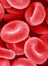
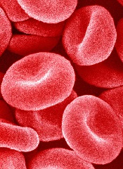
New research indicates that non-muscle myosin II-A (NMIIA) plays a key role in maintaining red blood cell (RBC) shape and deformability.
Researchers found evidence to suggest that NMIIA forms filaments in RBCs, and specialized regions at both ends of the filaments can pull on actin to control the stiffness of the cell membrane.
“You need active contraction on the cell membrane, similar to how muscles contract,” explained study author Velia Fowler, PhD, of The Scripps Research Institute in La Jolla, California.
“The myosin pulls on the actin to provide tension in the membrane, and then that tension maintains the biconcave shape.”
Dr Fowler and her colleagues described these findings in PNAS.
The researchers noted that RBC shape and deformability depend upon the membrane skeleton—a network of actin filaments (F-actin) cross-linked by spectrin tetramers.
And although NMII motors are known to exert force on F-actin networks to control cell shapes, no one had investigated a function for NMII contractility in the spectrin–F-actin network of RBCs. So Dr Fowler and her colleagues did just that.
The researchers said their work suggests NMIIA is the predominant RBC NMII isoform, and NMIIA motor activity regulates interactions with the spectrin–F-actin network to control RBCs’ shape and deformability.
Specifically, NMIIA forms bipolar filaments in RBCs, and these filaments associate with F-actins via their motor domains. It is through these interactions that NMIIA contractile forces promote membrane tension to maintain RBC shape and deformability.
To test these findings, the researchers treated RBCs with a compound called blebbistatin, which inhibits NMII motor activity.
After treatment, the team observed a decrease in NMIIA filaments associated with the RBC membrane and evidence of decreased membrane tension. The treated RBCs became elongated and showed a reduction in biconcavity as well as an increase in deformability.
The researchers believe this work could have applications for diseases in which RBCs are deformed. In fact, the team thinks inhibiting NMIIA in RBCs might prove useful for treating patients with sickle cell disease, as it could restore some elasticity to the patients’ RBCs.
Going forward, the researchers hope to learn more about what regulates NMIIA’s activity in RBCs and other cells.


