User login
Synthetic heparin poised for clinical trials, team says
Researchers say they have synthesized low molecular weight heparin (LMWH) that may someday replace animal-sourced heparin.
The team created heparin dodecasaccharides (12-mers) using a manufacturing method that yielded gram quantities—roughly 1000-fold more than previous approaches used to synthesize LMWHs.
One of these dodecasaccharides, called 12-mer-1, demonstrated safety and efficacy in animals models.
Robert Linhardt, PhD, of Rensselaer Polytechnic Institute in Troy, New York, and his colleagues detailed this research in Science Translational Medicine.
The researchers tested 12-mer-1 in a mouse model of venous thrombosis induced by stenosis of the inferior vena cava. Twenty-four hours after stenosis, 12-mer-1 had reduced clot weight by about 60% (P<0.05), when compared to phosphate-buffered saline.
The effects of 12-mer-1 were comparable to those achieved with enoxaparin. However, the dose of 12-mer-1 (1.5 mg/kg) was one-fifth the dose of enoxaparin (7.5 mg/kg). This suggests 12-mer-1 has “considerably higher antithrombotic potency” than enoxaparin, according to the researchers.
Dr Linhardt and his colleagues also tested 12-mer-1 in a mouse model of sickle cell disease. The team said the anticoagulant (given at 2.0 mg/kg every 8 hours for 7 days) “significantly attenuated the activation of coagulation” when compared to saline (P<0.05).
The researchers then tested the clearance of 12-mer-1 in mice with severe kidney failure.
The team observed significant impairment of clearance for both high-dose (1.5 mg/kg) and low-dose (0.3 mg/kg) 12-mer-1 (P<0.05). However, the impairment of 12-mer-1 clearance was dependent upon the severity of the kidney injury.
The researchers also performed toxicology studies of 12-mer-1 in rats. The animals received a single dose of 12-mer-1 at 3600 mg/kg per day for 7 consecutive days.
The rats experienced a decrease in white blood cell counts, but this was within the historical control data range. Two additional doses of 12-mer-1 (400 and 1200 mg/kg per day) produced similar results.
Therefore, Dr Linhardt and his colleagues concluded that 12-mer-1 was well-tolerated.
The researchers also noted that anticoagulation with 12-mer-1 was completely reversible via treatment with protamine, which could potentially reduce bleeding risk.
The team believes that, with substantial optimization, 12-mer-1 could be suitable for industrial-scale synthesis.
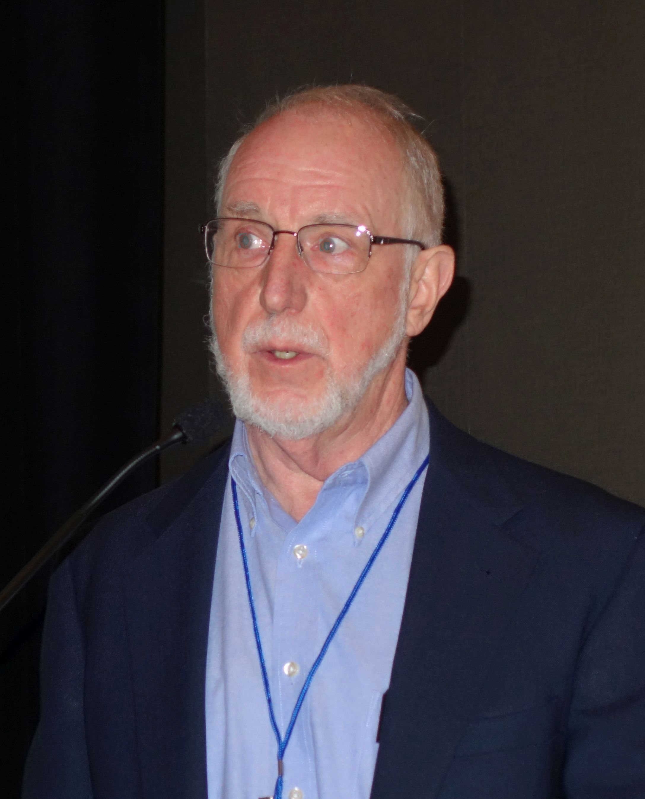
“This is at the cusp of clinical trials and commercial use,” Dr Linhardt said. “There is no question about the science. We have proven that this is a safer, more effective alternative to its natural counterpart, and what now determines its success or failure is the marketplace.” ![]()
Team identifies mutation that causes EPP
Researchers say they have discovered a genetic mutation that triggers erythropoietic protoporphyria (EPP).
The team performed genetic sequencing on members of a family from Northern France who had EPP of a previously unknown genetic signature.
The sequencing revealed a mutation in the gene CLPX that promotes EPP.
Barry Paw MD, PhD, of the Dana-Farber/Boston Children’s Cancer and Blood Disorders Center in Massachusetts, and his colleagues reported this discovery in PNAS.
About EPP
To produce heme, the body goes through porphyrin synthesis, which mainly occurs in the liver and bone marrow. Genetic defects can hinder the body’s ability to produce heme, and a decrease in heme production leads to a buildup of protoporphyrin components.
In the case of EPP, protoporphyrin IX accumulates in the red blood cells, plasma, and sometimes the liver.
When protoporphyrin IX is exposed to light, it produces chemicals that damage surrounding cells. As a result, people with EPP experience swelling, burning, and redness of the skin after exposure to sunlight.
“People with EPP are chronically anemic, which makes them feel very tired and look very pale, with increased photosensitivity because they can’t come out in the daylight,” Dr Paw said. “Even on a cloudy day, there’s enough ultraviolet light to cause blistering and disfigurement of the exposed body parts, ears, and nose.”
Although some genetic pathways leading to the build-up of protoporphyrin IX have already been described, many cases of EPP remain unexplained.
New discovery
Dr Paw and his colleagues noted that heme synthesis is controlled by the mitochondrial AAA+ unfoldase ClpX, which participates in the degradation and activation of δ-aminolevulinate synthase (ALAS).
In their analysis of the French family with EPP, the researchers discovered a dominant mutation in the ATPase active site of CLPX. This mutation—p.Gly298Asp—prompts the accumulation of protoporphyrin IX.
The researchers said the mutation partially inactivates CLPX, which increases the post-translational stability of ALAS. This increases ALAS protein and ALA levels and leads to the accumulation of protoporphyrin IX.
“This newly discovered mutation really highlights the complex genetic network that underpins heme metabolism,” Dr Paw said. “Loss-of-function mutations in any number of genes that are part of this network can result in devastating, disfiguring disorders.”
Dr Paw also noted that identifying the mutations that contribute to EPP and other porphyrias could pave the way for new methods of treating these disorders. ![]()
How thyroid hormone affects RBC production
Physicians have long known that patients with an underactive thyroid tend to have anemia because thyroid hormone stimulates red blood cell (RBC) production.
Now, researchers say they have determined how this occurs.
Xiaofei Gao, PhD, of Westlake Institute for Advanced Study in Hangzhou, Zhejiang Province, China, and his colleagues conducted this research and reported the results in PNAS.
The team began by studying the formation of human RBCs in culture. They wondered if something in the culture serum was essential for RBC maturation. So they ran the serum through a charcoal filter, which attracts and retains hydrophobic molecules.
Once filtered, the serum no longer supported RBC production. This validated the researchers’ theory that one of the hydrophobic molecules was key to RBC maturation.
In fact, the team found thyroid hormone was essential for the final step of RBC maturation.
When the researchers added thyroid hormone back to the serum, RBC progenitors once again started down the path to maturation.
If thyroid hormone was added at an earlier stage of development, the RBCs short-circuited their usual developmental processes and began turning into mature RBCs.
With further investigation, the researchers pinpointed the receptor inside maturing RBCs to which thyroid hormone binds—thyroid hormone receptor beta (TRβ).
From there, the team found that nuclear receptor coactivator 4 (NCOA4), a protein necessary for thyroid hormone stimulation, works with TRβ to regulate RBC development.
Finally, experiments showed that TRβ agonists could stimulate RBC development and alleviate anemic symptoms in a mouse model of chronic anemia.
The researchers therefore believe this work could lead to new therapies for anemic patients, including those with an underactive thyroid. ![]()
SCD drug receives rare pediatric disease designation
The US Food and Drug Administration (FDA) has granted rare pediatric disease designation to GBT440 for the treatment of sickle cell disease (SCD).
GBT440 is being developed by Global Blood Therapeutics, Inc. as a potentially disease-modifying therapy for SCD.
The drug works by increasing hemoglobin’s affinity for oxygen. Since oxygenated sickle hemoglobin does not polymerize, it is believed that GBT440 blocks polymerization and the resultant sickling of red blood cells.
If GBT440 can restore normal hemoglobin function and improve oxygen delivery, the therapy may be capable of modifying the progression of SCD.
The FDA previously granted GBT440 fast track and orphan drug designations.
About rare pediatric disease designation
Rare pediatric disease designation is granted to drugs that show promise to treat diseases affecting fewer than 200,000 patients in the US, primarily patients age 18 or younger.
The designation provides incentives to advance the development of drugs for rare disease, including access to the FDA’s expedited review and approval programs.
Under the FDA’s Rare Pediatric Disease Priority Review Voucher Program, if a drug with rare pediatric disease designation is approved, the drug’s developer may qualify for a voucher that can be redeemed to obtain priority review for any subsequent marketing application.
GBT440 trials
GBT440 is currently under investigation in a phase 1/2 trial (GBT440-001) of healthy subjects and adults with SCD. Data from this trial were presented at the 2016 ASH Annual Meeting.
At that time, there were 41 SCD patients who had been receiving GBT440 for up to 6 months.
All of these patients experienced a “profound and durable” reduction in hemolysis, as assessed by hemoglobin, reticulocytes, and/or bilirubin, according to Global Blood Therapeutics.
Patients treated with GBT440 for at least 90 days demonstrated a “clinically significant” increase in hemoglobin (greater than 1 g/dL increase) when compared with placebo-treated patients (46% vs 0%; P=0.006).
Patients treated with GBT440 also had a sustained reduction in irreversibly sickled cells when compared with placebo-treated patients (-76.6% vs +9.7%; P<0.001).
The most common treatment-related adverse events were grade 1/2 headache and gastrointestinal disorders. These events occurred in similar rates in the placebo and GBT440 arms. There were no drug-related serious or severe adverse events.
No sickle cell crises events occurred while participants were on GBT440. Exercise testing data showed normal tissue oxygen delivery (no change in oxygen consumption compared to placebo).
GBT440 is also under investigation in the phase 3 HOPE study, which includes SCD patients age 12 and older. And the drug is being tested in the phase 2 HOPE-KIDS 1 study, which includes pediatric patients (ages 6 to 17) with SCD. ![]()
Paper-based diagnostic device is like ‘portable lab’
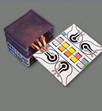
Researchers say they have developed self-powered, paper-based electrochemical devices (SPEDs) that can provide sensitive diagnostics in low-resource settings and at the point of care.
The SPEDs can detect biomarkers in the blood and identify conditions such as anemia by performing electrochemical analyses that are powered by the user’s touch.
The devices produce color-coded test results that are easy for non-experts to understand.
“You could consider this a portable laboratory that is just completely made out of paper, is inexpensive, and can be disposed of through incineration,” said Ramses V. Martinez, PhD, of Purdue University in West Lafayette, Indiana.
“We hope these devices will serve untrained people located in remote villages or military bases to test for a variety of diseases without requiring any source of electricity, clean water, or additional equipment.”
Dr Martinez and his colleagues developed the SPEDs and described them in a paper published in Advanced Materials Technologies.
SPED testing is initiated by placing a pinprick of blood in a circular feature on the device, which is less than 2-inches square. The SPEDs also contain “self-pipetting test zones” that can be dipped into a sample instead of using a finger-prick test.
The top layer of each SPED is made of untreated cellulose paper with patterned hydrophobic domains that define channels that wick up blood samples for testing. These microfluidic channels allow for assays that change color to indicate specific test results.
The researchers also created a machine-vision diagnostic application to identify and quantify each of these colorimetric tests from a digital image of the SPED, perhaps taken with a cell phone. This provides rapid results for the user and allows for consultation with a remote expert if necessary.
The bottom layer of the SPED is a triboelectric generator (TEG), which generates the electric current necessary to run the diagnostic test by rubbing or pressing it.
An inexpensive, hand-held device called a potentiostat can be plugged into the SPED to automate the diagnostic tests so they can be performed by untrained users. The battery powering the potentiostat can be recharged using the TEG built into the SPEDs.
“To our knowledge, this work reports the first self-powered, paper-based devices capable of performing rapid, accurate, and sensitive electrochemical assays in combination with a low-cost, portable potentiostat that can be recharged using a paper-based TEG,” Dr Martinez said.
SPEDs can perform multiplexed analyses, enabling the detection of various targets for a range of point-of-care testing applications. In addition, the devices are compatible with mass-printing technologies, such as roll-to-roll printing or spray deposition. And the SPEDs can be used to power other electronic devices to facilitate telemedicine applications in resource-limited settings.
Dr Martinez and his colleagues used the SPEDs to detect biomarkers such as glucose, uric acid and L-lactate, ketones, and white blood cells, which indicate factors related to liver and kidney function, malnutrition, and anemia.
The researchers said future versions of the technology will contain several additional layers for more complex assays to detect diseases such as malaria, dengue fever, yellow fever, hepatitis, and HIV. ![]()
Bacterial infection inhibits hematopoiesis
New research suggests that bacterial infection activates hematopoietic stem cells (HSCs) and significantly reduces their ability to produce blood by forcibly inducing proliferation.
These findings indicate that bacterial infections might cause dysregulated hematopoiesis like that which occurs in patients with anemias and leukemias.
Hitoshi Takizawa, PhD, of Kumamoto University’s International Research Center for Medical Sciences in Kumamoto, Japan, and his colleagues reported these findings in Cell Stem Cell.
The researchers gave lipopolysaccharide to mice to generate a bacterial infection model and analyzed the role of Toll-like receptors (TLRs) in HSCs.
The team found that lipopolysaccharides spread throughout the body, with some eventually reaching the bone marrow. This stimulated TLR4 and induced HSCs to proliferate.
However, the stimulus also induced stress on the HSCs.
So although HSCs proliferate temporarily upon TLR4 stimulation, their ability to successfully self-replicate decreases, resulting in diminished blood production.
The researchers observed similar results after infecting mice with Salmonella Typhimurium.
“Fortunately, we were able to confirm that this molecular reaction can be inhibited by drugs,” Dr Takizawa said.
Specifically, the researchers found that activation of reactive oxygen species and p38 through TLR4 ligation is key to HSC dysfunction.
And small-molecule inhibitors of reactive oxygen species and p38 were able to prevent HSC dysfunction.
“The medication maintains the production of blood and immune cells without weakening the immune reaction against pathogenic bacteria,” Dr Takizawa said. “It might be possible to simultaneously prevent blood diseases and many bacterial infections in the future.” ![]()
L-glutamine to prevent sickle cell complications featured in FDA podcast
The recent approval of L-glutamine, marketed as Endari, to reduce the acute complications of sickle cell disease in adult and pediatric patients 5 years of age and older is discussed in the Drug Information Soundcast in Clinical Oncology (DISCO) from a Food and Drug Adminstration podcast series that provides information about new product approvals, emerging safety information for cancer treatments, and other current topics in cancer drug development.
The basis for the approval was discussed in our coverage of the FDA’s Oncologic Drugs Advisory Committee meeting.
This episode of DISCO is hosted by Sanjeeve Bala, MD, and was developed by Abhilasha Nair, MD; Dr. Bala; Kathy M. Robie Suh, MD; Ann T. Farrell, MD; Kirsten B. Goldberg, and Richard Pazdur, MD. All are with the FDA’s Oncology Center of Excellence and the Office of Hematology and Oncology Products. Steven Jackson of the FDA’s Division of Drug Information was the sound producer.
Post-approval trials for accelerated drugs fall short
New research has revealed shortcomings of post-approval studies for drugs granted accelerated approval in the US.
Researchers found that, for drugs granted accelerated approval from 2009 to 2013, both pre-approval and post-approval trials had limitations in their design and the endpoints used.
“One might expect accelerated approval confirmatory trials to be much more rigorous than the pre-approval trials,” said study author Aaron S. Kesselheim, MD, of Brigham and Women’s Hospital in Boston, Massachusetts.
“But we found that there were few differences in these key design features of the trials conducted before or after approval.”
Dr Kesselheim and his colleagues reported these findings in JAMA.
The researchers examined pre- and post-approval clinical trials of drugs granted accelerated approval by the US Food and Drug Administration (FDA) between 2009 and 2013.
During that time, the FDA granted 22 drugs accelerated approval for 24 indications (15 of them for hematologic disorders).
Fourteen of the indications were approved on the basis of single-intervention-group studies that enrolled a median of 132 patients.
The FDA ordered 38 post-approval studies to confirm the safety and efficacy of the drugs.
Three years after the last drug’s approval, half of those studies (n=19) were not complete. Eight (42%) of the incomplete studies were either terminated or delayed by more than 1 year.
For 14 of the 24 indications (58%), results from the post-approval studies were not available after a median of 5 years of follow-up.
Study comparison
Published reports were available for 18 of the 19 completed post-approval studies. The characteristics of these studies did not differ much from the 30 pre-approval studies.
There were no statistically significant differences with regard to median patient enrollment (P=0.17), the use of randomized (P=0.31) or double-blind trials (P=0.17), the use of placebo as a comparator (P=0.17), or the lack of a comparator (P=0.21).
However, there was a significant difference in the use of an active comparator (P=0.02), with more post-approval studies using an active comparator.
The researchers also found that 17 of the 18 post-approval trials still used surrogate measures of effect as primary endpoints.
There was no significant difference between pre- and post-approval trials when it came to the use of disease response (P=0.17) or most other surrogate measures (P=0.21) as the trials’ primary endpoint.
The same was true for overall survival (P=0.20), although significantly more post-approval studies used progression-free survival (P=0.001) as a primary endpoint.
“It is important to use clinical endpoints in testing investigational drugs whenever possible because there are numerous cases of drugs approved on the basis of a surrogate measure that turn out to later not effect actual clinical outcomes—or even make them worse,” Dr Kesselheim said.
To address these issues and improve the quality of confirmatory studies, Dr Kesselheim suggested the FDA clearly describe the limitations in the pre-approval data that will need to be addressed in post-approval studies.
He also suggested the agency work with manufacturers to ensure that post-approval studies are conducted using design features that will be optimally useful for confirming the efficacy of the drug. ![]()
New research has revealed shortcomings of post-approval studies for drugs granted accelerated approval in the US.
Researchers found that, for drugs granted accelerated approval from 2009 to 2013, both pre-approval and post-approval trials had limitations in their design and the endpoints used.
“One might expect accelerated approval confirmatory trials to be much more rigorous than the pre-approval trials,” said study author Aaron S. Kesselheim, MD, of Brigham and Women’s Hospital in Boston, Massachusetts.
“But we found that there were few differences in these key design features of the trials conducted before or after approval.”
Dr Kesselheim and his colleagues reported these findings in JAMA.
The researchers examined pre- and post-approval clinical trials of drugs granted accelerated approval by the US Food and Drug Administration (FDA) between 2009 and 2013.
During that time, the FDA granted 22 drugs accelerated approval for 24 indications (15 of them for hematologic disorders).
Fourteen of the indications were approved on the basis of single-intervention-group studies that enrolled a median of 132 patients.
The FDA ordered 38 post-approval studies to confirm the safety and efficacy of the drugs.
Three years after the last drug’s approval, half of those studies (n=19) were not complete. Eight (42%) of the incomplete studies were either terminated or delayed by more than 1 year.
For 14 of the 24 indications (58%), results from the post-approval studies were not available after a median of 5 years of follow-up.
Study comparison
Published reports were available for 18 of the 19 completed post-approval studies. The characteristics of these studies did not differ much from the 30 pre-approval studies.
There were no statistically significant differences with regard to median patient enrollment (P=0.17), the use of randomized (P=0.31) or double-blind trials (P=0.17), the use of placebo as a comparator (P=0.17), or the lack of a comparator (P=0.21).
However, there was a significant difference in the use of an active comparator (P=0.02), with more post-approval studies using an active comparator.
The researchers also found that 17 of the 18 post-approval trials still used surrogate measures of effect as primary endpoints.
There was no significant difference between pre- and post-approval trials when it came to the use of disease response (P=0.17) or most other surrogate measures (P=0.21) as the trials’ primary endpoint.
The same was true for overall survival (P=0.20), although significantly more post-approval studies used progression-free survival (P=0.001) as a primary endpoint.
“It is important to use clinical endpoints in testing investigational drugs whenever possible because there are numerous cases of drugs approved on the basis of a surrogate measure that turn out to later not effect actual clinical outcomes—or even make them worse,” Dr Kesselheim said.
To address these issues and improve the quality of confirmatory studies, Dr Kesselheim suggested the FDA clearly describe the limitations in the pre-approval data that will need to be addressed in post-approval studies.
He also suggested the agency work with manufacturers to ensure that post-approval studies are conducted using design features that will be optimally useful for confirming the efficacy of the drug.
Folic acid fortification prevents millions of cases of anemia
DENVER – Mandatory food fortification with folic acid not only prevents neural tube defects, it also prevents an estimated 10 million cases of folate-deficiency anemia annually in the United States, James L. Mills, MD, reported at the annual meeting of the Teratology Society.
“We should have people be thinking about the fact that we’re preventing millions of cases of folate-deficiency anemia, not just thousands of cases of neural tube defects. That point does not seem to have reached the public health community. We need to correct the erroneous assumption that a small group are the only ones benefiting by exposing the entire population to folic acid,” said Dr. Mills, senior investigator at the Eunice Kennedy Shriver National Institute of Child Health and Human Development in Bethesda, Md.
Extrapolating from the nationally representative survey to the full U.S. population, Dr. Mills estimated that translates to roughly 10 million cases of folate-deficiency anemia prevented per year as a result of the mandatory fortification of grain introduced in 1998. That represents an enormous financial savings in avoided costs of diagnosis and treatment of this disorder.
The Food Fortification Initiative reports that 86 countries have embraced mandatory food fortification of wheat, maize, and/or rice. More than two dozen reports from around the world describe 40%-60% reductions in neural tube defect rates as a consequence. However, some of the world’s most populous nations are not on board. These include China, India, Russia, and the entire European Union.
Among the arguments raised by opponents of mandatory food fortification is the notion that it exposes the entire population to folic acid while benefiting only a small group of individuals who are spared having a neural tube defect. But the findings regarding prevention of folate-deficiency anemia demonstrate that argument is incorrect, Dr. Mills said.
Increased risks of asthma, cancer, and twinning as a consequence of mandatory food fortification have been proposed but are not supported by evidence. The only well-established adverse event is masking of vitamin B12 deficiency by correction of the anemia. But most reported cases have occurred after exposure to folic acid in milligram per day amounts, whereas the average U.S. exposure in women of childbearing age is just 163 mcg per day, less than half the recommended daily intake for that group. Also, no increase in cases of newly diagnosed vitamin B12 deficiency without anemia occurred in the U.S. after mandatory fortification was introduced, according to Dr. Mills.
Audience member Godfrey P. Oakley Jr., MD, noted that there is randomized trial evidence to indicate that folic acid supplementation has another important benefit: primary prevention of stroke in hypertensive adults. He cited the randomized, double-blind China Stroke Primary Prevention Trial, in which almost 21,000 hypertensive Chinese adults without a history of myocardial infarction or stroke were randomized to a single-pill combination of 10 mg of enalapril and 0.8 mg of folic acid daily or to a tablet containing 10 mg of enalapril alone.
During a median 4.5 years of follow-up, the enalapril/folic acid group had a 24% reduction in the risk of ischemic stroke and a 20% reduction in the composite of cardiovascular death, MI, and stroke (JAMA. 2015 Apr 7;313[13]:1325-35).
This is a potential game-changing finding which cries out for a confirmatory trial, he said. “There’s a lot going for that paper. I don’t know of a research agenda item that’s more important than trying to find out the relationship between folic acid fortification and stroke. I wish somebody would put some money into it,” said Dr. Oakley, research professor of epidemiology at Emory University in Atlanta.
Dr. Mills responded that he has reservations about the quality of the Chinese study, particularly in light of a Chinese government analysis that concluded that 80% of Chinese clinical trials were fraudulent (BMJ. 2016 Oct 5;355:i5396).
“That makes me want to see more data from a source I have a little bit more confidence in,” he added.
Another possible benefit of folic acid supplementation worthy of investigation is its theoretic potential for cancer prevention. “Folic acid provides one-carbon atoms for DNA repair,” Dr. Mills noted.
Dr. Mills reported having no relevant financial disclosures.
EXPERT ANALYSIS FROM TERATOLOGY SOCIETY 2017
VSTs can treat 5 different viral infections after HSCT
New research suggests virus-specific T cells (VSTs) can protect patients from severe viral infections that sometimes occur after hematopoietic stem cell transplant (HSCT).
The VSTs proved effective against 5 different viruses—Epstein-Barr virus (EBV), adenovirus (AdV), cytomegalovirus (CMV), BK virus (BKV), and human herpesvirus 6 (HHV-6).
Ifigeneia Tzannou, MD, of Baylor College of Medicine in Houston, Texas, and her colleagues reported these findings in the Journal of Clinical Oncology.
“In this study, we continued our previous work . . . in which we showed that patients who had developed an Epstein-Barr virus infection after a transplant . . . could be helped by receiving immune cells specialized in eliminating that particular virus,” Dr Tzannou said. “Then, we and others successfully targeted other viruses—namely, adenoviruses and cytomegalovirus.”
“The novel contribution of this study is that we have targeted additional viruses, the BK virus and the HHV-6 virus, which had not been targeted this way before,” added study author Bilal Omer, MD, of Baylor College of Medicine.
“This is important because the BK virus does not have an effective treatment, and the complications are significant, including severe pain and bleeding. These patients are in the hospital for weeks, months sometimes, and, now, we have a treatment option.”
The researchers tested their VSTs in a phase 2 trial of 38 HSCT recipients with at least 1 of the aforementioned viruses.
“[To prepare the VSTs,] we take blood from healthy donors who have already been exposed to these viruses and who we have confirmed have immune cells that can fight the infections,” Dr Tzannou said.
“We isolate the cells and let them multiply in culture. The final product is a mixture of cells that, together, can target all 5 viruses. We prepared 59 sets of virus-specific cells from different donors following this procedure.”
“Our strategy is to prepare a number of sets of virus-specific cells ahead of time and store them in a freezer, ready to use when a patient needs them,” Dr Omer noted. “To match patient and donor, we use elaborate matching algorithms.”
Patients
The trial included 38 patients who had undergone HSCT to treat acute myeloid leukemia/myelodysplastic syndromes (n=20), acute lymphoblastic leukemia (n=9), lymphoma/myeloma (n=3), or nonmalignant disorders (n=6).
These 38 patients had a total of 45 infections—CMV (n=17), EBV (n=2), AdV (n=7), BKV (n=16), and HHV-6 (n=3).
Response
The researchers monitored virus levels and other clinical responses in the 37 evaluable patients.
Six weeks after the first VST infusion, the overall response rate was 91.9%.
Seventeen patients received VSTs for persistent CMV. Sixteen of these patients (94.1%) responded, 6 with complete responses (CRs) and 10 with partial responses (PRs).
Two patients received VSTs for EBV, and both achieved a virologic CR.
Seven patients received VSTs for persistent AdV. The response rate was 71.4%. Four patients achieved a CR, 1 had a PR, and 2 patients did not respond.
Three patients received VSTs to treat HHV-6 reactivations. The response rate was 67%. Two patients had a PR, and 1 was not evaluable.
Sixteen patients received VSTs for BKV-associated hemorrhagic cystitis (n= 14) or BKV-associated nephritis (n=2).
All 16 patients responded. One had a clinical and virologic CR. Six had a clinical CR but a virologic PR. Seven had a virologic and clinical PR. And 2 patients had only a virologic PR.
A total of 15 patients received a second VST infusion—1 due to lack of response, 7 who had a PR, and 7 due to recurrence. Ten of these patients responded to the second infusion—1 with a CR and 9 with a PR.
Four patients received a third infusion of VSTs. Two achieved a CR, 1 had a PR, and 1 did not respond.
Toxicity
One patient developed an isolated fever within 24 hours of VST infusion, but the researchers did not observe any other immediate toxicities.
One of the patients with BKV-associated hemorrhagic cystitis experienced transient hydronephrosis and a decrease in renal function associated with a concomitant bacterial urinary tract infection.
Nineteen patients had prior grade 2 to 4 graft-versus-host disease (GVHD)—15 with grade 2 and 4 with grade 3. All GVHD was quiescent at the time of VST infusion.
One patient developed recurrent grade 3 gastrointestinal GVHD after VST infusion and rapid corticosteroid taper. Five patients developed recurrent (n=3) or de novo (n=2) grade 1 to 2 skin GVHD, which resolved with topical treatment (n=4) and reinitiation of corticosteroid treatment (n=1).
Two patients had a flare of upper-gastrointestinal GVHD, which resolved after a brief corticosteroid course.
“We didn’t have any significant toxicities,” Dr Tzannou said. “Taken together, the results of this trial suggest that it is reasonable to consider this treatment as an early option for these patients. We hope that the results of a future multicenter, phase 3 clinical trial will help raise awareness in both physicians and patients that this treatment, which is safe and effective, is available.”


