User login
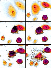
Olfactory receptor could be target for AML therapy

Investigators have discovered that an olfactory receptor in white blood cells responds to Sandalore, a synthetic odorant with a sandalwood note.
The team identified 7 olfactory receptors in a chronic myeloid leukemia (CML) cell line that were also present in white blood cells from patients with acute myeloid leukemia (AML).
One of the highest expressed receptors, OR2AT4, responded to Sandalore by fighting off the leukemia.
The investigators believe this finding could aid the development of new treatment for AML.
Hanns Hatt, PhD, DrMed, of the Ruhr-Universität Bochum in Germany, and his colleagues described this work in Cell Death Discovery.
The team found 7 olfactory receptors in the CML cell line K562—OR51B4, OR51B5, OR52D1, OR2W3, OR2B6, OR2AT4, and OR51I2. These receptors were also expressed in samples from AML patients.
The investigators then found that OR2AT4, one of the highest expressed olfactory receptors, is activated by Sandalore.
If Sandalore was used to activate the receptor, it inhibited leukemia cell proliferation and induced apoptosis in the leukemia cells. It also induced erythroid differentiation.
In 2014, Dr Hatt and his colleagues discovered that OR2AT4 is present in skin cells and that, by activating it with sandalwood aroma, wound healing is promoted. Through a series of tests, the team identified the signaling pathways underlying the observed effects.
With the current study, the investigators found that if Sandalore activates OR2AT4 in the context of CML or AML, processes similar to those in the olfactory cells in the nose start in blood cells.
The concentration of calcium ions in the cells increases. This, in turn, activates signaling pathways in which phosphate groups are transmitted to MAP kinases.
“This could be a new starting point for the development of leukemia treatment,” Dr Hatt said. “Acute myeloid leukemia, in particular, is a disease for which specific medication is not, as yet, available.” ![]()
When loved ones get cancer, people turn to the Web

chemotherapy
Photo by Rhoda Baer
Loved ones of cancer patients often turn to the Internet for further information about the disease, but they are less inclined to seek emotional support from social media forums, according to a study published in Computers, Informatics, Nursing.
It is fairly common for loved ones of cancer patients to develop depression or anxiety disorders as a result of the diagnosis, but there aren’t many studies focusing specifically on cancer patients’ caregivers and family members, said study author Carolyn Lauckner, PhD, of the University of Georgia in Athens.
“I think, sometimes, the loved ones and caregivers get forgotten about,” she said. “And that’s why I wanted to research this population to see if there are ways that we can better support these individuals.”
Dr Lauckner surveyed 191 people whose loved ones were diagnosed with cancer in the past year or who were currently acting as caregivers to someone with cancer. The motivation behind the research was personal for Dr Lauckner.
“I went through a period of time where I had 3 loved ones diagnosed within a short amount of time,” she said. “I had these experiences where I heard about the diagnosis and I would go online to look it up, and then I would immediately become terrified and freak out about all the stuff I read online.”
Like Dr Lauckner, more than 75% of the subjects she surveyed searched online for information on a loved one’s disease. Most looked for treatment options, prevention strategies and risk factors, and prognosis information.
“I was pleasantly surprised by the amount of people who said that they were looking for prevention information online and detection information,” Dr Lauckner said. “[T]hat shows that not only are they concerned for their loved one but they’re also concerned about how they themselves can avoid cancer, which, from a public health perspective, is great.”
Respondents were less inclined to view blogs or go online to hear about others’ cancer experiences. These kinds of sites were linked to negative emotions for participants, such as fear, sadness, and anger.
“A lot of people, especially in the cancer realm, they will use blogs or discussion posts to vent and to talk about the harsh realities of living with an illness,” Dr Lauckner said.
“And while I think that that is beneficial for both the person who is writing it and potentially for some people who want an idea of what to expect, when someone is dealing with the prospect of their loved one having to go through that experience, it can be extremely distressing.”
The most commonly visited websites were those of charitable organizations like the American Cancer Society, which were associated with positive emotions. Dr Lauckner said she found this information encouraging because it shows that participants were consulting reliable sources of information and not being swayed by personal accounts as much.
Dr Lauckner ultimately wants to build on the information gleaned in this study to determine the most effective use of social media and technology to distribute cancer prevention and risk reduction messages to the public. ![]()
Standard BMI inadequate for ALL patients

Photo by Bill Branson
New research suggests that body mass index (BMI) is an inadequate method for estimating changes in body fat and obesity in children with acute lymphoblastic leukemia (ALL).
Investigators found a discrepancy between BMI and body composition in this population, and the cause of this appeared to be increases in body fat with simultaneous loss of lean muscle mass during treatment.
The team reported these findings in Leukemia & Lymphoma.
With previous work, the investigators found that obese children diagnosed with high-risk ALL had a 50% greater risk of their disease recurring compared with children who were not obese.
“In my lab, we’ve seen a direct interaction between fat cells and leukemia cells that may help explain this increased risk of disease relapse,” said study author Steven Mittelman, MD, PhD, of Children’s Hospital Los Angeles in California.
“It appears that the fat cells ‘protect’ leukemia cells, making them less susceptible to chemotherapy and making an accurate measure of body fat essential.”
To determine if BMI accurately reflects body fat in ALL, the investigators analyzed 50 patients. They were predominantly Hispanic, between the ages of 10 to 21, and had newly diagnosed high-risk B-precursor ALL or T-cell ALL.
The team measured the percentage of total body fat and lean muscle mass at the time of diagnosis, at the end of induction, and at the end of delayed intensification. They also calculated BMI Z-score—a measure of how a given child’s BMI deviates from a population of children of the same age and sex—at these time points.
The investigators said sarcopenic obesity—gain in body fat percentage with loss of lean muscle mass—was “surprisingly common” during ALL treatment.
And sarcopenic obesity resulted in poor correlation between changes in BMI Z-score and body fat percentage overall (r=-0.05), within the time points (r=0.02), and within patients (r=-0.09, all not significant). BMI Z-score and body fat percentage changed in opposite directions in more than 50% of interval assessments.
“We found that change in BMI did not reflect changes in body fat or obesity,” said Etan Orgel, MD, of Children’s Hospital Los Angeles.
“In some patients, reaching a ‘healthy’ BMI was due solely to loss of muscle even while body fat continued to rise. Based on these results, we believe that evaluation of obesity in patients with leukemia should include direct measures of body composition.” ![]()
Incorporating cultural beliefs into cancer care

Photo by Daniel Sone
Understanding and integrating patients’ cultural beliefs into cancer treatment plans may help improve their acceptance of and adherence to treatment in multicultural settings, according to research published in the Journal of Global Oncology.
Researchers examined traditional Maya healers’ understanding of cancer and found that, although there are key differences between Maya and Western medicine perspectives, they also share many fundamental concepts.
“Maya healers understand cancer in remarkably similar ways to Western doctors,” said lead study author Mónica Berger-González, PhD, of the Institute for Environmental Decisions at ETH Zurich in Switzerland.
“Recognizing this is the first step to bridging the gap between cultures and ultimately providing better, more effective services for indigenous populations.”
Nearly half of the population in Guatemala (approximately 5.4 million people) relies on Maya medicine. Traditional healers have practiced in Guatemala for more than 2000 years, with the healing tradition passed down orally and through apprenticeship.
According to the authors, this is one of the first studies to explore the subject of Maya healers and cancer across several ethno-linguistic groups, and limited data exist on survival outcomes.
Dr Berger-González and her colleagues conducted in-depth interviews with 67 healers across various ethnic and language groups in Guatemala, exploring how its indigenous people define and treat cancer.
Of the Maya healers interviewed, 46% were illiterate. Although only 36% were able to define the word cancer, most (85%) were familiar with the term and identified malignancy as a core characteristic of the disease, explaining the concept of metastasis clearly.
The analysis also revealed that Maya healers understand the origins of cancer in ways that align closely with Western medical concepts.
When asked to identify the physical causes of cancer, 10 of 17 causes provided correlated directly with cancer risk factors as understood in Western medicine. Healers cited causes such as the consumption of harmful foods (46.3%), hereditary conditions (29.6%), and lifestyle factors such as smoking or working with toxic substances (29.6%).
One notable difference identified between the 2 perspectives is that Maya healers’ view of cancer is not limited to the physical body, but rather includes a complex imbalance of the emotions, mind, and spirit.
The Maya treatment of cancer is consequently holistic and seeks to restore that balance. This is achieved through a combination of methods—such as regulating diet, plant therapy, detoxifying baths—as well as social, psychological, and spiritual methods, the latter of which, the authors note, is harder to grasp in Western medicine.
“If healthcare professionals do not understand indigenous peoples’ conception of cancer, these patients are far less likely to accept and adhere to treatment in the public healthcare system,” Dr Berger-González said.
Many indigenous people in Guatemala do not have access to Western medicine services, cannot afford them, or prefer Maya medicine even when Western medical treatment is available. Yet Western medicine practitioners have little to no training in multicultural management or traditional indigenous medicine.
The authors offer 3 key recommendations to help address the challenges of providing care in multicultural settings:
- Adequate training of healthcare professionals on cultural and social perceptions of cancer
- Increasing evidence-based research on traditional medicine
- Establishing national regulations on integrating traditional and Western medicine—following other countries like Peru, Brazil, and Ecuador, which have successfully incorporated these aspects of care.
The authors plan to continue their transdisciplinary research with the goal of providing biomedical evidence that advances different aspects of Maya medicine. ![]()
Drug nets 3rd breakthrough designation from FDA

Image by Lance Liotta
The US Food and Drug Administration (FDA) has granted a third breakthrough therapy designation for the BCL-2 inhibitor venetoclax (ABT-199).
This time, the designation is for venetoclax in combination with hypomethylating agents to treat patients with treatment-naïve acute myeloid leukemia (AML) who are ineligible for standard induction therapy.
Venetoclax previously received breakthrough designation as a single agent for patients with relapsed or refractory chronic lymphocytic leukemia (CLL) and in combination with rituximab to treat patients with relapsed or refractory CLL and 17p deletion.
Breakthrough therapy designation is designed to accelerate the development and review of medicines that demonstrate early clinical evidence of a substantial improvement over current treatment options for serious diseases.
Venetoclax is currently under investigation in a phase 1/2 trial in combination with low-dose cytarabine for treatment-naïve patients with AML and in a phase 1b study in combination with decitabine or azacitidine for treatment-naïve AML patients.
A phase 2 study of single-agent venetoclax in AML has been completed. The results were presented at ASH 2014.
At that time, the trial had enrolled 32 patients, 30 of whom had relapsed or refractory disease. Patients had a median age of 71 (range, 19 to 84), and half were male.
The overall response rate was 15.5%, with 1 patient achieving a complete response (CR) and 4 patients achieving a CR with incomplete count recovery (CRi).
The researchers noted that 3 of the patients who had a CR/CRi had IDH mutations. Two of these patients also achieved minimal residual disease negativity.
The median bone marrow blast count in evaluable patients decreased 36% after treatment, and 6 patients (19%) had at least a 50% reduction in bone marrow blasts.
Common adverse events following treatment (occurring in at least 25% of patients) included nausea, diarrhea, fatigue, neutropenia, and vomiting.
Grade 3 and 4 adverse events (occurring in 3 or more patients) included febrile neutropenia, anemia, and pneumonia. No patient died as a result of treatment-related adverse events.
Venetoclax is being developed by AbbVie in partnership with Genentech and Roche. ![]()
Appalachia has higher cancer incidence than rest of US
receiving chemotherapy
Photo by Rhoda Baer
New research suggests that people living in the Appalachian region of the US are more likely to develop cancer than people in the rest of the country.
The study showed that Appalachians had a significantly higher incidence of cancer overall and higher rates of many solid tumor malignancies.
However, lymphoma rates were similar between Appalachians and non-Appalachians, and Appalachians had a significantly lower rate of myeloma.
This research was published in Cancer Epidemiology, Biomarkers & Prevention.
“The Appalachian region, which extends from parts of New York to Mississippi, spans 420 counties in 13 US states, and about 25 million people reside in this area,” said study author Reda Wilson, MPH, of the Centers for Disease Control and Prevention (CDC) in Atlanta, Georgia.
“This region is primarily made up of rural areas, with persistent poverty levels that are at least 20%, which is higher than the national average.”
In 2007, the CDC’s National Program of Cancer Registries (NPCR) published a comprehensive evaluation of cancer incidence rates in Appalachia between 2001 and 2003.
The data showed higher cancer rates in Appalachia than in the rest of the US. However, this publication had some shortcomings, including data that were not available for analysis.
“The current analyses reported here were performed to update the earlier evaluation by expanding the diagnosis years from 2004 to 2011 and including data on 100% of the Appalachian and non-Appalachian populations,” Wilson said.
For this study, Wilson and her colleagues used data from the NPCR and the National Cancer Institute’s Surveillance, Epidemiology, and End Results (SEER) program. Together, NPCR and SEER cover 100% of the US population.
The researchers analyzed the Appalachian population by region (north, central, and south Appalachia), gender, race (black and white only), and Appalachian Regional Commission-designated economic status (distressed, at-risk, transitional, competitive, and attainment). And the team compared these data with data on the non-Appalachian population.
The results showed that cancer incidence rates (IRs) were elevated among Appalachians regardless of how they were categorized. The IRs were per 100,000 people, age-adjusted to the 2000 US standard population.
The IR for all cancers was 565.8 for males in Appalachia and 543.0 for non-Appalachian males (P<0.05). And the cancer IRs for females were 428.7 in Appalachia and 418.2 outside the region (P<0.05).
There was no significant difference between the regions in IRs for lymphomas. The Hodgkin lymphoma IRs were 3.1 in Appalachian males, 3.2 in non-Appalachian males, and 2.5 for females in both regions.
The non-Hodgkin lymphoma IRs were 23.3 in Appalachian males, 23.4 in non-Appalachian males, 16.4 in Appalachian females, and 16.3 in non-Appalachian females.
Myeloma IRs were significantly lower in Appalachia (P<0.05). The myeloma IRs were 7.3 in Appalachian males, 7.5 in non-Appalachian males, 4.7 in Appalachian females, and 4.9 in non-Appalachian females.
There was no significant difference in leukemia IRs among males, but females in Appalachia had a significantly higher leukemia IR (P<0.05). The leukemia IRs were 16.9 in Appalachian males, 16.7 in non-Appalachian males, 10.4 in Appalachian females, and 10.2 in non-Appalachian females.
“Appalachia continues to have higher cancer incidence rates than the rest of the country,” Wilson said. “But a promising finding is that we’re seeing the gap narrow in the incidence rates between Appalachia and non-Appalachia since the 2007 analysis, with the exception of cancers of the oral cavity and pharynx, larynx, lung and bronchus, and thyroid.”
“This study helps identify types of cancer in the Appalachian region that could be reduced through more evidence-based screening and detection. Our study also emphasizes the importance of lifestyle changes needed to prevent and reduce cancer burden.”
The researchers noted that this study did not differentiate urban versus rural areas within each county, and data on screening and risk factors were based on self-reported responses.
Furthermore, cancer IRs were calculated for all ages combined and were not evaluated by age groups. Future analyses will be targeted toward capturing these finer details, Wilson said. ![]()
Acalabrutinib yields 95% overall response in relapsed CLL
Acalabrutinib, an oral drug that is a more specific Bruton tyrosine kinase (BTK) inhibitor related to ibrutinib, produced a high response rate and durable remissions at a median 14 months of follow-up in an uncontrolled phase I/II trial of 61 adults with relapsed chronic lymphocytic leukemia, according to a report published online Jan. 28 in the New England Journal of Medicine.
The study patients had received a median of three previous therapies for CLL; 31% had chromosome 17p13.1 deletions, and 75% had unmutated immunoglobulin heavy-chain variable genes.
At the analysis, one patient had died from pneumonia at 13 months and CLL had progressed at 16 months in another patient. The overall response rate among the 60 evaluable patients was 95%, with a partial response in 85% and a partial response with lymphocytosis in 10%. The rate of stable disease was 5%. Adverse events were mostly mild and self-limiting; eight patients (13%) discontinued treatment, said Dr. John C. Byrd of the division of hematology, Ohio State University, Columbus, and his associates.
All 18 patients with chromosome 17p13.1 deletions responded to acalabrutinib, with a partial response in 89% and a partial response with lymphocytosis in 11%. One patient with a chromosome 17p13.1 deletion had disease progression, and this patient had a C481S (major clone) mutation in BTK and an L845F (minor clone) mutation in PLCγ2.
No cases of Richter’s transformation occurred.
Patients were treated at six sites in the United States and the United Kingdom. Four different doses of oral acalabrutinib were used in the first phase of the study; the drug’s low toxicity permitted a twice-daily 100-mg dose in phase II of the study. Twice-daily dosing promoted continuous levels of drug binding to BTK, according to the researchers. It is hoped that this approach will decrease drug resistance and will perhaps lower the rate of transformation into large-cell lymphoma.
Among patients who had cytopenia at entry into the study, platelet count improved in 62%, hemoglobin levels improved in 76%, and absolute neutrophil count improved in 80%. Among patients who had B symptoms (weight loss, night sweats, and fever) at study entry, those symptoms resolved in 88% by the third cycle of treatment and in 100% by the ninth cycle, Dr. Byrd and his associates said (N Engl J Med. 2016 Jan 28. doi: 10.1056/NEJMoa1509981). The most common adverse events were headache (43% of patients), diarrhea (39%), weight gain (26%), pyrexia (23%), and upper respiratory tract infection (23%). Fewer than 2% of patients developed severe diarrhea, rash, arthralgia, myalgia, bruising, or bleeding.
These findings offered strong justification to further investigate the efficacy and safety of acalabrutinib for relapsed CLL, and a phase III trial is now underway, the investigators added.
FROM THE NEW ENGLAND JOURNAL OF MEDICINE
Key clinical point: Acalabrutinib, a more selective and therefore less-toxic relative of ibrutinib, produced a high response rate and durable remission in relapsed CLL.
Major finding: Acalabrutinib showed robust clinical activity, with an overall response rate of 95%, only one patient death, and only one case of CLL progression.
Data source: A multicenter phase I/II industry-sponsored clinical trial involving 61 patients followed for 14 months.
Disclosures: This trial was supported by Acerta Pharma, which was involved in study design and data analysis; it was also supported by the National Cancer Institute, the Leukemia and Lymphoma Society, the Four Winds Foundation, the Sullivan Chronic Lymphocytic Leukemia Research Fund, Mr. and Mrs. Michael Thomas, Al and Midge Lipkin, and the D. Warren Brown Foundation. Dr. Byrd reported receiving research grants from Acerta and serving as an unpaid consultant for Acerta, AbbVie, Genentech, Janssen, and Pharmacyclics; his associates reported ties to numerous industry sources.
Compounds could treat leukemia, lymphoma
apoptosis in cancer cells
Preclinical research suggests a new class of small-molecule inhibitors could potentially treat leukemias and lymphomas.
Researchers identified compounds targeting the Mdm2–MdmX RING–RING interaction as a new class of E3 ligase inhibitors.
Experiments showed that these compounds, dubbed MMRis, can induce apoptosis in leukemia and lymphoma cells.
The researchers detailed these experiments in Cell Death and Disease.
“We are excited about the unique activities of these compounds and will continue to focus our research efforts on development of their clinical potential,” said study author Xinjiang Wang, PhD, of Roswell Park Cancer Institute in Buffalo, New York.
“These compounds kill cancer cells. [They don’t] just stop cancer cell growth temporarily. These types of agents offer the promise of therapeutic benefit.”
Dr Wang and his colleagues found that MMRis specifically inhibit Mdm2–MdmX E3 ligase activity toward Mdm2 and p53 substrates.
The team said MMRis have an advantage over p53-activating agents that are currently in use as cancer therapies.
That is because MMRis activate the pro-apoptotic function of the p53 pathway, whereas current p53-activating agents temporarily prevent cancer growth but don’t damage existing cancer cells or prevent cancer growth long-term.
The researchers found that 2 MMRi compounds—MMRi6 and its analog, MMRi64—can disrupt Mdm2–MdmX interactions and activate p53 in vitro.
And MMRi64 selectively induces the apoptotic arm of the p53 pathway in leukemia and lymphoma cells.
The team noted that, unlike Nutlin3a, MMRi64 only induces expression of the pro-apoptotic gene PUMA, with minimal induction of the growth-arresting gene p21. However, combining MMRi64 and Nutlin3a produces a synergistic apoptotic effect.
“This study opens a new area for anticancer drug development,” Dr Wang said. “MMRi compounds also can be used as a tool for better understanding the anti-death mechanisms developed by cancer cells.”
“We are moving the research of MMRi compounds forward using both preclinical models and human cancer cell lines. Our hope is that further development of clinically useful MMRi will eventually provide a new treatment option for cancer patients.” ![]()
apoptosis in cancer cells
Preclinical research suggests a new class of small-molecule inhibitors could potentially treat leukemias and lymphomas.
Researchers identified compounds targeting the Mdm2–MdmX RING–RING interaction as a new class of E3 ligase inhibitors.
Experiments showed that these compounds, dubbed MMRis, can induce apoptosis in leukemia and lymphoma cells.
The researchers detailed these experiments in Cell Death and Disease.
“We are excited about the unique activities of these compounds and will continue to focus our research efforts on development of their clinical potential,” said study author Xinjiang Wang, PhD, of Roswell Park Cancer Institute in Buffalo, New York.
“These compounds kill cancer cells. [They don’t] just stop cancer cell growth temporarily. These types of agents offer the promise of therapeutic benefit.”
Dr Wang and his colleagues found that MMRis specifically inhibit Mdm2–MdmX E3 ligase activity toward Mdm2 and p53 substrates.
The team said MMRis have an advantage over p53-activating agents that are currently in use as cancer therapies.
That is because MMRis activate the pro-apoptotic function of the p53 pathway, whereas current p53-activating agents temporarily prevent cancer growth but don’t damage existing cancer cells or prevent cancer growth long-term.
The researchers found that 2 MMRi compounds—MMRi6 and its analog, MMRi64—can disrupt Mdm2–MdmX interactions and activate p53 in vitro.
And MMRi64 selectively induces the apoptotic arm of the p53 pathway in leukemia and lymphoma cells.
The team noted that, unlike Nutlin3a, MMRi64 only induces expression of the pro-apoptotic gene PUMA, with minimal induction of the growth-arresting gene p21. However, combining MMRi64 and Nutlin3a produces a synergistic apoptotic effect.
“This study opens a new area for anticancer drug development,” Dr Wang said. “MMRi compounds also can be used as a tool for better understanding the anti-death mechanisms developed by cancer cells.”
“We are moving the research of MMRi compounds forward using both preclinical models and human cancer cell lines. Our hope is that further development of clinically useful MMRi will eventually provide a new treatment option for cancer patients.”
Leukemia death rates expected to fall in EU

receiving chemotherapy
Photo by Rhoda Baer
Projections for 2016 suggest that leukemia deaths are on the decline in the European Union (EU), particularly for children and young adults.
Leukemia death rates are expected to fall 19% in men and 16% in women in 2016, when compared to 2007 data.
Rates are projected to fall 38% in boys and 20% in girls ages 0 to 14, 26% in men and 22% in women ages 15 to 44, and 19% in men and women ages 45 to 69.
Researchers said improvements in management, multidrug chemotherapy, immunotherapies, stem cell transplants, radiotherapy, and targeted therapies have all contributed to improvements in survival for leukemia patients.
However, some leukemias remain hard to treat, particularly those that are more common in adults and the elderly.
“Predictions of death rates from leukemia are complicated by the fact that leukemias are a varied collection of blood cancers, with some being more treatable than others,” said study author Carlo La Vecchia, MD, of the University of Milan in Italy.
“However, the important falls in overall death rates from this group of diseases are very encouraging and are a testament to the hard work of researchers and clinicians in developing and implementing better diagnosis and treatments.”
Dr Vecchia and his colleagues reported the results of this research in Annals of Oncology.
The researchers looked at cancer deaths in the EU as a whole and in the 6 largest countries—France, Germany, Italy, Poland, Spain, and the UK.
The team assessed mortality for all cancers and looked at individual data for cancers of the stomach, intestines, pancreas, lung, prostate, breast, and uterus (including cervix), as well as leukemias.
All cancers
The researchers’ projections suggest that total cancer mortality rates will decrease for both sexes in 2016, despite a rise in the absolute number of deaths due to the aging population. The team said the median age of the total EU population was 41 in 2011 and will be 42 in 2016.
So a total of 1,359,500 EU citizens are expected to die of cancer in 2016—753,600 men and 605,900 women. This is compared to 1,314,787 cancer deaths in 2011—734,259 men and 580,528 women.
The age-standardized mortality rates for 2016 are 133.5 per 100,000 men and 85.2 per 100,000 women, compared to 144.6 per 100,000 men and 88.1 per 100,000 women in 2011.
So that’s a 7.7% fall in cancer death rates for men and a 3.3% fall for women from 2011 through 2016, despite a 3.3% increase in the absolute number of cancer deaths.
Leukemias
The researchers predict that, in 2016, age-standardized leukemia mortality rates will be 3.95 per 100,000 men and 2.46 per 100,000 women.
In comparison, the age-standardized mortality rates for 2000-2004 were 5.23 for men and 3.20 for women. The rates for 2005-2009 were 4.85 for men and 2.92 for women.
For males ages 0 to 14, the age-standardized mortality rates are 1.10 for 2000-2004, 0.87 for 2005-2009, and 0.54 for 2016. For females in this age group, the rates are 0.85 for 2000-2004, 0.69 for 2005-2009, and 0.55 for 2016.
For males ages 15 to 44, the age-standardized mortality rates are 1.50 for 2000-2004, 1.28 for 2005-2009, and 0.95 for 2016. For females, the rates are 1.07 for 2000-2004, 0.92 for 2005-2009, and 0.72 for 2016.
For males ages 45 to 69, the age-standardized mortality rates are 9.19 for 2000-2004, 8.39 for 2005-2009, and 6.79 for 2016. For females, the rates are 5.74 for 2000-2004, 5.12 for 2005-2009, and 4.16 for 2016.
CAR T-cells induce remissions, avoid GVHD in relapsed B-cell malignancies
In patients with B-cell malignancies who did not obtain remissions after allogeneic hematopoietic stem-cell transplantation (alloHSCT), infusion of anti-CD19 chimeric antigen receptor (CAR19) T-cells induced remissions without causing graft-versus-host disease (GVHD).
Of 20 patients treated, 6 obtained complete remission and 2 obtained partial remissions, while none of the patients experienced new-onset acute GVHD.
“The increased antimalignancy potency of CAR19 T-cells allowed small doses of T-cells to eradicate malignancy without causing GVHD; therefore, this work demonstrates a solution to the central problem of alloHSCT, the separation of [graft-versus-malignancy] from GVHD,” wrote Dr. Jennifer Brudno of the National Cancer Institute, Bethesda, Md., and colleagues (J Clin Onc. 2016 Jan. 25. doi: 10.1200/JCO.2015.64.5929).
In general, CAR19 T-cells did not persist longer than 4 weeks, the median time it takes for GVHD to develop after donor lymphocyte infusion, which may be a factor in avoiding GVHD. Smaller doses may be another contributing factor: CAR19 T-cell doses administered in the trial ranged from 106/kg to 107/kg, which is 10-fold smaller than typical donor lymphocyte infusions.
In contrast to previous CAR T-cell studies, patients did not receive chemotherapy before CAR19 T-cell infusion, so endogenous T-cells and natural killer cells were not depleted. Evidence indicates that lymphocyte depletion enhances antitumor activity of adoptively transferred T-cells, but the current results indicate that prior lymphocyte depletion is not an absolute requirement.
The CAR19 T-cell infusion was especially effective in acute lymphoblastic leukemia (ALL), with four of the five ALL patients obtaining complete remission. Patients with chronic lymphocyte leukemia and lymphoma also obtained remissions.
Toxicities observed were consistent with previous CAR T-cell studies and included fever, tachycardia, and hypotension, indicative of cytokine release syndrome.
Peak levels of CAR19 T-cells were significantly higher in patients with complete or partial responses, and patients who did not obtain a complete or partial response were more likely to have undetectable or very low levels. Increasing peak blood levels of CAR19 T-cells in vivo is an important goal for future research, according to investigators. Since endogenous CD19+ cells may promote proliferation of CAR19 T- cells, CD19+ cellular vaccines might enhance CAR19 T-cell proliferation in patients with low levels, they suggested.
Administration of programmed cell death protein-1 (PD-1) antagonists may also offer improvements. High levels of PD-1 expression on CAR19 T-cells were observed at the time of peak blood CAR19 T-cell levels.
Dr. Brudno reported having no disclosures. Dr. Goy and Dr. Rosenberg reported ties to industry, and Dr. Kochenderfer reported patents pending on CAR T-cell therapy.
FROM JOURNAL OF CLINICAL ONCOLOGY
Key clinical point: In patients who relapsed after allogeneic hematopoietic stem-cell transplantation, anti-CD19 chimeric antigen receptor (CAR) T-cell therapy induced remissions without causing graft-versus-host disease (GVHD).
Major finding: Of 20 patients treated, 6 obtained complete remission and 2 obtained partial remissions; none experienced new-onset acute GVHD.
Data source: Patients with B-cell malignancies who had progressed after allogeneic hematopoietic stem-cell transplantation.
Disclosures: Dr. Brudno reported having no disclosures. Dr. Goy and Dr. Rosenberg reported ties to industry, and Dr. Kochenderfer reported patents pending on CAR T-cell therapy.