User login
Academic hospitals offer better AML survival
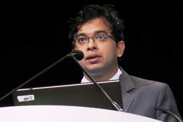
ORLANDO – Patients with acute myeloid leukemia (AML) initially treated at an academic center lived significantly longer than those treated at nonacademic centers, a database analysis shows.
Median overall survival increased from 7 months at a nonacademic center to 12.6 months at an academic center (P less than .001).
One-year overall survival rates were also significantly better at 51% vs. 39% (P less than .001).

The difference remained significant even after controlling for important confounders including age, comorbidity burden, receipt of chemotherapy, transplant, and delay between diagnosis and treatment, Mr. Smith Giri reported at the annual meeting of the American Society of Hematology.
“From a policy perspective, it may be useful to know whether these results are due to higher volume of cases, more advanced technology, expanded role of specialists, or greater, round-the-clock availability of resident physicians,” said Mr. Giri of the University of Tennessee Health Science Center in Memphis.
Prior studies in cancer have suggested better overall survival among breast cancer patients treated at academic centers, but this is the first study looking at outcomes in AML, the most common acute leukemia in adults.
Using the National Cancer Database Participant User File, the investigators identified 7,823 patients with AML who received their initial therapy at the reporting facility from 1998 to 2011. The database collects information from more than 1,500 Commission on Cancer (CoC)–accredited facilities. Of the 7,823 patients, 4,681 (60%) were treated at an AC (academic/research program) and 3,142 at a non-AC (community cancer program/comprehensive community cancer program).
Patients treated at an AC were significantly younger than those treated at a non-AC (median 62 years vs. 67 years), tended to be of nonwhite race, less educated, have a lower income, and more comorbidities.
Receipt of chemotherapy (97.4% vs. 94.5%) and transplant (9% vs. 2.4%) were significantly higher at an AC than a non-AC (both P less than .001).
Kaplan Meier survival curves suggested disparate survival curves between the two groups, mainly within the first 5 years of follow-up (P less than .001), Mr. Giri said.
In multivariate model analysis, the non-AC group had significantly worse risk adjusted 30-day mortality than the AC group (odds ratio, 1.52; 95% confidence interval 1.33-1.74; P less than .001) and worse overall survival (hazard ratio, 1.13; 95% CI 1.07-1.19; P less than .001).
The study (Ab. 533) findings should be interpreted with caution because of its limitations, including the lack of information on AML risk type in the database, the fact that administrative datasets are prone to coding errors, and because the analysis did not adjust for hospital volume, which has been shown to affect survival, he said. Also, because there are more than 3,500 non–Coc approved hospitals, the sample may not be representative of overall U.S. hospitals.
During a discussion of the results, Mr. Giri acknowledged that patients treated at academic centers may have greater access to clinical trials and experimental agents. Future analyses should also distinguish patients with a diagnosis of acute promyelocytic leukemia, a distinct subset of AML.
ORLANDO – Patients with acute myeloid leukemia (AML) initially treated at an academic center lived significantly longer than those treated at nonacademic centers, a database analysis shows.
Median overall survival increased from 7 months at a nonacademic center to 12.6 months at an academic center (P less than .001).
One-year overall survival rates were also significantly better at 51% vs. 39% (P less than .001).
The difference remained significant even after controlling for important confounders including age, comorbidity burden, receipt of chemotherapy, transplant, and delay between diagnosis and treatment, Mr. Smith Giri reported at the annual meeting of the American Society of Hematology.
“From a policy perspective, it may be useful to know whether these results are due to higher volume of cases, more advanced technology, expanded role of specialists, or greater, round-the-clock availability of resident physicians,” said Mr. Giri of the University of Tennessee Health Science Center in Memphis.
Prior studies in cancer have suggested better overall survival among breast cancer patients treated at academic centers, but this is the first study looking at outcomes in AML, the most common acute leukemia in adults.
Using the National Cancer Database Participant User File, the investigators identified 7,823 patients with AML who received their initial therapy at the reporting facility from 1998 to 2011. The database collects information from more than 1,500 Commission on Cancer (CoC)–accredited facilities. Of the 7,823 patients, 4,681 (60%) were treated at an AC (academic/research program) and 3,142 at a non-AC (community cancer program/comprehensive community cancer program).
Patients treated at an AC were significantly younger than those treated at a non-AC (median 62 years vs. 67 years), tended to be of nonwhite race, less educated, have a lower income, and more comorbidities.
Receipt of chemotherapy (97.4% vs. 94.5%) and transplant (9% vs. 2.4%) were significantly higher at an AC than a non-AC (both P less than .001).
Kaplan Meier survival curves suggested disparate survival curves between the two groups, mainly within the first 5 years of follow-up (P less than .001), Mr. Giri said.
In multivariate model analysis, the non-AC group had significantly worse risk adjusted 30-day mortality than the AC group (odds ratio, 1.52; 95% confidence interval 1.33-1.74; P less than .001) and worse overall survival (hazard ratio, 1.13; 95% CI 1.07-1.19; P less than .001).
The study (Ab. 533) findings should be interpreted with caution because of its limitations, including the lack of information on AML risk type in the database, the fact that administrative datasets are prone to coding errors, and because the analysis did not adjust for hospital volume, which has been shown to affect survival, he said. Also, because there are more than 3,500 non–Coc approved hospitals, the sample may not be representative of overall U.S. hospitals.
During a discussion of the results, Mr. Giri acknowledged that patients treated at academic centers may have greater access to clinical trials and experimental agents. Future analyses should also distinguish patients with a diagnosis of acute promyelocytic leukemia, a distinct subset of AML.
AT ASH 2015
Key clinical point: Academic hospitals tend to have better short- and long-term mortality for patients with AML than nonacademic hospitals.
Major finding: Median overall survival was 12.6 months at an academic center vs. 7 months at a nonacademic center (P less than .001).
Data source: Retrospective analysis of 7,823 patients with AML.
Disclosures: The research was supported in part by a grant from the University of Nebraska Medical Center. The National Cancer Database is jointly sponsored by the American College of Surgeons and American Cancer Society. Mr. Giri reported having no relevant conflicts of interest.
Surprising finding in upfront use of idelalisib monotherapy
Photo courtesy of ASH
ORLANDO, FL—Investigators have observed early fulminant hepatotoxicity in a subset of primarily younger chronic lymphocytic leukemia (CLL) patients treated with idelalisib monotherapy in the frontline setting.
In a phase 2 study of idelalisib plus ofatumumab, 52% of the 24 patients enrolled experienced grade 3 or higher hepatotoxicity shortly after idelalisib was started.
The investigators say this may occur because a proportion of regulatory T cells in the peripheral blood decreases while patients are on idelalisib. The team believes this early hepatotoxicity is immune-mediated.
Benjamin Lampson, MD, PhD, of the Dana-Farber Cancer Institute in Boston, Massachusetts, described these surprising findings at the 2015 ASH Annual Meeting as abstract 497.*
Study design and patient demographics
Patients received 150 mg of idelalisib twice daily as monotherapy on days 1 through 56. They then received combination therapy with idelalisib plus ofatumumab for 8 weekly infusions, followed by 4 monthly infusions through day 225, and then idelalisib monotherapy indefinitely.
The primary endpoint of overall response rate was assessed 2 months after the completion of combination therapy.
“This dosing strategy is slightly different than what has been previously used in trials combining these particular drugs,” Dr Lampson said. “Specifically, previously reported trials started these agents simultaneously without a lead-in period of monotherapy.”
The investigators monitored the patients weekly for toxicities during the 2-month monotherapy lead-in period.
Three-quarters of the patients are male. Their median age is 67.4 years (range, 57.6–84.9), 54% have unmutated IgHV, 17% have deletion 17p or TP53 mutation, 4% have deletion 11q, and 54% have deletion 13q.
The patients received no prior therapies.
Results
The trial is currently ongoing.
The 24 patients enrolled as of early November have been on therapy a median of 7.7 months, for a median follow-up time of 14.7 months.
“What we began to notice after enrolling just a few subjects on the trial was that severe hepatotoxicity was occurring shortly after initiating idelalisib,” Dr Lampson said.
In the first 2 months of therapy, 52% of patients developed transaminitis, and 13% developed colitis or diarrhea, all grade 3 or higher. Thirteen percent developed pneumonitis of any grade.
Younger age is a risk factor for early hepatotoxicity, Dr Lampson said, with a significance of P=0.02. All subjects age 65 or younger (n=7) required systemic steroids to treat their toxicities.
Hepatotoxicity developed in a median of 28 days, he said, “and the hepatotoxicity is typically occurring before the first dose of ofatumumab is administered at week 8, suggesting that idelalisib alone is the cause of the hepatotoxicity.”
Dr Lampson noted that toxicities resolved rapidly with steroids.
“I do want to point out that all subjects evaluable for a response have had a response,” he added. “Additionally, in all subjects where treatment has been discontinued, the discontinuation was due to adverse events rather than disease progression.”
Twelve patients with grade 2 or higher transaminitis were re-challenged with idelalisib after holding the drug for toxicity.
Five patients were re-challenged while off steroids, and 4 developed recurrent transaminitis within 4 days. Seven patients were re-challenged while on steroids, and 2 developed recurrent transaminitis within 4 days.
“In general, our experience has been, if idelalisib is resumed while the subject remains on steroids, the drug is more likely to be tolerated and the subject can eventually be taken off steroids,” Dr Lampson said.
Comparison with earlier studies
The investigators compared the frequency of toxicity in their trial to earlier studies of idelalisib (Brown, Blood 2014; Coutre, EHA 2015, abstr P588; O’Brien, Blood 2015).
They found that grade 3 or higher transaminitis (52%) and any grade pneumonitis (13%) were higher in their trial than in the 3 other trials.
Colitis/diarrhea was about the same in 2 of the 3 other trials. But in the paper by O’Brien et al, 42% of patients experienced grade 3 or greater colitis/diarrhea.
The lower rate of colitis in the present trial may be due to the shorter follow-up, Dr Lampson said, as colitis is a late adverse event.
The O’Brien trial was also an upfront study, so patients had no prior therapies. The investigators observed that toxicities appeared to be more common in less heavily pretreated patients.
“As the median number of prior therapies decreases,” Dr Lampson said, “the frequency of adverse events increases.”
He noted that, in the O’Brien trial, idelalisib was started simultaneously with the other drugs, perhaps accounting for its somewhat lower rate (21%) of grade 3 or higher transaminitis.
Additionally, the patient population in the O’Brien trial was older than the population in the current trial, which could account for the higher rate of transaminitis, as younger age is a risk factor.
Decrease in regulatory T cells
Investigators noted a decrease in regulatory T cells while patients were on therapy. Eleven of 15 patients (73%) with matched samples had a significant (P<0.05) decrease in the percentage of T cells over time.
This, they say, could provide a possible explanation for the development of early hepatotoxicity.
The trial is investigator-initiated and funded by Gilead Sciences. ![]()
*Data in the presentation differs from the abstract.
Genes can stop onset of AML, study suggests

Image by Lance Liotta
Two genes can stop the development of acute myeloid leukemia (AML), according to research published in the Journal of Experimental Medicine.
The work suggests that Hif-1α and Hif-2α work together to stop the formation of leukemic stem cells, and blocking either Hif-2α or both genes
accelerates AML development.
Investigators said these findings are surprising because previous research suggested that blocking Hif-1α or Hif-2α might stop AML progression.
But their study suggests that therapies designed to block these genes might worsen AML or at least have no impact on the disease.
Conversely, designing new therapies that promote the activity of Hif-1α and Hif-2α could help treat AML or stop relapse after chemotherapy.
“Our discovery that Hif-1α and Hif-2α molecules act together to stop leukemia development is a major milestone in our efforts to combat leukemia,” said study author Kamil R. Kranc, DPhil, of the University of Edinburgh in Scotland.
“We now intend to harness this knowledge to develop curative therapies that eliminate leukemic stem cells, which are the underlying cause of AML.” ![]()
Group finds inconsistencies in genome sequencing procedures

Photo courtesy of NIGMS
Researchers say they have identified substantial differences in the procedures and quality of cancer genome sequencing between sequencing centers.
And this led to dramatic discrepancies in the number and types of somatic mutations detected when using the same cancer genome sequences for analysis.
The group’s study involved 83 researchers from 78 institutions participating in the International Cancer Genomics Consortium (ICGC).
The ICGC is an international effort to establish a comprehensive description of genomic, transcriptomic, and epigenomic changes in 50 different tumor types and/or subtypes that are thought to be of clinical and societal importance across the globe.
The consortium is characterizing more than 25,000 cancer genomes and carrying out 78 projects supported by different national and international funding agencies.
For the current project, which was published in Nature Communications, researchers studied a patient with chronic lymphocytic leukemia and a patient with medulloblastoma.
The team analyzed the entire tumor genome of each patient and compared it to the normal genome of the same patient to decipher the molecular causes for these cancers.
The researchers said they saw “widely varying mutation call rates and low concordance among analysis pipelines.”
So they established a reference mutation dataset to assess analytical procedures. They said this “gold-set” reference database has helped the ICGC community improve procedures for identifying more true somatic mutations in cancer genomes and making fewer false-positive calls.
“The findings of our study have far-reaching implications for cancer genome analysis,” said Ivo Gut, of Centro Nacional de Analisis Genómico in Barcelona, Spain.
“We have found many inconsistencies in both the sequencing of cancer genomes and the data analysis at different sites. We are making our findings available to the scientific and diagnostic community so that they can improve their systems and generate more standardized and consistent results.” ![]()
Oncology 2015: new therapies and new transitions toward value-based cancer care
The past year has been an exciting one for new oncology and hematology drug approvals and the continued evolution of our oncology delivery system toward high quality and value. In all, at press time in mid-November, the US Food and Drug Administration (FDA) had approved or granted expanded indications for 24 drugs, compared with 19 in the 2 preceding years. Of those 24 approvals, 7 were accelerated and 6 were expanded approvals, and 3 alone were for the immunotherapeutic drug, nivolumab – 2 for non-small-cell lung cancer (NSCLC) and 1 for metastatic melanoma.
Click on the PDF icon at the top of this introduction to read the full article.
Combination offers ‘important new option’ for CLL, team says
Photo courtesy of ASH
ORLANDO, FL—Idelalisib, the first-in-class PI3Kδ inhibitor, combined with bendamustine and rituximab (BR) for relapsed/refractory chronic
lymphocytic leukemia (CLL) offers “an important new option over the standard of care,” according to Andrew Zelenetz, MD, a member of the
international research team that conducted the phase 3 study of this combination.
Patients who received idelalisib plus BR experienced a much longer progression-free survival (PFS) than those who received BR alone, 23.1
months versus 11.1 months, respectively.
“And the benefit was seen across risk groups,” Dr Zelenetz said.
He pointed out that the trial was stopped early in October because of the “overwhelming benefit” of idelalisib compared to the conventional therapy arm.
Dr Zelenetz, of Memorial Sloan Kettering Cancer Center in New York, New York, presented the findings at the 2015 ASH Annual Meeting as LBA-5.
Idelalisib had already been approved by the US Food and Drug Administration for the treatment of relapsed/refractory CLL.
“Many people refer to this [idelalisib] as a B-cell receptor drug,” Dr Zelenetz said, “but it is more than that. It is involved in signaling of very key pathways in cell survival and migration.”
The investigators hoped that by combining idelalisib with BR, they would be able to improve PFS and maintain tolerable toxicity. So they conducted Study 115 to find out.
Study 115 design and population
Study 115 was a double-blind, placebo-controlled phase 3 study.
The idelalisib arm consisted of 207 patients randomized to receive bendamustine at 70 mg/m2 on days 1 and 2 every 4 weeks for 6 cycles, rituximab at 375 mg/m2 during cycle 1 and 500 mg/m2 cycles 2 through 6, and idelalisib at 150 mg twice daily until progression.
The BR arm consisted of 209 patients randomized to the same BR regimen plus placebo twice daily until progression.
Investigators stratified patients according to 17p deletion and/or TP52 mutation, IGHV mutation status, and refractory versus relapsed disease.
The primary endpoint was PFS and the secondary endpoints were overall response rate (ORR), nodal response, overall survival (OS), and complete response (CR) rate.
Patients had to have disease progression within less than 36 months from their last therapy, measurable disease, and no history of CLL transformation. They could not have progressed in less than 6 months from their last bendamustine treatment and they could not have had any prior inhibitors of BTK, PI3Kδ, or SYK.
Patient disposition and demographics
One hundred fifteen patients (56%) in the idelalisib arm are still on study, and 52% are on treatment. In the BR arm, 63 patients (30%) are still on study, and 29% are on treatment.
Patient characteristics were well balanced between the arms. Most patients (76%) were male, 58% were younger than 65 years and 42% were 65 or older. About half were Rai stage III/IV and the median number of prior regimens was 2 (range, 1–13).
The most common prior regimens in both arms were fludarabine/cyclophosphamide/rituximab, fludarabine/cyclophosphamide, and chlorambucil. Fifteen percent of patients in the idelalisib arm and 8% in the BR arm had prior BR.
A third of patients in each arm had either 17p deletion or TP53 mutation, and two-thirds had neither. Most patients did not have IGHV mutation—84% in the idelalisib group and 83% in the BR group.
Thirty-one percent of the idelalisib-treated patients and 29% of the placebo patients had refractory disease, and 69% and 71%, respectively, had relapsed disease.
Efficacy
Median PFS, as assessed by independent review committee, “was highly statistically significant,” Dr Zelenetz said, at 23.1 months for idelalisib and 11.1 for BR (P<0.0001).
In addition, all subgroups analyzed favored idelalisib—refractory or relapsed disease, mutation status, cytogenetics, gender, age, and race.
Patients with neither deletion 17p nor TP53 had a hazard ratio of 0.22 favoring the idelalisib group, and patients with either one or the other of those mutations had a hazard ratio of 0.50 favoring idelalisib.
ORR was 68% and 45% for idelalisib and placebo, respectively, with 5% in the idelalisib arm and none in the placebo arm achieving a CR.
Dr Zelenetz pointed out that the CR rate was low largely due to missing confirmatory biopsies.
Ninety-six percent of patients in the idelalisib arm experienced 50% or more reduction in lymph nodes, compared with 61% in the placebo arm.
Patients in the idelalisib arm also experienced a significant improvement in OS of P=0.008 when stratified and P=0.023 when unstratified. Median OS has not been reached in either arm.
There was no difference in survival benefit in patients with refractory disease.
Safety
All patients in the idelalisib arm and 97% in the BR arm experienced an adverse event (AE), with 93% and 76% grade 3 or higher in the idelalisib and BR arms, respectively.
Serious AEs occurred in 66% of idelalisib-treated patients and 44% of placebo patients.
Fifty-four patients (26%) in the idelalisib arm discontinued the study drug due to AEs, and 22 (11%) required a study drug dose reduction. This was compared with 28 patients (13%) discontinuing and 13 patients requiring dose reductions in the placebo arm.
The most frequent AE occurring in more than 10% of patients was neutropenia. Grade 3 or higher neutropenia occurred in 60% of idelalisib patients and 46% of placebo patients.
Most AEs were higher in the idelalisib arm compared with the BR arm, including grade 3 or higher events, such as febrile neutropenia (20%, 6%), anemia (15%, 12%), thrombocytopenia (13%, 12%), pneumonia (11%, 6%), ALT increase (11%, <1%), pyrexia (7%, 3%), diarrhea (7%, 2%), and rash (3%, 0), among others.
Serious AEs occurring in more than 2% of patients were also higher in the idelalisib arm than the BR arm, and included febrile neutropenia (18%, 5%), pneumonia (14%, 6%), pyrexia (12%, 6%), neutropenia (4%, 1%), sepsis (4%, 1%), anemia (2%, 2%), lower respiratory tract infection (2%, 2%), diarrhea (4%, <1%), and neutropenic sepsis (1%, 3%).
The remainder of the serious AEs—urinary tract infection, bronchitis, septic shock, and squamous cell carcinoma—occurred in 2% or fewer patients in either arm.
Dr Zelenetz pointed out that the safety profile is consistent with previously reported studies.
Gilead Sciences developed idelalisib and funded Study 115. ![]()
*Data in the abstract differ slightly from data presented at the meeting.
Group recommends adding rituximab to ALL therapy
Photo courtesy of ASH
ORLANDO, FL—Investigators from the Group for Research on Adult Lymphoblastic Leukemia (GRAALL) recommend integrating rituximab into the treatment of adult patients with acute lymphoblastic leukemia (ALL) based on results of the GRAALL-R 2005 study.
Patients who received rituximab as part of their therapy had a median event-free survival (EFS) at 2 years of 65%, compared to 52% for patients who did not receive rituximab. After censoring for stem cell transplant in first complete remission, the benefit was even greater.
Sébastien Maury, MD, PhD, of Hȏpital Hénri Mondor in Creteil, France, presented the results during the plenary session of the 2015 ASH Annual Meeting as abstract 1.
Dr Maury said GRAALL-R 2005 is the first phase 3, randomized study to evaluate the role of rituximab in the treatment of B-cell precursor (BCP) ALL.
Only one previous study, he said, suggested a potential benefit of adding rituximab compared to historic controls of chemotherapy alone.
He explained that, because the CD20 antigen is expressed at diagnosis in 30% to 40% of patients with BCP-ALL, investigators undertook to evaluate whether adding the anti-CD20 monoclonal antibody rituximab to the ALL treatment regimen could be beneficial for newly diagnosed Ph-negative BCP-ALL patients.
Study design & population
Investigators randomized 105 patients to receive the pediatric-inspired GRAALL protocol plus rituximab and 104 patients to the same regimen without rituximab.
Patients had to have 20% or more CD20-positive leukemic blasts.
Patients in the rituximab arm received 375 mg/m2 during induction on days 1 and 7, during salvage reinduction (if needed) on days 1 and 7, during consolidation blocks (6 infusions), during late intensification on days 1 and 7, and during the first year of maintenance (6 infusions), for a total of 16 to 18 infusions.
“In this trial, allogeneic transplantation was offered in first remission to high-risk patients who were those patients with at least one of these baseline or response-related criteria,” Dr Maury said.
Investigators defined high-risk at baseline as having a white blood cell count of 30 x 109/L or higher, CNS involvement, CD10-negative disease, or unfavorable cytogenetics.
And response-related criteria for high-risk disease included poor peripheral blast clearance after the 1-week steroid pre-phase, poor bone marrow blast clearance after the first week of chemotherapy, or no hematologic complete response after the first induction course.
Patient characteristics were well balanced between the arms, with a median age for the entire group of 40.2 years. Rituximab-treated patients had 61% CD20-positive blasts, and the no-rituximab arm had 69%.
More patients in the rituximab arm had a better ECOG performance status, although the difference was not significant. Thirteen percent were assessed as being grade 2 or higher in the rituximab arm, compared with 18% in the no-rituximab arm (P=0.06).
“The proportion of high-risk patients was comparable in both arms,” Dr Maury said, “representing around two-thirds of the study population.”
In the rituximab arm, 70% were considered high-risk, compared with 64% in the no-rituximab arm (P=0.46).
“However, despite this,” he said, “a significantly higher proportion of patients received allo transplant at first remission in the rituximab arm, 34% versus 20%. And since this was not explained by a different proportion of high-risk patients, this was probably due to differences in donor availability.”
Dr Maury noted that compliance to treatment was “quite good.”
Efficacy
The median follow-up was 30 months, and the primary endpoint was EFS.
The EFS rate for rituximab-treated patients at 2 years was 65%, compared with 52% for the non-rituximab patients (hazard ratio=0.66, P=0.038).
EFS was also significantly better with rituximab when patients were censored at allogeneic transplant, with a hazard ratio of 0.59 and a significance of 0.021.
However, there were no significant differences in early complete response rates, minimal residual disease (MRD) after induction, and MRD after consolidation.
“[O]nly 40% of patients could be centrally analyzed [for MRD],” Dr Maury explained, “which may be the reason why we could not detect any impact of rituximab on MRD.”
The cumulative incidence of relapse at 2 years was 18% in the rituximab arm and 32% in the no-rituximab arm (hazard ratio=0.52, P=0.017). And after censoring for stem cell transplant in first complete remission, the hazard ratio was 0.49 in favor of rituximab (P=0.018).
Overall survival (OS) was not significantly different between the arms. Rituximab-treated patients had an OS rate of 71%, compared with 64% in the no-rituximab arm (P=0.095).
“However, this difference became significant when censoring patients at time of allo-transplant,” Dr Maury said.
There was a 12% cumulative incidence of death in first complete remission at 2 years in each arm.
Investigators performed multivariate analysis and found that treatment with rituximab (P=0.020), age (P=0.022), white blood cell count of 30 x 109/L or higher (P=0.005), and CNS involvement all significantly impacted EFS.
When they introduced stem cell transplant in first remission as a covariable, the same factors remained significant. Allogeneic stem cell transplant in first remission did not make a significant difference on EFS (P=0.62).
Safety
One hundred twenty-four patients reported 246 severe adverse events, the most frequent of which was infection—71 in the rituximab arm and 55 in the no-rituximab arm, a difference that was not significant (P=0.16).
Severe allergic events were significantly different between the arms, with 2 severe allergic events reported in the rituximab arm and 14 in the no-rituximab arm (P=0.002). Of these 16 events, all but one were due to asparaginase.
“We believe that this may reflect the protective effect of rituximab that might inhibit B-cell protection of antibodies against asparaginase,” Dr Maury said, although the investigators did not actually measure the antibodies.
Severe lab abnormalities, neurologic and pulmonary events, coagulopathy, cardiologic and gastrointestinal events were not significantly different between the arms.
Dr Maury emphasized that the addition of rituximab to standard intensive chemotherapy is well tolerated, significantly improves EFS, and prolongs OS in patients not receiving allogeneic transplant in first remission.
While the optimal dose schedule of rituximab still remains to be determined, the GRAALL investigators believe that “the addition of rituximab should be the new standard of care for these patients,” Dr Maury declared. ![]()
RATIFY trial results available after almost 10 years in the making
Photo courtesy of ASH
ORLANDO, FL—After almost 10 years, during which time no new drug for acute myeloid leukemia (AML) has been approved, results of the RATIFY trial are available.
In this trial, investigators evaluated midostaurin in combination with chemotherapy for younger patients with FLT3-mutated AML.
Patients who received midostaurin in their regimen instead of placebo experienced significantly longer overall survival (OS) and event-free survival (EFS).
“The primary endpoint of the trial was overall survival—which was very important—uncensored for transplant,” said Richard Stone, MD, of Dana-Farber Cancer Institute in Boston, Massachusetts.
“This is very much a ‘real-world’ trial. We just took patients from the start and saw how they did until the end.”
Dr Stone presented results from the trial during the plenary session of the 2015 ASH Annual Meeting (abstract 6*).
Midostaurin (PKC412), developed as a VEGF and protein kinase C inhibitor, is a known FLT3 inhibitor that specifically inhibits the growth of leukemic cell lines.
As a multi-kinase inhibitor, its potency against FLT3 may be limited, but it is active against both ITD and TKD mutations. Because AML is polyclonal at diagnosis, a multi-targeted inhibitor may have an advantage in newly diagnosed AML.
As a single agent, Dr Stone explained, midostaurin produced few remissions in advanced, mutant AML. But when combined with chemotherapy in a phase 1B study in newly diagnosed patients, it produced encouraging results.
So members of the Alliance for Clinical Trials in Oncology, formerly CALGB, and many other cooperative groups around the world—the AMLSG, CETLAM, ECOG, EORTC, GIMEMA, NCIC, OSHO, PETHEMA, SAL, and SWOG—conducted a phase 3, randomized, double-blind trial of induction and consolidation with or without midostaurin in patients younger than 60 with newly diagnosed FLT3-mutated AML.
The primary endpoint was OS, and secondary endpoints were OS censored and uncensored at the time of transplant, complete remission (CR) rates, EFS, disease-free survival (DFS), and adverse events.
Mark Levis, MD, of Johns Hopkins Sidney Kimmel Comprehensive Cancer Center in Baltimore, Maryland, who introduced the plenary presentation, suggested that OS may not be the best primary endpoint for an AML trial, given the long time it takes to complete a trial like this.
He pointed out that, “while we waited for RATIFY to accrue and mature, our approach to the disease changed.” Allogeneic transplant is now often the favored consolidation.
Eligibility criteria
Patients aged 18 to 60 with normal end-organ function and documented AML were eligible to enroll. Patients could not have had acute promyelocytic leukemia.
Their FLT3 mutation had to be centrally determined prior to enrollment by 1 of 9 academic labs around the world, and results had to be obtainable within 48 hours of sample collection.
Patients could receive up to 5 days of hydroxyurea prior to the start of treatment while awaiting the results of the mutation analysis.
Trial design
Investigators randomized patients to induction of daunorubicin and cytarabine with either midostaurin or placebo.
Daunorubicin was administered at 60 mg/m2 by intravenous push on days 1-3, cytarabine at 200 mg/m2 per day on days 1-7, and midostaurin at 50 mg orally twice a day on days 8-21.
A second cycle of induction was given based on day 21 bone marrow biopsy.
When patients achieved CR, they went on to consolidation with high-dose cytarabine (3 g/m2 over 3 hours every 12 hours on days 1, 3, and 5) and either midostaurin at the same induction dose or placebo.
After up to 4 doses of consolidation, patients went on to maintenance therapy for 12 months. This consisted of placebo or midostaurin at the same dose twice daily on days 1-28.
Patient population
The investigators screened 3279 patients younger than 60 years with newly diagnosed AML and found 27% (n=887) to be FLT3-positive.
They randomized 717 patients—81% of the FLT3-positive patients—to either the midostaurin arm (n=360) or the placebo arm (n=357). These 717 patients were evaluable for the intent-to-treat analysis.
The median age was 47.1 (range, 19.0–59.8) in the midostaurin arm and 48.6 (range, 18.0–60.9) in the placebo arm. There were significantly more males in the midostaurin arm (48.1%) than in the placebo arm (40.6%, P=0.045).
And the arms were almost identical for FLT3 stratification. In the midostaurin group, 22.5% had FLT3 TKD (no ITD), 47.5% had an ITD allelic ratio (+/- FLT3 TKD) less than 0.7, and 30.0% had a ratio of 0.7 or more.
In the placebo arm, 22.7% had FLT3 TKD, and 47.6% and 29.7% had ITD allelic ratios less than and greater than 0.7, respectively.
Safety
Rash/desquamation was the only significant difference in grade 3-4 non-hematologic events between the arms, with 13% in the midostaurin arm and 8% in the placebo arm (P=0.02).
Other grade 3-4 non-hematologic events occurring in 10% of patients or more included, in the midostaurin and placebo arms, respectively: febrile neutropenia (81%, 82%), infection (40%, 38%), diarrhea (15%, 16%), hypokalemia (13%, 17%), pain (13%, 13%), other infection (12%, 12%), ALT/SGPT (12%, 9%), and fatigue (9%, 11%).
There were 18 deaths (5%) in the midostaurin arm and 19 (5.3%) in the placebo arm during induction and consolidation treatment.
Efficacy
OS was superior for patients randomized to midostaurin. The median survival was 74.7 months (range, 31.7-not estimable) in the midostaurin arm and 25.6 months (range,18.6-42.9) in the placebo arm (P=0.0076).
Overall, there were 357 deaths, 171 and 186 in the midostaurin and placebo arms, respectively.
The 5-year survival rate was 50.9% for midostaurin and 43.9% for placebo.
EFS was also superior for patients randomized to midostaurin (P=0.0032), at a median of 8.0 months for midostaurin and 3.6 months for placebo. The 5-year EFS was 27.5% for midostaurin and 19.3% for placebo.
The CR rates by day 60 were not significantly different between the arms, at 59% and 53% for midostaurin and placebo, respectively. And the median time to CR was the same in each group, at 35 days.
The survival benefit, both overall and event-free, was consistent regardless of whether the patients were FLT3-ITD high, low, or FLT3-TKD.
The OS and EFS benefits were also consistent in censored and uncensored analyses, despite the high stem cell transplant rate. Four hundred eight patients (57%) received a transplant, 212 in the midostaurin arm and 196 in the placebo arm.
“The trial was positive whether you censored the patients at transplant or you kept following them for the whole time,” Dr Stone said.
He concluded that this type of international collaborative AML study based on genotype at diagnosis is feasible to do. But it took “a lot of collaboration, a lot of cooperation, a lot of persistence, because the trial took a long time to conceive of, conduct, and analyze.” ![]()
*Data in the presentation differ from the abstract.
FDA approves rapid-infusion bendamustine

Photo by Bill Branson
The US Food and Drug Administration (FDA) has approved the use of Bendeka, a liquid, low-volume (50 mL), 10-minute infusion formulation of bendamustine hydrochloride.
Bendeka is now approved to treat patients with chronic lymphocytic leukemia (CLL) and patients with indolent B-cell non-Hodgkin lymphoma (NHL) that has progressed during or within 6 months of treatment with rituximab or a rituximab-containing regimen.
The FDA previously granted Bendeka orphan drug designation for CLL and indolent B-cell NHL.
Under a license agreement with Eagle Pharmaceuticals, Inc., Teva Pharmaceutical Industries Ltd. is responsible for all US commercial activities for Bendeka.
Teva said it expects to make Bendeka commercially available to prescribers during the first quarter of 2016. For details on the drug, see the full prescribing information.
Teva also markets bendamustine hydrochloride under the trade name Treanda, which is FDA-approved to treat CLL and NHL and is available in 2 formulations:
- A solution of 45 mg/0.5 mL or 180 mg/2 mL in a single-dose vial
- A 25 mg or 100 mg lyophilized powder in a single-dose vial for reconstitution.
2nd-gen BTK inhibitor may be safer, team says

ORLANDO, FL—The second-generation BTK inhibitor acalabrutinib (ACP-196) can elicit durable partial responses in patients with chronic lymphocytic leukemia (CLL) while producing minimal side effects, according to researchers.
They said data suggest that, compared to the first-generation BTK inhibitor ibrutinib, acalabrutinib more selectively blocks the BTK pathway.
And it does so without disrupting other molecular pathways that are important for preserving platelet and immune function, thereby avoiding or minimizing certain side effects.
John C. Byrd, MD, of The Ohio State University Comprehensive Cancer Center in Columbus, and his colleagues reported data from an ongoing phase 1/2 trial of acalabrutinib in NEJM and at the 2015 ASH Annual Meeting (abstract 831). The study was sponsored by Acerta Pharma.
The researchers reported on 61 patients with relapsed CLL. They had a median age of 62 (range, 44-84) and a median of 3 prior therapies (range, 1-13).
Most patients had an ECOG performance status of 1 (59%) or 0 (36%). Most had high-risk (67%) or intermediate-risk disease (31%) according to Rai classification. Forty-six percent of patients had lymph nodes ≥ 5 cm in diameter, and 5% had lymph nodes ≥ 10 cm.
Seventy-five percent of patients had unmutated immunoglobulin variable-region heavy-chain gene, 31% had 17p deletion, 29% had 17q deletion, and 81% had β2-microglobulin > 3.5 mg/liter.
Patients enrolled in the phase 1 portion of the study received escalating doses of acalabrutinib, with a maximum dose of 400 mg once daily. Patients involved in the phase 2 portion of the study were treated with a 100 mg dose twice daily.
Adverse events and discontinuation
At a median follow-up of 14.3 months (range, 0.5 to 20), 53 patients are still receiving treatment.
The primary reasons for treatment discontinuation were investigator or patient decision (n=2), active autoimmune hemolytic anemia that required additional therapy (n=1), fatal pneumonia (n=1), CLL progression, and adverse events of diarrhea (n=1), gastritis (n=1), and dyspnea (n=1).
The most common adverse events of all grades (occurring in at least 20% of patients) were headache (43%), diarrhea (39%), increased weight (26%), pyrexia (23%), upper respiratory tract infection (23%), fatigue (21%), peripheral edema (21%), hypertension (20%), and nausea (20%).
Grade 3/4 adverse events included diarrhea (2%), increased weight (2%), pyrexia (3%), fatigue (3%), hypertension (7%), and arthralgia (2%).
Response
The overall response rate among the 60 evaluable patients was 95%. This included partial responses in 85% of patients and partial responses with lymphocytosis in 10%. The rate of stable disease was 5%.
The researchers noted that responses occurred in all dosing cohorts, and the response rate increased over time.
All 18 patients with 17p deletion experienced a partial response (89%) or partial response with lymphocytosis (11%). But 1 of these patients later progressed.
All 4 patients who previously received idelalisib responded to acalabrutinib, with partial responses in 75% and partial responses with lymphocytosis in 25%.
There were no cases of Richter’s transformation.
In all, 1 patient experienced progression at 16 months, and 1 patient died of pneumonia at 13 months.
“This data is very exciting because it illustrates that acalabrutinib is a highly potent and selective oral BTK inhibitor that can be given safely in patients with relapsed CLL,” Dr Byrd said. “What is particularly remarkable is how well patients are tolerating this therapy.”
Clinical trials of acalabrutinib in CLL are ongoing, including a phase 3 head-to-head comparison of ibrutinib and acalabrutinib.