User login
Mitchel is a reporter for MDedge based in the Philadelphia area. He started with the company in 1992, when it was International Medical News Group (IMNG), and has since covered a range of medical specialties. Mitchel trained as a virologist at Roswell Park Memorial Institute in Buffalo, and then worked briefly as a researcher at Boston Children's Hospital before pivoting to journalism as a AAAS Mass Media Fellow in 1980. His first reporting job was with Science Digest magazine, and from the mid-1980s to early-1990s he was a reporter with Medical World News. @mitchelzoler
Seeking an alteplase alternative for stroke
Where is a biosimilar when you need one?
A few weeks ago, I reported on an advisory committee of the Food and Drug Administration overwhelmingly recommending that the agency approve a biosimilar form of infliximab. If the FDA follows this recommendation, and every indication was that it would, it would become the second biosimilar drug approved for U.S. marketing, and a top agency official recently noted during Congressional testimony that the FDA biosimilar program had 59 additional agents working their way through the agency’s development program. A 2014 report cited more than 700 biosimilars under development worldwide.
But when analysts and journalists list the top reference drugs for biosimilars in the pipeline, one reference biologic agent conspicuously absent is alteplase, the thrombolytic also known as tissue plasminogen activator (TPA), which is approved for lysing clots that cause MIs and acute ischemic strokes.
Alteplase, marketed under the brand name Activase, is the only thrombolytic drug with U.S. approval for treating acute ischemic stroke. Although results from several studies published in 2015 rapidly boosted endovascular thrombectomy to physically remove clots causing acute ischemic strokes, this new intervention has by no means relegated intravenous alteplase to the sidelines. Experts I spoke with in February at the International Stroke Conference unanimously endorsed treatment with alteplase as the unshaken keystone of acute ischemic stroke treatment (for appropriate patients), even in the thrombectomy era. Thrombolysis is seen as a complement to thrombectomy rather than a treatment eclipsed by thrombectomy.
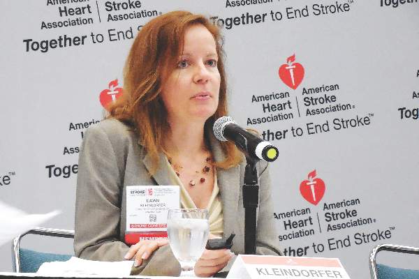
Although alteplase treatment remains central it’s also become increasingly expensive. According to a report at the Stroke Conference by Dr. Dawn Kleindorfer, a neurologist and co–medical director of the comprehensive stroke center at the University of Cincinnati, the price paid by Medicare for alteplase given to acute ischemic stroke patients more than doubled during 2005-2014. Upward pricing became especially dramatic starting in 2010, she noted, with a price tag of about $6,400/100 mg vial (enough for roughly one dose) by mid-2014. The cost for alteplase jumped from 27% of the amount hospitals got reimbursed by Medicare for treating a patient with acute ischemic stroke in 2006 to 53% of the reimbursement in 2013, Dr. Kleindorfer reported.
“I think this might impact the ability of hospitals to provide health care because the [alteplase] cost is so high and now more than half the reimbursement,” she warned. Despite its high price, alteplase “remains cost effective,” she noted, and use of the drug in recent years has increased, not dropped.
Dr. Kleindorfer was at a complete loss to explain why alteplase’s price has risen so much since 2010, and she noted that the drug received FDA approval in 1996 and is now off patent for the acute ischemic stroke indication (as well as the acute MI indication). Despite that, no biosimilar has appeared or seems to be under announced development, nor has any other thrombolytic drug received FDA approval for the stroke indication.
In fact, the history of thrombolytics for acute ischemic stroke is notably checkered. For example, an alternative thrombolytic, desmoteplase, showed early signs of promise for treating acute ischemic stroke, but the Danish drug company Lundbeck halted development of desmoteplase in late 2014 after the drug failed to achieve target primary endpoints in pivotal trials. Another thrombolytic, reteplase (Retavase), has had FDA approval for treating acute MI since 1996 but has never received approval for acute ischemic stroke. A spokesperson for Chiesi, the company that currently markets Retavase, declined to comment on why an indication for stroke was never pursued. A third thrombolytic similar to alteplase, tenecteplase, showed promise for treating acute ischemic stroke in a relatively recent phase II study, where it actually showed superiority to alteplase in a head-to-head comparison, but tenecteplase is owned and marketed by Genentech as TNKase (for acute MI only), the same company that also markets alteplase and so has little incentive to develop tenecteplase as an alternative stroke treatment.
“Even a doubling of the price [for TPA, alteplase] doesn’t change whether the drug is cost effective, but this effectively reduces hospital reimbursements that support acute stroke programs,” commented Dr. S. Claiborne Johnston in an interview. “This will ultimately impact the enthusiasm for and use of TPA,” predicted Dr. Johnston, dean of the Dell Medical School of the University of Texas in Austin and a researcher who has analyzed the economics of thrombolytic drugs for acute ischemic stroke.
“The lack of competition [to alteplase for treating acute ischemic stroke] is an example of how our current biopharma marketplace is dysfunctional,” Dr. Johnston told me. “Trials of new agents are difficult, expensive, and protracted, and there is some question whether any agent that works better [than alteplase] at lysing a clot would also produce a higher hemorrhage risk. We really need a biosimilar, but the path to develop this is not as easy as for [generic] drugs.” Although the higher price alteplase now commands might make development of a biosimilar even more attractive, Dr. Johnston said he was unaware of any company that so far is venturing down that path.
On Twitter @mitchelzoler
Where is a biosimilar when you need one?
A few weeks ago, I reported on an advisory committee of the Food and Drug Administration overwhelmingly recommending that the agency approve a biosimilar form of infliximab. If the FDA follows this recommendation, and every indication was that it would, it would become the second biosimilar drug approved for U.S. marketing, and a top agency official recently noted during Congressional testimony that the FDA biosimilar program had 59 additional agents working their way through the agency’s development program. A 2014 report cited more than 700 biosimilars under development worldwide.
But when analysts and journalists list the top reference drugs for biosimilars in the pipeline, one reference biologic agent conspicuously absent is alteplase, the thrombolytic also known as tissue plasminogen activator (TPA), which is approved for lysing clots that cause MIs and acute ischemic strokes.
Alteplase, marketed under the brand name Activase, is the only thrombolytic drug with U.S. approval for treating acute ischemic stroke. Although results from several studies published in 2015 rapidly boosted endovascular thrombectomy to physically remove clots causing acute ischemic strokes, this new intervention has by no means relegated intravenous alteplase to the sidelines. Experts I spoke with in February at the International Stroke Conference unanimously endorsed treatment with alteplase as the unshaken keystone of acute ischemic stroke treatment (for appropriate patients), even in the thrombectomy era. Thrombolysis is seen as a complement to thrombectomy rather than a treatment eclipsed by thrombectomy.
Although alteplase treatment remains central it’s also become increasingly expensive. According to a report at the Stroke Conference by Dr. Dawn Kleindorfer, a neurologist and co–medical director of the comprehensive stroke center at the University of Cincinnati, the price paid by Medicare for alteplase given to acute ischemic stroke patients more than doubled during 2005-2014. Upward pricing became especially dramatic starting in 2010, she noted, with a price tag of about $6,400/100 mg vial (enough for roughly one dose) by mid-2014. The cost for alteplase jumped from 27% of the amount hospitals got reimbursed by Medicare for treating a patient with acute ischemic stroke in 2006 to 53% of the reimbursement in 2013, Dr. Kleindorfer reported.
“I think this might impact the ability of hospitals to provide health care because the [alteplase] cost is so high and now more than half the reimbursement,” she warned. Despite its high price, alteplase “remains cost effective,” she noted, and use of the drug in recent years has increased, not dropped.
Dr. Kleindorfer was at a complete loss to explain why alteplase’s price has risen so much since 2010, and she noted that the drug received FDA approval in 1996 and is now off patent for the acute ischemic stroke indication (as well as the acute MI indication). Despite that, no biosimilar has appeared or seems to be under announced development, nor has any other thrombolytic drug received FDA approval for the stroke indication.
In fact, the history of thrombolytics for acute ischemic stroke is notably checkered. For example, an alternative thrombolytic, desmoteplase, showed early signs of promise for treating acute ischemic stroke, but the Danish drug company Lundbeck halted development of desmoteplase in late 2014 after the drug failed to achieve target primary endpoints in pivotal trials. Another thrombolytic, reteplase (Retavase), has had FDA approval for treating acute MI since 1996 but has never received approval for acute ischemic stroke. A spokesperson for Chiesi, the company that currently markets Retavase, declined to comment on why an indication for stroke was never pursued. A third thrombolytic similar to alteplase, tenecteplase, showed promise for treating acute ischemic stroke in a relatively recent phase II study, where it actually showed superiority to alteplase in a head-to-head comparison, but tenecteplase is owned and marketed by Genentech as TNKase (for acute MI only), the same company that also markets alteplase and so has little incentive to develop tenecteplase as an alternative stroke treatment.
“Even a doubling of the price [for TPA, alteplase] doesn’t change whether the drug is cost effective, but this effectively reduces hospital reimbursements that support acute stroke programs,” commented Dr. S. Claiborne Johnston in an interview. “This will ultimately impact the enthusiasm for and use of TPA,” predicted Dr. Johnston, dean of the Dell Medical School of the University of Texas in Austin and a researcher who has analyzed the economics of thrombolytic drugs for acute ischemic stroke.
“The lack of competition [to alteplase for treating acute ischemic stroke] is an example of how our current biopharma marketplace is dysfunctional,” Dr. Johnston told me. “Trials of new agents are difficult, expensive, and protracted, and there is some question whether any agent that works better [than alteplase] at lysing a clot would also produce a higher hemorrhage risk. We really need a biosimilar, but the path to develop this is not as easy as for [generic] drugs.” Although the higher price alteplase now commands might make development of a biosimilar even more attractive, Dr. Johnston said he was unaware of any company that so far is venturing down that path.
On Twitter @mitchelzoler

VIDEO: Hands-off yields best brain arteriovenous malformation outcomes
LOS ANGELES – Hang a do-not-disturb sign on brain arteriovenous malformations.
Patients who underwent invasive interventions to repair an unruptured arteriovenous malformation (AVM) in their brain faced a greater than two-fold increased rate of death or stroke during an average 4 years of follow-up, compared with patients who received medical treatment only with no active intervention, Dr. Christian Stapf reported at the International Stroke Conference.
When analyzed on an intention-to-treat basis, for every five AVM patients treated by endovascular surgery, conventional surgery, or radiotherapy, one additional patient died or had a stroke, compared with the death or stroke rate among control patients who received only medical management. When analyzed based on the treatments that patients actually received, the number-needed-to-harm fell to one excess death or stroke for every three AVM patients who underwent an invasive procedure, compared with control patients, reported Dr. Stapf, a professor in the department of neurosciences at the University of Montreal.
The results from A Randomized Trial of Unruptured Brain AVMs (ARUBA) “show us that we clearly have not been as good as we thought in helping patients against their stroke risk,” said Dr. Stapf in a video interview during the meeting. “Given that the risk of death or stroke was reduced three- to fivefold with no [invasive] treatment and leaving the AVM alone makes us think that we can’t recommend preventive intervention with currently-used techniques. Living with the AVM seems like the far better option.”
The ARUBA study, run at 39 centers in nine countries including 13 U.S. centers, randomized 226 patients with unruptured AVMs before the study’s data safety and monitoring board stopped study enrollment prematurely in April 2013. The study group included 110 patients randomized to receive medical interventions only and 116 randomized to medical intervention plus “best possible” AVM eradication. The exact type of eradication for each patient was left up to local clinicians, who tailored the intervention to address the size, location, and anatomy of each AVM. Medical management included steps such as treatment with antiepileptic drugs to treat seizures, various treatments for headaches, and physiotherapy for patients with neurologic deficits.
The study’s primary endpoint was the combined rate of death or stroke, which occurred in 41 of the 116 patients (35%) randomized to receive an invasive intervention and in 15 of the 110 (14%) randomized to medical treatment only during an average follow-up of 50 months, with many patients followed for 5 years.
When analyzed by the treatment patients actually received, 106 underwent an invasive intervention and 43 of these patients (41%) died or had a stroke, and 120 patients received medical treatments only, of whom 13 (11%) died or had a stroke.
A secondary endpoint was the rate of death or disability after 5-year follow-up, with disability defined as a modified Rankin Scale score of 2 or more. This occurred in 38% of the 45 patients who underwent AVM eradication and had this follow-up available, and in 18% of 51 patients who had medial treatment only and received this follow-up.
Interim results from the study came out 2 years ago, with an average follow-up of 33 months (Lancet. 2014 Feb 15;383[9917]:614-21), but the trial was designed to have 5-year follow-up, largely accomplished in the new data reported by Dr. Stapf.
Many clinicians had already abandoned invasive interventions to treat brain AVMs following release of the interim results, and Dr. Stapf predicted that this trend will further strengthen now that the final results are in and confirm the earlier indication of hazard. Until the ARUBA results became available, clinicians had presumed invasive interventions to resolve or minimize malformations were beneficial based on intuition. ARUBA is the first systematic comparison of procedures versus a hands-off approach for brain AVMs, he said.
“Neurologists will now be less likely to refer patients for intervention, and interventionalists will be less enthusiastic to perform procedures,” Dr. Stapf said during the meeting, sponsored by the American Heart Association. In addition, anyone now performing an intervention in routine practice would need to consider the possible legal implications if the patient were to have a bad outcome. Dr. Stapf also noted that some professional societies are now considering recommendations against routine interventions. He conceded that some invasive interventions might still occur for selected cases on an investigational basis, but the ARUBA results “set the bar very high against performing any new interventions,” he concluded.
Approximately 3,000 patients annually are newly diagnosed with an unruptured brain AVM in the United States and Canada, he estimated.
ARUBA received no commercial support. Dr. Stapf had no disclosures.
The video associated with this article is no longer available on this site. Please view all of our videos on the MDedge YouTube channel
On Twitter @mitchelzoler
AT THE INTERNATIONAL STROKE CONFERENCE
Key clinical point: The first direct comparison of invasive treatment and medical management only for intracerebral arteriovenous malformations showed substantial hazard from active intervention.
Major finding: The incidence of death or stroke was 35% with active intervention and 14% with medical management only.
Data source: ARUBA, a multicenter, prospective, international, randomized study with 226 patients.
Disclosures: ARUBA received no commercial support. Dr. Stapf had no disclosures.

ISC: Thrombectomy shown highly cost-effective for stroke
LOS ANGELES – Endovascular thrombectomy is not only clinically the best option for many patients with acute, ischemic strokes involving a proximal occlusion in a large cerebral artery; it’s also highly cost effective, based on follow-up analyses of two of the five randomized trials published in 2015 that collectively established thrombectomy as standard of care for these patients.
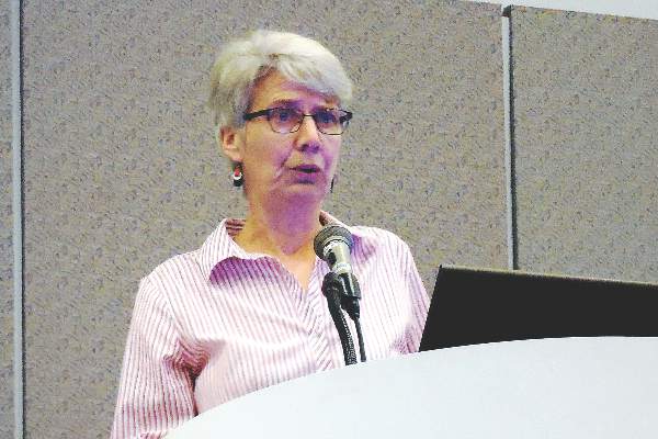
Thrombectomy plus administration of intravenous tissue plasminogen activator (TPA), compared with TPA only, “is highly cost effective and economically dominant with lower long-term cost and better outcomes,” Theresa I. Shireman, Ph.D., said at the International Stroke Conference.
And in an independent analysis of data from a totally different trial, endovascular thrombectomy on average reduced patients’ acute length of hospitalization, improved their survival and quality of life, and was cost saving when compared with treatment with intravenous TPA only, which had previously been the standard of care, Dr. Bruce C.V. Campbell reported at the meeting.
The analysis presented by Dr. Shireman used data collected in the SWIFT-PRIME trial, which randomized 196 patients at centers in the United States and Europe to treatment with either intravenous TPA plus endovascular thrombectomy or TPA alone. Average total costs during the index hospitalization ran to roughly $46,000 in the combined-treatment arm and about $29,000 in the TPA-only arm, a difference largely driven by a roughly $15,000 average incremental cost for the thrombectomy procedure, said Dr. Shireman, professor of health services research at Brown University in Providence, R.I.
However, the cost-effectiveness of thrombectomy began to kick in soon after. During the 90 days following index hospitalization, patients who underwent thrombectomy had substantial average reductions in their need for inpatient rehabilitation, time spent in skilled nursing facilities, and in outpatient rehabilitation. Overall, total medical costs during the first 90 days post discharge ran on average close to $5,000 less per patient following thrombectomy. In addition, based on their health status after 90 days, patients treated with thrombectomy were projected to have a greater than 1.7-year average life expectancy than those randomized to TPA only, with a projected net gain of 1.74 quality-adjusted life years (QALY) per patient and with a projected average decrease of roughly $23,000 in total lifetime medical costs.

Based on this average increase in QALYs and decreased long-term cost, adding thrombectomy to TPA for routine treatment of the types of patients enrolled in SWIFT-PRIME was economically dominant, Dr. Shireman said at the meeting sponsored by the American Heart Association. She also projected that despite the higher upfront cost for adding thrombectomy to treatment, the eventual savings in long-term care meant that thrombectomy began producing a net saving once patients survived for more than 22 months following their index hospitalization.
Dr. Campbell reported very similar findings in his analysis of data collected from the EXTEND-IA trial, which randomized 70 patients at 10 centers in Australia and New Zealand. During the first 90 days of treatment, including the index hospitalization, treatment with thrombectomy plus TPA saved an average of roughly $4,000 U.S.per patient, compared with TPA only, even though the average incremental cost for adding thrombectomy was nearly $11,000 U.S. The overall increased total 90-day costs with TPA only was largely driven by a substantially longer time spent hospitalized among the TPA-only patients, compared with those treated with thrombectomy plus TPA, said Dr. Campbell, a neurologist and head of hyperacute stroke at Royal Melbourne Hospital.
In addition, adding thrombectomy resulted in a projected average 4-year increase in life expectancy, and an average gain of about 3 QALYs per patient. Thrombectomy “is an incredibly powerful procedure, not just in terms of clinical response but also in terms of economics,” he concluded. Even when judged by the worst-case scenario of the analysis, “there is a 100% probability that the cost-effectiveness per QALY is less than $10,000 U.S., which is incredible value,” Dr. Campbell said.
SWIFT-PRIME was sponsored by Covidien/Medtronic. EXTEND-IA received partial funding through an unrestricted grant from Covidien/Medtronic. Dr. Shireman and Dr. Campbell had no personal disclosures.
On Twitter @mitchelzoler
LOS ANGELES – Endovascular thrombectomy is not only clinically the best option for many patients with acute, ischemic strokes involving a proximal occlusion in a large cerebral artery; it’s also highly cost effective, based on follow-up analyses of two of the five randomized trials published in 2015 that collectively established thrombectomy as standard of care for these patients.
Thrombectomy plus administration of intravenous tissue plasminogen activator (TPA), compared with TPA only, “is highly cost effective and economically dominant with lower long-term cost and better outcomes,” Theresa I. Shireman, Ph.D., said at the International Stroke Conference.
And in an independent analysis of data from a totally different trial, endovascular thrombectomy on average reduced patients’ acute length of hospitalization, improved their survival and quality of life, and was cost saving when compared with treatment with intravenous TPA only, which had previously been the standard of care, Dr. Bruce C.V. Campbell reported at the meeting.
The analysis presented by Dr. Shireman used data collected in the SWIFT-PRIME trial, which randomized 196 patients at centers in the United States and Europe to treatment with either intravenous TPA plus endovascular thrombectomy or TPA alone. Average total costs during the index hospitalization ran to roughly $46,000 in the combined-treatment arm and about $29,000 in the TPA-only arm, a difference largely driven by a roughly $15,000 average incremental cost for the thrombectomy procedure, said Dr. Shireman, professor of health services research at Brown University in Providence, R.I.
However, the cost-effectiveness of thrombectomy began to kick in soon after. During the 90 days following index hospitalization, patients who underwent thrombectomy had substantial average reductions in their need for inpatient rehabilitation, time spent in skilled nursing facilities, and in outpatient rehabilitation. Overall, total medical costs during the first 90 days post discharge ran on average close to $5,000 less per patient following thrombectomy. In addition, based on their health status after 90 days, patients treated with thrombectomy were projected to have a greater than 1.7-year average life expectancy than those randomized to TPA only, with a projected net gain of 1.74 quality-adjusted life years (QALY) per patient and with a projected average decrease of roughly $23,000 in total lifetime medical costs.
Based on this average increase in QALYs and decreased long-term cost, adding thrombectomy to TPA for routine treatment of the types of patients enrolled in SWIFT-PRIME was economically dominant, Dr. Shireman said at the meeting sponsored by the American Heart Association. She also projected that despite the higher upfront cost for adding thrombectomy to treatment, the eventual savings in long-term care meant that thrombectomy began producing a net saving once patients survived for more than 22 months following their index hospitalization.
Dr. Campbell reported very similar findings in his analysis of data collected from the EXTEND-IA trial, which randomized 70 patients at 10 centers in Australia and New Zealand. During the first 90 days of treatment, including the index hospitalization, treatment with thrombectomy plus TPA saved an average of roughly $4,000 U.S.per patient, compared with TPA only, even though the average incremental cost for adding thrombectomy was nearly $11,000 U.S. The overall increased total 90-day costs with TPA only was largely driven by a substantially longer time spent hospitalized among the TPA-only patients, compared with those treated with thrombectomy plus TPA, said Dr. Campbell, a neurologist and head of hyperacute stroke at Royal Melbourne Hospital.
In addition, adding thrombectomy resulted in a projected average 4-year increase in life expectancy, and an average gain of about 3 QALYs per patient. Thrombectomy “is an incredibly powerful procedure, not just in terms of clinical response but also in terms of economics,” he concluded. Even when judged by the worst-case scenario of the analysis, “there is a 100% probability that the cost-effectiveness per QALY is less than $10,000 U.S., which is incredible value,” Dr. Campbell said.
SWIFT-PRIME was sponsored by Covidien/Medtronic. EXTEND-IA received partial funding through an unrestricted grant from Covidien/Medtronic. Dr. Shireman and Dr. Campbell had no personal disclosures.
On Twitter @mitchelzoler
AT THE INTERNATIONAL STROKE CONFERENCE
Key clinical point: Adding endovascular thrombectomy to TPA treatment for selected patients with acute, ischemic stroke proved highly cost effective on the basis of data collected in two independent randomized trials.
Major finding: In SWIFT-PRIME, thrombectomy saved a projected average of $23,000 in lifetime health care costs and added 1.74 QALYs.
Data source: SWIFT-PRIME, an international, multicenter, randomized trial that enrolled 196 patients.
Disclosures: SWIFT-PRIME was sponsored by Covidien/Medtronic. EXTEND-IA received partial funding through an unrestricted grant from Covidien/Medtronic. Dr. Shireman and Dr. Campbell had no personal disclosures.

ISC: Imaging supplants clocks for targeting stroke reperfusion
LOS ANGELES – Can brain imaging surpass the clock for identifying acute ischemic stroke patients who will benefit from thrombolytic or thrombectomy treatment?
That’s what experts now envision, based on early findings from several studies. Although the evidence is not yet definitive enough to justify using imaging as a replacement for time-from-stroke-onset in routine practice, the results so far are encouraging and have led to the start or planning of several phase III trials that will try to nail down a role for imaging, either CT or MR, to identify acute ischemic stroke patients who qualify for reperfusion therapy.

“What we’re trying to do is move away from using the clock as a surrogate marker and instead use imaging as the surrogate,” Dr. Jenny P. Tsai said in an interview at the International Stroke Conference.
The linchpin of this new approach is that clocking time elapsed from the onset of stroke symptoms to initiation of thrombolytic or endovascular treatment makes no allowance for patient-to-patient variations in collateral cerebral circulation, a factor that appears to make a big difference in whether patients can be many more hours removed from the start of their stroke and still have salvageable brain tissue. And relying on time since stroke symptom onset gives a seriously flawed estimate of a stroke’s duration when patients have an unwitnessed stroke.
The alternative is to use either CT perfusion imaging or a combination of MR perfusion and diffusion-weighted imaging “to get effective reperfusion treatments to patients who present at later time windows,” said Dr. Gregory W. Albers, professor of neurology at Stanford (Calif.) University and director of the Stanford Stroke Center. He called these two new approaches to gauging a patient’s suitability for reperfusion therapy “the next big thing in imaging” for stroke.
Using CT perfusion to assess target mismatch
Dr. Tsai presented an analysis of thrombectomy reperfusion done in 181 patients enrolled in the CT Perfusion to Predict Response to Recanalization in Ischemic Stroke Project (CRISP), which included a total of 201 acute ischemic stroke patients with large cerebral-artery occlusions treated at six U.S. centers. Her analysis excluded nine patients who presented more than 18 hours after their stroke onset, six patients who did not have successful CT perfusion assessment, and five additional patients excluded for other reasons. The 181 patients analyzed included 125 with a target mismatch in CT perfusion, indicating that salvageable tissue remained in the area of their stroke. Among these 125 patients, 111 underwent successful reperfusion by thrombectomy.
The researchers identified good treatment outcomes as patients with a modified Rankin Scale score of 2 or less 90 days after treatment. A multivariate analysis showed that among these 111 patients, achievement of a good 90-day outcome had no statistically significant relationship with time-to-treatment out to at least the first 8 hours following stroke onset, reported Dr. Tsai, a neurologist at the Stanford Stroke Center. The data also showed a nonsignificant relationship between good outcomes and time from stroke onset to treatment beyond 8 hours, but the confidence interval for this nonsignificant relationship became very wide at later times as the analysis focused on fewer and fewer patients.
“In patients with large-artery occlusions who have a target mismatch profile [on perfusion CT] and achieve successful reperfusion there is no significant association between onset-to-reperfusion-time and the probability of a good functional outcome,” suggesting that “CT perfusion is a biomarker of good outcomes beyond 6 hours” after stroke onset, Dr. Tsai concluded. CT perfusion has the advantage of being more widely available than MRI is, she added. Last year, her associates at Stanford reported similar findings using perfusion-diffusion mismatch in the cerebral area around a stroke visualized with MRI (Neurology. 2015 Aug 25;85[8]:708-15).
“MRI is not readily available” at all U.S. centers that treat stroke patients, while CT perfusion imaging is much more ubiquitous, but the findings reported by Dr. Tsai require confirmation in the several prospective, randomized trials now underway and testing this approach, Dr. Albers said.
Using MRI in unwitnessed strokes
A second challenge for using reperfusion therapy in stroke patients are patients who have unwitnessed strokes, with a completely unknown elapsed time from onset to presentation. The ability of MRI to help identify patients with unwitnessed stroke who can safely receive intravenous thrombolytic therapy with alteplase (tissue plasminogen activator, Activase) underwent testing in the MR WITNESS (Study of Intravenous Thrombolysis With Alteplase in MRI-Selected Patients) trial.
The study enrolled 80 patients with an unwitnessed acute, ischemic stroke at 10 U.S. centers. Patients had to be in a position to receive alteplase within 4.5 hours of when their stroke was first identified; 57 (71%) of the participants had wake-up strokes. All patients underwent two types of MRI to identify them as potential candidates for safe administration of alteplase: diffusion weighted imaging, to identify that a stroke had occurred, and fluid-attenuated inversion recovery (FLAIR) assessment, to identify strokes that had occurred during the prior 4 hours.
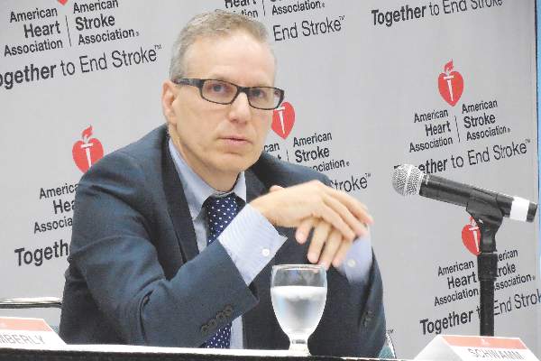
This phase II study’s primary safety endpoint was the incidence of symptomatic intracerebral hemorrhage following alteplase treatment, which occurred in 1 of the 80 patients (1.25%), not a statistically significant difference when compared with the historical standard, the 2.4% rate seen in stroke patients treated with intravenous alteplase 3 to 4.5 hours after their known stroke onset in the ECASS III (European Cooperative Acute Stroke Study) (N Engl J Med. 2008 Sept 25;359[13]:1317-29). The rate of asymptomatic intracerebral hemorrhage was not significantly different between the new study and ECASS III, reported Dr. Lee H. Schwamm, director of TeleStroke & Acute Stroke Services at the Massachusetts General Hospital in Boston.
Based on this result, “we know this approach is safe, and we saw a signal of efficacy, but we really don’t know how effective it will be” until this approach to assessing unwitnessed-stroke patients by imaging undergoes testing in a phase III trial, cautioned Dr. Schwamm at the meeting, sponsored by the American Heart Association.
Future work will also examine whether similar results can be obtained by CT imaging, which would “open this approach to every U.S. emergency department,” Dr. Schwamm said. Although the MR diffusion-weighted imaging and FLAIR analyses used in the current study do not require anything more than standard MRI equipment and software, it remains less available than CT imaging at most U.S. hospitals, he said.
The video associated with this article is no longer available on this site. Please view all of our videos on the MDedge YouTube channel
On Twitter @mitchelzoler
Traditionally, we have used a clock to identify patients who are eligible to receive reperfusion therapy, but now researchers are trying to extend patient eligibility with imaging. The Stanford (Calif.) team is trying to identify good candidates for reperfusion treatment who present with acute ischemic strokes beyond the traditional time window of 6 hours. The researchers at Massachusetts General Hospital, Boston, and their collaborators are trying to apply a similar approach to patients who have unwitnessed strokes and so have an unknown elapsed time from the start of their stroke.
The rationale behind the Stanford work is that some patients will have salvageable neurons beyond the traditional treatment time window and that this tissue can be identified by MR and CT perfusion imaging.
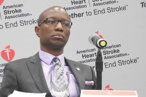 |
| Mitchel L. Zoler/Frontline Medical News Dr. Bruce I. Ovbiagele |
The workers in Boston and their collaborators used MR diffusion weighted imaging to confirm that a patient has had a stroke, and then use MR fluid attenuated inversion recovery (FLAIR) to determine if the stroke had occurred within the previous 4 hours. If a stroke has not been going on long enough to produce a positive FLAIR image, it means that the patient is still eligible for thrombolytic therapy. This could be huge for U.S. clinical practice because so many patients have unwitnessed strokes. We definitely need a larger efficacy study, but the results Dr. Schwamm reported are highly encouraging.
CT is more widely available right now in U.S. practice than is MRI, so ideally we would like to be able to use CT imaging.
Dr. Bruce I. Ovbiagele is professor and chairman of neurology at the Medical University of South Carolina in Charleston. He had no disclosures. He made these comments in an interview.
Traditionally, we have used a clock to identify patients who are eligible to receive reperfusion therapy, but now researchers are trying to extend patient eligibility with imaging. The Stanford (Calif.) team is trying to identify good candidates for reperfusion treatment who present with acute ischemic strokes beyond the traditional time window of 6 hours. The researchers at Massachusetts General Hospital, Boston, and their collaborators are trying to apply a similar approach to patients who have unwitnessed strokes and so have an unknown elapsed time from the start of their stroke.
The rationale behind the Stanford work is that some patients will have salvageable neurons beyond the traditional treatment time window and that this tissue can be identified by MR and CT perfusion imaging.
|
| Mitchel L. Zoler/Frontline Medical News Dr. Bruce I. Ovbiagele |
The workers in Boston and their collaborators used MR diffusion weighted imaging to confirm that a patient has had a stroke, and then use MR fluid attenuated inversion recovery (FLAIR) to determine if the stroke had occurred within the previous 4 hours. If a stroke has not been going on long enough to produce a positive FLAIR image, it means that the patient is still eligible for thrombolytic therapy. This could be huge for U.S. clinical practice because so many patients have unwitnessed strokes. We definitely need a larger efficacy study, but the results Dr. Schwamm reported are highly encouraging.
CT is more widely available right now in U.S. practice than is MRI, so ideally we would like to be able to use CT imaging.
Dr. Bruce I. Ovbiagele is professor and chairman of neurology at the Medical University of South Carolina in Charleston. He had no disclosures. He made these comments in an interview.
LOS ANGELES – Can brain imaging surpass the clock for identifying acute ischemic stroke patients who will benefit from thrombolytic or thrombectomy treatment?
That’s what experts now envision, based on early findings from several studies. Although the evidence is not yet definitive enough to justify using imaging as a replacement for time-from-stroke-onset in routine practice, the results so far are encouraging and have led to the start or planning of several phase III trials that will try to nail down a role for imaging, either CT or MR, to identify acute ischemic stroke patients who qualify for reperfusion therapy.
“What we’re trying to do is move away from using the clock as a surrogate marker and instead use imaging as the surrogate,” Dr. Jenny P. Tsai said in an interview at the International Stroke Conference.
The linchpin of this new approach is that clocking time elapsed from the onset of stroke symptoms to initiation of thrombolytic or endovascular treatment makes no allowance for patient-to-patient variations in collateral cerebral circulation, a factor that appears to make a big difference in whether patients can be many more hours removed from the start of their stroke and still have salvageable brain tissue. And relying on time since stroke symptom onset gives a seriously flawed estimate of a stroke’s duration when patients have an unwitnessed stroke.
The alternative is to use either CT perfusion imaging or a combination of MR perfusion and diffusion-weighted imaging “to get effective reperfusion treatments to patients who present at later time windows,” said Dr. Gregory W. Albers, professor of neurology at Stanford (Calif.) University and director of the Stanford Stroke Center. He called these two new approaches to gauging a patient’s suitability for reperfusion therapy “the next big thing in imaging” for stroke.
Using CT perfusion to assess target mismatch
Dr. Tsai presented an analysis of thrombectomy reperfusion done in 181 patients enrolled in the CT Perfusion to Predict Response to Recanalization in Ischemic Stroke Project (CRISP), which included a total of 201 acute ischemic stroke patients with large cerebral-artery occlusions treated at six U.S. centers. Her analysis excluded nine patients who presented more than 18 hours after their stroke onset, six patients who did not have successful CT perfusion assessment, and five additional patients excluded for other reasons. The 181 patients analyzed included 125 with a target mismatch in CT perfusion, indicating that salvageable tissue remained in the area of their stroke. Among these 125 patients, 111 underwent successful reperfusion by thrombectomy.
The researchers identified good treatment outcomes as patients with a modified Rankin Scale score of 2 or less 90 days after treatment. A multivariate analysis showed that among these 111 patients, achievement of a good 90-day outcome had no statistically significant relationship with time-to-treatment out to at least the first 8 hours following stroke onset, reported Dr. Tsai, a neurologist at the Stanford Stroke Center. The data also showed a nonsignificant relationship between good outcomes and time from stroke onset to treatment beyond 8 hours, but the confidence interval for this nonsignificant relationship became very wide at later times as the analysis focused on fewer and fewer patients.
“In patients with large-artery occlusions who have a target mismatch profile [on perfusion CT] and achieve successful reperfusion there is no significant association between onset-to-reperfusion-time and the probability of a good functional outcome,” suggesting that “CT perfusion is a biomarker of good outcomes beyond 6 hours” after stroke onset, Dr. Tsai concluded. CT perfusion has the advantage of being more widely available than MRI is, she added. Last year, her associates at Stanford reported similar findings using perfusion-diffusion mismatch in the cerebral area around a stroke visualized with MRI (Neurology. 2015 Aug 25;85[8]:708-15).
“MRI is not readily available” at all U.S. centers that treat stroke patients, while CT perfusion imaging is much more ubiquitous, but the findings reported by Dr. Tsai require confirmation in the several prospective, randomized trials now underway and testing this approach, Dr. Albers said.
Using MRI in unwitnessed strokes
A second challenge for using reperfusion therapy in stroke patients are patients who have unwitnessed strokes, with a completely unknown elapsed time from onset to presentation. The ability of MRI to help identify patients with unwitnessed stroke who can safely receive intravenous thrombolytic therapy with alteplase (tissue plasminogen activator, Activase) underwent testing in the MR WITNESS (Study of Intravenous Thrombolysis With Alteplase in MRI-Selected Patients) trial.
The study enrolled 80 patients with an unwitnessed acute, ischemic stroke at 10 U.S. centers. Patients had to be in a position to receive alteplase within 4.5 hours of when their stroke was first identified; 57 (71%) of the participants had wake-up strokes. All patients underwent two types of MRI to identify them as potential candidates for safe administration of alteplase: diffusion weighted imaging, to identify that a stroke had occurred, and fluid-attenuated inversion recovery (FLAIR) assessment, to identify strokes that had occurred during the prior 4 hours.
This phase II study’s primary safety endpoint was the incidence of symptomatic intracerebral hemorrhage following alteplase treatment, which occurred in 1 of the 80 patients (1.25%), not a statistically significant difference when compared with the historical standard, the 2.4% rate seen in stroke patients treated with intravenous alteplase 3 to 4.5 hours after their known stroke onset in the ECASS III (European Cooperative Acute Stroke Study) (N Engl J Med. 2008 Sept 25;359[13]:1317-29). The rate of asymptomatic intracerebral hemorrhage was not significantly different between the new study and ECASS III, reported Dr. Lee H. Schwamm, director of TeleStroke & Acute Stroke Services at the Massachusetts General Hospital in Boston.
Based on this result, “we know this approach is safe, and we saw a signal of efficacy, but we really don’t know how effective it will be” until this approach to assessing unwitnessed-stroke patients by imaging undergoes testing in a phase III trial, cautioned Dr. Schwamm at the meeting, sponsored by the American Heart Association.
Future work will also examine whether similar results can be obtained by CT imaging, which would “open this approach to every U.S. emergency department,” Dr. Schwamm said. Although the MR diffusion-weighted imaging and FLAIR analyses used in the current study do not require anything more than standard MRI equipment and software, it remains less available than CT imaging at most U.S. hospitals, he said.
The video associated with this article is no longer available on this site. Please view all of our videos on the MDedge YouTube channel
On Twitter @mitchelzoler
AT THE INTERNATIONAL STROKE CONFERENCE
Key clinical point: New CT and MR techniques provide alternatives to time-from-stroke-onset to select acute ischemic stroke patients for reperfusion therapies.
Major finding: CT perfusion identified ischemic stroke patients who could undergo effective thrombectomy more than 6 hours after their stroke onset.
Data source: The CRISP study, which enrolled 201 patients at six U.S. centers.
Disclosures: Dr. Tsai and Dr. Schwamm had no disclosures. Dr. Albers has been a consultant to iSchemaView and Covidien/Medtronic and has an ownership interest in iSchemaView.

ISC: Carotid Surgery, Stenting Offer Patients Balanced Alternatives
LOS ANGELES – The equipoise between carotid stenting and endarterectomy received a further boost in 10-year results from the landmark Carotid Revascularization Endarterectomy versus Stenting Trial (CREST) that compared the two options head-to-head.
Reported the day after results from another big trial that pitted carotid stenting against surgery, the Asymptomatic Carotid Trial (ACT I), the new long-term results from the CREST study mean that deciding among the options relies largely on patient preference although individual clinical characteristics might favor one approach or the other, experts said.

The big remaining unknown and wild card is whether doing no procedural intervention at all and relying entirely on optimal, contemporary medical treatment works just as well as endarterectomy or carotid stenting. The role for stand-alone medical therapy against carotid surgery or stenting (on top of medical therapy) is currently undergoing a formal, direct comparison in the randomized Carotid Revascularization and Medical Management for Asymptomatic Carotid Stenosis Trial (CREST-2).
Taking the 5-year outcome results from ACT I and the 10-year outcome results from CREST both into account, “we now have a lot of evidence that both carotid stenting and surgery are safe and durable. The results support both options” for either patients with symptomatic carotid artery stenosis or asymptomatic patients with carotid stenosis as extensive as in the patients enrolled in these trials, said Dr. Thomas G. Brott at the International Stroke Conference.
“In routine practice, we lay out the options of endarterectomy, carotid stenting, or no intervention with just medical treatment to patients and let them decide,” noted Dr. Brott, professor of neurology and director of research at the Mayo Clinic in Jacksonville, Fla.
CREST randomized 2,502 symptomatic or asymptomatic patients with significant carotid stenosis during 2000-2008 at 117 U.S. and Canadian centers. From this group, 1,607 consented and were available for long-term follow-up, done at a median of 7.4 years and as long as 10 years after follow-up.
The study’s primary, long-term endpoint was stroke, MI, or death during the periprocedural period (30 days after treatment or 36 days after enrollment depending on when the procedural intervention occurred) plus the rate of ipsilateral stroke during up to 10 years of follow-up. This combined endpoint occurred in 10% of the patients who underwent endarterectomy and in 12% of those who had stenting, a difference that was not statistically significant, Dr. Brott reported. Concurrent with his presentation at the meeting, sponsored by the American Heart Association, the results also were published online (N Engl J Med. 2016 Feb 18. doi: 10.1056/NEJMoa1505215).
The results included a secondary endpoint that showed a significant benefit for endarterectomy. The tally of periprocedural strokes or deaths plus ipsilateral strokes during 10-year follow-up was 8% for the surgical group and 11% for those who received a stent, a 37% excess hazard with stenting.
Dr. Brott attributed this secondary difference between the two arms of the study to a statistically significant excess of stroke or death during the periprocedural period in the patients treated by stenting, and more specifically an excess of strokes. The rate of total periprocedural strokes was 4% with stenting and 2% with endarterectomy, a statistically significant difference. Although an embolic protection device was used “when feasible” during stenting, this protection can be fallible, Dr. Brott noted. In contrast, the results from the ACT I trial showed no statistically significant difference in the rate of periprocedural total strokes between the stented and endarterectomy patients.
Dr. Brott had no relevant disclosures. The CREST trial received partial funding from Abbott Vascular.
The 10-year CREST results are good news for patients with carotid disease because they show the durability of both interventions we can offer patients. Having these data and the results from ACT I allows physicians to have an informed discussion with patients about their treatment options. I also hope that with these results from both trials, reimbursement will cease to be a deciding factor and that both surgery and stenting will be on a level playing field for insurance coverage.
Although on a population level stenting and surgery appear to produce comparable results, individual patient characteristics can make one option more appropriate. These include the morphology of a patient’s carotid arteries and stenotic lesions that can make stenting a technical challenge, and a patient’s medical condition and comorbidities which could put them at higher risk for general anesthesia and surgery. Also, a big concern for many patients is how long they will require hospitalization.
 |
Dr. Mark J. Alberts |
A major unresolved question now about treating carotid disease is whether medical treatment alone is an equally good third alternative for asymptomatic patients. We are in a relatively new era of medical therapy, with more options for smoking cessation, better and more diverse drugs for blood pressure and hyperglycemia control, and wider use of high-dose statins. Some patients are eager to avoid any intervention and already opt for medical management only, but only after CREST-2 is completed will we know whether they will truly fare as well as patients who have a procedure performed.
Another issue that needs to be considered when extrapolating the results from CREST and ACT I to routine practice is that the surgeons and interventionalists who performed the procedures in these trials were highly selected and experienced. One cannot assume that the results in these trials will be replicated by any surgeon or interventionalist in the community. I suggest that patients investigate the track record of their community hospitals and operators by consulting the performance information that is increasingly posted on the Internet.
Dr. Mark J. Alberts is professor of neurology and medical director of the neurology service at the University of Texas Southwestern Medical Center in Dallas. He had no disclosures. He made these comments in an interview.
The 10-year CREST results are good news for patients with carotid disease because they show the durability of both interventions we can offer patients. Having these data and the results from ACT I allows physicians to have an informed discussion with patients about their treatment options. I also hope that with these results from both trials, reimbursement will cease to be a deciding factor and that both surgery and stenting will be on a level playing field for insurance coverage.
Although on a population level stenting and surgery appear to produce comparable results, individual patient characteristics can make one option more appropriate. These include the morphology of a patient’s carotid arteries and stenotic lesions that can make stenting a technical challenge, and a patient’s medical condition and comorbidities which could put them at higher risk for general anesthesia and surgery. Also, a big concern for many patients is how long they will require hospitalization.
|
Dr. Mark J. Alberts |
A major unresolved question now about treating carotid disease is whether medical treatment alone is an equally good third alternative for asymptomatic patients. We are in a relatively new era of medical therapy, with more options for smoking cessation, better and more diverse drugs for blood pressure and hyperglycemia control, and wider use of high-dose statins. Some patients are eager to avoid any intervention and already opt for medical management only, but only after CREST-2 is completed will we know whether they will truly fare as well as patients who have a procedure performed.
Another issue that needs to be considered when extrapolating the results from CREST and ACT I to routine practice is that the surgeons and interventionalists who performed the procedures in these trials were highly selected and experienced. One cannot assume that the results in these trials will be replicated by any surgeon or interventionalist in the community. I suggest that patients investigate the track record of their community hospitals and operators by consulting the performance information that is increasingly posted on the Internet.
Dr. Mark J. Alberts is professor of neurology and medical director of the neurology service at the University of Texas Southwestern Medical Center in Dallas. He had no disclosures. He made these comments in an interview.
LOS ANGELES – The equipoise between carotid stenting and endarterectomy received a further boost in 10-year results from the landmark Carotid Revascularization Endarterectomy versus Stenting Trial (CREST) that compared the two options head-to-head.
Reported the day after results from another big trial that pitted carotid stenting against surgery, the Asymptomatic Carotid Trial (ACT I), the new long-term results from the CREST study mean that deciding among the options relies largely on patient preference although individual clinical characteristics might favor one approach or the other, experts said.
The big remaining unknown and wild card is whether doing no procedural intervention at all and relying entirely on optimal, contemporary medical treatment works just as well as endarterectomy or carotid stenting. The role for stand-alone medical therapy against carotid surgery or stenting (on top of medical therapy) is currently undergoing a formal, direct comparison in the randomized Carotid Revascularization and Medical Management for Asymptomatic Carotid Stenosis Trial (CREST-2).
Taking the 5-year outcome results from ACT I and the 10-year outcome results from CREST both into account, “we now have a lot of evidence that both carotid stenting and surgery are safe and durable. The results support both options” for either patients with symptomatic carotid artery stenosis or asymptomatic patients with carotid stenosis as extensive as in the patients enrolled in these trials, said Dr. Thomas G. Brott at the International Stroke Conference.
“In routine practice, we lay out the options of endarterectomy, carotid stenting, or no intervention with just medical treatment to patients and let them decide,” noted Dr. Brott, professor of neurology and director of research at the Mayo Clinic in Jacksonville, Fla.
CREST randomized 2,502 symptomatic or asymptomatic patients with significant carotid stenosis during 2000-2008 at 117 U.S. and Canadian centers. From this group, 1,607 consented and were available for long-term follow-up, done at a median of 7.4 years and as long as 10 years after follow-up.
The study’s primary, long-term endpoint was stroke, MI, or death during the periprocedural period (30 days after treatment or 36 days after enrollment depending on when the procedural intervention occurred) plus the rate of ipsilateral stroke during up to 10 years of follow-up. This combined endpoint occurred in 10% of the patients who underwent endarterectomy and in 12% of those who had stenting, a difference that was not statistically significant, Dr. Brott reported. Concurrent with his presentation at the meeting, sponsored by the American Heart Association, the results also were published online (N Engl J Med. 2016 Feb 18. doi: 10.1056/NEJMoa1505215).
The results included a secondary endpoint that showed a significant benefit for endarterectomy. The tally of periprocedural strokes or deaths plus ipsilateral strokes during 10-year follow-up was 8% for the surgical group and 11% for those who received a stent, a 37% excess hazard with stenting.
Dr. Brott attributed this secondary difference between the two arms of the study to a statistically significant excess of stroke or death during the periprocedural period in the patients treated by stenting, and more specifically an excess of strokes. The rate of total periprocedural strokes was 4% with stenting and 2% with endarterectomy, a statistically significant difference. Although an embolic protection device was used “when feasible” during stenting, this protection can be fallible, Dr. Brott noted. In contrast, the results from the ACT I trial showed no statistically significant difference in the rate of periprocedural total strokes between the stented and endarterectomy patients.
Dr. Brott had no relevant disclosures. The CREST trial received partial funding from Abbott Vascular.
AT THE INTERNATIONAL STROKE CONFERENCE

ISC: Carotid surgery, stenting offer patients balanced alternatives
LOS ANGELES – The equipoise between carotid stenting and endarterectomy received a further boost in 10-year results from the landmark Carotid Revascularization Endarterectomy versus Stenting Trial (CREST) that compared the two options head-to-head.
Reported the day after results from another big trial that pitted carotid stenting against surgery, the Asymptomatic Carotid Trial (ACT I), the new long-term results from the CREST study mean that deciding among the options relies largely on patient preference although individual clinical characteristics might favor one approach or the other, experts said.

The big remaining unknown and wild card is whether doing no procedural intervention at all and relying entirely on optimal, contemporary medical treatment works just as well as endarterectomy or carotid stenting. The role for stand-alone medical therapy against carotid surgery or stenting (on top of medical therapy) is currently undergoing a formal, direct comparison in the randomized Carotid Revascularization and Medical Management for Asymptomatic Carotid Stenosis Trial (CREST-2).
Taking the 5-year outcome results from ACT I and the 10-year outcome results from CREST both into account, “we now have a lot of evidence that both carotid stenting and surgery are safe and durable. The results support both options” for either patients with symptomatic carotid artery stenosis or asymptomatic patients with carotid stenosis as extensive as in the patients enrolled in these trials, said Dr. Thomas G. Brott at the International Stroke Conference.
“In routine practice, we lay out the options of endarterectomy, carotid stenting, or no intervention with just medical treatment to patients and let them decide,” noted Dr. Brott, professor of neurology and director of research at the Mayo Clinic in Jacksonville, Fla.
CREST randomized 2,502 symptomatic or asymptomatic patients with significant carotid stenosis during 2000-2008 at 117 U.S. and Canadian centers. From this group, 1,607 consented and were available for long-term follow-up, done at a median of 7.4 years and as long as 10 years after follow-up.
The study’s primary, long-term endpoint was stroke, MI, or death during the periprocedural period (30 days after treatment or 36 days after enrollment depending on when the procedural intervention occurred) plus the rate of ipsilateral stroke during up to 10 years of follow-up. This combined endpoint occurred in 10% of the patients who underwent endarterectomy and in 12% of those who had stenting, a difference that was not statistically significant, Dr. Brott reported. Concurrent with his presentation at the meeting, sponsored by the American Heart Association, the results also were published online (N Engl J Med. 2016 Feb 18. doi: 10.1056/NEJMoa1505215).
The results included a secondary endpoint that showed a significant benefit for endarterectomy. The tally of periprocedural strokes or deaths plus ipsilateral strokes during 10-year follow-up was 8% for the surgical group and 11% for those who received a stent, a 37% excess hazard with stenting.
Dr. Brott attributed this secondary difference between the two arms of the study to a statistically significant excess of stroke or death during the periprocedural period in the patients treated by stenting, and more specifically an excess of strokes. The rate of total periprocedural strokes was 4% with stenting and 2% with endarterectomy, a statistically significant difference. Although an embolic protection device was used “when feasible” during stenting, this protection can be fallible, Dr. Brott noted. In contrast, the results from the ACT I trial showed no statistically significant difference in the rate of periprocedural total strokes between the stented and endarterectomy patients.
Dr. Brott had no relevant disclosures. The CREST trial received partial funding from Abbott Vascular.
On Twitter @mitchelzoler
The 10-year CREST results are good news for patients with carotid disease because they show the durability of both interventions we can offer patients. Having these data and the results from ACT I allows physicians to have an informed discussion with patients about their treatment options. I also hope that with these results from both trials, reimbursement will cease to be a deciding factor and that both surgery and stenting will be on a level playing field for insurance coverage.
Although on a population level stenting and surgery appear to produce comparable results, individual patient characteristics can make one option more appropriate. These include the morphology of a patient’s carotid arteries and stenotic lesions that can make stenting a technical challenge, and a patient’s medical condition and comorbidities which could put them at higher risk for general anesthesia and surgery. Also, a big concern for many patients is how long they will require hospitalization.
|
Dr. Mark J. Alberts |
A major unresolved question now about treating carotid disease is whether medical treatment alone is an equally good third alternative for asymptomatic patients. We are in a relatively new era of medical therapy, with more options for smoking cessation, better and more diverse drugs for blood pressure and hyperglycemia control, and wider use of high-dose statins. Some patients are eager to avoid any intervention and already opt for medical management only, but only after CREST-2 is completed will we know whether they will truly fare as well as patients who have a procedure performed.
Another issue that needs to be considered when extrapolating the results from CREST and ACT I to routine practice is that the surgeons and interventionalists who performed the procedures in these trials were highly selected and experienced. One cannot assume that the results in these trials will be replicated by any surgeon or interventionalist in the community. I suggest that patients investigate the track record of their community hospitals and operators by consulting the performance information that is increasingly posted on the Internet.
Dr. Mark J. Alberts is professor of neurology and medical director of the neurology service at the University of Texas Southwestern Medical Center in Dallas. He had no disclosures. He made these comments in an interview.
LOS ANGELES – The equipoise between carotid stenting and endarterectomy received a further boost in 10-year results from the landmark Carotid Revascularization Endarterectomy versus Stenting Trial (CREST) that compared the two options head-to-head.
Reported the day after results from another big trial that pitted carotid stenting against surgery, the Asymptomatic Carotid Trial (ACT I), the new long-term results from the CREST study mean that deciding among the options relies largely on patient preference although individual clinical characteristics might favor one approach or the other, experts said.
The big remaining unknown and wild card is whether doing no procedural intervention at all and relying entirely on optimal, contemporary medical treatment works just as well as endarterectomy or carotid stenting. The role for stand-alone medical therapy against carotid surgery or stenting (on top of medical therapy) is currently undergoing a formal, direct comparison in the randomized Carotid Revascularization and Medical Management for Asymptomatic Carotid Stenosis Trial (CREST-2).
Taking the 5-year outcome results from ACT I and the 10-year outcome results from CREST both into account, “we now have a lot of evidence that both carotid stenting and surgery are safe and durable. The results support both options” for either patients with symptomatic carotid artery stenosis or asymptomatic patients with carotid stenosis as extensive as in the patients enrolled in these trials, said Dr. Thomas G. Brott at the International Stroke Conference.
“In routine practice, we lay out the options of endarterectomy, carotid stenting, or no intervention with just medical treatment to patients and let them decide,” noted Dr. Brott, professor of neurology and director of research at the Mayo Clinic in Jacksonville, Fla.
CREST randomized 2,502 symptomatic or asymptomatic patients with significant carotid stenosis during 2000-2008 at 117 U.S. and Canadian centers. From this group, 1,607 consented and were available for long-term follow-up, done at a median of 7.4 years and as long as 10 years after follow-up.
The study’s primary, long-term endpoint was stroke, MI, or death during the periprocedural period (30 days after treatment or 36 days after enrollment depending on when the procedural intervention occurred) plus the rate of ipsilateral stroke during up to 10 years of follow-up. This combined endpoint occurred in 10% of the patients who underwent endarterectomy and in 12% of those who had stenting, a difference that was not statistically significant, Dr. Brott reported. Concurrent with his presentation at the meeting, sponsored by the American Heart Association, the results also were published online (N Engl J Med. 2016 Feb 18. doi: 10.1056/NEJMoa1505215).
The results included a secondary endpoint that showed a significant benefit for endarterectomy. The tally of periprocedural strokes or deaths plus ipsilateral strokes during 10-year follow-up was 8% for the surgical group and 11% for those who received a stent, a 37% excess hazard with stenting.
Dr. Brott attributed this secondary difference between the two arms of the study to a statistically significant excess of stroke or death during the periprocedural period in the patients treated by stenting, and more specifically an excess of strokes. The rate of total periprocedural strokes was 4% with stenting and 2% with endarterectomy, a statistically significant difference. Although an embolic protection device was used “when feasible” during stenting, this protection can be fallible, Dr. Brott noted. In contrast, the results from the ACT I trial showed no statistically significant difference in the rate of periprocedural total strokes between the stented and endarterectomy patients.
Dr. Brott had no relevant disclosures. The CREST trial received partial funding from Abbott Vascular.
On Twitter @mitchelzoler
AT THE INTERNATIONAL STROKE CONFERENCE
Key clinical point: Long-term follow-up of the CREST trial out to 10 years showed no statistically significant difference between endarterectomy or carotid stenting for patients with carotid artery stenosis.
Major finding: The primary, long-term endpoint occurred in 10% of endarterectomy patients and 12% of stented patients, a nonsignificant difference.
Data source: The CREST trial, which followed 1,607 patients for up to 10 years after their randomized intervention.
Disclosures: Dr. Brott had no relevant disclosures. The CREST trial received partial funding from Abbott Vascular.
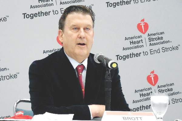
VIDEO: Octogenarians benefit from ischemic stroke thrombectomy
LOS ANGELES – Clot removal to recanalize the occluded intracerebral arteries of acute ischemic stroke patients was as effective for producing good outcomes in patients aged 80 years or older as it was in younger patients, according to results from a pooled analysis of 1,287 patients in five separate but similar randomized trials.
This unprecedented evidence for the safety and efficacy of thrombectomy (also known as embolectomy) in octogenarians experiencing an acute occlusive, large-vessel, proximal anterior-circulation stroke was one of several new findings from the pooled analysis that should help further push thrombectomy to the forefront of acute care for patients undergoing this type of ischemic stroke, predicted Dr. Wade S. Smith in a video interview at the International Stroke Conference.
“By looking at all the data, we have much more refined information on the robustness of the treatment across age groups, which is quite important, especially patients in the 80-plus age group,” commented Dr. Smith, professor of neurology and chief of the neurovascular division at the University of California, San Francisco.
Until now, during the year following the reports in early 2015 from all five studies, “age had been a limiting factor” in applying the practicing-changing intervention of thrombectomy to patients, he noted.
“This [the new pooled analysis] will change that,” Dr. Smith predicted. “It does not apply to patients who were infirm prior to their stroke – but for patients who were otherwise healthy, with a modified Rankin scale level of 0 or 1 at initial presentation, it appears that they benefit [from thrombectomy] regardless of their age.” In the pooled analysis, 198 of the 1,287 total patients (15%) were at least 80 years old.
“It removes age discrimination. A healthy 80-year-old may do extremely well with this treatment,” Dr. Smith said.
The consistency of benefit across a wide range of stroke severity that showed up in the trials as four distinct strata of NIH Stroke Scale scores prior to treatment was another important finding that could not previously be definitively made by analyzing each of the five trials individually.
In patients with stroke-severity scores that ranged from 10 or less (the least severely affected) to patients with scores of 21 or greater, all had post-thrombectomy improvements that clustered around the overall average number-needed-to-treat of 2.6 patients to reduce the disability of one patient at follow-up by at least one level on the modified Rankin scale.
Other notable findings from the pooled analysis were that thrombectomy also produced a consistent benefit to patients across every other subgroup examined, including sex, specific occlusion site, whether or not patients also received thrombolytic treatment with tissue plasminogen activator, and time to thrombectomy treatment (5 or fewer hours from stroke onset or more than 5 hours), reported Dr. Michael D. Hill and Dr. Tudor G. Jovin in a joint presentation at the meeting, sponsored by the American Heart Association.
Their pooled analysis, known as HERMES, pooled data from the MR CLEAN, ESCAPE, REVASCAT, SWIFT PRIME, and EXTEND IA trials, all run during 2010-2014.
“Endovascular treatment is a highly effective treatment across all subgroups,” concluded Dr. Hill and Dr. Jovin as they completed their talk. “These data may provide additional support for endovascular treatment in subgroups not addressed in the individual trials.”
Concurrent with their report at the meeting, the results appeared in a paper published online (Lancet. 2016 Feb 18;doi: 10.1016/S0140-6736(16)00163-X).
Both Dr. Jovin and Dr. Hill shared the enthusiasm of Dr. Smith and others in the packed meeting room about the age finding.
“Older patients seemed to benefit even more” from thrombectomy, compared with younger patients, noted Dr. Jovin, chief of the stroke division at the University of Pittsburgh and a coinvestigator on SWIFT PRIME. “There is no reason to deny this treatment to appropriately selected patients based on age,” he said.
“There is no upper age limit,” agreed Dr. Hill, professor of neurology and director of the stroke unit at the University of Calgary (Alta.) and a coinvestigator on the ESCAPE trial. “If it’s an otherwise healthy 90-year-old who is living independently, you can surely consider them for this treatment.”
HERMES received fundings through an unrestricted grant from Medtronic. Dr. Hill and Dr. Jovin had no personal disclosures. Dr. Smith served on the data safety and monitoring board for a trial funded by Stryker.
The video associated with this article is no longer available on this site. Please view all of our videos on the MDedge YouTube channel
On Twitter @mitchelzoler
AT THE INTERNATIONAL STROKE CONFERENCE
Key clinical point: A pooled analysis of five recent trials of thrombectomy for acute ischemic stroke should help further propel its widespread U.S. adoption.
Major finding: Intracerebral, transcatheter clot removal in acute ischemic stroke was equally effective in octogenarian and younger patients.
Data source: HERMES, a pooled analysis of data from 1,287 acute ischemic stroke patients randomized in five separate but similar trials.
Disclosures: HERMES received fundings through an unrestricted grant from Medtronic. Dr. Hill and Dr. Jovin had no personal disclosures. Dr. Smith served on the data safety and monitoring board for a trial funded by Stryker.

STS: Lactate dehydrogenase of 1,150 IU/L flags LVAD thrombosis
PHOENIX – A blood lactate dehydrogenase level of at least 1,150 IU/L may be the best cutoff for identifying pump thrombosis in patients who have received a left ventricular assist device based on findings from a combined series of 502 pump recipients.
Using this cutoff correlated with a positive predictive value of 74% and a negative predictive value of 92%, Dr. Vikram Sood said at the annual meeting of the Society of Thoracic Surgeons.
Results from several prior studies had established an elevated blood level of lactate dehydrogenase (LDH) as perhaps the best early flag of significant thrombus inside of a left ventricular assist device (LVAD), a condition called pump thrombosis. But prior studies had not identified a specific cutoff value to use that could confidently diagnose pump thrombosis. A level of 1,150 IU/L is about five times the upper limit of normal, noted Dr. Francis D. Pagani, professor of surgery and surgical director of adult heart transplantation at the University of Michigan, Ann Arbor, and a senior author on this report.
Dr. Sood and his associates reviewed records for 502 patients who received a HeartMate II LVAD at either the University of Michigan or at the Mayo Clinic in Rochester, Minn., during 2004-2014. During that time, 58 of the patients required a total of 70 LVAD explants. Fifty-two of the explants were for suspected pump thrombosis, 12 for infection, and 6 for lead fracture. The average time from LVAD placement to explant was 12 months.
Among the 52 LVADs explanted for suspected pump thrombosis, 47 actually had thrombosis. In addition, six of the LVADs that were explanted for other reasons also had a significant amount of thrombus, resulting in 53 total explanted LVAD with confirmed pump thrombosis.
The researchers analyzed blood levels of LDH during the 6 months prior to explant. Among the patients with the 53 LVADs explanted with confirmed pump thrombosis, the median of all LDH levels during the period was 1,061 IU/L and the median maximum LDH level for each patient was 1,940 IU/L, reported Dr. Sood, a cardiac surgeon at the University of Michigan. In contrast, among the patients who had a total of 17 LVADs explanted without pump thrombosis, the median value for all LDH measures during the 6 months prior to explant was 533 IU/L, and their medium maximum level was 504 IU/L.
Analysis of LDH levels prior to explant and subsequent confirmation of pump thrombosis identified a cut off of 1,150 IU/L as able to identify pump thrombosis with a sensitivity of 78% and a specificity of 90%, Dr. Sood reported.
On Twitter @mitchelzoler
AT THE STS ANNUAL MEETING
Key clinical point: In patients who have received a left ventricular assist device, a blood level of lactate dehydrogenase of at least 1,150 IU/L had a good positive and negative predictive value for pump thrombosis.
Major finding: A 1,150 IU/L cutoff for LDH had a positive predictive value of 74% and a negative predictive value of 92%.
Data source: Retrospective review of 502 patients who received a HeartMate II LVAD at either of two U.S. centers during 2004-2014.
Disclosures: Dr. Sood and Dr. Pagani had no disclosures.

STS: Minimizing LVAD pump thrombosis poses new challenges
PHOENIX – Cardiothoracic surgeons who implant left ventricular assist devices in patients with failing hearts remain at a loss to fully explain why they started seeing a sharp increase in thrombus clogging in these devices in 2012, but nevertheless they are gaining a better sense of how to minimize the risk.
Three key principles for minimizing thrombosis risk are selecting the right patients to receive left ventricular assist devices (LVAD), applying optimal management strategies once patients receive a LVAD, and maintaining adequate flow of blood through the pump, Dr. Francis D. Pagani said in a talk at a session devoted to pump thrombosis at the annual meeting of the Society of Thoracic Surgeons.

Other critical aspects include optimal implantation technique, quick work-up of patients to rule out reversible LVAD inflow or outflow problems once pump thrombosis is suspected, and ceasing medical therapy of the thrombosis if it proves ineffective and instead progress to surgical pump exchange, pump explantation, or heart transplant when necessary, said Dr. Ahmet Kilic, a cardiothoracic surgeon at the Ohio State University, Columbus.
Another key issue is that, now that the pump thrombosis incidence is averaging about 10% of LVAD recipients, with an incidence rate during 2-year follow-up as high as 24% reported from one series, surgeons and physicians who care for LVAD patients must have a high index of suspicion and routinely screen LVAD recipients for early signs of pump thrombosis. The best way to catch pump thrombosis early seems to be by regularly measuring patients’ serum level of lactate dehydrogenase (LDH), said Dr. Robert L. Kormos, professor of surgery and director of the artificial heart program at the University of Pittsburgh.
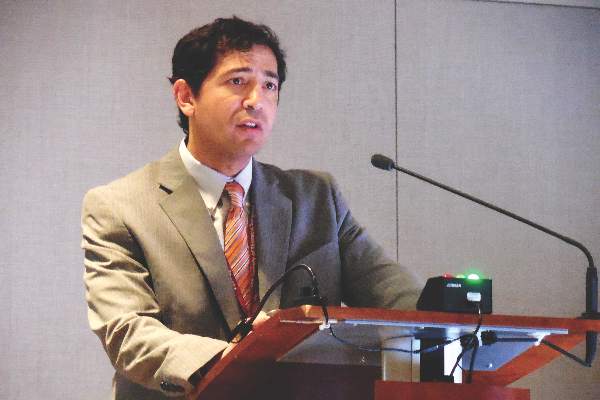
“We measure LDH on most clinic visits, whether or not the patient has an indication of pump thrombosis. We need to screen [LDH levels] much more routinely than we used to,” he said during the session. “Elevated LDH is probably the first and most reliable early sign, but you need to also assess LDH isoenzymes because we’ve had patients with an elevation but no sign of pump thrombosis, and their isoenzymes showed that the increased LDH was coming from their liver,” Dr. Kormos said in an interview.
Although serial measurements and isoenzyme analysis can establish a sharp rise in heart-specific LDH in an individual patient, a report at the meeting documented that in a series of 53 patients with pump thrombosis treated at either of two U.S. centers, an LDH level of at least 1,155 IU/L flagged pump thrombosis with a fairly high sensitivity and specificity. This LDH level is roughly five times the upper limit of normal, noted Dr. Pagani, professor of surgery and surgical director of adult heart transplantation at the University of Michigan, Ann Arbor, and a senior author on this report.
But prior to this report Dr. Kormos said that he regarded a LDH level of 600-800 IU/L as enough of an elevation above normal to prompt concern and investigation. And he criticized some LVAD programs that allow LDH levels to rise much higher.
“I know of clinicians who see a LDH of 1,500-2,000 IU/L but the patient seems okay and they wonder if they should change out the pump. For me, it’s a no brainer. Others try to list a patient like this for a heart transplant so they can avoid doing a pump exchange. I think that’s dangerous; it risks liver failure or renal failure. I would not sit on any LVAD that is starting to produce signs of hemolysis syndrome, but some places do this,” Dr. Kormos said in an interview.
“Pump thrombosis probably did not get addressed in as timely a fashion as it should have been” when it was first seen on the rise in 2012, noted Dr. James K. Kirklin, professor of surgery and director of cardiothoracic surgery at the University of Alabama, Birmingham. “It is now being addressed, and we realize that this is not just a pump problem but also involves patient factors and management factors that we need to learn more about. We are quite ignorant of the patient factors and understanding their contributions to bleeding and thrombosis,” said Dr. Kirklin. He also acknowledged that whatever role the current generation of LVAD pumps play in causing thrombosis will not quickly resolve.
“I’m looking forward to a new generation of pumps, but the pumps we have today will probably remain for another 3-5 years.”
The issue of LVAD pump thrombosis first came into clear focus with publication at the start of 2014 of a report that tracked its incidence from 2004 to mid-2013 at three U.S. centers that had placed a total of 895 LVADs in 837 patients. The annual rate of new episodes of pump thrombosis jumped from about 1%-2% of LVAD recipients throughout the first part of the study period through the end of 2011, to an annual rate of about 10% by mid 2013 (N Engl J Med. 2014 Jan 2;370[1]:33-40).
“The inflection occurred in about 2012,” noted Dr. Nicholas G. Smedira, a cardiothoracic surgeon at the Cleveland Clinic. “No one has figured out why” the incidence suddenly spiked starting in 2012 and intensified in 2013, he said. This epidemic of pump thrombosis has produced “devastating complications” that have led to multiple readmissions and reduced cost-effectiveness of LVADs and has affected how the heart transplant community allocates hearts, Dr. Smedira said during his talk at the session. He noted that once the surge in pump thrombosis started, the timing of the appearance of significant thrombus shifted earlier, often occurring within 2-3 months after LVAD placement. There now is “increasing device-related pessimism” and increasing demoralization among clinicians because of this recurring complication, he said.
More recent data show the trend toward increasingly higher rates of pump thrombosis continuing through the end of 2013, with the situation during 2014 a bit less clear. Late last year, data from 9,808 U.S. patients who received an LVAD and entered the Interagency Registry for Mechanically Assisted Circulatory Support (INTERMACS) showed that the incidence of pump thrombosis during the first 6 months following an implant rose from 1% in 2008 to 2% in 2009 and in 2010, 4% in 2011, 7% in 2012, 8% in 2013, and then eased back to 5% in the first half of 2014 (J Heart Lung Transplant. 2015 Dec;34[12]:1515-26). The annual rate rose from 2% in 2008 to a peak of 11% in 2013, with 12-month data from 2014 not yet available at the time of this report.
“The modest reduction of observed pump thrombosis at 6 months during 2014 has occurred in a milieu of heightened intensity of anti-coagulation management, greater surgical awareness of optimal pump implantation and positioning and pump speed management. Thus, one may speculate that current thrombosis risk-mitigation strategies have contributed to reducing but not eliminating the increased thrombosis risk observed since 2011,” concluded the authors of the report.
Surgeons and cardiologists must now have a high index of suspicion for pump thrombosis in LVAD recipients, and be especially on the lookout for four key flags of a problem, said Dr. Kormos. The first is a rising LDH level, but additional flags include an isolated power elevation that doesn’t correlate with anything else, evidence of hemolysis, and new-onset heart failure symptoms. These can occur individually or in some combination. He recommended following a diagnostic algorithm first presented in 2013 that remains very valid today (J Heart Lung Transplant. 2013 July;32[7]:667-70).
Dr. Kormos also highlighted that the presentation of pump thrombosis can differ between the two LVADs most commonly used in U.S. practice, the HeartMate II and the HeartWare devices. A LDH elevation is primarily an indicator for HeartMate II, while both that model and the HeartWare device show sustained, isolated power elevations when thrombosis occurs.
Dr. Pagani, Dr. Kirklin, and Dr. Smedira had no disclosures. Dr. Kormos has received travel support from HeartWare. Dr. Kilic has been a consultant to Thoratec and a speaker on behalf of Baxter International.
On Twitter @mitchelzoler
Dr. Hossein Almassi, FCCP, comments: With improvements in technology and development of rotary pumps, there has been a significant growth in the use of mechanical circulatory support (MCS) for treatment of end stage heart failure with a parallel improvement in patients’ survival and the quality of life.
 |
| Dr. Hossein Almassi |
The authors of this report presented at the 2016 annual meeting of the STS, are authorities in the field of MCS outlining the observed increase in pump thrombosis noted in 2012. The sharp increase in the thrombosis rate is different from the lower incidence seen in the preapproval stage of the pump trial.
It should be noted that the report is related mainly to the HeatMate II left ventricular assist device (LVAD) and not the more recently implanted HeartWare device.
The diagnostic algorithm outlined in the accompanying reference (J Heart Lung Transplant. 2013 July;32[7]:667-70) regarding the diagnosis and management of suspected pump thrombosis is worth reading with the main criteria heralding a potential pump thrombosis being 1)sustained pump power elevation, 2) elevation of cardiac LDH or plasma-free hemoglobin, 3) hemolysis, and 4) symptoms of heart failure.
With further refinements in technology, the field of MCS is awaiting the development of newer LVAD devices that would mitigate the serious problem of pump thrombosis.
Dr. Hossein Almassi, FCCP, comments: With improvements in technology and development of rotary pumps, there has been a significant growth in the use of mechanical circulatory support (MCS) for treatment of end stage heart failure with a parallel improvement in patients’ survival and the quality of life.
|
| Dr. Hossein Almassi |
The authors of this report presented at the 2016 annual meeting of the STS, are authorities in the field of MCS outlining the observed increase in pump thrombosis noted in 2012. The sharp increase in the thrombosis rate is different from the lower incidence seen in the preapproval stage of the pump trial.
It should be noted that the report is related mainly to the HeatMate II left ventricular assist device (LVAD) and not the more recently implanted HeartWare device.
The diagnostic algorithm outlined in the accompanying reference (J Heart Lung Transplant. 2013 July;32[7]:667-70) regarding the diagnosis and management of suspected pump thrombosis is worth reading with the main criteria heralding a potential pump thrombosis being 1)sustained pump power elevation, 2) elevation of cardiac LDH or plasma-free hemoglobin, 3) hemolysis, and 4) symptoms of heart failure.
With further refinements in technology, the field of MCS is awaiting the development of newer LVAD devices that would mitigate the serious problem of pump thrombosis.
PHOENIX – Cardiothoracic surgeons who implant left ventricular assist devices in patients with failing hearts remain at a loss to fully explain why they started seeing a sharp increase in thrombus clogging in these devices in 2012, but nevertheless they are gaining a better sense of how to minimize the risk.
Three key principles for minimizing thrombosis risk are selecting the right patients to receive left ventricular assist devices (LVAD), applying optimal management strategies once patients receive a LVAD, and maintaining adequate flow of blood through the pump, Dr. Francis D. Pagani said in a talk at a session devoted to pump thrombosis at the annual meeting of the Society of Thoracic Surgeons.
Other critical aspects include optimal implantation technique, quick work-up of patients to rule out reversible LVAD inflow or outflow problems once pump thrombosis is suspected, and ceasing medical therapy of the thrombosis if it proves ineffective and instead progress to surgical pump exchange, pump explantation, or heart transplant when necessary, said Dr. Ahmet Kilic, a cardiothoracic surgeon at the Ohio State University, Columbus.
Another key issue is that, now that the pump thrombosis incidence is averaging about 10% of LVAD recipients, with an incidence rate during 2-year follow-up as high as 24% reported from one series, surgeons and physicians who care for LVAD patients must have a high index of suspicion and routinely screen LVAD recipients for early signs of pump thrombosis. The best way to catch pump thrombosis early seems to be by regularly measuring patients’ serum level of lactate dehydrogenase (LDH), said Dr. Robert L. Kormos, professor of surgery and director of the artificial heart program at the University of Pittsburgh.
“We measure LDH on most clinic visits, whether or not the patient has an indication of pump thrombosis. We need to screen [LDH levels] much more routinely than we used to,” he said during the session. “Elevated LDH is probably the first and most reliable early sign, but you need to also assess LDH isoenzymes because we’ve had patients with an elevation but no sign of pump thrombosis, and their isoenzymes showed that the increased LDH was coming from their liver,” Dr. Kormos said in an interview.
Although serial measurements and isoenzyme analysis can establish a sharp rise in heart-specific LDH in an individual patient, a report at the meeting documented that in a series of 53 patients with pump thrombosis treated at either of two U.S. centers, an LDH level of at least 1,155 IU/L flagged pump thrombosis with a fairly high sensitivity and specificity. This LDH level is roughly five times the upper limit of normal, noted Dr. Pagani, professor of surgery and surgical director of adult heart transplantation at the University of Michigan, Ann Arbor, and a senior author on this report.
But prior to this report Dr. Kormos said that he regarded a LDH level of 600-800 IU/L as enough of an elevation above normal to prompt concern and investigation. And he criticized some LVAD programs that allow LDH levels to rise much higher.
“I know of clinicians who see a LDH of 1,500-2,000 IU/L but the patient seems okay and they wonder if they should change out the pump. For me, it’s a no brainer. Others try to list a patient like this for a heart transplant so they can avoid doing a pump exchange. I think that’s dangerous; it risks liver failure or renal failure. I would not sit on any LVAD that is starting to produce signs of hemolysis syndrome, but some places do this,” Dr. Kormos said in an interview.
“Pump thrombosis probably did not get addressed in as timely a fashion as it should have been” when it was first seen on the rise in 2012, noted Dr. James K. Kirklin, professor of surgery and director of cardiothoracic surgery at the University of Alabama, Birmingham. “It is now being addressed, and we realize that this is not just a pump problem but also involves patient factors and management factors that we need to learn more about. We are quite ignorant of the patient factors and understanding their contributions to bleeding and thrombosis,” said Dr. Kirklin. He also acknowledged that whatever role the current generation of LVAD pumps play in causing thrombosis will not quickly resolve.
“I’m looking forward to a new generation of pumps, but the pumps we have today will probably remain for another 3-5 years.”
The issue of LVAD pump thrombosis first came into clear focus with publication at the start of 2014 of a report that tracked its incidence from 2004 to mid-2013 at three U.S. centers that had placed a total of 895 LVADs in 837 patients. The annual rate of new episodes of pump thrombosis jumped from about 1%-2% of LVAD recipients throughout the first part of the study period through the end of 2011, to an annual rate of about 10% by mid 2013 (N Engl J Med. 2014 Jan 2;370[1]:33-40).
“The inflection occurred in about 2012,” noted Dr. Nicholas G. Smedira, a cardiothoracic surgeon at the Cleveland Clinic. “No one has figured out why” the incidence suddenly spiked starting in 2012 and intensified in 2013, he said. This epidemic of pump thrombosis has produced “devastating complications” that have led to multiple readmissions and reduced cost-effectiveness of LVADs and has affected how the heart transplant community allocates hearts, Dr. Smedira said during his talk at the session. He noted that once the surge in pump thrombosis started, the timing of the appearance of significant thrombus shifted earlier, often occurring within 2-3 months after LVAD placement. There now is “increasing device-related pessimism” and increasing demoralization among clinicians because of this recurring complication, he said.
More recent data show the trend toward increasingly higher rates of pump thrombosis continuing through the end of 2013, with the situation during 2014 a bit less clear. Late last year, data from 9,808 U.S. patients who received an LVAD and entered the Interagency Registry for Mechanically Assisted Circulatory Support (INTERMACS) showed that the incidence of pump thrombosis during the first 6 months following an implant rose from 1% in 2008 to 2% in 2009 and in 2010, 4% in 2011, 7% in 2012, 8% in 2013, and then eased back to 5% in the first half of 2014 (J Heart Lung Transplant. 2015 Dec;34[12]:1515-26). The annual rate rose from 2% in 2008 to a peak of 11% in 2013, with 12-month data from 2014 not yet available at the time of this report.
“The modest reduction of observed pump thrombosis at 6 months during 2014 has occurred in a milieu of heightened intensity of anti-coagulation management, greater surgical awareness of optimal pump implantation and positioning and pump speed management. Thus, one may speculate that current thrombosis risk-mitigation strategies have contributed to reducing but not eliminating the increased thrombosis risk observed since 2011,” concluded the authors of the report.
Surgeons and cardiologists must now have a high index of suspicion for pump thrombosis in LVAD recipients, and be especially on the lookout for four key flags of a problem, said Dr. Kormos. The first is a rising LDH level, but additional flags include an isolated power elevation that doesn’t correlate with anything else, evidence of hemolysis, and new-onset heart failure symptoms. These can occur individually or in some combination. He recommended following a diagnostic algorithm first presented in 2013 that remains very valid today (J Heart Lung Transplant. 2013 July;32[7]:667-70).
Dr. Kormos also highlighted that the presentation of pump thrombosis can differ between the two LVADs most commonly used in U.S. practice, the HeartMate II and the HeartWare devices. A LDH elevation is primarily an indicator for HeartMate II, while both that model and the HeartWare device show sustained, isolated power elevations when thrombosis occurs.
Dr. Pagani, Dr. Kirklin, and Dr. Smedira had no disclosures. Dr. Kormos has received travel support from HeartWare. Dr. Kilic has been a consultant to Thoratec and a speaker on behalf of Baxter International.
On Twitter @mitchelzoler
EXPERT ANALYSIS FROM THE STS ANNUAL MEETING
VIDEO: Migraine plus aura doubles ischemic stroke rate
LOS ANGELES – Adults who have migraine with aura had twice the rate of ischemic strokes, compared with people with migraine without aura in a review of 11,663 older Americans followed for an average of 18 years.
Migraine with aura appeared to increase the rate of cardioembolic strokes particularly, and the findings highlighted that middle-aged and older adults diagnosed with migraine with aura require special attention to interventions that could reduce their cardiovascular disease risks, Dr. Souvik Sen said in a video interview during the International Stroke Conference.
While results from prior studies have implicated migraine with aura as a risk factor for ischemic stroke, the new analysis was notable for involving an especially large number of community-dwelling older Americans, suggesting that the finding is broadly applicable to the general population of people with an average age of 58-60 years, said Dr. Sen, professor of clinical neurology and chairman of the department of neurology at the University of South Carolina in Columbia.
“It’s been known that migraine can increase the risk of stroke and that certain subgroups of migraine patients are especially important, such as patients with aura,” commented Dr. Ralph Sacco, professor and chairman of neurology at the University of Miami. “When I see patients with migraine I think about treating their migraine effectively and getting it under control, and putting even greater focus on controlling stroke risk factors. I also warn women with migraine about their increased stroke risk from using estrogen-containing oral contraceptives or hormone replacement,” Dr. Sacco noted.
Dr. Sen noted that the new analysis included people enrolled in the Atherosclerosis Risk in Communities (ARIC) study, which enrolled a random sample of more than 15,000 participants from four U.S. communities in 1987. The analysis focused on 11,663 people with migraine information collected during the third ARIC examination during 1993-1995, which identified 458 people (4%) who had migraine with aura, 1,158 (10%) who had migraine without aura, and 10,047 (86%) who were migraine free. During 18 years of follow-up the incidence of ischemic stroke among those with migraine with aura was twofold higher than among those with migraine without aura after adjustment for baseline rate of stroke risk factors, a statistically significant difference, he reported at the meeting sponsored by the American Heart Association. This difference seemed largely driven by a significant increase in the rate of cardioembolic strokes among the people with migraine with aura.
One hypothesis explaining the increased stroke rate in patients with aura is that, in patients with this presentation of migraine, the neurologic activity of migraine appears to spread to cerebral blood vessels and may affect blood flow within those vessels, Dr. Sen explained. It is also possible that migraine with aura may be a risk marker for or may predispose people to more extensive cardiovascular disease including atrial fibrillation.
The primary clinical implication of the finding is that people with migraine with aura should especially undergo a thorough assessment for cardiovascular disease risk factors, and should receive especially aggressive risk factor management, including controlling hypertension and smoking cessation, assessing ECG and atrial fibrillation, and avoiding estrogen treatment, Dr. Sen said.
The video associated with this article is no longer available on this site. Please view all of our videos on the MDedge YouTube channel
On Twitter @mitchelzoler
AT THE INTERNATIONAL STROKE CONFERENCE
Key clinical point: Older adults with migraine and aura had twice the ischemic strokes, compared with patients with aura-free migraines.
Major finding: Strokes occurred twofold more often in patients with migraine with aura, compared with patients with migraine alone after adjustments.
Data source: Review of 11,663 older adult Americans enrolled in the Atherosclerosis Risk in Communities study.
Disclosures: Dr. Sen had no disclosures.