User login
Cancer patients with incidental VTE have high risk of recurrent thrombi
SAN FRANCISCO – Unless there is a clear contraindication to anticoagulation therapy, cancer patients who have an incidental pulmonary embolism discovered during imaging studies should receive chronic anticoagulation, preferably with a low-molecular-weight heparin, based on study results presented at the annual meeting of the American Society of Hematology.

Without anticoagulation therapy, the risk for a recurent venous thromboembolic event was fourfold higher among 926 cancer patients who had a pulmonary embolism discovered during CT imaging for reasons other than suspicion of pulmonary embolism (PE), according to Dr. Tom van der Hulle of Leiden (the Netherlands) University Medical Center.
Among cancer patients who did receive an anticoagulant, low-molecular-weight heparin (LMWH) was significantly better than warfarin at preventing recurrent venous thromboembolism (VTE) or PE. Compared with warfarin, LMWH also was associated with a significantly lower risk for bleeding.
“Our study confirms the high risk of recurrent [VTE] in patients with cancer-associated incidental pulmonary embolism, but it also provides outcomes if patients are left untreated,” Dr. van der Hulle said at a briefing prior to his presentation of the data.
Risk for primary and recurrent VTE is particularly high for patients with pancreatic cancer, lung cancer, metastatic adenocarcinoma of gastroinestinal or gynecologic origin, and brain tumors, noted Dr. Agnes Lee of the University of British Columbia in Vancouver. Dr. Lee also presented data at the briefing but was not involved with the study.
“Once the patients are started on treatment for their clotting episode, for a DVT or PE, a minimum of 3-6 months of anticoagulation is recommended. After that, we look at patients to see whether they have ongoing reasons to develop recurrent thrombosis. So this would be patients who have active cancer – they’re still receiving chemotherapy, they have obvious evidence of cancer or metastatic disease, or they’re sick for other reasons. These are patients who would then continue on anticoagulant therapy,” she added.
Dr. van der Hulle and his colleagues conducted a pooled meta-analysis of 926 patients with cancer who had an incidental PE discovered during a CT scan. The researchers compared results for patients treated with LMWH (79%), a vitamin K antagonist (predominantly warfarin, 11%), and for those who did not receive prophylaxis with an anticoagualant because of contraindications.
They looked at recurrent VTE, major bleeding episodes, and death at 6 months and conducted subgroup analyses based on patient management and thrombus location.
Patients with metastatic disease comprised 74% of those treated with LMWH, 62% of those treated with a vitamin K antagonist, and 73% of untreated patients.
The overall pooled 6-month risk of symptomatic recurrent VTE was 5.8%. The risk of major bleeding was 4.7%, and the risk of death was 37%.
Risk of recurrent VTE was 6.2% for patients treated with LMWH and 6.4% for those treated with a vitamin K antagonist. Recurrent VTE risk was 12% for untreated patients.
Major bleeding was seen in 3.9% of LMWH-treated patients and in 13% of vitamin K antagonist–treated patients (hazard ratio 3.9; 95% confidence interval, 1.6-10).The 6-month mortality rates were 37% for patients treated with LMWH, 28% for those treated with warfarin or a related drug, and 47% for untreated patients.
When the authors looked at the location of the thrombus – isolated subsegmental incidental PE vs. central PE – they found that the risk of recurrent isolated embolism was 7.8% and for central embolism was 5.5%.
Dr. van der Hulle said that the findings of this observational study should preferably be confirmed in a randomized trial, but acknowledged that, given the risk of no treatment, a placebo-controlled trial might be considered unethical.
The study was internally supported. Dr. van der Hulle and Dr. Lee reported no relevant conflicts of interest.
SAN FRANCISCO – Unless there is a clear contraindication to anticoagulation therapy, cancer patients who have an incidental pulmonary embolism discovered during imaging studies should receive chronic anticoagulation, preferably with a low-molecular-weight heparin, based on study results presented at the annual meeting of the American Society of Hematology.
Without anticoagulation therapy, the risk for a recurent venous thromboembolic event was fourfold higher among 926 cancer patients who had a pulmonary embolism discovered during CT imaging for reasons other than suspicion of pulmonary embolism (PE), according to Dr. Tom van der Hulle of Leiden (the Netherlands) University Medical Center.
Among cancer patients who did receive an anticoagulant, low-molecular-weight heparin (LMWH) was significantly better than warfarin at preventing recurrent venous thromboembolism (VTE) or PE. Compared with warfarin, LMWH also was associated with a significantly lower risk for bleeding.
“Our study confirms the high risk of recurrent [VTE] in patients with cancer-associated incidental pulmonary embolism, but it also provides outcomes if patients are left untreated,” Dr. van der Hulle said at a briefing prior to his presentation of the data.
Risk for primary and recurrent VTE is particularly high for patients with pancreatic cancer, lung cancer, metastatic adenocarcinoma of gastroinestinal or gynecologic origin, and brain tumors, noted Dr. Agnes Lee of the University of British Columbia in Vancouver. Dr. Lee also presented data at the briefing but was not involved with the study.
“Once the patients are started on treatment for their clotting episode, for a DVT or PE, a minimum of 3-6 months of anticoagulation is recommended. After that, we look at patients to see whether they have ongoing reasons to develop recurrent thrombosis. So this would be patients who have active cancer – they’re still receiving chemotherapy, they have obvious evidence of cancer or metastatic disease, or they’re sick for other reasons. These are patients who would then continue on anticoagulant therapy,” she added.
Dr. van der Hulle and his colleagues conducted a pooled meta-analysis of 926 patients with cancer who had an incidental PE discovered during a CT scan. The researchers compared results for patients treated with LMWH (79%), a vitamin K antagonist (predominantly warfarin, 11%), and for those who did not receive prophylaxis with an anticoagualant because of contraindications.
They looked at recurrent VTE, major bleeding episodes, and death at 6 months and conducted subgroup analyses based on patient management and thrombus location.
Patients with metastatic disease comprised 74% of those treated with LMWH, 62% of those treated with a vitamin K antagonist, and 73% of untreated patients.
The overall pooled 6-month risk of symptomatic recurrent VTE was 5.8%. The risk of major bleeding was 4.7%, and the risk of death was 37%.
Risk of recurrent VTE was 6.2% for patients treated with LMWH and 6.4% for those treated with a vitamin K antagonist. Recurrent VTE risk was 12% for untreated patients.
Major bleeding was seen in 3.9% of LMWH-treated patients and in 13% of vitamin K antagonist–treated patients (hazard ratio 3.9; 95% confidence interval, 1.6-10).The 6-month mortality rates were 37% for patients treated with LMWH, 28% for those treated with warfarin or a related drug, and 47% for untreated patients.
When the authors looked at the location of the thrombus – isolated subsegmental incidental PE vs. central PE – they found that the risk of recurrent isolated embolism was 7.8% and for central embolism was 5.5%.
Dr. van der Hulle said that the findings of this observational study should preferably be confirmed in a randomized trial, but acknowledged that, given the risk of no treatment, a placebo-controlled trial might be considered unethical.
The study was internally supported. Dr. van der Hulle and Dr. Lee reported no relevant conflicts of interest.
Key clinical point: Cancer patients who have incidental pulmonary emboli discovered during imaging should receive chronic anticoagulation with low-molecular-weight heparin.
Major finding: The risk for a recurrent venous thromboembolism was 12% in untreated patients and about 6% in those treated with anticoagulation therapy.
Data source: Pooled meta-analysis of 926 cancer patients with incidentally discovered pulmonary emboli.
Disclosures: The study was internally supported. Dr. van der Hulle and Dr. Lee reported no relevant conflicts of interest.

Nilotinib plus chemotherapy pays off for older patients with Ph+ALL
SAN FRANCISCO– The study was small but encouraging: Among 47 older patients with newly diagnosed acute lymphoblastic leukemia positive for the Philadelphia chromosome, 41 had a complete hematologic response to a combination of chemotherapy and the targeted agent nilotinib (Tasigna), report investigators from a European consortium.
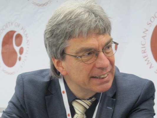
“The data I have presented show that the combination of nilotinib with this age-adapted chemotherapy is highly effective. We do have quite a reasonable overall survival estimate at 2 years of just more than 70%,” said Dr. Oliver Ottmann of Goethe University in Frankfurt, on behalf of colleagues in the European Working Group for Adult ALL (EWALL).
The study also shows that although some centers are reluctant to offer allogeneic stem cell transplantation (SCT) to older patients, it is still a viable treatment option in this population, Dr. Ottmann said at a briefing at the annual meeting of the American Society of Hematology.
Although older patients with newly diagnosed Philadelphia-positive (Ph+) ALL have a high complete hematologic response rate (CHR) with imatinib (Gleevec), they generally have a poor prognosis because of a high rate of relapse.
Because nilotinib, a potent inhibitor of the ABL kinase, has good efficacy in the chronic and accelerated phase of Ph+ chronic myeloid leukemia, the EWALL investigators initiated a study to evaluate it in combination with chemotherapy in the front-line setting.
Adults aged 55 years and older with ALL positive for the Philadelphia chromosome and/or BCR-ABL1 fusion who were treatment naive or had not received therapy other than corticosteroids, single-dose vincristine, or three doses of cyclophosphamide were eligible.
Details of the combination regimen are available online.
Briefly, following a prephase with dexamethasone and optional cyclophosphamide, patients receive nilotinib 400 mg twice daily starting with induction and continuously thereafter. During induction, nilotinib is given with intravenous injections of vincristine and dexamethasone for 4 weeks, followed by consolidation with nilotinib, methotrexate, asparaginase and cytarabine. Maintenance consists of nilotinib, 6-mercaptopurine, and methotrexate once weekly for 1 month then every other month, and dexamethasone and vincristine in 2 month intervals up to 24 months.
The data Dr. Ottmann reported come from an interim analysis of the ongoing study. As of August 2014, data on 47 of 56 patients was available for an efficacy analysis, As noted before, the rate of CHR was 87%, occurring in 41 of 47 patients. The treatment evoked a partial response or no response in 2 patients, and there was one death during the induction phase. Additionally, three patients discontinued therapy early and were not included in the assessment, but at least one had a complete response later on, Dr. Ottmann noted.
The median time to a complete response (CR) was 41 days, but CRs occurred as early as 25 days and as late as 62 days after the start of therapy. The remissions at the time of data cutoff appeared to be durable, but follow-up is still early, he said.
Overall survival at a median follow-up for all patients of 8.6 months was 72.7% at 30 months for patients who did not undergo SCT (allowed under the protocol), and 67.1% at 30 months for patients who underwent SCT. This difference was not significant, but only nine patients at the time of data cutoff had undergone transplantation.
“It will be interesting to see how this will proceed if the transplant-free patients will do as well as the others,” Dr. Ottmann said.
An analysis of molecular response by minimal residual disease (MRD) time point showed a significant further increase with the consolidation chemotherapy and kinase inhibitor, emphasizing that “continuing the treatment in this form emphasizes the depth of response. If we then look at the rate of MRD negativity using high quality assays, then a quarter of the patients have undetectable polymerase chain reaction during the consolidation cycles, and approximately 80% achieves something that we call a major molecular response,” he said.
Dr. Ottmann did not provide updated safety data, but at the time of the data cutoff, there had been 34 serious adverse events reported, 11 of which occurred during induction, 16 during consolidation, 6 during maintenance, and 1 following discontinuation. The most-common adverse events were infections and neutropenic fevers. Single serious adverse events included metabolic, cardiovascular, neurologic, renal, and hepatic events.
The trial is expected to be completed in the next few months.
SAN FRANCISCO– The study was small but encouraging: Among 47 older patients with newly diagnosed acute lymphoblastic leukemia positive for the Philadelphia chromosome, 41 had a complete hematologic response to a combination of chemotherapy and the targeted agent nilotinib (Tasigna), report investigators from a European consortium.
“The data I have presented show that the combination of nilotinib with this age-adapted chemotherapy is highly effective. We do have quite a reasonable overall survival estimate at 2 years of just more than 70%,” said Dr. Oliver Ottmann of Goethe University in Frankfurt, on behalf of colleagues in the European Working Group for Adult ALL (EWALL).
The study also shows that although some centers are reluctant to offer allogeneic stem cell transplantation (SCT) to older patients, it is still a viable treatment option in this population, Dr. Ottmann said at a briefing at the annual meeting of the American Society of Hematology.
Although older patients with newly diagnosed Philadelphia-positive (Ph+) ALL have a high complete hematologic response rate (CHR) with imatinib (Gleevec), they generally have a poor prognosis because of a high rate of relapse.
Because nilotinib, a potent inhibitor of the ABL kinase, has good efficacy in the chronic and accelerated phase of Ph+ chronic myeloid leukemia, the EWALL investigators initiated a study to evaluate it in combination with chemotherapy in the front-line setting.
Adults aged 55 years and older with ALL positive for the Philadelphia chromosome and/or BCR-ABL1 fusion who were treatment naive or had not received therapy other than corticosteroids, single-dose vincristine, or three doses of cyclophosphamide were eligible.
Details of the combination regimen are available online.
Briefly, following a prephase with dexamethasone and optional cyclophosphamide, patients receive nilotinib 400 mg twice daily starting with induction and continuously thereafter. During induction, nilotinib is given with intravenous injections of vincristine and dexamethasone for 4 weeks, followed by consolidation with nilotinib, methotrexate, asparaginase and cytarabine. Maintenance consists of nilotinib, 6-mercaptopurine, and methotrexate once weekly for 1 month then every other month, and dexamethasone and vincristine in 2 month intervals up to 24 months.
The data Dr. Ottmann reported come from an interim analysis of the ongoing study. As of August 2014, data on 47 of 56 patients was available for an efficacy analysis, As noted before, the rate of CHR was 87%, occurring in 41 of 47 patients. The treatment evoked a partial response or no response in 2 patients, and there was one death during the induction phase. Additionally, three patients discontinued therapy early and were not included in the assessment, but at least one had a complete response later on, Dr. Ottmann noted.
The median time to a complete response (CR) was 41 days, but CRs occurred as early as 25 days and as late as 62 days after the start of therapy. The remissions at the time of data cutoff appeared to be durable, but follow-up is still early, he said.
Overall survival at a median follow-up for all patients of 8.6 months was 72.7% at 30 months for patients who did not undergo SCT (allowed under the protocol), and 67.1% at 30 months for patients who underwent SCT. This difference was not significant, but only nine patients at the time of data cutoff had undergone transplantation.
“It will be interesting to see how this will proceed if the transplant-free patients will do as well as the others,” Dr. Ottmann said.
An analysis of molecular response by minimal residual disease (MRD) time point showed a significant further increase with the consolidation chemotherapy and kinase inhibitor, emphasizing that “continuing the treatment in this form emphasizes the depth of response. If we then look at the rate of MRD negativity using high quality assays, then a quarter of the patients have undetectable polymerase chain reaction during the consolidation cycles, and approximately 80% achieves something that we call a major molecular response,” he said.
Dr. Ottmann did not provide updated safety data, but at the time of the data cutoff, there had been 34 serious adverse events reported, 11 of which occurred during induction, 16 during consolidation, 6 during maintenance, and 1 following discontinuation. The most-common adverse events were infections and neutropenic fevers. Single serious adverse events included metabolic, cardiovascular, neurologic, renal, and hepatic events.
The trial is expected to be completed in the next few months.
Key clinical point: Combining a kinase inhibitor with an intensive chemotherapy regimen produces a high complete response rate in patients age 55 and older who have Ph+All.
Major finding: The complete hematologic response rate for the combination of chemotherapy and nilotinib was 87%.
Data source: Investigator initiated study in 56 patients with acute lymphoblastic leukemia positive for the Philadelphia chromosome or BCR/ABL fusion.
Disclosures: The study was sponsored by participating institution. Dr. Ottmann disclosed consultancy, honoraria, and research funding from Novartis, maker of nilotinib.

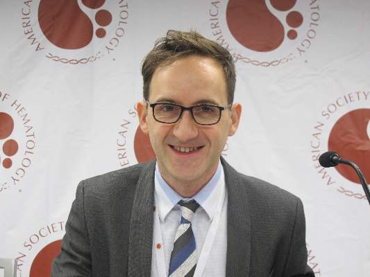
Sorafenib boosts event-free survival of adult AML
SAN FRANCISCO – Adding a kinase inhibitor to a standard regimen for acute myeloid leukemia can prolong event-free and relapse-free survival in young adult patients, but the effect on overall survival is still unclear, investigators reported.
In a randomized controlled trial, 3-year event-free survival (EFS), the primary endpoint, was 40% among patients treated with chemotherapy and sorafenib (Nexavar), compared with 22% for patients treated with chemotherapy and a placebo (P = .013). The median EFS was 21 months and 9 months, respectively, reported Dr. Christoph Röllig of University Hospital in Dresden, Germany.
Relapse-free survival (RFS) at 3 years was 56% in the sorafenib-treated group, compared with 38% in the placebo group (P = .017). The median RFS was not reached in the sorafenib group, vs. 23 months in the placebo group, Dr. Röllig reported at the annual meeting of the American Society of Hematology.
“These data constitute the first randomized evidence that actually kinase inhibitors work in AML. What we judge is that, according to evidence-based medicine principles, a comparatory trial would be desirable in order to establish sorafenib in AML treatment,” he said at a briefing prior to his presentation of the data in a plenary session.
Dr. Röllig and colleagues in 25 centers enrolled patients from 18 to 60 years with newly diagnosed AML. Of the 276 enrolled, 267 went on to receive study treatment: 134 were assigned to sorafenib and 133 to placebo. The study drug was given along a chemotherapy regimen consisting of two cycles of induction with daunorubicin and cytarabine followed by three cycles of high-dose cytarabine consolidation. Patients who did not have a response after the first cycle of daunorubicin induction underwent a second induction attempt with cytarabine and mitoxantrone.
The assigned study medication was given on days 10-19 of induction cycles one and two, from day 8 of each consolidation cycle until 3 days before the start of the next consolidation cycle, and as maintenance for 12 months after the end of consolidation.
As noted before, EFS in an intention-to-treat analysis censored for stem-cell therapy favored the sorafenib-treated patients, as did RFS. In addition, there was evidence to suggest a benefit trend toward prolonged RFS and overall survival with sorafenib among patients positive for the FLT3-ITD mutation, which has been shown to be sensitive to kinase inhibitors.
Patients in the sorafenib arm had significantly more episodes of fever, bleeding events, and the hand-foot syndrome, but there were no significant differences in other adverse events, Dr. Röllig said.
In an interview, he said that the investigators chose sorafenib because of its good track record and its efficacy against multiple kinases. His center is also involved in clinical trials exploring whether a different kinase inhibitor, quizartinib, has similar or better efficacy against AML.
The study was supported by Bayer. Dr. Röllig reported having no relevant disclosures.
Key clinical point: This study is the first randomized trial to show efficacy of kinase inhibition in AML.
Major finding: 3-year event-free survival was 40% among patients treated with chemotherapy and sorafenib, compared with 22% for patients treated with chemotherapy and a placebo.
Data source: Randomized controlled trial involving 267 adults.
Disclosures: The study was supported by Bayer. Dr. Röllig reported having no relevant disclosures.

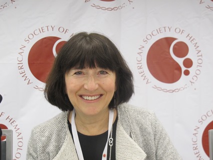
Young adults with ALL have better survival with pediatric regimens
SAN FRANCISCO– Teens and young adults don’t like being treated like children, but they should make an exception when it comes to acute lymphoblastic leukemia therapy, because pediatric ALL regimens are associated with significantly better event-free and overall survival among patients with ALL from the ages of 16 to 40 years.
Those findings come from a clinical trial 14 years in the making, in which 296 adolescents and young adults (AYA) with ALL were treated with an intensive pediatric chemotherapy combination regiment rather than a less-intensive adult regimen. At 2-year follow-up, the rate of overall survival (OS) was 78%, with the median overall survival not yet reached, and the event-free survival (EFS) rate was 66%, reported Dr. Wendy Stock from the University of Chicago Medical Center.
In contrast, EFS rates among AYA treated with adult regimens have historically ranged from 35-40%, Dr. Stock said at a press briefing at the annual meeting of the American Society of Hematology.
“These data really started 14 years ago at this ASH meeting when we presented data showing that young adults ages 16 to 20 who were treated on adult cooperative group studies in the United States fared much worse than the same age group who were treated on pediatric studies,” she said.
In 2008, Dr. Stock and her colleagues published a study (Blood 2008;112:1646-54) showing that AYAs treated under Children’s Cancer Group (CCG) protocols had an overall survival rate at 7 years of 67% and an EFS rate of 63%. In contrast, AYAs treated under Cancer and Leukemia Group B (CALGB) protocols had an OS of 46% and EFS of only 34%. The risk for worse outcomes was approximately twofold among adolescents treated with adult regimens, compared with those treated with pediatric regimens. The findings were similar in studies from France, the United Kingdom, and the Netherlands, Dr. Stock noted.
The investigators determined at that time that, under the CCG regimens, the teen and young adult patients received earlier and more intensive central nervous system prophylaxis and higher doses of nonmyelosuppressive drugs, especially glucocorticoids, vincristine, and pegylated asparignase, while those on the CALGB regimens received higher doses of myelosuppressive agents such as anthracyclines.
Because their original findings came from a retrospective study, the investigators decided to launch a prospective study,US Intergroup trial C10403, designed to evaluate outcomes among patients with ALL from the ages of 16-40 years when they were treated with a pediatric regimen by adult hematologists/oncologists in the cooperative group setting.
A total of 296 eligible patients with a median age of 25 years were enrolled. The patients had newly diagnosed ALL of T-cell or B-cell lineage; patients with Burkitt’s type ALL or ALL positive for the Philadelphia chromosome were excluded. The patients were treated with a regimen identical to the Capizzi methotrexate arm of the Children’s Oncology Group AALL0232 study. The regimen consisted of four intensive courses: remission induction, remission consolidation, interim maintenance and delayed intensification, and prolonged maintenance therapy. Patients who had an M2 marrow response after remission induction (more than 5% but less than 25% lymphoblasts) received an extended remission induction course of therapy.
As noted before, the EFS rate was 66% and the median EFS duration was 59 months. The 2-year overall survival rate was 79%. EFS rates were similar between patients with B-cell lineage (65%) and T-cell lineage (68%) ALL, and there were no significant differences in EFS or OS by sex or by age.
There were five (2%) treatment-related deaths during protocol therapy, including two cases of liver failure, both occurring during induction; two infections (one in the induction phase and one in the consolidation phase); and one ventricular arrhythmia (during induction). Treatment toxicities in general were similar to those seen in the standard therapy of the COG AALL0232 trial, although patients in the current study had an increase in risk for thrombosis and early hyperbilirubinemia.
“Our outcomes are similar to other prospective international studies which apply pediatric regimens to the young adult population of acute lymphoblastic leukemia,” Dr. Stock said.
In analyses of biological factors that affect outcome, the investigators found that white blood cell counts above 30,000/uL were associated with worse EFS and OS, and that the presence of a BCR-ABL1-like signature and overexpression of the gene CRLF2 were common and associated with significantly worse survival.
The investigators plan to use the study as a basis for future studies incorporating target antibodies and kinase inhibitors in an attempt to improve survival further by eradicating minimal residual disease, Dr. Stock said.
Key clinical point: Adolescents and young adults with acute lymphoblastic leukemia should be treated with a pediatric rather than an adult ALL regimen.
Major finding: Overall survival was 78% and event-free survival rate was 66%, compared with 46% and 34% for young adults treated with adult-style regimens in the past.
Data source: Prospective trial with 296 patients from the ages of 16-40 years with ALL.
Disclosures: The study is supported by the National Institutes of Health. Dr. Stock disclosed serving as an advisor and receiving research funding from Sigma-Tau Pharmaceuticals.

Early Discoid Lupus Onset Protective Against Nephritis
BOSTON - Among 839 patients with systemic lupus erythematosus (SLE) and no evidence of lupus nephritis at the time of diagnosis, the presence of discoid lupus erythematosus (DLE) at SLE onset was associated with a 64% reduction in risk for developing lupus nephritis.
“Our data suggest that while SLE patients who have DLE preceding the diagnosis of SLE are at low risk of further lupus nephritis, SLE patients without discoid lupus at onset should be monitored for early diagnosis of lupus nephritis and adequate treatment,” said Dr. Guillermo Javier Pons-Estel from the department of autoimmune disease, L’Institut Clínic de Medicina i Dermatologia, Hospital Clinic, in Barcelona, Spain.
He reported results of a study comparing lupus nephritis rates in patients enrolled in the GLADEL (Grupo di Latino Americano De Estudio de Lupus) longitudinal study of Latin American patients with SLE.
DLE is characterized by the appearance of scarring inflammatory plaques that heal but leave central scars, atrophy, and dyspigmentation. It occurs at onset in approximately 5%-11% of all patients with SLE, and after a delay of 3-5 years from onset in a small proportion of patients, he said at the annual meeting of the American College of Rheumatology.
There is conflicting evidence for DLE protecting against nephritis in patients with SLE, Dr. Pons-Estel said. He pointed to a study published in 2012 of a multiethnic cohort of patients with SLE in which the authors found that patients with DLE were less likely to have severe renal involvement or end-stage renal disease (ESRD) (Arthritis Care Res. 2012;64:704-12). On the other hand, a 2013 study of 1,043 patients, 117 of whom had DLE, found no association, either positive or negative, between DLE and either lupus nephritis or ESRD (J. Am. Acad. Dermatol. 2013;69:19-24).
To further explore a possible link between time of DLE onset and risk for lupus nephritis, the investigators looked at a sample of patients with SLE from 34 centers in nine Central and South American nations.
They defined lupus nephritis as at least one of the following: proteinuria on two or more occasions or the presence of cellular casts; renal biopsy compatible with lupus nephritis histopathology class II-IV according to World Health Organization criteria; serum creatinine greater than 2.0 mg/dL; and/or ESRD.
The main predictor in the study was DLE onset, and the investigators also included in their multivariate models the potential confounding variables of sex, ethnicity, socioeconomic status, delay in diagnosis, insurance status, and level of education. They also analyzed the contribution of potential clinical confounding variables that included disease activity as assessed by the SLE Disease Activity Index, Systemic Lupus International Collaborating Clinics/ACR Damage Index, and medications such as antimalarial agents, immunosuppressants, methylprednisolone pulse, and oral glucocorticoids at doses greater than 60 mg per day.
In Cox univariate analysis, the only factors significantly associated with lower risk for lupus nephritis included DLE onset before SLE diagnosis (hazard ratio [HR], 0.37; P = .0007), age at diagnosis (HR, 0.91; P = .0013), and years of education (HR, 0.91; P = .0360).
In multivariate proportional regression analysis, the only significant predictors for lower risk were DLE onset in combination with either sociodemographic confounders (HR, 0.37; P = .0008), treatment confounders (HR, 0.37; P = .0009), or clinical disease activity assessments (HR, 0.37; P = .0007).
The association held up in a final prediction multivariate model in which the investigators controlled for sociodemographics, age at diagnosis, ethnicity (Caucasian vs. other), and clinical assessment at diagnosis. In this model, the HR for DLE onset was 0.36 (P = .0006; 98.2% reliability).
The cumulative incidence of lupus nephritis at 1 and 5 years for patients with DLE at or before SLE diagnosis were 7% and 16%, respectively, compared with 16% and 37% for patients with no DLE at diagnosis.
Dr. Pons-Estel acknowledged that the study was limited by a lack of data on skin activity and extension of dermatologic lesions and by missing information on lupus nephritis class for more than two-thirds of patients with the condition.
“Our data point out the need for investigations to unravel immune and environmental factors implicated in the pathways associated with the protective role of DLE onset against lupus nephritis,” he said.
Session co-moderator Dr. Cynthia Aranow, a rheumatologist at the Feinstein Institute for Medical Research in Manhasset, N.Y., who was not involved in the study, questioned whether the protective effective seen with DLE could have come from the antimalarial agent hydroxychloroquine, and whether patients with DLE might be more compliant with the medication.
Dr. Ponss-Estel replied that more than 75% of the patients in the study used an antimalarial agent, but that the investigators found no significant differences in nephritis between patients with DLE who were taking hydroxychloroquine and those who were not.
The study was funded by member institutions. Dr. Pons-Estel and Dr. Aranow reported having no relevant disclosures.
AT THE ACR ANNUAL MEETING
Early discoid lupus onset protective against nephritis
BOSTON - Among 839 patients with systemic lupus erythematosus (SLE) and no evidence of lupus nephritis at the time of diagnosis, the presence of discoid lupus erythematosus (DLE) at SLE onset was associated with a 64% reduction in risk for developing lupus nephritis.
“Our data suggest that while SLE patients who have DLE preceding the diagnosis of SLE are at low risk of further lupus nephritis, SLE patients without discoid lupus at onset should be monitored for early diagnosis of lupus nephritis and adequate treatment,” said Dr. Guillermo Javier Pons-Estel from the department of autoimmune disease, L’Institut Clínic de Medicina i Dermatologia, Hospital Clinic, in Barcelona, Spain.
He reported results of a study comparing lupus nephritis rates in patients enrolled in the GLADEL (Grupo di Latino Americano De Estudio de Lupus) longitudinal study of Latin American patients with SLE.
DLE is characterized by the appearance of scarring inflammatory plaques that heal but leave central scars, atrophy, and dyspigmentation. It occurs at onset in approximately 5%-11% of all patients with SLE, and after a delay of 3-5 years from onset in a small proportion of patients, he said at the annual meeting of the American College of Rheumatology.
There is conflicting evidence for DLE protecting against nephritis in patients with SLE, Dr. Pons-Estel said. He pointed to a study published in 2012 of a multiethnic cohort of patients with SLE in which the authors found that patients with DLE were less likely to have severe renal involvement or end-stage renal disease (ESRD) (Arthritis Care Res. 2012;64:704-12). On the other hand, a 2013 study of 1,043 patients, 117 of whom had DLE, found no association, either positive or negative, between DLE and either lupus nephritis or ESRD (J. Am. Acad. Dermatol. 2013;69:19-24).
To further explore a possible link between time of DLE onset and risk for lupus nephritis, the investigators looked at a sample of patients with SLE from 34 centers in nine Central and South American nations.
They defined lupus nephritis as at least one of the following: proteinuria on two or more occasions or the presence of cellular casts; renal biopsy compatible with lupus nephritis histopathology class II-IV according to World Health Organization criteria; serum creatinine greater than 2.0 mg/dL; and/or ESRD.
The main predictor in the study was DLE onset, and the investigators also included in their multivariate models the potential confounding variables of sex, ethnicity, socioeconomic status, delay in diagnosis, insurance status, and level of education. They also analyzed the contribution of potential clinical confounding variables that included disease activity as assessed by the SLE Disease Activity Index, Systemic Lupus International Collaborating Clinics/ACR Damage Index, and medications such as antimalarial agents, immunosuppressants, methylprednisolone pulse, and oral glucocorticoids at doses greater than 60 mg per day.
In Cox univariate analysis, the only factors significantly associated with lower risk for lupus nephritis included DLE onset before SLE diagnosis (hazard ratio [HR], 0.37; P = .0007), age at diagnosis (HR, 0.91; P = .0013), and years of education (HR, 0.91; P = .0360).
In multivariate proportional regression analysis, the only significant predictors for lower risk were DLE onset in combination with either sociodemographic confounders (HR, 0.37; P = .0008), treatment confounders (HR, 0.37; P = .0009), or clinical disease activity assessments (HR, 0.37; P = .0007).
The association held up in a final prediction multivariate model in which the investigators controlled for sociodemographics, age at diagnosis, ethnicity (Caucasian vs. other), and clinical assessment at diagnosis. In this model, the HR for DLE onset was 0.36 (P = .0006; 98.2% reliability).
The cumulative incidence of lupus nephritis at 1 and 5 years for patients with DLE at or before SLE diagnosis were 7% and 16%, respectively, compared with 16% and 37% for patients with no DLE at diagnosis.
Dr. Pons-Estel acknowledged that the study was limited by a lack of data on skin activity and extension of dermatologic lesions and by missing information on lupus nephritis class for more than two-thirds of patients with the condition.
“Our data point out the need for investigations to unravel immune and environmental factors implicated in the pathways associated with the protective role of DLE onset against lupus nephritis,” he said.
Session co-moderator Dr. Cynthia Aranow, a rheumatologist at the Feinstein Institute for Medical Research in Manhasset, N.Y., who was not involved in the study, questioned whether the protective effective seen with DLE could have come from the antimalarial agent hydroxychloroquine, and whether patients with DLE might be more compliant with the medication.
Dr. Ponss-Estel replied that more than 75% of the patients in the study used an antimalarial agent, but that the investigators found no significant differences in nephritis between patients with DLE who were taking hydroxychloroquine and those who were not.
The study was funded by member institutions. Dr. Pons-Estel and Dr. Aranow reported having no relevant disclosures.
AT THE ACR ANNUAL MEETING
Key clinical point: Discoid lupus erythematosus diagnosed at or before a systemic lupus diagnosis predicts lower risk of lupus nephritis.
Major finding: The hazard ratio for developing subsequent lupus nephritis in patients with early discoid lupus was 0.36 (P = .0006).
Data source: A longitudinal cohort of 839 Latin American patients with SLE and no evidence of lupus nephritis at the time of diagnosis.
Disclosures: The study was funded by member institutions. Dr. Pons-Estel and Dr. Aranow reported having no relevant disclosures.
PD-1 checkpoint inhibitors show mettle against relapsed Hodgkin’s lymphoma
SAN FRANCISCO– PD-1 checkpoint inhibitors, which have shown remarkable efficacy against advanced malignant melanoma, appear to hold similar promise in the treatment of relapsed or refractory Hodgkin’s lymphoma, results from two early studies suggest.
In a phase I study, the PD-1 blocking antibody nivolumab produced an 87% response rate in 23 heavily pre-treated patients with relapsed Hodgkin’s lymphoma (HL). In a separate phase Ib study, pembrolizumab, which blocks the PD-1 and PD-2 ligands, produced a 66% overall response rate, 21% complete remission rate, and 86% clinical benefit rate among 29 patients with HL for whom therapy with brentuximab vedotin (Adcetris) had failed.
The studies were presented at a media briefing prior to the presentation of data in oral sessions at the annual meeting of the American Society of Hematology.
“Classical Hodgkin lymphoma appears to be a tumor with genetically determined vulnerability to PD-1 blockade. We hope that PD-1 blockade in the future can become an important part of the treatment of patients with Hodgkin lymphoma,” said Dr. Phillipe Armand from the Dana-Farber Cancer Institute in Boston, an investigator for the nivolumab study.

Evidence from preclinical studies suggests that the Reed-Sternberg malignant cells characteristic of HL may use the PD-1 (programmed death 1) pathway to evade detection by immune cells, as suggested by pathologic studies showing the cells surrounded by an extensive but ineffective infiltrate of inflammatory cells.
“We’ve wondered for a long time how Hodgkin lymphoma could attract such a brisk immune response and yet have this immune response fail to kill the tumor,” he said.
Genetic Achilles heel
Genetic analyses had shown that HL frequently has a mutation that results in amplification of a region on chromosome 9 (9p24.1) which leads to increased expression of PD-1 ligands 1 and 2, and leads to a downregulation or weakening of the immune response. The mutation appears to work through the Janus kinase (JAK)-signal transducer and activator transcription (STAT) signalling. These findings suggested to researchers that classical HL has a genetically driven and, ideally, targetable dependence on the PD-1 pathway for survival, Dr. Armand explained.
To test this idea, he and colleagues studied 23 patients with relapsed or refractory HL that had been heavily pre-treated who were part of an independent expansion cohort of a study of nivolumab in hematologic malignancies. Of these patients, 78% were enrolled after a relapse following autologous stem cell transplantation, and 22% after treatment with brentuximab vedotin had failed.
The patients received nivolumab 3 mg/kg every 2 weeks until they had either a complete response, tumor progression, or excessive side effects. In all, 20 of the 23 patients (87%) had an objective response to the single-agent therapy, including 4 (17%) complete responses and 16 (70%) partial responses. The remaining three patients (13%) had stable disease.
The longest time on study at the data cutoff point was 72 weeks. Among all responders, 60% had a response by 8 weeks of therapy, 48% are ongoing, and 43% of patients are still on treatment.
Drug-related adverse events were reported in 18 patients, most commonly rash and decreased platelet count. Five patients had grade 3 events. There were no drug-related grade 4 events or deaths.
In an editorial accompanying the study, which was also published online in The New England Journal of Medicine, Dr. Mario Sznoll and Dr. Dan L. Longo from the Yale University School of Medicine in New Haven, Connecticut write that “with recent data showing impressive clinical activity of PD-1 or PD-L1 antagonists in subgroups of patients with a variety of different cancers, the critical and foundational role of immune interventions in cancer treatment is no longer deniable,” (NEJM, Dec. 6, 2014 [DOI: 10.1056/NEJMoa1411087]).
Pembrolizumab trial
Dr. Craig H. Moskowitz from Memorial Sloan-Kettering Cancer Center in New York City discussed results of the second study, dubbed KEYNOTE-013 (A Phase Ib Multi-Cohort Trial of MK-3475 in Subjects With Hematologic Malignancies).
In this study, patients with HL who were not transplant eligible or for whom transplant had failed and who either had a relapse or were refractory to therapy with brentuximab vedotin received 19 mg/kg IV infusion of pembrolizumab every 2 weeks until complete response, partial response/stable disease, or disease progression.

Of the 31 patients enrolled, 29 were available for the analysis. As of the data cutoff in November 2014, 6 patients (21%) had achieved a complete remission, and 13 (45%) had a partial response, for an overall response rate of 66%. The median time to response was 12 weeks, and as of the data cutoff 17 of 19 patients had ongoing responses. The median response duration has not yet been reached. An additional 6 patients (21%) had stable disease, leading to an overall clinical benefit rate (responses plus stable disease) of 86%.
The patients generally tolerated the drug well. There were 4 treatment-related adverse events in 3 patients, including axillary pain, hypoxia, joint swelling, and pneumonitis. There were no grade 4 treatment-related events or deaths.
Of the tumor samples evaluable, all expressed PD-L1, supporting the rationale for PD-1 blockade in this population, Dr. Moskowitz said.
The results of both his and Dr. Armand’s study support the continued development of PD-1 inhibitors in various subsets of patients with classical Hodgkin’s lymphoma, he said.
SAN FRANCISCO– PD-1 checkpoint inhibitors, which have shown remarkable efficacy against advanced malignant melanoma, appear to hold similar promise in the treatment of relapsed or refractory Hodgkin’s lymphoma, results from two early studies suggest.
In a phase I study, the PD-1 blocking antibody nivolumab produced an 87% response rate in 23 heavily pre-treated patients with relapsed Hodgkin’s lymphoma (HL). In a separate phase Ib study, pembrolizumab, which blocks the PD-1 and PD-2 ligands, produced a 66% overall response rate, 21% complete remission rate, and 86% clinical benefit rate among 29 patients with HL for whom therapy with brentuximab vedotin (Adcetris) had failed.
The studies were presented at a media briefing prior to the presentation of data in oral sessions at the annual meeting of the American Society of Hematology.
“Classical Hodgkin lymphoma appears to be a tumor with genetically determined vulnerability to PD-1 blockade. We hope that PD-1 blockade in the future can become an important part of the treatment of patients with Hodgkin lymphoma,” said Dr. Phillipe Armand from the Dana-Farber Cancer Institute in Boston, an investigator for the nivolumab study.
Evidence from preclinical studies suggests that the Reed-Sternberg malignant cells characteristic of HL may use the PD-1 (programmed death 1) pathway to evade detection by immune cells, as suggested by pathologic studies showing the cells surrounded by an extensive but ineffective infiltrate of inflammatory cells.
“We’ve wondered for a long time how Hodgkin lymphoma could attract such a brisk immune response and yet have this immune response fail to kill the tumor,” he said.
Genetic Achilles heel
Genetic analyses had shown that HL frequently has a mutation that results in amplification of a region on chromosome 9 (9p24.1) which leads to increased expression of PD-1 ligands 1 and 2, and leads to a downregulation or weakening of the immune response. The mutation appears to work through the Janus kinase (JAK)-signal transducer and activator transcription (STAT) signalling. These findings suggested to researchers that classical HL has a genetically driven and, ideally, targetable dependence on the PD-1 pathway for survival, Dr. Armand explained.
To test this idea, he and colleagues studied 23 patients with relapsed or refractory HL that had been heavily pre-treated who were part of an independent expansion cohort of a study of nivolumab in hematologic malignancies. Of these patients, 78% were enrolled after a relapse following autologous stem cell transplantation, and 22% after treatment with brentuximab vedotin had failed.
The patients received nivolumab 3 mg/kg every 2 weeks until they had either a complete response, tumor progression, or excessive side effects. In all, 20 of the 23 patients (87%) had an objective response to the single-agent therapy, including 4 (17%) complete responses and 16 (70%) partial responses. The remaining three patients (13%) had stable disease.
The longest time on study at the data cutoff point was 72 weeks. Among all responders, 60% had a response by 8 weeks of therapy, 48% are ongoing, and 43% of patients are still on treatment.
Drug-related adverse events were reported in 18 patients, most commonly rash and decreased platelet count. Five patients had grade 3 events. There were no drug-related grade 4 events or deaths.
In an editorial accompanying the study, which was also published online in The New England Journal of Medicine, Dr. Mario Sznoll and Dr. Dan L. Longo from the Yale University School of Medicine in New Haven, Connecticut write that “with recent data showing impressive clinical activity of PD-1 or PD-L1 antagonists in subgroups of patients with a variety of different cancers, the critical and foundational role of immune interventions in cancer treatment is no longer deniable,” (NEJM, Dec. 6, 2014 [DOI: 10.1056/NEJMoa1411087]).
Pembrolizumab trial
Dr. Craig H. Moskowitz from Memorial Sloan-Kettering Cancer Center in New York City discussed results of the second study, dubbed KEYNOTE-013 (A Phase Ib Multi-Cohort Trial of MK-3475 in Subjects With Hematologic Malignancies).
In this study, patients with HL who were not transplant eligible or for whom transplant had failed and who either had a relapse or were refractory to therapy with brentuximab vedotin received 19 mg/kg IV infusion of pembrolizumab every 2 weeks until complete response, partial response/stable disease, or disease progression.
Of the 31 patients enrolled, 29 were available for the analysis. As of the data cutoff in November 2014, 6 patients (21%) had achieved a complete remission, and 13 (45%) had a partial response, for an overall response rate of 66%. The median time to response was 12 weeks, and as of the data cutoff 17 of 19 patients had ongoing responses. The median response duration has not yet been reached. An additional 6 patients (21%) had stable disease, leading to an overall clinical benefit rate (responses plus stable disease) of 86%.
The patients generally tolerated the drug well. There were 4 treatment-related adverse events in 3 patients, including axillary pain, hypoxia, joint swelling, and pneumonitis. There were no grade 4 treatment-related events or deaths.
Of the tumor samples evaluable, all expressed PD-L1, supporting the rationale for PD-1 blockade in this population, Dr. Moskowitz said.
The results of both his and Dr. Armand’s study support the continued development of PD-1 inhibitors in various subsets of patients with classical Hodgkin’s lymphoma, he said.
Key clinical point: PD-1 checkpoint inhibition appears to be an effective strategy against treatment-refractory Hodgkin’s lymphoma.
Major finding: Nivolumab produced an 87% objective response rate and pembrolizumab a 66% response rate in patients with heavily pre-treated Hodgkin’s lymphoma.
Data source: A phase I study with 23 patients and a phase Ib study with 29 patients with relapsed or refractory Hodgkin’s lymphoma.
Disclosures: Dr. Armand’s study is supported by Bristol-Myers Squibb. He reported grant support from Bristol-Myers Squibb during the conduct of the study and personal fees from Merck outside the study. Dr. Moskowitz’ study is supported by Merck. He reported receiving research funding from the company. Dr. Sznol reported personal fees from Bristol-Myers Squibb, Dr. Longo reported no relevant disclosures.
ASPIRE: Carfilzomib-Len-Dex ‘a new standard of care’ for relapsed multiple myeloma
SAN FRANCISCO – New standards of care are hard to come by in the treatment of relapsed/refractory multiple myeloma, but interim results of a phase III trial of the combination of carfilzomib, lenalidomide, and dexamethasone suggest that it might just fit the bill, investigators say.
In a randomized controlled trial comparing the combination, the comination, labeled KRd (the K is for carfilzomib’s tradename, Kyrpolis), was associated with significantly better progression-free survival (PFS) and a trend toward better overall survival than lenalidomide (Revlimid) and dexamethasone combined (RD), reported Dr. A. Keith Stewart, principal investigator and dean for research at the Mayo Clinic in Scottsdale, Arizona, at the annual meeting of the American Society of Hematology.
“Dr. Stewart’s study will, I think, establish a new standard of care in this patient population,” commented Dr. Brad Kahl of the University of Wisconsin School of Medicine and Public Health in Madison, who moderated a briefing where the data were presented. They were published simultaneously online in the New England Journal of Medicine (NEJM, Dec. 6, 2014 [DOI: 10.1056/NEJMoa1411321]).
Among 792 patients with relapsed/refractory multiple myeloma, PFS with patients randomized to receive KRd was 26.3 months, compared with 17.6 months for patients randomized to RD. The hazard ratio for progression or death with KRd was 0.69 (P = .0001, crossing the pre-specified stopping boundary). Median overall survival had not been reached for either group. Kaplan-Meier 24 month overall survival rates were 73.3% and 65.0%, respec-tively, trending in favor of KRd, but with a P value (.04) that did not meet the pre-specified stopping boundary for survival (P = .005).
“In this same population of patients – this is patients who have relapsed one, two or three times – the best result ever reported before with any combina-tion of chemotherapy was about 19 months, with a very similar cocktail of bortezomib, thalidomide and dexamethasone,” Dr. Stewart said at the briefing.
The KRd combination also appeared to be safe.
“Despite the fact that we added a third drug and patients were on treatment significantly longer, there was a fairly well balanced ratio of patients who had to discontinue treatment due to side effects. It’s important to note that cardiac and renal events which have been reported in some studies of heavily pre-treated patients [with carfilzomib] in the past, were marginally higher in the three-drug regimen, but overall were very consistent or even lower than had previously been reported [with single-agent carilzomib],” Dr. Stewart said.
Carfilzomib is an epoxyketone proteasome inhibitor that binds selectively to the constitutive proteasome and immunoproteasome. Unlike the first-in-class proteasome inhibitor, bortezomib (Velcade), carfilzomib irreversibly binds to and disables its targets, Dr. Stewart said in an interview.
In the ASPIRE (Carfilzomib, Lenalidomide, and Dexamethasone versus Lenalidomide and Dexamethasone for the Treatment of Patients with Relapsed Multiple Myeloma) trial, investigators enrolled 792 adults with relapsed multiple myeloma from 20 countries in North America, Europe, and the Middle East. The patients, who had previously received 1-3 prior lines of therapy, were randomly assigned to receive either KRd (carfilzomib 20 mg/m2 on days 1 and 2 of the first 28-day cycle, then 27 mg/m2 for days 8,9,15, and 16, and all days of subsequent cycles), oral lenolidamide 25 mg days 1-21, and oral dexamethasone days 1, 8, 15 and 22 of each cycle; or RD, which consisted of lenalidomide and dexamethasone in the same doses and on the same schedule as in KRd.
At the time of the data cutoff for the interim analysis in June 2014, 118 of 396 patients assigned to KRd were still on treatment, as were 86 of 396 in the RD group.
At the time of the analysis, 431 PFS events had been documented, and the study met its primary endpoint of superior PFS with the addition of carfilzomib.
Common adverse events, including diarrhea, cough, fever and hypertension were reported more frequently in the KRd group.
Remissions were more durable among patients who received carfilzomib, and these patients reported higher quality-of-life scores on the QLQ-C30 Global Health Status and Quality of Life scale, Dr. Stewart noted.
AT ASH 2014
Key clinical point: Adding the proteasome inhibitor carfilzomib to lenalidomide and dexamethasone resulted in a significantly better PFS in patients with relapsed/refractory multiple myeloma.
Major finding: Progresion-free survival was 26.3 months for the 3-drug combo compared with 17.6 months for len-dex alone.
Data source: Phase III open-label randomized controlled trial in 792 patients with relapsed/refractory multiple myeloma.
Disclosures: The study was supported by Onyx Pharmaceuticals. Dr. Stewart reports support from Onyx during the conduct of the study, and grant support from Onyx and personal fees from Celgene outside the study.
Few psoriatic arthritis patients achieve minimal disease activity on methotrexate
BOSTON – It’s time to test whether methotrexate is really up to snuff as a first-line therapy for psoriatic arthritis, Canadian investigators say.
They base that recommendation on findings from a retrospective study showing that fewer than 18% of patients with psoriatic arthritis treated with methotrexate achieved minimal disease activity (MDA) after 6 months.

They also found evidence to suggest that physicians may overestimate the benefits of methotrexate for psoriatic arthritis.
“Physician-dependent measures reveal good response to methotrexate at 6 months, but patient-reported measures are less responsive, possibly due to side effects, back pain, disease, [and/or] disability,” said Dr. Barry J. Sheane of the division of rheumatology in the department of medicine at the University of Toronto.
The data on the effects of methotrexate in psoriatic arthritis are considerably less impressive than are those seen with tumor necrosis factor–alpha inhibitors (TNFi), Dr. Sheane said. For example, at 24 weeks, 39% of patients treated with adalimumab (Humira) and 52% treated with infliximab (Remicade) had achieved MDA, he noted.
“We suggest that a randomized controlled trial of methotrexate compared to a TNF inhibitor is warranted,” Dr. Sheane said at the annual meeting of the American College of Rheumatology.
He and his colleagues reviewed records on 204 consecutive patients treated for psoriatic arthritis with methotrexate from January 2004 through April 2014. The patients, who had a mean duration of psoriatic arthritis of 6.2 years, were all naive to biological antirheumatic drugs and were initiating methotrexate therapy.
Of the 204 patients, 167 had data sufficient for a 6-month analysis.
The investigators defined MDA after 6 months on methotrexate, the primary endpoint, as the presence of at least five out of the following seven domains: 0-1 tender joints, 0-1 swollen joints, Psoriasis Area Severity Index (PASI) 1 or less or body surface area involved 3% or less, 0-1 tender entheseal points, Health Assessment Questionnaire (HAQ) score of 0.5 or less, patient global disease activity Visual Analog Scale score of 20 or lower, and patient pain Visual Analog Scale score of 15 or lower.
They found that 17.4% of patients (29 of 167) met the physician-rated criteria for MDA at 6 months, even though 82.6% of the patients (138) had a PASI score of 1 or lower, and 58.1% (97) had a swollen joint count of 1 or fewer.
When the patients were asked for their assessment of global disease activity, 13.2% (22) rated disease activity with a prespecified score of 20 or lower. Only 12% of patients (20) reported an HAQ score of 0.5 or less.
The mean methotrexate dose was similar between patients who achieved MDA (17.8 mg/week) and those who did not (17.3 mg/week). The median dose was 17.5 mg/week in each group.
After controlling for sex, baseline sacroiliitis, and duration of psoriasis and psoriatic arthritis at the start of methotrexate in a multivariate model, the authors found that only dactylitis, inflammatory back pain, and mechanical back pain were significantly associated with a lower probability of patients reaching MDA. The variables that did not prove to be associated with achieving MDA after 6 months on methotrexate included erythrocyte sedimentation rate, presence of nail disease, number of clinically damaged joints, and body mass index.
The study was internally funded. Dr. Sheane reported having no relevant disclosures.
BOSTON – It’s time to test whether methotrexate is really up to snuff as a first-line therapy for psoriatic arthritis, Canadian investigators say.
They base that recommendation on findings from a retrospective study showing that fewer than 18% of patients with psoriatic arthritis treated with methotrexate achieved minimal disease activity (MDA) after 6 months.
They also found evidence to suggest that physicians may overestimate the benefits of methotrexate for psoriatic arthritis.
“Physician-dependent measures reveal good response to methotrexate at 6 months, but patient-reported measures are less responsive, possibly due to side effects, back pain, disease, [and/or] disability,” said Dr. Barry J. Sheane of the division of rheumatology in the department of medicine at the University of Toronto.
The data on the effects of methotrexate in psoriatic arthritis are considerably less impressive than are those seen with tumor necrosis factor–alpha inhibitors (TNFi), Dr. Sheane said. For example, at 24 weeks, 39% of patients treated with adalimumab (Humira) and 52% treated with infliximab (Remicade) had achieved MDA, he noted.
“We suggest that a randomized controlled trial of methotrexate compared to a TNF inhibitor is warranted,” Dr. Sheane said at the annual meeting of the American College of Rheumatology.
He and his colleagues reviewed records on 204 consecutive patients treated for psoriatic arthritis with methotrexate from January 2004 through April 2014. The patients, who had a mean duration of psoriatic arthritis of 6.2 years, were all naive to biological antirheumatic drugs and were initiating methotrexate therapy.
Of the 204 patients, 167 had data sufficient for a 6-month analysis.
The investigators defined MDA after 6 months on methotrexate, the primary endpoint, as the presence of at least five out of the following seven domains: 0-1 tender joints, 0-1 swollen joints, Psoriasis Area Severity Index (PASI) 1 or less or body surface area involved 3% or less, 0-1 tender entheseal points, Health Assessment Questionnaire (HAQ) score of 0.5 or less, patient global disease activity Visual Analog Scale score of 20 or lower, and patient pain Visual Analog Scale score of 15 or lower.
They found that 17.4% of patients (29 of 167) met the physician-rated criteria for MDA at 6 months, even though 82.6% of the patients (138) had a PASI score of 1 or lower, and 58.1% (97) had a swollen joint count of 1 or fewer.
When the patients were asked for their assessment of global disease activity, 13.2% (22) rated disease activity with a prespecified score of 20 or lower. Only 12% of patients (20) reported an HAQ score of 0.5 or less.
The mean methotrexate dose was similar between patients who achieved MDA (17.8 mg/week) and those who did not (17.3 mg/week). The median dose was 17.5 mg/week in each group.
After controlling for sex, baseline sacroiliitis, and duration of psoriasis and psoriatic arthritis at the start of methotrexate in a multivariate model, the authors found that only dactylitis, inflammatory back pain, and mechanical back pain were significantly associated with a lower probability of patients reaching MDA. The variables that did not prove to be associated with achieving MDA after 6 months on methotrexate included erythrocyte sedimentation rate, presence of nail disease, number of clinically damaged joints, and body mass index.
The study was internally funded. Dr. Sheane reported having no relevant disclosures.
AT THE ACR ANNUAL MEETING
Key clinical point: It may be time to rethink methotrexate’s role as first-line therapy for psoriatic arthritis.
Major finding: Only 17.4% of patients with psoriatic arthritis treated with methotrexate achieved minimal disease activity at 6 months.
Data source: Retrospective study of 204 patients, 167 of whom had data sufficient for an efficacy and dosing analysis at 6 months.
Disclosures: The study was internally funded. Dr. Sheane reported having no relevant disclosures.

Tapering TNFi’s safe and much cheaper in RA
BOSTON – Tapering but perhaps not completely stopping tumor necrosis factor inhibitors can result in good control of rheumatoid arthritis with considerable cost savings, according to investigators in two studies.
Dr. Noortje van Herwaarden and colleagues from Sint Maartenskliniek in Nijmegen, the Netherlands, conducted a randomized, open-label study of dose-reduction vs. usual care in 180 patients with stable, well-controlled rheumatoid arthritis (RA) on a tumor necrosis factor inhibitor (TNFi) – either adalimumab (Humira) or etanercept (Enbrel) – plus in some cases a stable disease-modifying antirheumatic drug (DMARD) or glucocorticoid.

“This is the first study to demonstrate that disease activity–guided tapering of adalimumab and etanercept in RA patients doing well is noninferior to usual care regarding major flare,” Dr. van Herwaarden said at the annual meeting of the American College of Rheumatology.
She and her colleagues randomly assigned the patients on a 2:1 basis to TNFi dose reduction or usual care. Dose reduction was accomplished with a stepwise increase in the interval between injections every 3 months until flare. Flare was defined as an increase greater than 1.2 in the 28-joint Disease Activity Score calculated using C-reactive protein (DAS28-CRP) or a DAS28-CRP increase greater than 0.6 with a current DAS28-CRP score of 3.2 or greater, compared with baseline. Follow-up was for 18 months with visits every 3 months or any time a patient reported more symptoms.
Among 169 patients available for a per-protocol analysis, the cumulative incidence of major DAS28-CRP flares lasting more than 3 months (the primary endpoint) was 12% in patients randomized to dose reduction, compared with 10% in the usual-care group, a difference that was not statistically significant. The absolute difference was 2% and the 95% confidence interval of –12% to 12% fell below the prespecified margin of 20% for noninferiority of dose reduction, Dr. van Herwaarden said.
However, there was a significant difference in the cumulative incidence of short-term flares lasting 3 months or less, which occurred in 73% of patients on dose reduction, compared with 27% of those on usual care (P < .001).
Measures of disease activity, functioning, and quality of life were generally similar throughout the study, the investigators found.
In the dose-reduction group, 43% of patients successfully tapered the TNFi, and 20% stopped altogether, but for the remaining 37% of patients, no dose reduction was possible. There were no between-group differences in the use of DMARDs or glucocorticoids.
Minor radiographic progression
Looking at radiographic progression, the investigators found no differences in minimal clinically important change or smallest detectable change between the dose-reduction and usual-care groups. But significantly more patients in the dose-reduction group met stricter cutoff for progression of 0.5%, compared with the usual care group (32 patients vs. 15 patients, respectively). The total modified Sharp-van der Heijde score and joint-space narrowing scores also favored usual care, but the erosion score was not different between the groups.
Adverse events were similar between the groups, but there were more serious adverse events in the dose-reduction group, most of which were attributable to planned surgery among these patients, Dr. van Herwaarden said.
Mean total per-patient costs for the 18-month follow-up were almost $11,000 lower in the dose-reduction group ($15,728 vs. $26,576). The decremental cost-effectiveness ratio was calculated to be $488,116 – the amount that is saved when 1 quality-adjusted life-year (QALY) is lost. The difference is about five times higher than the widely accepted amount that society is willing to pay for 1 QALY gained, Dr. van Herwaarden said.
Reduce yes, stop no
In a separate study, Japanese researchers found that stopping a biological agent altogether in patients with RA in remission was associated with a high rate of failure.

The investigators looked at a Japanese rheumatic diseases registry data and found that among 31 patients who discontinued biological DMARDs while in remission in typical clinical practice, 71% by 1 year and 76% by 2 years had a treatment failure, defined as restart of the biological agent, glucocorticoid dose escalation, nonbiological DMARD addition, or loss of remission on the clinical disease activity index.
“Maintenance of disease activity after discontinuation is relatively difficult in established RA patients in the typical clinical practice setting. This may suggest that a reduction in dose intensity – either by prolongation of treatment intervals or by dose induction intervals – may be a more feasible option in established RA patients,” said Dr. Kazuki Yoshida, currently a research fellow in the section of clinical sciences in the division of rheumatology, immunology, and allergy at Brigham and Women’s Hospital in Boston.
Dr. van Herwaarden’s study was internally funded. Dr. Yoshida’s study was supported by a grant to Kameda Medical Center, Kamogawa City, Japan. Both physicians reported having no relevant disclosures.
BOSTON – Tapering but perhaps not completely stopping tumor necrosis factor inhibitors can result in good control of rheumatoid arthritis with considerable cost savings, according to investigators in two studies.
Dr. Noortje van Herwaarden and colleagues from Sint Maartenskliniek in Nijmegen, the Netherlands, conducted a randomized, open-label study of dose-reduction vs. usual care in 180 patients with stable, well-controlled rheumatoid arthritis (RA) on a tumor necrosis factor inhibitor (TNFi) – either adalimumab (Humira) or etanercept (Enbrel) – plus in some cases a stable disease-modifying antirheumatic drug (DMARD) or glucocorticoid.
“This is the first study to demonstrate that disease activity–guided tapering of adalimumab and etanercept in RA patients doing well is noninferior to usual care regarding major flare,” Dr. van Herwaarden said at the annual meeting of the American College of Rheumatology.
She and her colleagues randomly assigned the patients on a 2:1 basis to TNFi dose reduction or usual care. Dose reduction was accomplished with a stepwise increase in the interval between injections every 3 months until flare. Flare was defined as an increase greater than 1.2 in the 28-joint Disease Activity Score calculated using C-reactive protein (DAS28-CRP) or a DAS28-CRP increase greater than 0.6 with a current DAS28-CRP score of 3.2 or greater, compared with baseline. Follow-up was for 18 months with visits every 3 months or any time a patient reported more symptoms.
Among 169 patients available for a per-protocol analysis, the cumulative incidence of major DAS28-CRP flares lasting more than 3 months (the primary endpoint) was 12% in patients randomized to dose reduction, compared with 10% in the usual-care group, a difference that was not statistically significant. The absolute difference was 2% and the 95% confidence interval of –12% to 12% fell below the prespecified margin of 20% for noninferiority of dose reduction, Dr. van Herwaarden said.
However, there was a significant difference in the cumulative incidence of short-term flares lasting 3 months or less, which occurred in 73% of patients on dose reduction, compared with 27% of those on usual care (P < .001).
Measures of disease activity, functioning, and quality of life were generally similar throughout the study, the investigators found.
In the dose-reduction group, 43% of patients successfully tapered the TNFi, and 20% stopped altogether, but for the remaining 37% of patients, no dose reduction was possible. There were no between-group differences in the use of DMARDs or glucocorticoids.
Minor radiographic progression
Looking at radiographic progression, the investigators found no differences in minimal clinically important change or smallest detectable change between the dose-reduction and usual-care groups. But significantly more patients in the dose-reduction group met stricter cutoff for progression of 0.5%, compared with the usual care group (32 patients vs. 15 patients, respectively). The total modified Sharp-van der Heijde score and joint-space narrowing scores also favored usual care, but the erosion score was not different between the groups.
Adverse events were similar between the groups, but there were more serious adverse events in the dose-reduction group, most of which were attributable to planned surgery among these patients, Dr. van Herwaarden said.
Mean total per-patient costs for the 18-month follow-up were almost $11,000 lower in the dose-reduction group ($15,728 vs. $26,576). The decremental cost-effectiveness ratio was calculated to be $488,116 – the amount that is saved when 1 quality-adjusted life-year (QALY) is lost. The difference is about five times higher than the widely accepted amount that society is willing to pay for 1 QALY gained, Dr. van Herwaarden said.
Reduce yes, stop no
In a separate study, Japanese researchers found that stopping a biological agent altogether in patients with RA in remission was associated with a high rate of failure.
The investigators looked at a Japanese rheumatic diseases registry data and found that among 31 patients who discontinued biological DMARDs while in remission in typical clinical practice, 71% by 1 year and 76% by 2 years had a treatment failure, defined as restart of the biological agent, glucocorticoid dose escalation, nonbiological DMARD addition, or loss of remission on the clinical disease activity index.
“Maintenance of disease activity after discontinuation is relatively difficult in established RA patients in the typical clinical practice setting. This may suggest that a reduction in dose intensity – either by prolongation of treatment intervals or by dose induction intervals – may be a more feasible option in established RA patients,” said Dr. Kazuki Yoshida, currently a research fellow in the section of clinical sciences in the division of rheumatology, immunology, and allergy at Brigham and Women’s Hospital in Boston.
Dr. van Herwaarden’s study was internally funded. Dr. Yoshida’s study was supported by a grant to Kameda Medical Center, Kamogawa City, Japan. Both physicians reported having no relevant disclosures.
AT THE ACR ANNUAL MEETING
Key clinical point: Tapering TNFi therapy based on RA activity can save money and doesn’t raise the rate of major flares or adverse events, but it does increase minor flares.
Major finding: The cumulative incidence of major DAS28-CRP flares lasting more than 3 months was 12% in patients randomized to dose reduction, compared with 10% in the usual care group.
Data source: Randomized, open-label study comparing dose-reduction vs. usual care in 180 patients with RA; Japanese registry study of 744 patients.
Disclosures: Dr. van Herwaarden’s study was internally funded. Dr. Yoshida’s study was supported by a grant to Kameda Medical Center, Kamogawa City, Japan. Both physicians reported having no relevant disclosures.