User login
Neuropilin-1 surpasses AFP as HCC diagnostic marker
A transmembrane glycoprotein labeled neuropilin-1 may be a diagnostic biomarker for hepatocellular carcinoma.
In a series of experiments using HCC tissues and cell lines, as well as serum samples from patients with other malignancies or hepatitis, Jiafei Lin, MD, from Shanghai Jiaotong University, Shanghai, China, and colleagues found that neuropilin-1 (NRP1) was up-regulated in hepatocellular carcinoma (HCC) and promotes tumor growth.
“Notably, the concentrations of serum NRP1 in the HCC patients were much higher than those of hepatitis B, hepatitis C, cirrhosis, breast cancer, colon cancer, gastric cancer, and lung cancer patients,” they wrote in the journal Clinica Chimica Acta.
They also found that NRP1 has a high degree of sensitivity and specificity for HCC, and suggested that NRP1 could replace alpha fetoprotein (AFP) for early clinical diagnosis of HCC.
They first showed that NRP1 was directly regulated by TEAD, a family of transcription factors essential for developmental processes. The experiments in HCC cell lines showed that messenger RNA levels of NRP1 were increased when TEAD was overexpressed, and decreased when TEAD was knocked down. The experiments also suggested that TEAD binds directly to the promoter of NRP1 to stimulate its transcription in HCC cells.
The investigators then sought to demonstrate that NRP1 promotes tumor development and growth in HCC by testing expression of the protein in both normal liver and HCC tissue samples.
“NRP1 was found highly up-regulated in HCC tissues compared to normal tissues. Moreover, NRP1 was recruited to the membrane in HCC tissues, whereas this protein was not detected in normal tissues,” they wrote.
Furthermore, when they zeroed in on NRP1 using two different short hairpin RNAs to silence its expression, they found that knocking down NRP1 suppressed the viability of tumor cells and inhibited colony formation while also ramping up programmed cell death. Taken together, the data indicate that NRP1 is highly expressed in HCC and promotes tumorigenesis.
They then showed that NRP1 serum concentrations were significantly higher in samples from patients with HCC than in those from healthy individuals or patients with hepatitis B, hepatitis C, cirrhosis, breast cancer, colon cancer, gastric cancer, or lung cancer. In addition, higher NRP1 concentrations were significantly associated with higher HCC tumor stages.
The investigators then looked at the relationship between serum NRP1 and standard liver function markers in HCC, and found that NRP1 serum levels significantly correlated with gamma-glutamyltransferase, albumin, bile acid, ALT, AST, AFP, and prealbumin levels, but not total bilirubin or total protein levels.
Finally, they demonstrated that serum NRP1 is a better diagnostic marker than AFP, with an area under the receiver operating characteristic curve of 0.971, compared with 0.862 for AFP. At an NRP1 cutoff of 68 pg/mL, NRP1 had a sensitivity of 93.7%, and a specificity of 98.7%. Combining NRP1 with AFP only slightly improved the diagnostic accuracy.
“These results indicate that the single use of NRP1 is a promising choice for the diagnosis of HCC,” the investigators wrote.
They noted that the most of the study subjects were of Han Chinese origin, and that the results need to be validated in people of other ethnicities.
The study was supported by the National Natural Science Foundation of China. Potential conflicts of interest were not reported.
SOURCE: Lin J et al. Clin Chim Acta. 2018;485:158-65.
FROM CLINICA CHIMICA ACTA
Key clinical point: The transmembrane glycoprotein neuropilin-1 may be a better diagnostic marker for hepatocellular carcinoma than alpha fetoprotein.
Major finding: Serum levels of neuropilin-1 were significantly higher in patients with HCC, compared with those with normal liver tissues, other liver diseases, or other malignancies.
Study details: A basic science investigation using HCC tissues and cell lines, as well as serum samples.
Disclosures: The study was supported by the National Natural Science Foundation of China. Potential conflicts of interest were not reported.
Source: Lin J et al. Clin Chim Acta. 2018;485:158-65.
Rapid venetoclax dose escalation aids relapsed CLL
STOCKHOLM – Patients with chronic lymphocytic leukemia (CLL) who experience relapse after therapy with a B-cell receptor signaling inhibitor tend to have a swiftly progressive disease course that requires immediate intervention. For these patients, a rapid venetoclax dose-escalation protocol may be a safe way to quickly regain disease control, and possibly bridge to salvage therapies, investigators reported.
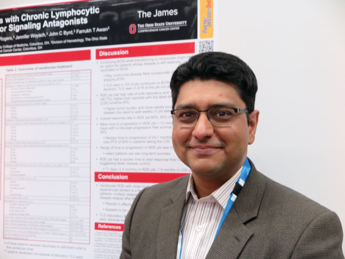
Of 15 patients with CLL who relapsed after treatment with a B-cell receptor inhibitor (BCRi), all were able to get to their target dose of venetoclax under close inpatient monitoring at a median of 12 days, compared with the 35 days usually required for venetoclax dose escalation, reported Farrukh T. Awan, MD, of Ohio State University Comprehensive Cancer Center in Columbus, and his colleagues.
Only two patients developed clinical tumor lysis syndrome (TLS), a common occurrence with venetoclax therapy, and this adverse event was manageable, Dr. Awan said at the annual congress of the European Hematology Association.
“The reason why we have been doing a slow ramp up on venetoclax is the original toxicity issues that we saw early on,” he said in an interview. “But unfortunately, a lot of patients are progressing on these new agents and have very rapid disease progression, and what we have seen is that if you stop the ibrutinib, the disease progresses very quickly, and by the time they can get up to the effective dose of venetoclax, they’re too sick to continue, or they might even die from disease progression.”
To combat this problem, Dr. Awan and his colleagues developed a rapid dose escalation protocol that would ramp up from 20 mg to 400 mg, with increases every 1 or 2 days depending on tolerability and incident TLS. Lab tests for TLS were evaluated every 4-8 hours.
All patients were closely monitored in the hospital, and all were started on allopurinol or other uric acid–lowering agents before starting on venetoclax.
The investigators reported safety and efficacy outcomes for the patients in a retrospective analysis.
The median age of the patients, 12 men and 3 women, was 65 years (range, 58-86 years). Seven patients had Eastern Cooperative Oncology Group Performance Status of 0, seven had an ECOG score of 1, and one had a score of 2-4.
Ten patients had most recently been treated with a BCRi, either a Bruton’s tyrosine kinase inhibitor (ibrutinib or acalabrutinib), idelalisib, or entospletinib. Three patients received ibrutinib plus chemotherapy, and two received rituximab and dexamethasone followed by rituximab maintenance.
The median time to full venetoclax dose was 12 days (range, 5-21 days) and all 15 patients reached the target dose. The mean length of stay during the ramp-up period was 9.5 days (range, 6-22 days).
The incidence of clinical TLS was 13.2%, occurring in two patients, one at the initial 20-mg dose, and one at the 200-mg dose level. Another five patients had asymptomatic TLS. Other treatment-related adverse events were anemia in seven patients, neutropenia in six patients, thrombocytopenia in five patients, and lung infection in one patient.
Twelve patients had a partial response, one had stable disease, and two had progressive disease. The mean time to best response was 71 days.
One-year progression-free survival was 49%, and 1-year overall survival was 68%.
The investigators found that for patients who still have some disease control with a BCRi, it may be possible to keep them on that drug while transitioning to venetoclax. The rapid dose escalation protocol should only be attempted in highly experience comprehensive cancer centers, Dr. Awan said.
“Under very close monitoring in an experienced inpatient setting, where the nurses are very used to doing this on a weekly basis in a very high volume center, I think that our data show that we could do this without affecting toxicity significantly or mortality,” he said.
Venetoclax therapy could buy enough time for patients to bridge to other options, such as chimeric antigen receptor (CAR) T-cell therapy or allogeneic stem cell transplant, he noted.
“But if we had waited 4 weeks, most of these patients would not have made it,” he said.
The study was internally funded. Dr. Awan reported research funding from Gilead, Pharmacyclics, AbbVie, and Janssen.
SOURCE: Koenig K et al. EHA Congress, Abstract PF357.
STOCKHOLM – Patients with chronic lymphocytic leukemia (CLL) who experience relapse after therapy with a B-cell receptor signaling inhibitor tend to have a swiftly progressive disease course that requires immediate intervention. For these patients, a rapid venetoclax dose-escalation protocol may be a safe way to quickly regain disease control, and possibly bridge to salvage therapies, investigators reported.
Of 15 patients with CLL who relapsed after treatment with a B-cell receptor inhibitor (BCRi), all were able to get to their target dose of venetoclax under close inpatient monitoring at a median of 12 days, compared with the 35 days usually required for venetoclax dose escalation, reported Farrukh T. Awan, MD, of Ohio State University Comprehensive Cancer Center in Columbus, and his colleagues.
Only two patients developed clinical tumor lysis syndrome (TLS), a common occurrence with venetoclax therapy, and this adverse event was manageable, Dr. Awan said at the annual congress of the European Hematology Association.
“The reason why we have been doing a slow ramp up on venetoclax is the original toxicity issues that we saw early on,” he said in an interview. “But unfortunately, a lot of patients are progressing on these new agents and have very rapid disease progression, and what we have seen is that if you stop the ibrutinib, the disease progresses very quickly, and by the time they can get up to the effective dose of venetoclax, they’re too sick to continue, or they might even die from disease progression.”
To combat this problem, Dr. Awan and his colleagues developed a rapid dose escalation protocol that would ramp up from 20 mg to 400 mg, with increases every 1 or 2 days depending on tolerability and incident TLS. Lab tests for TLS were evaluated every 4-8 hours.
All patients were closely monitored in the hospital, and all were started on allopurinol or other uric acid–lowering agents before starting on venetoclax.
The investigators reported safety and efficacy outcomes for the patients in a retrospective analysis.
The median age of the patients, 12 men and 3 women, was 65 years (range, 58-86 years). Seven patients had Eastern Cooperative Oncology Group Performance Status of 0, seven had an ECOG score of 1, and one had a score of 2-4.
Ten patients had most recently been treated with a BCRi, either a Bruton’s tyrosine kinase inhibitor (ibrutinib or acalabrutinib), idelalisib, or entospletinib. Three patients received ibrutinib plus chemotherapy, and two received rituximab and dexamethasone followed by rituximab maintenance.
The median time to full venetoclax dose was 12 days (range, 5-21 days) and all 15 patients reached the target dose. The mean length of stay during the ramp-up period was 9.5 days (range, 6-22 days).
The incidence of clinical TLS was 13.2%, occurring in two patients, one at the initial 20-mg dose, and one at the 200-mg dose level. Another five patients had asymptomatic TLS. Other treatment-related adverse events were anemia in seven patients, neutropenia in six patients, thrombocytopenia in five patients, and lung infection in one patient.
Twelve patients had a partial response, one had stable disease, and two had progressive disease. The mean time to best response was 71 days.
One-year progression-free survival was 49%, and 1-year overall survival was 68%.
The investigators found that for patients who still have some disease control with a BCRi, it may be possible to keep them on that drug while transitioning to venetoclax. The rapid dose escalation protocol should only be attempted in highly experience comprehensive cancer centers, Dr. Awan said.
“Under very close monitoring in an experienced inpatient setting, where the nurses are very used to doing this on a weekly basis in a very high volume center, I think that our data show that we could do this without affecting toxicity significantly or mortality,” he said.
Venetoclax therapy could buy enough time for patients to bridge to other options, such as chimeric antigen receptor (CAR) T-cell therapy or allogeneic stem cell transplant, he noted.
“But if we had waited 4 weeks, most of these patients would not have made it,” he said.
The study was internally funded. Dr. Awan reported research funding from Gilead, Pharmacyclics, AbbVie, and Janssen.
SOURCE: Koenig K et al. EHA Congress, Abstract PF357.
REPORTING FROM THE EHA CONGRESS
Key clinical point:
Major finding: All patients reached the target dose of venetoclax, with only two cases of manageable clinical tumor lysis syndrome.
Study details: Retrospective analysis of outcomes for 15 patients with CLL who relapsed after treatment with a B-cell receptor signaling inhibitor.
Disclosures: The study was internally funded. Dr. Awan reported research funding from Gilead, Pharmacyclics, AbbVie, and Janssen.
Source: Koenig K et al. EHA Congress, Abstract PF357.
Concomitant drugs may explain PEG-ASP liver toxicities in ALL
STOCKHOLM – Liver toxicities in adults with acute lymphoblastic leukemia (ALL) treated with a pediatric-type regimen containing pegylated asparaginase (PEG-ASP) may be related to concomitant use of other hepatotoxic drugs, investigators cautioned.

A retrospective review of records on 26 adult ALL patients treated with PEG-ASP since 2013 showed that concomitant use of vincristine, idarubicin, and vancomycin was associated with an increased risk for grade 3 or 4 hepatotoxicity, reported Fabio Guolo, MD, from the University of Genoa (Italy) and his colleagues.
In contrast, patients who received other chemotherapy drugs or antimicrobial agents did not have significant liver toxicities, Dr. Guolo said in an interview at the annual congress of the European Hematology Association.
“Increased toxicity from therapy prevents delivery of the most active therapy, and asparaginase is one of the keys to the success of pediatric trials in ALL, so we have tried to push the dose of asparaginase as high as we could in adult patients,” he said.
“We asked why some patients will develop toxicity while receiving a relatively low dose of asparaginase, whereas other patients who received higher doses did not,” Dr. Guolo added.
In recent years, investigators have found that adults with ALL tend to have better outcomes when they were treated with standard pediatric ALL regimens, which includes high-dose PEG-ASP.
To identify factors related to potential PEG-ASP toxicity in adults with ALL, the investigators combed through records of 26 adults patients, 19 of whom had received PEG-ASP in the frontline setting, and 7 of whom received it during treatment of relapsed or refractory disease.
The investigators looked at each course of PEG-ASP as an independent event (51 total episodes), paying special attention to concomitant chemotherapy and the use of both antimycotic and antibiotic agents.
Five of the patients had grade 3 hepatotoxicity, and three had grade 4 hepatotoxicity. The patients with grade 4 events had unexplained severe weight gain and painful hepatomegaly. Ultrasonography in these patients revealed acute steatosis similar to that seen with sinusoidal occlusive disease. All three patients had received concomitant idarubicin, vincristine, and vancomycin.
In univariate analysis, neither being older than 45 years, administration of PEG-ASP during an active leukemia phase, nor having a body mass index greater than 25 kg/m2 were significantly associated with increased incidence of grade 3 or 4 hepatotoxicity.
When the investigators looked at concomitant chemotherapy drugs, however, they found that liver toxicity was significantly higher with idarubicin cumulative doses of 20 mg/m2 or greater (hazard ratio, 1.49; P = .047) and that vincristine doses of 2 mg/m2 or greater were associated with a borderline increase in risk (HR, 4.75; P = .055).
There was no increased risk for liver toxicities with either steroids, daunorubicin, cyclophosphamide, cytarabine, methotrexate, or 6-mercaptopurine.
Additionally, concomitant vancomycin was also linked to increased hepatotoxicity (HR, 1.86; P =.009). In contrast, neither carbapenem-class anti-infectives nor azole were significantly associated with liver toxicities.
“Notably, none of the patients undergoing full pediatric induction, which contains higher cumulative doses of PEG-ASP, experienced grade 4 hepatotoxicity regardless of age,” Dr. Guolo and his colleagues wrote in their poster presentation.
In multivariate analysis controlling for age, BMI, drug regimen, and concomitant therapies, idarubicin remained a significant risk factor for severe hepatotoxicity (P = .004), and vancomycin remained as a borderline risk (P = .054).
Dr. Guolo acknowledged that the investigators could not account for the potential contribution of over-the-counter medications with known risk for hepatotoxicity, such as acetaminophen.
He noted that in his group’s experience, the toxicity profile of PEG-ASP in adults, including high-dose regimens, was manageable without excess toxicities as long as clinicians paid close attention to the use of concomitant agents.
The study was internally funded. The authors reported having no relevant conflicts of interest.
SOURCE: Minetto P et al. EHA Congress, Abstract PS934.
STOCKHOLM – Liver toxicities in adults with acute lymphoblastic leukemia (ALL) treated with a pediatric-type regimen containing pegylated asparaginase (PEG-ASP) may be related to concomitant use of other hepatotoxic drugs, investigators cautioned.
A retrospective review of records on 26 adult ALL patients treated with PEG-ASP since 2013 showed that concomitant use of vincristine, idarubicin, and vancomycin was associated with an increased risk for grade 3 or 4 hepatotoxicity, reported Fabio Guolo, MD, from the University of Genoa (Italy) and his colleagues.
In contrast, patients who received other chemotherapy drugs or antimicrobial agents did not have significant liver toxicities, Dr. Guolo said in an interview at the annual congress of the European Hematology Association.
“Increased toxicity from therapy prevents delivery of the most active therapy, and asparaginase is one of the keys to the success of pediatric trials in ALL, so we have tried to push the dose of asparaginase as high as we could in adult patients,” he said.
“We asked why some patients will develop toxicity while receiving a relatively low dose of asparaginase, whereas other patients who received higher doses did not,” Dr. Guolo added.
In recent years, investigators have found that adults with ALL tend to have better outcomes when they were treated with standard pediatric ALL regimens, which includes high-dose PEG-ASP.
To identify factors related to potential PEG-ASP toxicity in adults with ALL, the investigators combed through records of 26 adults patients, 19 of whom had received PEG-ASP in the frontline setting, and 7 of whom received it during treatment of relapsed or refractory disease.
The investigators looked at each course of PEG-ASP as an independent event (51 total episodes), paying special attention to concomitant chemotherapy and the use of both antimycotic and antibiotic agents.
Five of the patients had grade 3 hepatotoxicity, and three had grade 4 hepatotoxicity. The patients with grade 4 events had unexplained severe weight gain and painful hepatomegaly. Ultrasonography in these patients revealed acute steatosis similar to that seen with sinusoidal occlusive disease. All three patients had received concomitant idarubicin, vincristine, and vancomycin.
In univariate analysis, neither being older than 45 years, administration of PEG-ASP during an active leukemia phase, nor having a body mass index greater than 25 kg/m2 were significantly associated with increased incidence of grade 3 or 4 hepatotoxicity.
When the investigators looked at concomitant chemotherapy drugs, however, they found that liver toxicity was significantly higher with idarubicin cumulative doses of 20 mg/m2 or greater (hazard ratio, 1.49; P = .047) and that vincristine doses of 2 mg/m2 or greater were associated with a borderline increase in risk (HR, 4.75; P = .055).
There was no increased risk for liver toxicities with either steroids, daunorubicin, cyclophosphamide, cytarabine, methotrexate, or 6-mercaptopurine.
Additionally, concomitant vancomycin was also linked to increased hepatotoxicity (HR, 1.86; P =.009). In contrast, neither carbapenem-class anti-infectives nor azole were significantly associated with liver toxicities.
“Notably, none of the patients undergoing full pediatric induction, which contains higher cumulative doses of PEG-ASP, experienced grade 4 hepatotoxicity regardless of age,” Dr. Guolo and his colleagues wrote in their poster presentation.
In multivariate analysis controlling for age, BMI, drug regimen, and concomitant therapies, idarubicin remained a significant risk factor for severe hepatotoxicity (P = .004), and vancomycin remained as a borderline risk (P = .054).
Dr. Guolo acknowledged that the investigators could not account for the potential contribution of over-the-counter medications with known risk for hepatotoxicity, such as acetaminophen.
He noted that in his group’s experience, the toxicity profile of PEG-ASP in adults, including high-dose regimens, was manageable without excess toxicities as long as clinicians paid close attention to the use of concomitant agents.
The study was internally funded. The authors reported having no relevant conflicts of interest.
SOURCE: Minetto P et al. EHA Congress, Abstract PS934.
REPORTING FROM EHA CONGRESS
Key clinical point:
Major finding: Idarubicin was associated with a higher risk of grade 3 or 4 hepatotoxicity, and vincristine was associated with a borderline risk.
Study details: Retrospective review of 51 PEG-ASP dosing events in 26 adult patients with ALL.
Disclosures: The study was internally funded. The authors reported having no relevant conflicts of interest.
Source: Minetto P et al. EHA Congress, Abstract PS934.
MicroRNAs flag liver damage in HIV-, HCV-infected persons
BOSTON – In persons infected with HIV-1, with or without hepatitis C coinfections, specific circulating microRNAs may signal the presence of liver injury and progression, investigators stated.
An analysis of small RNA expression in plasma samples from 144 HIV-infected patients showed that two microRNAs (miRNAs) in the same family of RNA fragments were significantly upregulated in patients with HIV-1 and HCV coinfections that progressed to liver cirrhosis, despite the patients having no evidence of liver fibrosis at the time of plasma sampling, reported Miguel Angel Martinez, PhD, of IrsiCaixa AIDS Research Institute in Badalona, Spain.
“Our results reveal that HIV-1 infection impacts liver miRNA metabolism and upregulated plasma levels of miRNAs that were previously associated with liver damage, even in the absence of an HCV coinfection,” he said at the Conference on Retroviruses & Opportunistic Infections. He reported the results in a themed discussion and scientific poster session.
Dr. Martinez and his colleagues performed large-scale deep sequencing analyses of miRNAs in plasma from 144 patients with HIV-1 who had elevated alanine aminotransferase (ALT), focal nodular hyperplasia, or HCV coinfections, and compared results with those from healthy blood donors and HCV mono-infected persons.
They identified 1,425 different mature miRNAs in the study samples. Compared with healthy donors, patients with HIV infections showed significantly dysregulated expression of 25 miRNAs, and 19 of these miRNAs were also found in patients with HCV monoinfection. All but 1 of 14 upregulated miRNAs in patients with HCV monoinfections were also upregulated in patients with HIV monoinfections.
Of these 13 upregulated miRNAs, 11 significantly and positively correlated with ALT and aspartate aminotransferase (AST) levels in most of the study samples, including those from healthy donors, Dr. Martinez noted.
“These results indicate that HIV mono-infection is able to dysregulate microRNAs related with liver injury and damage,” he said.
Of the 13 miRNAs, two, labeled miR-99a-5p and miR-100-5p, which belong to the same family of miRNAs, were found to be significantly upregulated in patients with HIV and HCV coinfections that later progressed to liver cirrhosis “even those these patients exhibited no liver fibrosis at the time of sampling,” he said
The two culprit miRNAs were significantly correlated with ALT and AST levels, as well as the degree of liver fibrosis.
A comparison of samples from patients with HIV monoinfection who had elevated ALT or focal nodular hyperplasia with those of patients with HIV infection but normal ALT levels showed that two other miRNAs, miR-122-3p and miR-193b-5p, were highly and significantly upregulated, and correlated with both aminotransferase and liver fibrosis levels.
“This study demonstrates the potential of microRNAs as biomarkers of liver injury progression in HIV-1 infected patients,” Dr. Martinez concluded.
The Spanish Instituto de Salud Carlos III and the Spanish AIDS network funded the study. Dr. Martinez reported having no conflicts of interest.
SOURCE: Martinez MA et al. CROI 2018, abstract 639.
REPORTING FROM CROI
Key clinical point: Specific circulating microRNAs appear to be biomarkers for liver injury and progression in persons with HIV and/or HCV infections.
Major finding: Two microRNAs correlated with elevated liver enzymes and liver fibrosis in patients with HIV and HCV coinfection, and two correlated with liver injury in patients with HIV monoinfection.
Study details: Analysis of plasma samples from 144 persons with HIV with elevated ALT, focal nodular hyperplasia, or HCV coinfections, with control samples from healthy donors and HCV monoinfected individuals.
Disclosures: The Spanish Instituto de Salud Carlos III and the Spanish AIDS network funded the study. Dr. Martinez reported having no conflicts of interest.
Source: Martinez MA et al. CROI 2018, abstract 639.
Hints of nivolumab efficacy seen in biliary tract cancers
BARCELONA – The immune checkpoint inhibitor nivolumab (Opdivo) shows activity against biliary tract cancers (BTC) that have progressed on prior systemic therapies, investigators report.
Among 27 patients with intra- and extrahepatic cholangiocarcinoma and cancers of the gallbladder for whom at least one prior line of therapy had failed, the overall response rate with nivolumab monotherapy was 18.5%, reported Richard Kim, MD of Moffitt Cancer Center, in Tampa.
“Nivolumab demonstrated clinical efficacy in BTC patients. It was very well tolerated, with few grade 3 or 4 adverse events,” he said at the European Society of Medical Oncology World Congress on Gastrointestinal Cancer.
The worldwide incidence of biliary tract cancers has grown over the last 4 decades.
“It is a very aggressive disease, with 5-year overall survival rate of advance disease of less than 2%,” he said.
The standard of care for first-line treatment of advanced disease is gemcitabine and cisplatin, but there is no standard treatment available for patients for whom first-line therapy fails.
Median survival of patients with biliary tract cancers who are receiving second- or third-line therapies is approximately 6-7 months, Dr. Kim said.
The rationale for using nivolumab in this setting comes from evidence suggesting that cholangiocarcinoma is related to dysregulated immunity, with carcinogenesis linked to autoimmune conditions such as primary sclerosing cholangitis, and to chronic parasitic infections.
“Immune regulatory protein PD-1 is upregulated more in intrahepatic cholangiocarcinoma tissues than in adjacent normal tissue, and patients with memory CD8 T cells had longer relapse-free survival and overall survival in extrahepatic cholangiocarcinoma after resection,” he explained.
To see whether the use of an immune checkpoint inhibitor could provide clinically meaningful benefit in patients with advance biliary tract cancers, the investigators conducted a phase 2, two-stage study. They first accrued 18 patients with histologically confirmed, treatment-refractory biliary tract malignancies and treated them with nivolumab 240 mg IV every 2 weeks for 16 weeks, followed by 480 mg IV every 4 weeks.
According to the study protocol, if one or more patients had a complete or partial response, additional patients would be enrolled. As of May 2018, 34 patients had been treated.
The median patient age was 64.5 years. Two-thirds of the patients (64.7%) had intrahepatic cholangiocarcinoma, 2.9% had extrahepatic cholangiocarcinoma, and 32.4% had tumors of the gallbladder.
Twenty patients were failed by their first-line therapies, and 14 were failed by two or more lines of therapy. All 34 received at least one dose of nivolumab.
Of this group, 10 patients remained on study at the time of Dr. Kim’s presentation. Fifteen were withdrawn for progressive disease according to Response Evaluation Criteria in Solid Tumors (RECIST) revision 1.1, and 9 due to clinical progression.
Of 27 patients evaluable for investigator-assessed overall responses – the primary endpoint – 5 patients (18.5%) had a partial response, and 11 (40.7%) had stable disease, for a disease-control rate of 59.3%. The remaining 11 evaluable patients had progressive disease.
“Of interest, of our five patients who had a partial response, three had a diagnosis of intrahepatic cholangiocarcinoma, and two had a diagnosis of a gallbladder tumor,” Dr. Kim said.
All five patients remained on treatment at the time of the presentation, with response duration ranging from 24 to 64 weeks. The median duration of response in these patients has not been reached.
Median progression-free survival for all 34 patients treated with at least one dose was 3.5 months. Overall survival with a median follow-up of 9.9 months has not been reached. The 6-months overall survival rate was 73.5%.
Approximately 20% of patients experienced grade 3 or 4 treatment-related adverse events. There were no grade 4 events and no treatment-related deaths.
The most common grade 3 events were hyponatremia in three patients (8.8%), and lymphopenia, colitis, and hyperbilirubinemia in one patient each (2.9%).
The investigators have collected tissues from all patients and plan to present data from biomarker studies at future meetings. Based on the results of this study, they plan to add 20 more patients to the phase 2 trial to confirm efficacy of nivolumab in this setting.
REPORTING FROM ESMO GI 2018
Key clinical point: Nivolumab monotherapy appears to have activity in treatment-refractory biliary tract cancers.
Major finding: Five of 27 evaluable patients had partial responses to nivolumab.
Study details: Two-stage phase 2 trial of 34 patients with intrahepatic or extrahepatic cholangiocarcinomas or gallbladder tumors.
Disclosures: Bristol-Myers Squibb sponsored the study. Dr. Kim disclosed honoraria and institutional research funding from that company and others.
Source: Kim R et al. European Society of Medical Oncology World Congress on Gastrointestinal Cancer. Abstract O-009.
Trifluridine/tipiracil improves survival of metastatic gastric cancer
BARCELONA – In patients with heavily pretreated metastatic gastric cancer, trifluridine/tipiracil was associated with a brief but statistically significant survival benefit, compared with placebo.
In the randomized, controlled TAGS (TAS-102 Gastric Study), median overall survival, the primary endpoint, was 5.7 months for patients assigned to receive trifluridine/tipiracil, compared with 3.6 months for patients randomized to placebo, reported Josep Tabernero, MD, PhD, of Vall d’Hebron Institute of Oncology in Barcelona.
Trifluridine/tipiracil (Lonsurf) is a combination of an oral thymidine analog (trifluridine, or FTD) and a thymidine phosphorylase inhibitor (tipiracil, or TPI).
It was approved by the Food and Drug Administration in 2015 for the treatment of patients with metastatic colorectal cancer who have been previously treated with chemotherapy and biological therapy.
In a phase 2 study conducted in Japan with 29 patients with metastatic gastric cancer that had progressed after chemotherapy with fluoropyrimidine, platinum, taxanes, or irinotecan, FTD/TPI was associated with a median overall survival of 8.7 months and an investigator-assessed disease control rate of 65.5%, Dr. Tabernero noted.
In the TAGS trial, patients with metastatic gastric cancer, including cancers of the gastroesophageal junction who had disease progression after two or more prior lines of therapy were randomized on a 2:1 basis to receive either oral FTD/TPI 35 mg/m2 twice daily on days 1-5 and 8-12 of each 28-day cycle or to receive placebo. In both arms, patients also received best supportive care.
As noted, the trial met its primary endpoint, with a 2.1 month improvement in median survival over placebo, which translated into a hazard ratio for death with FTD/TPI of 0.69 (P = .0003).
The 12-month overall survival rate for the FTD/TPI group was 21%, compared with 13% for the placebo group.
Median progression-free survival, a secondary endpoint, was also slightly but significantly better with FTD/TPI at 2.0 vs. 1.8 months (HR, 0.57; P less than .0001). Six-month progression-free survival rates were 15% and 6%, respectively.
Grade 3 or greater adverse events occurred in 80% of patients on FTD/TPI versus 58% of those on placebo. The drug combination was associated with more treatment-related adverse events (81% vs. 57%) but fewer adverse events leading to discontinuation (13% vs. 17%) and fewer treatment-related deaths (0.3 % vs. 0.6%).
FTD/TPI was also associated with more cases of grade 3 or 4 neutropenia, leukopenia, lymphocytopenia, anemia, and thrombocytopenia. Six patients (2%) treated with FTD/TPI had grade 3 or 4 febrile neutropenia, compared with none in the placebo group.
“The safety profile of trifluridine/tipiracil was predictable, manageable, and comparable to the population previously evaluated with metastatic colorectal cancer, with no new safety signals,” Dr. Tabernero said.
SOURCE: Tabernero J et al. ESMO GI 2018, Abstract LBA 002.
REPORTING FROM ESMO GI 2018
Key clinical point: Trifluridine/tipiracil (FTD/TIP) produced a small but significant survival benefit in patients with advanced gastric cancer.
Major finding: Median overall survival was 5.7 months with trifluridine/tipiracil versus 3.6 months with placebo.
Study details: Randomized, placebo-controlled trial in 506 patients with metastatic gastric cancer that progressed on at least two prior lines of therapy.
Disclosures: The study was sponsored by Taiho Oncology. Dr. Tabernero disclosed a consulting or advisory role for the company and others.
Source: Tabernero J et al. ESMO GI 2018, Abstract LBA 002.
Pembrolizumab does not surpass paclitaxel for gastric cancer
BARCELONA – The immune checkpoint inhibitor pembrolizumab (Keytruda) did not significantly improve overall survival of advanced/metastatic gastric or gastroesophageal junction cancer compared with paclitaxel, the results of the KEYNOTE-061 study show.
Among 395 patients with gastric or gastroesophageal junction (GEJ) cancer who had expression of the programmed death ligand 1 (PD-L1) on 1% or more of their tumor cells, lymphocytes, and macrophages, median overall survival (OS) for patients treated with pembrolizumab was 9.1 months, compared with 8.3 months. This translated into a hazard ratio (HR) for death in the pembrolizumab arm of 0.82, but with the 95% confidence interval crossing 1.00, and a P value (.04205) that did not meet the prespecified threshold for significance (P equal to or less than .0135).
Despite the failure of the trial to reach its primary endpoint, “these results may support further research to identify patients likely to benefit from pembrolizumab monotherapy and ongoing development of pembrolizumab-based combination therapy for gastric cancer,” he said at the European Society of Medical Oncology World Congress on Gastrointestinal Cancer.
Pembrolizumab is approved by the Food and Drug Administration for the treatment of patients with recurrent locally advanced or metastatic gastric or GEJ adenocarcinoma with tumors confirmed to carry PD-L1 for whom two or more prior lines of therapy had failed.
The approval was based on results of the nonrandomized, open label KEYNOTE-059 trial, which enrolled 259 patients with gastric or GEJ adenocarcinoma that progressed on at least two prior systemic treatments for advanced disease. Of the enrollees, 143 patients had tumors with a PD-L1 Combined Positive Score (CPS) of 1 or greater. The primary trial outcome, the objective response rate for these 143 patients, was 13.3% (95% confidence interval; 8.2-20), with a complete response rate of 1.4% and a partial response rate of 11.9%. The duration of response ranged from at least 2.8 months to at least 19.4 months.
The drug is also approved for unresectable of metastatic gastric tumors with high levels of microsatellite instability that progressed on prior therapy.
The KEYNOTE-061 study was designed to test the proposition that pembrolizumab could improve on paclitaxel for treatment of patients with adenocarcinoma of the stomach or GEJ that was metastatic or locally advanced and unresectable, and for which first-line therapy with a platinum agent and fluoropyrimidine had failed.
A total of 592 patients from Europe, Israel, North America, Asia, and Australia were enrolled and randomly assigned to receive either pembrolizumab 200 mg every 3 weeks for 35 cycles, or paclitaxel 80 mg/m2 on days 1, 8, and 15 of each 4-week cycle. Each treatment was continued until the maximum number of cycles (for pembrolizumab) or until confirmed disease progression, intolerable toxicity, patient withdrawal, or investigator’s decision.
As noted, OS for patients with a CPS score of 1 or greater, the primary endpoint, did not differ between treatment arms, but there was a numerical tilt that appeared to be in favor of pembrolizumab.
For example, the 12-month OS rates were 39.8% in the pembrolizumab groups vs. 27.1% in the paclitaxel group, and respective 18-month OS rates were 25.7% and 14.8%.
In an analysis of protocol-specified subgroups, there were general trends slightly favoring the checkpoint inhibitor over the taxane, but the only significant difference was among patients with GEJ cancers as the primary tumor location (HR, 0.61, 95% confidence interval, 0.41-0.90).
Pembrolizumab offered a small but significant survival advantage among patients with Eastern Cooperative Oncology Group performance status of 0, with a median OS of 12.3 months vs. 9.3 months (HR, 0.69, 95% CI, 0.49-0.97).
Analyses of PFS and OS by CPS score groups (less than 1, 1-10, or 10 and higher) showed no significant differences, however.
Treatment-related adverse events were more frequent with paclitaxel (84.1% vs. 52.7% of patients on pembrolizumab), but pembrolizumab was associated with more treatment-related deaths (three vs. one). Two of the deaths in the pembrolizumab arm were immune mediated. Grade 3 or greater adverse events occurred in 14.3% vs. 34.8%, respectively.
In the question-and-response following Dr. Shitara’s presentation, session comoderator David Cunningham, MD, of Royal Marsden NHS Foundation Trust in Sutton, England, asked why the KEYNOTE-061 investigators did not pit pembrolizumab against the combination of paclitaxel and ramucirumab (Cyramza) “since that’s what many people would use in this situation.”
Dr. Shitara replied that ramucirumab was not available or accepted as an option in many countries when the trial was first planned in 2014. He added that paclitaxel and ramucirumab should be the control arm for clinical trials going forward.
SOURCE: Shitara K. et al. ESMO World Congress on Gastrointestinal Cancer 2018. Abstract LBA-005.
REPORTING FROM ESMO GI 2018
Key clinical point: KEYNOTE-061 did not meet its primary endpoint of a significant survival advantage with pembrolizumab over paclitaxel.
Major finding: There was no significant difference in overall survival between patients with gastric or gastroesophageal junction cancers treated with pembrolizumab or paclitaxel.
Study details: Phase 3 randomized open-label trial in 592 patients with advanced or metastatic and unresectable tumors of the stomach of GEJ.
Disclosures: The trial was supported by Merck. Dr. Shitara has disclosed honoraria from Abbvie, Novartis and Yakult, consulting or advising with Astellas, BMS. Lilly, Ono Pharmaceutical, Pfizer, and Takeda, and institutional research funding from other companies. Dr. Cunningham has disclosed institutional research finding from Merck Serono and others.
Source: Shitara K et al. ESMO World Congress on Gastrointestinal Cancer 2018. Abstract LBA-005.
Ramucirumab improves HCC survival after sorafenib failure
BARCELONA – For patients with hepatocellular carcinoma (HCC) with elevated alpha-fetoprotein (AFP) levels who have disease progression following first-line sorafenib (Nexavar), the antiangiogenic agent ramucirumab was associated with a modest but significant improvement in overall survival, compared with placebo, a pooled analysis of clinical trial data showed.
Among 316 patients assigned to receive ramucirumab in the phase 3 REACH and REACH-2 trials, median overall survival was 8.1 months, compared with 5.0 months for placebo, an absolute difference of 3.1 months that translated into a hazard ratio favoring ramucirumab of 0.694 (P = .0002), reported Andrew X. Zhu, MD, PhD of the Massachusetts General Hospital Cancer Center in Boston.
Ramucirumab (Cyramza) is a human IgG1 monoclonal antibody directed against ligand activation of the vascular endothelial growth factor receptor 2.
The REACH trial did not meet its primary endpoint of an improvement in overall survival with ramucirumab in an intention-to-treat population. (Lancet Oncol. 2015 Jul;16(7):859-70). REACH-2, however, results of which were presented at the 2018 annual meeting of the American Society of Clinical Oncology (ASCO 2018, Abstract 4003), met its primary endpoint of overall survival with ramucirumab, with a median overall survival of 8.5 months versus 7.3 months for placebo (HR, 0.71; P = .0199) in patients with AFP levels of 400 ng/mL or greater who had experienced disease progression after first-line sorafenib.
The pooled analysis Dr. Zhu presented was designed to provide a better assessment of the efficacy and safety of ramucirumab as a second-line agent in this high-risk population.
In REACH, 250 patients with advanced HCC and AFP 400 ng/mL or greater were enrolled and randomized on a 1:1 basis to ramucirumab or placebo. In REACH 2, patients were randomized on a 2:1 basis to either ramucirumab 8 mg/kg intravenously every 2 weeks (the same dose as in REACH) or placebo. The pooled analysis included 316 patients on ramucirumab and 226 placebo-treated controls.
In each trial, treatment was continued until disease progression or unacceptable toxicity. The primary endpoint was overall survival, and secondary endpoints were progression-free survival, overall response rate, safety, and patient-reported outcomes.
Overall survival in the pooled analysis was as noted before. The benefits of ramucirumab were significantly better for men, for patients younger than 65 years, for patients of white and Asian race, for those with non–hepatitis B or C etiology of liver disease, and in patients with extrahepatic metastases. In addition, those without microvascular invasion, patients with Barcelona Clinic Liver Cancer score C versus B, those with excellent baseline performance status, those who received prior locoregional therapy, and patients who discontinued sorafenib because of disease progression also saw benefit from ramucirumab.
“Ramucirumab’s overall survival treatment benefit was consistent and robust across all subgroups. Sensitivity analyses, include random effect models, were consistent,” Dr. Zhu said.
The median progression-free survival with ramucirumab was 2.8 months, compared with 1.5 months for placebo (P less than .0001). The overall response rate, including all complete and partial responses, was 5.4% for ramucirumab versus 0.9% for placebo (P = .0064). The respective disease control rates, including patients with stable disease, were 56.3% versus 37.2% (P less than .0001).
Adverse events of special interest included grade 3 or greater liver injury or failure in 19.9% of patients on ramucirumab versus 26.5% on placebo, bleeding and/or hemorrhagic events in 4.7% versus 6.7%, and hypertension in 12.7% versus 3.6%.
The REACH trials were supported by Eli Lilly. Dr. Zhu disclosed consulting/advising for Eli Lilly and others, and institutional research from Eli Lilly and others.
SOURCE: Zhu AX et al. ESMO GI 2018, Abstract LBA-001.
REPORTING FROM ESMO GI 2018
Key clinical point: Second-line therapy with a vascular endothelial growth factor receptor 2 inhibitor modestly improved survival of patients with advanced hepatocellular carcinoma that progressed after first-line sorafenib.
Major finding: Median overall survival was 8.1 months with ramucirumab versus 5 months for placebo.
Study details: An analysis of pooled data from two phase 3 clinical trials with a total of 316 patients treated with ramucirumab and 226 treated with placebo.
Disclosures: The REACH trials were supported by Eli Lilly. Dr. Zhu disclosed consulting/advising for Eli Lilly and others, and institutional research from Eli Lilly and others.
Source: Zhu AX et al. ESMO GI 2018, Abstract LBA-001.
Dual-targeting CAR T active against AML in mice and one man
STOCKHOLM – A novel compound chimeric antigen receptor (cCAR) T-cell construct directed against two different targets may one day serve as a standalone therapy, as a supplement to chemotherapy, or as a bridge to transplant for patients with refractory acute myeloid leukemia (AML), investigators asserted.
To date, however, only one patient – a man with treatment-refractory AML – has been treated with the cCAR T, which contains two independent complete units, one directed against CD33 to target bulky disease and the other targeted against CLL1 on leukemic stem cells.
The patient was a 44-year-old man with AML who remained refractory after four cycles of chemotherapy and had 20% bone marrow blasts. He achieved a complete response after infusion with the cCAR T cells and went on to bone marrow transplant with no evidence of minimal residual disease (MRD) at 3 months of follow-up, Dr. Liu said.
Although anti-CD19 CAR T cells have been demonstrated to have significant efficacy in relapsed or refractory B-cell acute lymphoblastic leukemia, AML is a tougher problem to solve because the heterogeneity of myeloid leukemia cells allows some cells to escape targeting by enhanced T cells, which leads to eventual relapse.
To get around this problem, the investigators created a CAR T with a one-two punch, with one component targeting the antigen CLL1, which is expressed on leukemic stem cells, and a second, separate component targeting CD33, a myeloid marker expressed on bulk AML disease cells in a majority of patients.
They first tested the cCAR T cells against several AML cell lines and primary human AML samples, then in mouse models of human AML.
In vitro assays showed that the construct had specific antitumor activity against cell lines engineered to express either of the target antigens and also against samples from AML patients. In mouse models created with engineered CLL1 or CD33 expressing cell lines and an AML cell line, the cCAR T cells caused significant reductions in tumor burden and led to prolonged survival, Dr. Liu said.
Since CAR T-cell therapy is associated with serious or life-threatening side effects, such as the cytokine-release syndrome, the investigators built an “off switch” into the cCAR T construct that could be activated by CAMPATH, a monoclonal antibody directed against CD52. Introducing this agent into the mice quickly neutralized the cCAR T therapy, Dr. Liu said.
Finally, the investigators tested the construct in the human patient. He received the cCAR T construct after conditioning with fludarabine and cyclophosphamide; he had a complete remission by day 19 after receiving the cells and was MRD negative. He went on to an allogeneic stem cell transplant on day 44, and he remained MRD negative 3 months after transplant.
Side effects associated with the treatment were a grade 1 cytokine release syndrome event, manifesting in fever and chills, lung infection, and red blood cell transfusion dependence but also platelet transfusion independence.
The investigators have initiated a phase 1 trial and plan to enroll 20 patients to further evaluate the efficacy and safety of the cCAR T construct.
The study was supported by iCell Gene Therapeutics. Dr. Liu reported having no conflicts of interest.
SOURCE: Liu F et al. EHA Congress, Abstract S149.
REPORTING FROM THE EHA CONGRESS
Key clinical point:
Major finding: The only human patient treated with the construct had a complete remission and successful bridge to transplant.
Study details: Preclinical study plus phase 1 data on one patient.
Disclosures: The study was supported by iCell Gene Therapeutics. Dr. Liu reported having no conflicts of interest.
Source: Liu F et al. EHA Congress, Abstract S149.
Anti-CD22 CAR T rescues kids with ALL after CD19 failure
STOCKHOLM, SWEDEN – A novel chimeric antigen receptor T-cell construct directed against CD22 was able to rescue children with relapsed or refractory B-cell acute lymphoblastic leukemia for whom CD19-directed CAR T therapy had failed, investigators from China reported.
Thirty days after CAR T cell infusion, 12 of 15 children (80%) treated with the unnamed CD22 CAR T product had a complete response (CR) and one (6.7%) had a partial response (PR), for an overall response rate (ORR) of 86.7%, said Jing Pan, MD, from the Beijing Boren Hospital in China.
Surprisingly for a therapy of its type, the CD22 CAR T was well tolerated, with only mild cases of the cytokine release syndrome (CRS), and it could be delivered safely in children with relapsed disease after hematopoietic cell transplants (HCT), she reported in a briefing and in an oral abstract session at the annual congress of the European Hematology Association.
“CD22 CAR T immunotherapy brings hope for patients with refractory or relapsed B-ALL who failed on CD19 CAR T immunotherapy, and we think it’s quite safe. No children died and no children had severe side effects during the study, even in post-HCT patients,” Dr. Pan said.
She noted that in a previous clinical trial by her group, some patients experienced relapses and were resistant to retreatment with CD19-directed CAR T therapy due to mutations or loss of the CD19 antigen (Leukemia. 2017 Dec;31[12]:2587-93).
Since CD22 is highly expressed on leukemic cells in children with B-ALL, the investigators decided to evaluate the safety and efficacy of a CD22 CAR T as a rescue strategy. They enrolled 15 patients who either experienced relapse or did not have a response to CD19 CAR T immunotherapy. The CAR T construct they used contains an anti-CD22 single-chain variable fragment derived from a humanized CD22 antibody.
Patient conditioning with fludarabine and cyclophosphamide was performed simultaneously with CAR T transfection and expansion. After about 7 days, the expanded and transformed CAR T cells were infused at a dose of 8.2 x 10⁵/kg in patients who had not undergone HCT, and 0.9 x 10⁵ in patients who had received a transplant.
The patients ranged from 2 to 18 years old (median 8 years), had a median disease course of 21 months (range 5-84 months), and had a median of 42% bone marrow blasts (range 5%-95.5%).
Four of the 15 patients had relapses following allogeneic HCT, and the remaining 11 had relapses following chemotherapy. Two of the patients were found to be minimal residual disease (MRD)-positive by flow cytometry. Two patients had extramedullary disease only at relapse.
Ten of 11 patients who had experienced a hematologic relapse had either a CR or CR with incomplete recovery of counts (CRi), and of these 10 patients, nine were determined on follow-up to be MRD-negative by flow cytometry.
One of the two patients with extramedullary disease had a CR, and the other had a partial response.
Although two patients had no response to CD22 CAR T therapy, expression of the antigen was strong on leukemia cells from these patients, Dr. Pan said.
All patients experienced CRS, but none had greater than grade 2, Dr. Pan said, although she did not provide give specific numbers. Two patients had grade 1 neurotoxicity, two patients had grade 2 hypoxemia, and one patient had grade 2 liver enzyme elevation.
At a median follow-up of 108 days, six patients had been bridged to allogeneic HCT, and eleven of 12 patients who had a CR or CRi at 30 days had no evidence of disease progression. The remaining patient with an initial CR or CRi had a relapse at day 50. The 6-month progression-free survival rate was 91.7%.
Anton Hagenbeek, MD, PhD, from the Academic Medical Center at the University of Amsterdam, the Netherlands, who moderated the briefing but was not involved in the study, commented that given the high leukemia burden of the patients and the apparent efficacy of the therapy, one would expect to see higher grades of CRS, and asked Dr. Pan whether she could account for the CRS findings in her study, compared with those of trials for CD19-directed CAR T therapy.
It may have to do with differences in density of CD22 expression, compared with CD19 expression on leukemia cells, or on differences in the antibody used to target the cells, Dr. Pan said.
SOURCE: Pan J et al. EHA Congress, Abstract S832.
REPORTING FROM THE EHA CONGRESS
Key clinical point: A CD22-directed chimeric antigen receptor T cell construct may be a highly effective salvage therapy in children with acute lymphoblastic leukemia that has relapsed after, or is resistant to, CD19-direct CAR T therapy.
Major finding: In total, 12 of 15 children (80%) treated with the CD22 CAR T product had a complete response 30 days after infusion.
Study details: Open-label, single-center, single-arm study in 15 children with ALL that is resistant to, or relapsed after, prior CD19 CAR T therapy.
Disclosures: Dr. Pan and colleagues did not disclose a funding source for the study. She reported having no conflicts of interest.
Source: Pan J et al. EHA Congress, Abstract S832.