User login
Prevention Strategies Can Reduce Falls in Older Adults
SAN FRANCISCO – Falls are the main cause of hip fractures, and proven prevention strategies should be in every clinician's toolbox.
Physicians should ask patients aged 75 years or older if they have had any falls in the prior year or if they have balance or gait difficulties and observe them walking and getting into and out of a chair, said Dr. Edgar Pierluissi, who is medical director of the Acute Care for Elders Unit at San Francisco General Hospital.
A fall in the previous year increases the risk for a fall in the future by three- to fourfold.
Studies suggest that approximately 30% of U.S. adults over 65 years of age who are living in the community and half of adults over age 80 years will fall in the next year. Falls in adults aged 65 years or older cause injury in approximately 31%. Among those injured, 56% go to an emergency department and 38% visit a medical clinic, he said at the conference, sponsored by the University of California, San Francisco.
An exercise program with balance and strength training might be appropriate for older patients who've had only one or no falls and who don't have balance or gait difficulties, various guidelines suggest. If a patient reports two or more falls or has balance or gait difficulties, do a “falls evaluation,” an assessment of predisposing or precipitating factors that can point to appropriate preventive interventions, he said.
“We can perhaps make a difference” in many of the most common risk factors for falls that have been identified in 16 studies, Dr. Pierluissi said.
Muscle weakness quadruples the risk for a fall. A gait deficit, balance deficit, or use of an assistive device nearly triples the risk for falling. A visual deficit, arthritis, depression, or impaired activities of daily living more than double the risk for a fall. Cognitive impairment, use of some types of medications, or age older than 80 years each nearly doubles the risk for falling, he said.
To conduct a falls evaluation, get a good history of the patient's falls and their circumstances. Do a cardiovascular examination, medication review, neurological examination, and assessment for cognitive impairment. Assess gait, balance and mobility, muscle weakness, visual impairment, home hazards that might precipitate a fall, and the patient's perceived functional ability and fear related to falling (because many people who fear falling restrict their activity, which can lead to deconditioning and increased risk of falling).
A Cochrane Review of 111 randomized, controlled trials involving 55,303 participants identified effective interventions to reduce the risk of falling (Cochrane Database Syst. Rev. 2009 [doi:10.1002/14651858.CD007146.pub2]).
A number of forms of exercise reduced both the number of people who fell and the number of falls. Group tai chi exercise or individually prescribed exercise programs at home were effective. Multiple-component group exercise was effective if it targeted at least two of the following: strength, balance, flexibility, and endurance.
Conducting a multifactorial falls evaluation reduces the number of falls. In patients with visual impairment and a high risk of falling, assessing and modifying home hazards were effective, Dr. Pierluissi noted.
Withdrawing psychotropic medications and educating primary care physicians about the risk of falls association with drug therapy reduced the number of falls but not the number who fell. In patients with cardioinhibitory carotid sinus hypersensitivity, cardiac pacing reduced the number of falls.
Vitamin D supplementation may reduce falls in people with low vitamin D levels, but it's unclear whether this helps people with adequate vitamin D levels. Other preventive strategies of unknown effectiveness include correction of visual deficiency, hormone replacement therapy, or modifying home hazards for people who have not fallen.
The Cochrane Review suggested that wearing hip protectors may provide some marginally significant benefit to frail, older adults in institutional care but not for older people who remain ambulant in the community, Dr. Pierluissi said.
One randomized, controlled trial of 1,042 residents in 37 nursing homes found a high rate of adherence to wearing hip protectors (74%) but these did not reduce the risk for hip fracture during the 20-month study. Residents served as their own controls by wearing hip protectors with padding on one hip but not the other. Investigators stopped the study early because of lack of efficacy, with hip fractures on 3.1% of the protected hips and 2.5% of unprotected hips, a statistically nonsignificant difference (JAMA 2007;298:413-22).
Dr. Pierluissi said that he had no relevant conflicts of interest.
Expert Analysis from a Meeting on Osteoporosis
Denosumab's Effect Greater on Femoral Neck
Major Finding: Denosumab decreased nonvertebral fractures by 35% in patients with a femoral neck bone mineral density T score of −2.5 or lower and by only 3% in patients with higher femoral neck T scores, compared with patients in those subgroups who received placebo.
Data Source: Subgroup analysis in the original study of 7,808 postmenopausal women aged 60-80 years with osteoporosis who received every 6 months either a subcutaneous injection of denosumab (60 mg) or placebo along with daily calcium and vitamin D supplements.
Disclosures: Dr. Cummings has been a consultant to Amgen Pharmaceuticals, which markets denosumab; to Merck, which markets alendronate; and to Eli Lilly and Company.
SAN FRANCISCO – The pivotal clinical trial of denosumab showed a 20% decrease in nonvertebral fractures compared with placebo treatment, but a new subgroup analysis shows the protective effect is significantly higher in patients with femoral neck osteoporosis.
The preplanned subgroup analysis of data from the FREEDOM trial found that denosumab decreased nonvertebral fractures by 35% in patients with a femoral neck bone mineral density T score of −2.5 or lower and by only 3% in patients with higher femoral neck T scores, compared with patients in those subgroups who received placebo, Dr. Steven R. Cummings said.
The report of a 20% reduction in nonvertebral fractures in the overall trial for denosumab “underestimates its efficacy for those patients that we're most interested in treating with this drug – those with osteoporosis,” he said at the conference, sponsored by the University of California, San Francisco.
The analysis is one of several preplanned subgroup analyses being conducted, though this one is “the most interesting result for clinical care,” said Dr. Cummings, emeritus professor of medicine and epidemiology and biostatistics at the university.
The original FREEDOM study (Fracture Reduction Evaluation of Denosumab in Osteoporosis Every 6 Months) enrolled 7,808 postmenopausal women aged 60-80 years with osteoporosis to receive every 6 months either a subcutaneous injection of denosumab (60 mg) or placebo along with daily calcium and vitamin D supplements. All subjects had bone mineral density T scores less than −2.5 but not less than −4.0 at the lumbar spine or total hip. At 36 months, denosumab was associated with reductions of 68% in vertebral fracture and 40% in hip fracture (N. Engl. J. Med. 2009;361:756-65).
The FREEDOM results were the basis of the Food and Drug Administration's approval of denosumab in June 2010.
Data for 2,343 patients who continued denosumab for another 2 years and 2,207 patients who switched from placebo to denosumab in an ongoing extension of the trial suggest that the incidence of nonvertebral fractures continues to decline in the first 5 years of denosumab use. The 5-year results have been submitted for publication, he said (“Denosumab's Bone Benefits Persist at 5 Years of Therapy,”
For nonvertebral fractures, the incidence decreased from 2.6% in the denosumab group in the first year of the FREEDOM trial to 2.1% in year 2 and 2.2% in year 3. Nonvertebral fractures were seen in 1.4% of patients in year 4 and 1.1% of patients in year 5, extension study data show. Similar rates were seen for vertebral fractures.
The extension study did not include a placebo comparison, so “we did a pretty rigorous estimate of what the rates would be if the placebo group had continued out to 5 years,” Dr. Cummings said. They estimated that nonvertebral or vertebral fracture rates would be 2.6% in the placebo group in years 4 and 5.
Expert Analysis from a Meeting on Osteoporosis
More Nuanced Directions for Deep Brain Stimulation
PARIS – Deep brain stimulation could help a majority of patients with treatment-resistant major depression, preliminary studies suggest, but it’s only a start to dealing with patients’ problems.
The conceptual framework of deep brain stimulation is shifting from the idea of a global treatment for depression to targeted treatment of individual aspects of depression. There’s also a growing recognition that deep brain stimulation should be part of a holistic treatment plan, after two patients who achieved remission in early trials later committed suicide.

"These were people who had reached remission, who had actually done quite well, but were having trouble over time with other issues in their life," Dr. Helen S. Mayberg said at the annual congress of the European College of Neuropsychopharmacology. "This is not a cure, this is not a panacea, and we still have to deal with these elements of these patients."
Part of the evolving concepts of deep brain stimulation involves reframing expectations. When patients are severely depressed, what they most want is to make the psychic pain go away. Deep brain stimulation may lift that pain, but then the patient has other needs – a job, a friend, a direction in life.
"Recovery takes more than a stimulator," said Dr. Mayberg, professor of psychiatry and neurology at Emory University, Atlanta. As one patient described it, "we reset the system. We took the parking brake off the car that’s not moving," but then rehabilitation strategies are needed to help the patient drive to full recovery.
Another evolving concept involves the goal of deep brain stimulation. Dr. Mayberg likened it to the treatment of Parkinson’s disease. Physicians don’t try to treat the whole Parkinson’s syndrome; they fairly successfully treat tremor or rigidity and less successfully target gait and other aspects of the disease. "And they make no apologies about it," she said.
For major depression, it’s unclear which of the following characteristics deep brain stimulation should target: anhedonia, psychic pain, sleep disturbance, or suicidality. It’s probably unrealistic to think that deep brain stimulation can "get all of it," she said. "Having an idea in mind about which things are primary and which things are secondary might be important for this."
Preliminary studies targeting the subcallosal cingulate for stimulation hoped to affect negative mood, with a 60% response rate. Stimulation of the nucleus accumbens in other early studies focused on anhedonia, with a 50% response rate. Case reports of deep brain stimulation of the lateral habenula hoped to affect negative reward signals. Other brain sites have been targeted using different logical rationales.
Dr. Mayberg said she became preoccupied with the subcallosal cingulate in her first experiments with deep brain stimulation for depression, as evidence converged to support the hypothesis that resistance to conventional treatments was attributable to an inability to regulate this region. If the subcallosal cingulate couldn’t be talked into cooperation, drugged into cooperation, or shocked into cooperation, perhaps it could be targeted strategically with brain stimulation to modulate it.
In her pilot study in six patients who’d had treatment-resistant depression for nearly 6 years, deep brain stimulation produced a response in four patients and remission in three after 6 months (Neuron 2005;45:651-60).
She and her associates then implanted devices in 14 more patients and followed the total cohort of 20 for 1 year, at which time 55% were responding to the treatment (Biol. Psych. 2008;64:461-6). Long-term follow-up for 3-6 years showed that response and remission rates seemed to improve and stabilize over time, with an average response rate of 75% at 3 years and an overall 64% response rate at an average follow-up of 42 months. Remission rates averaged 50% at 3 years and 42% in the average 42 months of total follow-up (Am. J. Psych. 2011;168:502-10).
No late-developing side effects were seen, "so there doesn’t seem to be a price to pay for brain stimulation in this region," she said. Most meaningful was that many of the responders returned to work and meaningful activities after 5-6 years of severe illness.
Three devices were explanted because patients did not improve and because two patients in the original cohort developed infections. In addition, two patients committed suicide 3 years and 6.5 years after implantation, and not because their depression did not get better.
"Recovery takes more than a stimulator."
More recent, unpublished work by Dr. Mayberg and her associates replicated results in a 1-month, sham-controlled trial in 17 patients that also showed a difference in efficacy in patients with unipolar or bipolar depression. Average remission rates were 18% at 6 months, 36% at 1 year, and 58% at 2 years. The treatment did not induce mania or hypomania in any patients.
That study included preplanned periods in which the stimulation was turned off in a blinded fashion in patients who had responded to treatment. That led to a slow, steady relapse over approximately a 2-week period. Improvements returned after stimulation was resumed, but recovery was not immediate.
Dr. Mayberg said she imagines a day when further research will identify the critical brain circuits to target with deep brain stimulation, imaging will guide electrode placement to effective sites for stimulation, and new devices will tune the current flow to get into the tracts that mediate the acute effects of depression.
Dr. David J. Nutt, chair of neuropsychopharmacology at Imperial College London, said at the meeting that Dr. Mayberg’s research "does tell us something very fundamental about the way we should think about this. You can, with a small number of people, well studied, well characterized with brain imaging, make huge insights. You may actually be showing us a new way of doing other kinds of interventions, not just deep brain stimulation."
Dr. Mayberg discussed the off-label, experimental use of two devices for deep brain stimulation. She has an interest in related patents and has been a consultant for St. Jude Medical, which donated devices for some of the research. Her research has been funded by grants from the National Institute of Mental Health, the Brain and Behavior Research Foundation (formerly known as the National Alliance for Research on Schizophrenia and Depression, or NARSAD), the Dana Foundation, the Woodruff Foundation, and the Stanley Medical Research Institute.
PARIS – Deep brain stimulation could help a majority of patients with treatment-resistant major depression, preliminary studies suggest, but it’s only a start to dealing with patients’ problems.
The conceptual framework of deep brain stimulation is shifting from the idea of a global treatment for depression to targeted treatment of individual aspects of depression. There’s also a growing recognition that deep brain stimulation should be part of a holistic treatment plan, after two patients who achieved remission in early trials later committed suicide.
"These were people who had reached remission, who had actually done quite well, but were having trouble over time with other issues in their life," Dr. Helen S. Mayberg said at the annual congress of the European College of Neuropsychopharmacology. "This is not a cure, this is not a panacea, and we still have to deal with these elements of these patients."
Part of the evolving concepts of deep brain stimulation involves reframing expectations. When patients are severely depressed, what they most want is to make the psychic pain go away. Deep brain stimulation may lift that pain, but then the patient has other needs – a job, a friend, a direction in life.
"Recovery takes more than a stimulator," said Dr. Mayberg, professor of psychiatry and neurology at Emory University, Atlanta. As one patient described it, "we reset the system. We took the parking brake off the car that’s not moving," but then rehabilitation strategies are needed to help the patient drive to full recovery.
Another evolving concept involves the goal of deep brain stimulation. Dr. Mayberg likened it to the treatment of Parkinson’s disease. Physicians don’t try to treat the whole Parkinson’s syndrome; they fairly successfully treat tremor or rigidity and less successfully target gait and other aspects of the disease. "And they make no apologies about it," she said.
For major depression, it’s unclear which of the following characteristics deep brain stimulation should target: anhedonia, psychic pain, sleep disturbance, or suicidality. It’s probably unrealistic to think that deep brain stimulation can "get all of it," she said. "Having an idea in mind about which things are primary and which things are secondary might be important for this."
Preliminary studies targeting the subcallosal cingulate for stimulation hoped to affect negative mood, with a 60% response rate. Stimulation of the nucleus accumbens in other early studies focused on anhedonia, with a 50% response rate. Case reports of deep brain stimulation of the lateral habenula hoped to affect negative reward signals. Other brain sites have been targeted using different logical rationales.
Dr. Mayberg said she became preoccupied with the subcallosal cingulate in her first experiments with deep brain stimulation for depression, as evidence converged to support the hypothesis that resistance to conventional treatments was attributable to an inability to regulate this region. If the subcallosal cingulate couldn’t be talked into cooperation, drugged into cooperation, or shocked into cooperation, perhaps it could be targeted strategically with brain stimulation to modulate it.
In her pilot study in six patients who’d had treatment-resistant depression for nearly 6 years, deep brain stimulation produced a response in four patients and remission in three after 6 months (Neuron 2005;45:651-60).
She and her associates then implanted devices in 14 more patients and followed the total cohort of 20 for 1 year, at which time 55% were responding to the treatment (Biol. Psych. 2008;64:461-6). Long-term follow-up for 3-6 years showed that response and remission rates seemed to improve and stabilize over time, with an average response rate of 75% at 3 years and an overall 64% response rate at an average follow-up of 42 months. Remission rates averaged 50% at 3 years and 42% in the average 42 months of total follow-up (Am. J. Psych. 2011;168:502-10).
No late-developing side effects were seen, "so there doesn’t seem to be a price to pay for brain stimulation in this region," she said. Most meaningful was that many of the responders returned to work and meaningful activities after 5-6 years of severe illness.
Three devices were explanted because patients did not improve and because two patients in the original cohort developed infections. In addition, two patients committed suicide 3 years and 6.5 years after implantation, and not because their depression did not get better.
"Recovery takes more than a stimulator."
More recent, unpublished work by Dr. Mayberg and her associates replicated results in a 1-month, sham-controlled trial in 17 patients that also showed a difference in efficacy in patients with unipolar or bipolar depression. Average remission rates were 18% at 6 months, 36% at 1 year, and 58% at 2 years. The treatment did not induce mania or hypomania in any patients.
That study included preplanned periods in which the stimulation was turned off in a blinded fashion in patients who had responded to treatment. That led to a slow, steady relapse over approximately a 2-week period. Improvements returned after stimulation was resumed, but recovery was not immediate.
Dr. Mayberg said she imagines a day when further research will identify the critical brain circuits to target with deep brain stimulation, imaging will guide electrode placement to effective sites for stimulation, and new devices will tune the current flow to get into the tracts that mediate the acute effects of depression.
Dr. David J. Nutt, chair of neuropsychopharmacology at Imperial College London, said at the meeting that Dr. Mayberg’s research "does tell us something very fundamental about the way we should think about this. You can, with a small number of people, well studied, well characterized with brain imaging, make huge insights. You may actually be showing us a new way of doing other kinds of interventions, not just deep brain stimulation."
Dr. Mayberg discussed the off-label, experimental use of two devices for deep brain stimulation. She has an interest in related patents and has been a consultant for St. Jude Medical, which donated devices for some of the research. Her research has been funded by grants from the National Institute of Mental Health, the Brain and Behavior Research Foundation (formerly known as the National Alliance for Research on Schizophrenia and Depression, or NARSAD), the Dana Foundation, the Woodruff Foundation, and the Stanley Medical Research Institute.
EXPERT ANALYSIS FROM THE ANNUAL CONGRESS OF THE EUROPEAN COLLEGE OF NEUROPSYCHO-PHARMACOLOGY
Clinicians, Parents Still Confused About Vitamin D
SAN FRANCISCO – Dermatologists don’t need to check the serum vitamin D levels of every patient, according to Dr. Shelia Fallon Friedlander.
The test costs $100. Unless someone is in a high-risk group, which includes the elderly, dark-skinned, or obese patients; has fat malabsorption; or has limited sun exposure, there is no need to check vitamin D levels, she said at the Women’s and Pediatric Dermatology Seminar, sponsored by Skin Disease Education Foundation (SDEF).

"I don’t think every person of color who walks into your office needs to get blood levels" checked, but if they have other risk factors, order the test, she said. Many clinicians now routinely check blood levels in the elderly, she added.
Besides dermatologists, patients are seeking clarity about vitamin D supplementation. Among the questions they most frequently ask their physicians:
• Does everyone need supplementation? It is not an absolute, she said. Alaskans who are eating salmon day and night may not need to be supplemented, but the Institute of Medicine (IOM) suggests that supplementation is reasonable for everyone unless it is clear the person does not need it, said Dr. Friedlander of the University of California, San Diego.
• What kind of supplement is best? Some foods can be great sources for vitamin D, especially salmon, shiitake mushrooms, and milk. Vitamin D3 (cholecalciferol) supplements are thought to be better than vitamin D2 at raising blood levels, and have a longer shelf life.
• How much is best? For now, stick with the IOM recommendations of 400 IU/day for ages younger than 1 year, 600 IU/day for ages 1-70 years, and 800 IU/day for older people, except for people in high-risk groups, Dr. Friedlander advised at the seminar.
All breast-fed infants also are considered to have a higher risk for insufficient vitamin D because often the vitamin D status of the mother is unknown. Supplementation with 400 IU/day of vitamin D is recommended and is "not a bad idea for all children," she said.
For people aged 9-70 years who are at high risk, "1,000 IU/day is the number that has been batted around" for supplementation, though the IOM recommendations allow up to 4,000 IU/day, she said.
• Is more vitamin D better? No one knows, and there are suggestions in some studies of a detrimental effect from too much vitamin D. It’s clear, however, that too little vitamin D is a problem and a moderate amount of vitamin D is good for the body. But the data in 30-40 studies on vitamin D’s effects on risks for cancer and other problems aren’t clear enough yet to say whether a lot of vitamin D is helpful or harmful, Dr. Friedlander said.
Studies looking at colorectal cancer are supportive but not confirming of the beneficial effects of high levels of vitamin D, while studies on prostate and breast cancer are "really scary," she said. Many studies suggest a protective effect of high levels of vitamin D, but some show increased risks, including a small study showing a higher risk for kidney stones. "There’s a lot of controversy in the literature," she said.
The IOM based its recommendations on the principles of doing no harm and not relying on imprecise, suboptimal data. Dr. Friedlander reminded listeners of what happened with beta carotene supplementation: It was thought to decrease skin cancer risk until data showed it increased the risk for other cancers.
"Vitamin D has a beneficial effect until you hit a certain number. Nobody knows what the number is. In certain situations, more vitamin D may have a negative effect on your health," she said.
• Does vitamin D support bone health? Good studies make it absolutely clear that it does. Supplement levels above the recommended 600 IU/day for most people would be even better for bone health, "but there’s nothing to show that you need to get a gazillion units a day," she said.
• Does vitamin D reduce the risk for other diseases? The evidence that vitamin D may protect against multiple sclerosis, cardiovascular disease, and cancer is imprecise, inconclusive, inconsistent, and insufficient. This will change as more randomized, controlled trials are conducted, she said. "We may find that everyone needs to be on 1,000 IU/day, but right now the data aren’t there," she said.
• Should we get vitamin D from the sun? "No, no, no," Dr. Friedlander stressed. Dermatologists have advised patients to stay out of the sun for good reason. "This is a pseudocontroversy. Sunlight is a known carcinogen. You can’t make vitamin D in your skin without inducing DNA damage." People can get sufficient vitamin D from incidental sun exposure, a reasonable diet, and supplements.
Dr. Friedlander said she has no relevant financial disclosures.
SDEF and this news organization are owned by Elsevier.
SAN FRANCISCO – Dermatologists don’t need to check the serum vitamin D levels of every patient, according to Dr. Shelia Fallon Friedlander.
The test costs $100. Unless someone is in a high-risk group, which includes the elderly, dark-skinned, or obese patients; has fat malabsorption; or has limited sun exposure, there is no need to check vitamin D levels, she said at the Women’s and Pediatric Dermatology Seminar, sponsored by Skin Disease Education Foundation (SDEF).
"I don’t think every person of color who walks into your office needs to get blood levels" checked, but if they have other risk factors, order the test, she said. Many clinicians now routinely check blood levels in the elderly, she added.
Besides dermatologists, patients are seeking clarity about vitamin D supplementation. Among the questions they most frequently ask their physicians:
• Does everyone need supplementation? It is not an absolute, she said. Alaskans who are eating salmon day and night may not need to be supplemented, but the Institute of Medicine (IOM) suggests that supplementation is reasonable for everyone unless it is clear the person does not need it, said Dr. Friedlander of the University of California, San Diego.
• What kind of supplement is best? Some foods can be great sources for vitamin D, especially salmon, shiitake mushrooms, and milk. Vitamin D3 (cholecalciferol) supplements are thought to be better than vitamin D2 at raising blood levels, and have a longer shelf life.
• How much is best? For now, stick with the IOM recommendations of 400 IU/day for ages younger than 1 year, 600 IU/day for ages 1-70 years, and 800 IU/day for older people, except for people in high-risk groups, Dr. Friedlander advised at the seminar.
All breast-fed infants also are considered to have a higher risk for insufficient vitamin D because often the vitamin D status of the mother is unknown. Supplementation with 400 IU/day of vitamin D is recommended and is "not a bad idea for all children," she said.
For people aged 9-70 years who are at high risk, "1,000 IU/day is the number that has been batted around" for supplementation, though the IOM recommendations allow up to 4,000 IU/day, she said.
• Is more vitamin D better? No one knows, and there are suggestions in some studies of a detrimental effect from too much vitamin D. It’s clear, however, that too little vitamin D is a problem and a moderate amount of vitamin D is good for the body. But the data in 30-40 studies on vitamin D’s effects on risks for cancer and other problems aren’t clear enough yet to say whether a lot of vitamin D is helpful or harmful, Dr. Friedlander said.
Studies looking at colorectal cancer are supportive but not confirming of the beneficial effects of high levels of vitamin D, while studies on prostate and breast cancer are "really scary," she said. Many studies suggest a protective effect of high levels of vitamin D, but some show increased risks, including a small study showing a higher risk for kidney stones. "There’s a lot of controversy in the literature," she said.
The IOM based its recommendations on the principles of doing no harm and not relying on imprecise, suboptimal data. Dr. Friedlander reminded listeners of what happened with beta carotene supplementation: It was thought to decrease skin cancer risk until data showed it increased the risk for other cancers.
"Vitamin D has a beneficial effect until you hit a certain number. Nobody knows what the number is. In certain situations, more vitamin D may have a negative effect on your health," she said.
• Does vitamin D support bone health? Good studies make it absolutely clear that it does. Supplement levels above the recommended 600 IU/day for most people would be even better for bone health, "but there’s nothing to show that you need to get a gazillion units a day," she said.
• Does vitamin D reduce the risk for other diseases? The evidence that vitamin D may protect against multiple sclerosis, cardiovascular disease, and cancer is imprecise, inconclusive, inconsistent, and insufficient. This will change as more randomized, controlled trials are conducted, she said. "We may find that everyone needs to be on 1,000 IU/day, but right now the data aren’t there," she said.
• Should we get vitamin D from the sun? "No, no, no," Dr. Friedlander stressed. Dermatologists have advised patients to stay out of the sun for good reason. "This is a pseudocontroversy. Sunlight is a known carcinogen. You can’t make vitamin D in your skin without inducing DNA damage." People can get sufficient vitamin D from incidental sun exposure, a reasonable diet, and supplements.
Dr. Friedlander said she has no relevant financial disclosures.
SDEF and this news organization are owned by Elsevier.
EXPERT ANALYSIS FROM SDEF WOMEN’S & PEDIATRIC DERMATOLOGY SEMINAR
Be Prepared for Troublesome Tattoos, Problematic Piercings
SAN FRANCISCO – Body modifications are all the rage, so physicians need to know that troublesome tattoos can interfere with MRIs, and should keep a pair of pliers handy to deal with problematic piercings.
Approximately 36% of Americans aged 25-29 years have one or more tattoos. Piercings are most common among 16- to 20-year-olds, 47% of whom have rings, anchors, studs, or other metallic objects poking through various body parts. Some 10% of Americans aged 12-15 years and 27% of people aged 21-25 years have body piercings, Dr. Rachel L. Chin said at the annual meeting of the American College of Emergency Physicians.
Patients aren’t the only ones favoring the fashion.
"I don’t think I can name a single night nurse who doesn’t have more than one piercing other than in the ear," said Dr. Chin of San Francisco General Hospital and a professor of emergency medicine at the University of California, San Francisco.
Tattoo pigments may contain heavy metals and iron that act as conductors during MRI and can cause a burning sensation and intense pain. Although a 2002 survey of 1,032 patients with tattoos who underwent MRI reported no serious soft-tissue reactions or adverse events, the popularity of tattoos has grown astronomically in the past decade, and there now are many case reports of severe burning associated with tattoos and MRI, Dr. Chin said.
If this happens, apply a cool compress or ice packs, she suggested.
It’s difficult to know what’s in an individual’s tattoo. There are no federal regulations of tattoo pigments or studios that offer tattoos, and international pigment suppliers rarely produce lists of ingredients. Tattoo artists also may mix their own colors, and some use printer’s ink or automobile paint, she said. Most tattoo artists have had no formal training in anatomy, infection control, or universal precautions.
The Red Cross prohibits blood donations from anyone who has gotten a tattoo or piercing in the past 12 months unless the tattoo was applied by a studio certified by the Association of Professional Piercers or the Alliance of Professional Tattooists.
Tattoo recipients may show up with another common problem: infection. One study of 766 college students found that tattoos were associated with infection in 45%, local skin reactions in 39%, and hepatitis in two cases (Clin. Nurs. Res. 1999;8:368-85). Infection with hepatitis B and C, HIV, Pseudomonas, Staphylococcus, tetanus, and syphilis have been associated with tattoos. Tattoos from unlicensed artists were associated with outbreaks of methicillin-resistant S. aureus in Ohio, Kentucky, and Vermont.
Tattoo infections can be deadly. If you see a febrile patient with a recent tattoo and no other source of infection, consider the possibility of infective endocarditis, Dr. Chin said.
Another body modification technique called scarification is legal in some states, and regulations of shops vary by county or city. Customers may be branded using an electrocautery knife or may undergo scarring via a chemical burn, a tattoo gun without ink, or a scalpel to remove the outer layer of skin tissue. The design result relies solely on how well the body scars.
Piercing is a common practice in many cultures. In the United States, it most often involves rings in the earlobes, but rings, posts, rods, and dermal anchors are increasingly frequent in other body parts, including the nose, tongue, eyelid, lips, ear cartilage, nipples, belly, genitals, and more. When pop artist Lady Gaga showed up at the Grammy Awards sporting subdermal implants on her forehead, some fans began copying her.
One survey of 225 adolescents with piercings who were seen at an urban hospital found associated infection in 74%, bleeding in 30%, allergic reactions in 26%, and keloids in 19%, Dr. Chin said.
The upper ear cartilage is largely avascular, and thus prone to poor healing and more serious infection from piercing. In such cases, treat infection for staphylococcus or streptococcus infection (as well as methicillin-resistant S. aureus, if it’s a significant infection in your geographical area), but if the patient doesn’t improve after a few days of treatment, switch to ciprofloxacin and assess for Pseudomonas aeruginosa infection, she advised.
Tongue piercings can chip or fracture teeth and significantly increase the risk for gingival recession requiring gum surgery. Aspiration of tongue piercings have been reported during contact sports. Tongue piercings also have been associated with blood-borne infections (such as hepatitis and HIV), endocarditis, significant blood loss, and lingering pain, including trigeminal neuralgia.
Systemic infections that have been seen from body piercings include tetanus, acute poststreptococcal glomerulonephritis, streptococcal septicemia, staphylococcal toxic shock syndrome, and pseudomonal abscesses.
Any piercing can cause a traumatic laceration, which may be sutured just like any other laceration, she said.
Some penile piercings can lead to paraphimosis, and without prompt treatment the tissue ischemia may cause gangrene and autoamputation of the distal penis. Urethral injuries, infections, prolonged priapism, and recurrent condyloma acuminatum are other risks from male genital piercing. Female genital piercings increase the risk for vaginal lacerations, sexually transmitted diseases, and urinary tract infections.

Remind any patients with genital piercings to use condoms for any sexual contact, and to be aware that condoms may be torn by the body jewelry, Dr. Chin said.
Learn how to remove the most common kinds of body piercing jewelry, she suggested. These include barbell studs, labret studs, and bead rings, which may be removed by unscrewing and/or pulling on parts of the jewelry. A captive bead ring is most easily removed using ring expanding pliers or external snap ring pliers.
Pearling is another body modification technique in which small objects of various materials are placed beneath the skin of the penis, hands, or other body parts. The risks and healing characteristics are similar to those of any subdermal implants, and rejection can occur but is rare. Migration of the implanted material is common, however, both during and after healing.
At Dr. Chin’s institution, it’s not uncommon to see problematic penis rings, in which a metal ring that the patient has placed over his penis to keep an erection becomes stuck and interferes with blood flow. "We call the fire department" to come cut off the ring, she said. These patients can’t be given too much sedation, because the room must be free of excess oxygen when the fire department’s saw sends sparks flying.
If you see this problem and your fire department won’t come, call the orthopedics department. "They have the right tools" for the job, she said.
Dr. Chin said she has no relevant conflicts of interest.
SAN FRANCISCO – Body modifications are all the rage, so physicians need to know that troublesome tattoos can interfere with MRIs, and should keep a pair of pliers handy to deal with problematic piercings.
Approximately 36% of Americans aged 25-29 years have one or more tattoos. Piercings are most common among 16- to 20-year-olds, 47% of whom have rings, anchors, studs, or other metallic objects poking through various body parts. Some 10% of Americans aged 12-15 years and 27% of people aged 21-25 years have body piercings, Dr. Rachel L. Chin said at the annual meeting of the American College of Emergency Physicians.
Patients aren’t the only ones favoring the fashion.
"I don’t think I can name a single night nurse who doesn’t have more than one piercing other than in the ear," said Dr. Chin of San Francisco General Hospital and a professor of emergency medicine at the University of California, San Francisco.
Tattoo pigments may contain heavy metals and iron that act as conductors during MRI and can cause a burning sensation and intense pain. Although a 2002 survey of 1,032 patients with tattoos who underwent MRI reported no serious soft-tissue reactions or adverse events, the popularity of tattoos has grown astronomically in the past decade, and there now are many case reports of severe burning associated with tattoos and MRI, Dr. Chin said.
If this happens, apply a cool compress or ice packs, she suggested.
It’s difficult to know what’s in an individual’s tattoo. There are no federal regulations of tattoo pigments or studios that offer tattoos, and international pigment suppliers rarely produce lists of ingredients. Tattoo artists also may mix their own colors, and some use printer’s ink or automobile paint, she said. Most tattoo artists have had no formal training in anatomy, infection control, or universal precautions.
The Red Cross prohibits blood donations from anyone who has gotten a tattoo or piercing in the past 12 months unless the tattoo was applied by a studio certified by the Association of Professional Piercers or the Alliance of Professional Tattooists.
Tattoo recipients may show up with another common problem: infection. One study of 766 college students found that tattoos were associated with infection in 45%, local skin reactions in 39%, and hepatitis in two cases (Clin. Nurs. Res. 1999;8:368-85). Infection with hepatitis B and C, HIV, Pseudomonas, Staphylococcus, tetanus, and syphilis have been associated with tattoos. Tattoos from unlicensed artists were associated with outbreaks of methicillin-resistant S. aureus in Ohio, Kentucky, and Vermont.
Tattoo infections can be deadly. If you see a febrile patient with a recent tattoo and no other source of infection, consider the possibility of infective endocarditis, Dr. Chin said.
Another body modification technique called scarification is legal in some states, and regulations of shops vary by county or city. Customers may be branded using an electrocautery knife or may undergo scarring via a chemical burn, a tattoo gun without ink, or a scalpel to remove the outer layer of skin tissue. The design result relies solely on how well the body scars.
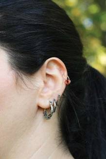
Piercing is a common practice in many cultures. In the United States, it most often involves rings in the earlobes, but rings, posts, rods, and dermal anchors are increasingly frequent in other body parts, including the nose, tongue, eyelid, lips, ear cartilage, nipples, belly, genitals, and more. When pop artist Lady Gaga showed up at the Grammy Awards sporting subdermal implants on her forehead, some fans began copying her.
One survey of 225 adolescents with piercings who were seen at an urban hospital found associated infection in 74%, bleeding in 30%, allergic reactions in 26%, and keloids in 19%, Dr. Chin said.
The upper ear cartilage is largely avascular, and thus prone to poor healing and more serious infection from piercing. In such cases, treat infection for staphylococcus or streptococcus infection (as well as methicillin-resistant S. aureus, if it’s a significant infection in your geographical area), but if the patient doesn’t improve after a few days of treatment, switch to ciprofloxacin and assess for Pseudomonas aeruginosa infection, she advised.
Tongue piercings can chip or fracture teeth and significantly increase the risk for gingival recession requiring gum surgery. Aspiration of tongue piercings have been reported during contact sports. Tongue piercings also have been associated with blood-borne infections (such as hepatitis and HIV), endocarditis, significant blood loss, and lingering pain, including trigeminal neuralgia.
Systemic infections that have been seen from body piercings include tetanus, acute poststreptococcal glomerulonephritis, streptococcal septicemia, staphylococcal toxic shock syndrome, and pseudomonal abscesses.
Any piercing can cause a traumatic laceration, which may be sutured just like any other laceration, she said.
Some penile piercings can lead to paraphimosis, and without prompt treatment the tissue ischemia may cause gangrene and autoamputation of the distal penis. Urethral injuries, infections, prolonged priapism, and recurrent condyloma acuminatum are other risks from male genital piercing. Female genital piercings increase the risk for vaginal lacerations, sexually transmitted diseases, and urinary tract infections.
Remind any patients with genital piercings to use condoms for any sexual contact, and to be aware that condoms may be torn by the body jewelry, Dr. Chin said.
Learn how to remove the most common kinds of body piercing jewelry, she suggested. These include barbell studs, labret studs, and bead rings, which may be removed by unscrewing and/or pulling on parts of the jewelry. A captive bead ring is most easily removed using ring expanding pliers or external snap ring pliers.
Pearling is another body modification technique in which small objects of various materials are placed beneath the skin of the penis, hands, or other body parts. The risks and healing characteristics are similar to those of any subdermal implants, and rejection can occur but is rare. Migration of the implanted material is common, however, both during and after healing.
At Dr. Chin’s institution, it’s not uncommon to see problematic penis rings, in which a metal ring that the patient has placed over his penis to keep an erection becomes stuck and interferes with blood flow. "We call the fire department" to come cut off the ring, she said. These patients can’t be given too much sedation, because the room must be free of excess oxygen when the fire department’s saw sends sparks flying.
If you see this problem and your fire department won’t come, call the orthopedics department. "They have the right tools" for the job, she said.
Dr. Chin said she has no relevant conflicts of interest.
SAN FRANCISCO – Body modifications are all the rage, so physicians need to know that troublesome tattoos can interfere with MRIs, and should keep a pair of pliers handy to deal with problematic piercings.
Approximately 36% of Americans aged 25-29 years have one or more tattoos. Piercings are most common among 16- to 20-year-olds, 47% of whom have rings, anchors, studs, or other metallic objects poking through various body parts. Some 10% of Americans aged 12-15 years and 27% of people aged 21-25 years have body piercings, Dr. Rachel L. Chin said at the annual meeting of the American College of Emergency Physicians.
Patients aren’t the only ones favoring the fashion.
"I don’t think I can name a single night nurse who doesn’t have more than one piercing other than in the ear," said Dr. Chin of San Francisco General Hospital and a professor of emergency medicine at the University of California, San Francisco.
Tattoo pigments may contain heavy metals and iron that act as conductors during MRI and can cause a burning sensation and intense pain. Although a 2002 survey of 1,032 patients with tattoos who underwent MRI reported no serious soft-tissue reactions or adverse events, the popularity of tattoos has grown astronomically in the past decade, and there now are many case reports of severe burning associated with tattoos and MRI, Dr. Chin said.
If this happens, apply a cool compress or ice packs, she suggested.
It’s difficult to know what’s in an individual’s tattoo. There are no federal regulations of tattoo pigments or studios that offer tattoos, and international pigment suppliers rarely produce lists of ingredients. Tattoo artists also may mix their own colors, and some use printer’s ink or automobile paint, she said. Most tattoo artists have had no formal training in anatomy, infection control, or universal precautions.
The Red Cross prohibits blood donations from anyone who has gotten a tattoo or piercing in the past 12 months unless the tattoo was applied by a studio certified by the Association of Professional Piercers or the Alliance of Professional Tattooists.
Tattoo recipients may show up with another common problem: infection. One study of 766 college students found that tattoos were associated with infection in 45%, local skin reactions in 39%, and hepatitis in two cases (Clin. Nurs. Res. 1999;8:368-85). Infection with hepatitis B and C, HIV, Pseudomonas, Staphylococcus, tetanus, and syphilis have been associated with tattoos. Tattoos from unlicensed artists were associated with outbreaks of methicillin-resistant S. aureus in Ohio, Kentucky, and Vermont.
Tattoo infections can be deadly. If you see a febrile patient with a recent tattoo and no other source of infection, consider the possibility of infective endocarditis, Dr. Chin said.
Another body modification technique called scarification is legal in some states, and regulations of shops vary by county or city. Customers may be branded using an electrocautery knife or may undergo scarring via a chemical burn, a tattoo gun without ink, or a scalpel to remove the outer layer of skin tissue. The design result relies solely on how well the body scars.
Piercing is a common practice in many cultures. In the United States, it most often involves rings in the earlobes, but rings, posts, rods, and dermal anchors are increasingly frequent in other body parts, including the nose, tongue, eyelid, lips, ear cartilage, nipples, belly, genitals, and more. When pop artist Lady Gaga showed up at the Grammy Awards sporting subdermal implants on her forehead, some fans began copying her.
One survey of 225 adolescents with piercings who were seen at an urban hospital found associated infection in 74%, bleeding in 30%, allergic reactions in 26%, and keloids in 19%, Dr. Chin said.
The upper ear cartilage is largely avascular, and thus prone to poor healing and more serious infection from piercing. In such cases, treat infection for staphylococcus or streptococcus infection (as well as methicillin-resistant S. aureus, if it’s a significant infection in your geographical area), but if the patient doesn’t improve after a few days of treatment, switch to ciprofloxacin and assess for Pseudomonas aeruginosa infection, she advised.
Tongue piercings can chip or fracture teeth and significantly increase the risk for gingival recession requiring gum surgery. Aspiration of tongue piercings have been reported during contact sports. Tongue piercings also have been associated with blood-borne infections (such as hepatitis and HIV), endocarditis, significant blood loss, and lingering pain, including trigeminal neuralgia.
Systemic infections that have been seen from body piercings include tetanus, acute poststreptococcal glomerulonephritis, streptococcal septicemia, staphylococcal toxic shock syndrome, and pseudomonal abscesses.
Any piercing can cause a traumatic laceration, which may be sutured just like any other laceration, she said.
Some penile piercings can lead to paraphimosis, and without prompt treatment the tissue ischemia may cause gangrene and autoamputation of the distal penis. Urethral injuries, infections, prolonged priapism, and recurrent condyloma acuminatum are other risks from male genital piercing. Female genital piercings increase the risk for vaginal lacerations, sexually transmitted diseases, and urinary tract infections.
Remind any patients with genital piercings to use condoms for any sexual contact, and to be aware that condoms may be torn by the body jewelry, Dr. Chin said.
Learn how to remove the most common kinds of body piercing jewelry, she suggested. These include barbell studs, labret studs, and bead rings, which may be removed by unscrewing and/or pulling on parts of the jewelry. A captive bead ring is most easily removed using ring expanding pliers or external snap ring pliers.
Pearling is another body modification technique in which small objects of various materials are placed beneath the skin of the penis, hands, or other body parts. The risks and healing characteristics are similar to those of any subdermal implants, and rejection can occur but is rare. Migration of the implanted material is common, however, both during and after healing.
At Dr. Chin’s institution, it’s not uncommon to see problematic penis rings, in which a metal ring that the patient has placed over his penis to keep an erection becomes stuck and interferes with blood flow. "We call the fire department" to come cut off the ring, she said. These patients can’t be given too much sedation, because the room must be free of excess oxygen when the fire department’s saw sends sparks flying.
If you see this problem and your fire department won’t come, call the orthopedics department. "They have the right tools" for the job, she said.
Dr. Chin said she has no relevant conflicts of interest.
EXPERT ANALYSIS FROM THE ANNUAL MEETING OF THE AMERICAN COLLEGE OF EMERGENCY PHYSICIANS
Dispel Myths to Recognize Child Abuse
SAN FRANCISCO – The color of a bruise indicates its age. You’ll almost always see bruising when a child has a fracture. Sexual abuse leaves behind physical exam findings.
These are all myths that can get in the way of physicians recognizing abuse of an infant or child. Physicians are required by law to report all suspicions of nonaccidental trauma, a catch-all term for child abuse, shaken baby syndrome, and battered-child syndrome.

Physicians can meet that obligation by ignoring these myths, recognizing red flags for nonaccidental trauma, and being familiar with signs of accidental trauma or medical conditions that can mimic the physical findings of nonaccidental trauma, Dr. Maureen D. McCollough said at the annual meeting of the American College of Emergency Physicians.
Myth: The age of bruises can be accurately determined by their color – red, purple, yellow, green, or brown. In reality, there is no predictable order or chronology of color in bruising, and even in the same person bruises of similar ages may have different colors, said Dr. McCollough of the University of Southern California, Los Angeles, and director of pediatric emergency medicine at Los Angeles County USC Medical Center.
Studies have shown poor interobserver reliability in assessing bruise coloring and poor physician accuracy in characterizing coloring.
Red flags of suspicion should go up if you see multiple bruises or lacerations, or see them in unusual locations. Accidental toddler tumbles can produce multiple bruises, but generally these are on bony prominences. Unusual locations for pediatric bruising include the lower back, buttocks, cheeks, ears, or neck. Bruising anywhere in an infant who is not yet mobile is suspicious.
"Remember, if you don’t cruise, you don’t bruise," she said.
Be suspicious if the pattern of the marks, bruises, or lacerations remind you of an object like a hand, hairbrush, belt, or buckle. Bruises around wrists or extremities may be from the child being tied up. Tight elastic socks can leave a mark around an infant’s leg that mimics this, in which case the parent should be able to provide a sock with dimensions that match the bruising.
Visible injuries around a baby’s mouth or frenulum should raise a red flag for forced feeding. Genital injuries may indicate forced toilet training. Hair pulling produces characteristic marks of traumatic alopecia – an incompletely bald child with diffuse alopecia, broken hairs, and no loose hairs at the periphery.
A wide variety of problems can mimic the visual signs of nonaccidental bruising, including dermal melanosis, vitamin K deficiency, leukemia, hemophilia, millipede secretions, Ehlers-Danlos syndrome, dermatitis, lice, and more.
An equally impressive array of events can mimic the look of abusive burns, bullae, and erythema. These include the cultural practices of coining, cupping, spooning, or moxibustion, skin infections, allergic reactions, herpes or varicella infection, diaper dermatitis, impetigo, and more.
Accidental burns usually have a typical "splash" pattern if liquid is involved, or a child who grasps something hot will have burns on the volar aspect of the fingers and palm. Accidental cigarette burns usually have a streaky appearance.
If there are no splash marks, or there is a sharp line of demarcation, or burns are limited to the perineum, consider that the child may have been forcibly immersed in something hot. Intentional cigarette burns tend to be similar in size – often 5-mm circles – and create injuries from bullae to deep craters that scab over. These usually are on the palms or soles but can be anywhere on the body. Again, be suspicious if you see a burn mark that looks like an object, such as a radiator or an iron.
Myth: Fractures usually are associated with overlying bruising. In fact, children with inflicted skeletal fractures often have no associated bruising. Bruising is present in only 43% of skull fractures and less than 20% of lower extremity fractures in cases of abuse, Dr. McCollough said.
Infants who can’t walk shouldn’t fracture. Spiral fractures caused by the twisting of a long bone such as the femur suggest nonaccidental trauma. Toddler spiral fractures of the tibia, on the other hand, are very common, caused when a leg is trapped under the body during a fall, such as getting a leg caught in a couch. "This is not abuse," she said.
Raise the red flags when you see swelling of a body part that is out of proportion to a described injury; this may indicate an underlying fracture. A diaphyseal (midshaft) fracture in a child less than 3 years old is suspect, and metaphyseal or epiphyseal fractures beyond the newborn period (also called corner fractures or bucket handle fractures) are virtually diagnostic of abuse.
The posterior ribs are the most common area of nonaccidental rib fractures.
Suspect head injuries and possible abuse if the child has unexplained seizures, vomiting, changes in neurological or mental status, or large scalp hematomas. Be suspicious if the parents’ explanation changes over time, if there is intracranial bleeds after "minimal" trauma, or if you find retinal hemorrhages outside of the newborn period, she said.
Myth: Sexual abuse leaves physical findings. More myths: A colposcope is needed to detect sexual abuse, and some girls are born without hymens.
Although hymens come in a wide variety of shapes and sizes, a study of more than 1,100 newborn girls showed that all of them had one, she noted. Reviews of cases of sexual abuse show that physical exam findings of pediatric sexual abuse are rare because the tissue is very elastic and heals quickly.
Physical evidence will be more likely if force was used, if the child resisted, if there are great differences in the sizes and ages of the perpetrator and victim, and if a foreign object was forced into the mouth, vagina, or anus. Bruising or bite marks on a child’s penis may suggest nonaccidental trauma from forced toilet training.
When you see visible clues to what may be abuse, photograph or draw what you see and include something in the image to show size or scale. Don’t just rely on written notes, she said.
SAN FRANCISCO – The color of a bruise indicates its age. You’ll almost always see bruising when a child has a fracture. Sexual abuse leaves behind physical exam findings.
These are all myths that can get in the way of physicians recognizing abuse of an infant or child. Physicians are required by law to report all suspicions of nonaccidental trauma, a catch-all term for child abuse, shaken baby syndrome, and battered-child syndrome.
Physicians can meet that obligation by ignoring these myths, recognizing red flags for nonaccidental trauma, and being familiar with signs of accidental trauma or medical conditions that can mimic the physical findings of nonaccidental trauma, Dr. Maureen D. McCollough said at the annual meeting of the American College of Emergency Physicians.
Myth: The age of bruises can be accurately determined by their color – red, purple, yellow, green, or brown. In reality, there is no predictable order or chronology of color in bruising, and even in the same person bruises of similar ages may have different colors, said Dr. McCollough of the University of Southern California, Los Angeles, and director of pediatric emergency medicine at Los Angeles County USC Medical Center.
Studies have shown poor interobserver reliability in assessing bruise coloring and poor physician accuracy in characterizing coloring.
Red flags of suspicion should go up if you see multiple bruises or lacerations, or see them in unusual locations. Accidental toddler tumbles can produce multiple bruises, but generally these are on bony prominences. Unusual locations for pediatric bruising include the lower back, buttocks, cheeks, ears, or neck. Bruising anywhere in an infant who is not yet mobile is suspicious.
"Remember, if you don’t cruise, you don’t bruise," she said.
Be suspicious if the pattern of the marks, bruises, or lacerations remind you of an object like a hand, hairbrush, belt, or buckle. Bruises around wrists or extremities may be from the child being tied up. Tight elastic socks can leave a mark around an infant’s leg that mimics this, in which case the parent should be able to provide a sock with dimensions that match the bruising.
Visible injuries around a baby’s mouth or frenulum should raise a red flag for forced feeding. Genital injuries may indicate forced toilet training. Hair pulling produces characteristic marks of traumatic alopecia – an incompletely bald child with diffuse alopecia, broken hairs, and no loose hairs at the periphery.
A wide variety of problems can mimic the visual signs of nonaccidental bruising, including dermal melanosis, vitamin K deficiency, leukemia, hemophilia, millipede secretions, Ehlers-Danlos syndrome, dermatitis, lice, and more.
An equally impressive array of events can mimic the look of abusive burns, bullae, and erythema. These include the cultural practices of coining, cupping, spooning, or moxibustion, skin infections, allergic reactions, herpes or varicella infection, diaper dermatitis, impetigo, and more.
Accidental burns usually have a typical "splash" pattern if liquid is involved, or a child who grasps something hot will have burns on the volar aspect of the fingers and palm. Accidental cigarette burns usually have a streaky appearance.
If there are no splash marks, or there is a sharp line of demarcation, or burns are limited to the perineum, consider that the child may have been forcibly immersed in something hot. Intentional cigarette burns tend to be similar in size – often 5-mm circles – and create injuries from bullae to deep craters that scab over. These usually are on the palms or soles but can be anywhere on the body. Again, be suspicious if you see a burn mark that looks like an object, such as a radiator or an iron.
Myth: Fractures usually are associated with overlying bruising. In fact, children with inflicted skeletal fractures often have no associated bruising. Bruising is present in only 43% of skull fractures and less than 20% of lower extremity fractures in cases of abuse, Dr. McCollough said.
Infants who can’t walk shouldn’t fracture. Spiral fractures caused by the twisting of a long bone such as the femur suggest nonaccidental trauma. Toddler spiral fractures of the tibia, on the other hand, are very common, caused when a leg is trapped under the body during a fall, such as getting a leg caught in a couch. "This is not abuse," she said.
Raise the red flags when you see swelling of a body part that is out of proportion to a described injury; this may indicate an underlying fracture. A diaphyseal (midshaft) fracture in a child less than 3 years old is suspect, and metaphyseal or epiphyseal fractures beyond the newborn period (also called corner fractures or bucket handle fractures) are virtually diagnostic of abuse.
The posterior ribs are the most common area of nonaccidental rib fractures.
Suspect head injuries and possible abuse if the child has unexplained seizures, vomiting, changes in neurological or mental status, or large scalp hematomas. Be suspicious if the parents’ explanation changes over time, if there is intracranial bleeds after "minimal" trauma, or if you find retinal hemorrhages outside of the newborn period, she said.
Myth: Sexual abuse leaves physical findings. More myths: A colposcope is needed to detect sexual abuse, and some girls are born without hymens.
Although hymens come in a wide variety of shapes and sizes, a study of more than 1,100 newborn girls showed that all of them had one, she noted. Reviews of cases of sexual abuse show that physical exam findings of pediatric sexual abuse are rare because the tissue is very elastic and heals quickly.
Physical evidence will be more likely if force was used, if the child resisted, if there are great differences in the sizes and ages of the perpetrator and victim, and if a foreign object was forced into the mouth, vagina, or anus. Bruising or bite marks on a child’s penis may suggest nonaccidental trauma from forced toilet training.
When you see visible clues to what may be abuse, photograph or draw what you see and include something in the image to show size or scale. Don’t just rely on written notes, she said.
EXPERT ANALYSIS FROM THE ANNUAL MEETING OF THE AMERICAN COLLEGE OF EMERGENCY PHYSICIANS
Definition of Giant Congenital Nevus Refined
SAN FRANCISCO – A new definition of giant congenital nevi should help physicians identify infants with supersized moles that are at risk for melanoma, neurocutaneous melanocytosis, liposarcoma, and other tumors.
For many years pediatric dermatologists sized congenital nevi as small, medium, or large, with "large" comprising nevi that are estimated to be 20 cm or greater in diameter when the patient has grown into adulthood. Then, physicians started referring to some of the large nevi as "giant" congenital nevi, with no universal agreement on what defines a giant nevus, Dr. Sheila Fallon Friedlander said at the Women’s & Pediatric Dermatology Seminar sponsored by Skin Disease Education Foundation (SDEF).
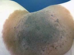
"Here’s our new definition that you’re going to be seeing in the literature," said Dr. Friedlander of the University of California, San Diego. Congenital nevi with diameters less than 1.5 cm after the patient is fully grown are considered small, nevi with diameters of at least 1.5 cm but less than 20 cm are medium size, and large nevi are at least 20 cm but less than 40 cm in diameter. Giant congenital nevi are those with diameters of 40 cm or greater when the patient is fully grown.
"When you are examining a baby in a nursery, how does that help you? That’s an adult-type figure," she noted. To estimate the grown size of a congenital nevus, measure the nevus on the baby and multiply by a scaling factor that’s based on different growth rates for different parts of the body. Lower extremities grow the most, so multiply the size of nevi located there by 3.3. Multiply the size of a nevus on the torso or an upper extremity by 2.8, and a nevus on the head by 1.7.
For example, a congenital nevus 7 cm in diameter on a baby’s leg qualifies as a large congenital nevus (7 x 3.3 = 23.1 cm), "so you need to worry more" about risks for that baby, she said.
Estimates of risk have been evolving with the terminology. Most studies have lumped large and giant categories together and referred to them as large congenital nevi. The true risks in studies of registry data are clouded by the fact that many families choose to excise or partially excise large congenital nevi.
Between 1% and 11% of patients with large congenital nevi will develop malignant melanoma or neurocutaneous melanocytosis, a 52-1,000 times greater risk than for someone without the lesion, according to the results of a New York University study of 160 patients (Pediatrics 2000;106:736-41). Large congenital nevi also are associated with increased risks for liposarcoma, rhabdomyosarcoma, and (in some body locations) tethered spinal cord syndrome.
In the New York University study, 14 of 15 patients who developed melanoma or neurocutaneous melanocytosis had lesions greater than 50 cm in diameter, which now would be considered giant. The risk is highest for lesions on the torso. The melanoma often can occur deep in the dermal-epidermal junction, making them hard to find, Dr. Friedlander said. The melanoma also can occur underneath the lesion in the CNS or mesentery.
Although melanomas in the general population are unlikely to develop until adolescence or adulthood, in children with large congenital nevi who develop melanoma, 70% of the cancers appear by age 10 years, and 50% appear by age 5. "So this is something that you can’t put off. You really need to deal with it," she said.

Studies report that 7% of patients with large congenital nevi will develop neurocutaneous melanocytosis, an aberrant migration of melanocytes in leptomeninges that creates a risk for ventricular obstruction. The presence of more than 20 satellite lesions around a large congenital nevus increases the risk for neurocutaneous melanocytosis five-fold and warrants an MRI evaluation, she said.
"Five years ago I was taught that posterior axial location may have a higher risk for neurocutaneous melanocytosis, but recent data shows that it’s the satellite lesions rather than the location" that matters, Dr. Friedlander said.
A posterior axial location is, however, a risk factor for cutaneous melanoma in patients with large congenital nevi. Nevus size is a risk factor for both melanoma and neurocutaneous melanocytosis, said Dr. Friedlander; "20 cm is bad, and 40 cm is worse."
Dr. Friedlander disclosed that she has been a speaker, consultant, or researcher for Pierre Fabre, Onset Therapeutics, Johnson & Johnson, and Galderma.
SDEF and this news organization are owned by Elsevier.
SAN FRANCISCO – A new definition of giant congenital nevi should help physicians identify infants with supersized moles that are at risk for melanoma, neurocutaneous melanocytosis, liposarcoma, and other tumors.
For many years pediatric dermatologists sized congenital nevi as small, medium, or large, with "large" comprising nevi that are estimated to be 20 cm or greater in diameter when the patient has grown into adulthood. Then, physicians started referring to some of the large nevi as "giant" congenital nevi, with no universal agreement on what defines a giant nevus, Dr. Sheila Fallon Friedlander said at the Women’s & Pediatric Dermatology Seminar sponsored by Skin Disease Education Foundation (SDEF).
"Here’s our new definition that you’re going to be seeing in the literature," said Dr. Friedlander of the University of California, San Diego. Congenital nevi with diameters less than 1.5 cm after the patient is fully grown are considered small, nevi with diameters of at least 1.5 cm but less than 20 cm are medium size, and large nevi are at least 20 cm but less than 40 cm in diameter. Giant congenital nevi are those with diameters of 40 cm or greater when the patient is fully grown.
"When you are examining a baby in a nursery, how does that help you? That’s an adult-type figure," she noted. To estimate the grown size of a congenital nevus, measure the nevus on the baby and multiply by a scaling factor that’s based on different growth rates for different parts of the body. Lower extremities grow the most, so multiply the size of nevi located there by 3.3. Multiply the size of a nevus on the torso or an upper extremity by 2.8, and a nevus on the head by 1.7.
For example, a congenital nevus 7 cm in diameter on a baby’s leg qualifies as a large congenital nevus (7 x 3.3 = 23.1 cm), "so you need to worry more" about risks for that baby, she said.
Estimates of risk have been evolving with the terminology. Most studies have lumped large and giant categories together and referred to them as large congenital nevi. The true risks in studies of registry data are clouded by the fact that many families choose to excise or partially excise large congenital nevi.
Between 1% and 11% of patients with large congenital nevi will develop malignant melanoma or neurocutaneous melanocytosis, a 52-1,000 times greater risk than for someone without the lesion, according to the results of a New York University study of 160 patients (Pediatrics 2000;106:736-41). Large congenital nevi also are associated with increased risks for liposarcoma, rhabdomyosarcoma, and (in some body locations) tethered spinal cord syndrome.
In the New York University study, 14 of 15 patients who developed melanoma or neurocutaneous melanocytosis had lesions greater than 50 cm in diameter, which now would be considered giant. The risk is highest for lesions on the torso. The melanoma often can occur deep in the dermal-epidermal junction, making them hard to find, Dr. Friedlander said. The melanoma also can occur underneath the lesion in the CNS or mesentery.
Although melanomas in the general population are unlikely to develop until adolescence or adulthood, in children with large congenital nevi who develop melanoma, 70% of the cancers appear by age 10 years, and 50% appear by age 5. "So this is something that you can’t put off. You really need to deal with it," she said.
Studies report that 7% of patients with large congenital nevi will develop neurocutaneous melanocytosis, an aberrant migration of melanocytes in leptomeninges that creates a risk for ventricular obstruction. The presence of more than 20 satellite lesions around a large congenital nevus increases the risk for neurocutaneous melanocytosis five-fold and warrants an MRI evaluation, she said.
"Five years ago I was taught that posterior axial location may have a higher risk for neurocutaneous melanocytosis, but recent data shows that it’s the satellite lesions rather than the location" that matters, Dr. Friedlander said.
A posterior axial location is, however, a risk factor for cutaneous melanoma in patients with large congenital nevi. Nevus size is a risk factor for both melanoma and neurocutaneous melanocytosis, said Dr. Friedlander; "20 cm is bad, and 40 cm is worse."
Dr. Friedlander disclosed that she has been a speaker, consultant, or researcher for Pierre Fabre, Onset Therapeutics, Johnson & Johnson, and Galderma.
SDEF and this news organization are owned by Elsevier.
EXPERT ANALYSIS FROM THE SDEF WOMEN'S & PEDIATRIC DERMATOLOGY SEMINAR
Simple Leg Raise Assesses 'Tank' in Septic Shock
SAN FRANCISCO – Before reaching for vasopressors to treat a patient in septic shock, physicians want to know that the patient has received enough fluid. Two traditional ways of assessing the patient’s "tank" both have limitations, but a relatively simple bedside procedure can predict the need for a fluid bolus.
Observing respiratory variations in arterial wave form analysis while health care workers raise a supine patient’s legs into a sitting position helps assess the volume status, also known as the pre-load or "the tank," in a patient with septic shock, Dr. Peter M. C. DeBlieux said at the annual meeting of the American College of Emergency Physicians.
"What that requires is a simple arterial line placed in either the radial or femoral artery, and then that’s transduced" and observed on a monitor with the heart rate, he said. Changes greater than 12%-15% in the pulse amplitude of the arterial wave form during either physiologic or mechanical breathing predicts that the patient would benefit from a fluid bolus, said Dr. DeBlieux, professor of medicine at Louisiana State University and professor of surgery at Tulane University, both in New Orleans.
This is more easily seen by raising the legs of a supine patient with septic shock. The patient’s head may be at a 45-degree angle, but the legs should start flat on the table. Two health care workers on either side of the bed should raise the patient’s legs to 90-degree angles at the hips and knees, as if the patient was in a sitting position, while observing the arterial tracing. If the arterial tracing shows at least a 12%-15% increase in pulse pressure, that warrants a fluid bolus.
"It’s a pretty simple thing to do at the bedside that changes management," he said.
Previously, volume status in patients with septic shock has been assessed by central venous pressure monitoring. Bedside ultrasound also has been used, focusing on the inferior vena cava and the collapsibility of the vena cava during normal respiratory cycles. A collapse of 20%-50% indicates that a fluid bolus would be beneficial. Both of these measures can give false elevations, however, if the patient has preexisting acute or chronic heart or lung disease.
Although an elevated central venous pressure (CVP) or collapsing inferior vena cava (IVC) on ultrasound during respiration indicated the need for a fluid bolus, "once the CVP is elevated or if you have a plethoric IVC, you’re left scratching your head," Dr. DeBlieux said. "Remember that any cardiac or pulmonary dysfunction gets immediately transmitted to the great vein," so the CVP may be high, or the IVC may look plethoric on ultrasound, but the patient may still need a fluid bolus.
"You would say that the tank is full, but you don’t know. I don’t know. We don’t know," he said.
Managing the tank corresponds with the first of three steps in the Surviving Sepsis Campaign's perfusion goals in early goal-directed therapy in acute care of septic shock. Dr. DeBlieux and some other educators refer to these as the tank, the hose and the pump for teaching purposes.
The tank goal is a CVP of 8-12 mm Hg through treatment with serial crystalloid fluid infusions.
The hose is marked by systemic vascular resistance. After addressing the tank, if the mean arterial blood pressure is lower than 65 mmHg, administer norepinephrine, dopamine or epinephrine to bring it up to 65 mm Hg. The medical literature supports any one of those three choices, but norepinephrine will produce fewer tachyarrhythmic events, he said.
In reality, managing the tank and hose may happen simultaneously because physicians may not want to wait for hours until the CVP gets to goal before administering vasopressors, he added.
The pump reflects oxygen delivery and utilization. After addressing the tank and hose, if the lactate level is greater than 4 mmol and central venous oxygen saturation (ScvO2) is less than 70%, consider giving dobutamine in the setting of left ventricular dysfunction, start invasive mechanical ventilation, and/or blood transfusion if the hemoglobin/hematocrit is less than 10/30 within 6 hours.
Dr. DeBlieux said he has no relevant conflicts of interest.
EXPERT ANALYSIS FROM THE ANNUAL MEETING OF THE AMERICAN COLLEGE OF EMERGENCY PHYSICIANS
Five or More Infantile Hemangiomas? Order Liver Ultrasound
SAN FRANCISCO – Infants with five or more hemangiomas should undergo a liver ultrasound according to new research, said Dr. Ilona J. Frieden.
Clinicians know that when they see infants with multiple cutaneous hemangiomas, they need to worry about internal hemangiomas, said Dr. Frieden at the seminar, sponsored by Skin Disease Education Foundation (SDEF). And now, for the first time, a prospective, multicenter study identified how many infantile hemangiomas should trigger alarm.

The study screened infants younger than 6 months in the United States, Canada, and Spain with abdominal ultrasound and found that 16% of 151 infants with five or more skin hemangiomas had liver hemangiomas, compared with no liver hemangiomas in a control group of infants with one to four skin hemangiomas, she said.
"We certainly recommend doing liver ultrasound in children with five or more hemangiomas," said Dr. Frieden, professor of dermatology and pediatrics at the University of California, San Francisco. "Five does seem to be the right threshold for doing this screening test."
Interestingly, only 2 of the 24 infants with hepatic hemangiomas received treatment specifically for the hepatic hemangiomas (Pediatr. Dermatol. 2011;28:245-53). "Liver hemangiomas are probably a lot like skin hemangiomas – they don’t all need treatment. That’s an important fact," she said.
In general, segmental hemangiomas are more likely to develop complications, and facial segmental hemangiomas are more likely to need systemic treatment. The main indication for treating cutaneous infantile hemangiomas is a risk of scarring and disfigurement, especially with facial segmental hemangiomas, ulcerated hemangiomas, and hemangiomas located on the ear or on any prominent facial site but especially the nasal tip or perioral areas.
The study found that 16% of 151 infants with five or more skin hemangiomas had liver hemangiomas.
Treatment should also be considered in patients with periocular or liver hemangiomas, hemangiomas of the airway, spinal dysraphism or genitourinary complications, or in PHACE syndrome, she said.
A previous study by Dr. Frieden and her associates showed that 30% of infants with large facial hemangiomas were shown to have PHACE syndrome (Pediatrics 2010;126:e418-26).
Dr. Frieden, who played a leading role in defining PHACE syndrome, estimated it to be a more common neurocutaneous syndrome than Sturge-Weber: "Not super-rare, but not common," she said.
Any infant with a facial hemangioma measuring 5 cm or more in diameter probably deserves cardiac echocardiography to look for transverse coarctation of the aorta, MRI and magnetic resonance angiography to look for arterial anomalies, and an eye exam to look for structural eye abnormalities even if there is no periocular hemangioma, she advised.
Infants with lumbosacral hemangiomas are at greater risk for having a tethered spinal cord or other problems such as genitourinary abnormalities. Approximately half of infants with typical lumbosacral hemangiomas will have a tethered spinal cord, prospective studies suggest.
Lumbosacral hemangiomas tend to stay flatter and be less bulky than hemangiomas on other body areas and so may not raise the suspicion of problems that would be warranted. Dr. Frieden has diagnosed tethered spinal cord in several infants with lumbosacral hemangiomas who were referred only after they developed ulcerations.
Dr. Frieden has been an advisor or consultant to Pierre Fabre and Topaz Pharmaceuticals.
SDEF and this news organization are owned by Elsevier.
SAN FRANCISCO – Infants with five or more hemangiomas should undergo a liver ultrasound according to new research, said Dr. Ilona J. Frieden.
Clinicians know that when they see infants with multiple cutaneous hemangiomas, they need to worry about internal hemangiomas, said Dr. Frieden at the seminar, sponsored by Skin Disease Education Foundation (SDEF). And now, for the first time, a prospective, multicenter study identified how many infantile hemangiomas should trigger alarm.
The study screened infants younger than 6 months in the United States, Canada, and Spain with abdominal ultrasound and found that 16% of 151 infants with five or more skin hemangiomas had liver hemangiomas, compared with no liver hemangiomas in a control group of infants with one to four skin hemangiomas, she said.
"We certainly recommend doing liver ultrasound in children with five or more hemangiomas," said Dr. Frieden, professor of dermatology and pediatrics at the University of California, San Francisco. "Five does seem to be the right threshold for doing this screening test."
Interestingly, only 2 of the 24 infants with hepatic hemangiomas received treatment specifically for the hepatic hemangiomas (Pediatr. Dermatol. 2011;28:245-53). "Liver hemangiomas are probably a lot like skin hemangiomas – they don’t all need treatment. That’s an important fact," she said.
In general, segmental hemangiomas are more likely to develop complications, and facial segmental hemangiomas are more likely to need systemic treatment. The main indication for treating cutaneous infantile hemangiomas is a risk of scarring and disfigurement, especially with facial segmental hemangiomas, ulcerated hemangiomas, and hemangiomas located on the ear or on any prominent facial site but especially the nasal tip or perioral areas.
The study found that 16% of 151 infants with five or more skin hemangiomas had liver hemangiomas.
Treatment should also be considered in patients with periocular or liver hemangiomas, hemangiomas of the airway, spinal dysraphism or genitourinary complications, or in PHACE syndrome, she said.
A previous study by Dr. Frieden and her associates showed that 30% of infants with large facial hemangiomas were shown to have PHACE syndrome (Pediatrics 2010;126:e418-26).
Dr. Frieden, who played a leading role in defining PHACE syndrome, estimated it to be a more common neurocutaneous syndrome than Sturge-Weber: "Not super-rare, but not common," she said.
Any infant with a facial hemangioma measuring 5 cm or more in diameter probably deserves cardiac echocardiography to look for transverse coarctation of the aorta, MRI and magnetic resonance angiography to look for arterial anomalies, and an eye exam to look for structural eye abnormalities even if there is no periocular hemangioma, she advised.
Infants with lumbosacral hemangiomas are at greater risk for having a tethered spinal cord or other problems such as genitourinary abnormalities. Approximately half of infants with typical lumbosacral hemangiomas will have a tethered spinal cord, prospective studies suggest.
Lumbosacral hemangiomas tend to stay flatter and be less bulky than hemangiomas on other body areas and so may not raise the suspicion of problems that would be warranted. Dr. Frieden has diagnosed tethered spinal cord in several infants with lumbosacral hemangiomas who were referred only after they developed ulcerations.
Dr. Frieden has been an advisor or consultant to Pierre Fabre and Topaz Pharmaceuticals.
SDEF and this news organization are owned by Elsevier.
EXPERT ANALYSIS FROM THE SDEF WOMEN’S & PEDIATRIC DERMATOLOGY SEMINAR
Major Finding: Sixteen percent of infants with five or more hemangiomas had hepatic hemangiomas.
Data Source: Prospective, multicenter study of 151 infants younger than 6 months of age with at least 5 infantile hemangiomas and a control group of infants with 1-4 hemangiomas.
Disclosures: Dr. Frieden has been an advisor or consultant to Pierre Fabre and Topaz Pharmaceuticals. SDEF and this news organization are owned by Elsevier.