User login
Audio - A survey of liability claims against obstetric providers highlights major areas of contention
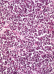
Tool identifies CNAs other algorithms miss

Image courtesy of NIGMS
A new tool can detect genetic alterations that have proven difficult to identify, according to research published in Nature Methods.
The tool is an algorithm called CONSERTING (Copy Number Segmentation by Regression Tree in Next-Generation Sequencing).
Researchers created CONSERTING to improve their ability to detect copy number alterations (CNAs) in the information generated by whole-genome sequencing techniques.
The group showed that CONSERTING could detect CNAs with better accuracy and sensitivity than other techniques, including 4 published algorithms used to recognize CNAs in whole-genome sequencing data.
The data the team analyzed encompassed the normal and tumor genomes from 43 children and adults with leukemia, brain tumors, melanoma, and retinoblastoma.
“CONSERTING helped us identify alterations that other algorithms missed, including previously undetected chromosomal rearrangements and copy number alterations present in a small percentage of tumor cells,” said study author Xiang Chen, PhD, of St. Jude Children’s Research Hospital in Memphis, Tennessee.
“[C]ONSERTING identified copy number alterations in children with 100 times greater precision and 10 times greater precision in adults,” added Jinghui Zhang, PhD, also of St. Jude.
Using CONSERTING, the researchers discovered genetic alterations driving pediatric leukemia, low-grade glioma, glioblastoma, and retinoblastoma.
The algorithm also helped the team identify genetic changes that are present in a small percentage of a tumor’s cells. The alterations may be the key to understanding why tumors sometimes return after treatment, they said.
Dr Zhang said CONSERTING should make it easier to track the evolution of tumors with complex genetic rearrangements.
St. Jude has made CONSERTING available to researchers free of charge. The software, user manual, and related data can be downloaded from http://www.stjuderesearch.org/site/lab/zhang.
St. Jude researchers have also developed a cloud version of CONSERTING and related tools that can be accessed through Amazon Web Services. Instead of downloading CONSERTING, scientists can upload data for analysis.
Creating CONSERTING
Work on CONSERTING began in 2010, shortly after the St. Jude Children’s Research Hospital-Washington University Pediatric Cancer Genome Project was launched. The Pediatric Cancer Genome Project used next-generation, whole-genome sequencing to study some of the most aggressive and least understood childhood cancers.
Early on in the project, researchers realized that existing analytic methods often missed duplications or deletions of DNA segments, particularly small changes that involve a handful of genes and provide insight into the origins of a patient’s cancer.
CONSERTING has now been used to analyze data for the Pediatric Cancer Genome Project. The project includes the normal and cancer genomes of 700 pediatric cancer patients with 21 different cancer subtypes.
CONSERTING combines a method of data analysis called regression tree, which is a machine-learning algorithm, with next-generation, whole-genome sequencing. Machine learning capitalizes on advances in computing to design algorithms that repeatedly and rapidly analyze large, complex sets of data and unearth unexpected insights.
“This combination has provided us with a powerful tool for recognizing copy number alterations, even those present in relatively few cells or in tumor samples that include normal cells along with tumor cells,” Dr Zhang said.
Next-generation, whole-genome sequencing involves breaking the human genome into about 1 billion pieces that are copied and reassembled using the normal genome as a template.
CONSERTING software compensates for gaps and variations in sequencing data. The sequencing data is integrated with information about the chromosomal rearrangements to find CNAs and identify their origins in the genome. ![]()
Agent preferentially targets FLT3-ITD AML

PHILADELPHIA—Preclinical research suggests a novel agent has preferential activity in acute myeloid leukemia (AML) with FMS-like tyrosine kinase 3 internal tandem duplication (FLT3-ITD) mutations.
The agent, VNLG-152, proved more cytotoxic in AML cell lines and patient samples with FLT3-ITD than in samples and cell lines with wild-type FLT3.
Exactly how and why this occurs remains somewhat of a mystery, however.
Sheetal Karne, MD, of the University of Maryland School of Medicine in Baltimore, and her colleagues detailed this mystery in a poster presentation at the AACR Annual Meeting 2015 (abstract 5408*).
Dr Karne noted that VNLG-152 targets translation by promoting the degradation of MAPK-interacting kinases (Mnks).
“[VNLG-152] has been previously published as functioning in Mnk degradation, which has been shown in triple-negative breast cancer and prostate cancer—in vivo and in vitro,” she said. “Our hypothesis was that, since [the drug] worked via decreasing translation, it would function in leukemia cells and, specifically, in leukemic cells with ITD mutations.”
So the investigators tested VNLG-152 in samples from AML patients, as well as both murine and human cell lines. They found that VNLG-152 was more cytotoxic in the presence of FLT3-ITD mutations, as evidenced by low micromolar IC50 concentrations.
The IC50 concentration was 3.4 μM in Ba/F3-ITD cells and 5.8 μM in Ba/F3-WT cells, which are murine cells transfected with human FLT3-ITD and wild-type FLT3, respectively. Similarly, the IC50 concentration was 1.8 μM in 32D-ITD cells and 18.2 μM in 32D-WT cells.
In the human FLT3-ITD AML cell lines MV4-11 and MOLM-14, IC50 concentrations were 2.3 μM and 4.2 μM, respectively. But concentrations were greater than 10 µM in the wild-type FLT3 human cell lines HL60 and U937.
In patient samples, the IC50 concentration was 1.0 μM in FLT3-ITD AML and 7.5 μM in AML with wild-type FLT3.
In additional tests with murine cell lines, the investigators found that VNLG-152 inhibits the growth of Ba/F3-ITD and Ba/F3-WT cells. But the drug induces apoptosis in these cell lines only when given in high concentrations.
Looking into the mechanism of VNLG-152, the investigators found that the drug decreased Mnk-1 expression in Ba/F3-ITD and Ba/F3-WT cell lines.
“We saw that VNLG-152 worked via degradation of Mnk, but it was the same in both wild-type and ITD, so we’re still looking for an explanation as to what caused this difference,” Dr Karne said.
She and her colleagues believe the Mnk degradation inhibits the phosphorylation of eukaryotic translation initiation factor 4E (eIF4E), a downstream target of FLT3-ITD. But they are still investigating that possibility.
The team is also hoping to test VNLG-152 in combination with other drugs, such as FLT3 inhibitors or chemotherapeutic agents. ![]()
*Information in the abstract differs from that presented at the meeting.
Inhibitor gets breakthrough designation for CLL
Though a safety issue previously slowed development of the BCL-2 inhibitor venetoclax (ABT-199), the drug is now moving through the pipeline.
The US Food and Drug Administration (FDA) has granted venetoclax breakthrough therapy designation to treat patients with relapsed or refractory chronic lymphocytic leukemia (CLL), including those with 17p deletion.
The drug has proven active against CLL and other hematologic malignancies.
However, it is also known to induce tumor lysis syndrome (TLS). In fact, TLS-related deaths temporarily halted enrollment in trials of venetoclax. But researchers discovered ways to reduce the risk of TLS, and the trials continued.
Now, the FDA has granted the drug breakthrough designation, which is intended to expedite the development and review of drugs indicated for serious or life-threatening conditions.
The criteria for breakthrough designation include preliminary clinical evidence suggesting the drug may offer substantial improvement on at least one clinically significant endpoint compared to available therapy.
Venetoclax in CLL/SLL
Results presented at the 2014 EHA Congress suggested that venetoclax can be effective in patients with CLL/small lymphocytic lymphoma (SLL), and certain measures can reduce the risk of TLS.
Researchers reported that modifying the dosing schedule of venetoclax, administering TLS prophylaxis, and monitoring patients can decrease or eliminate the risk of TLS. And venetoclax can produce responses in patients with high-risk disease.
In a phase 1 trial, the researchers tested venetoclax monotherapy in 105 patients with high-risk CLL/SLL. Seventy-eight patients were evaluable for treatment response as of April 2014. Nineteen of these patients had del (17p), 41 were fludarabine-refractory, and 24 had unmutated IGHV.
The response rate was 77% overall, 79% among patients with del (17p), 76% in patients who were fludarabine-refractory, and 75% in those with unmutated IGHV. The complete response rates were 23%, 26%, 22%, and 29%, respectively.
The median progression-free survival was about 18 months overall, but the median progression-free survival had not been reached for patients treated at or above 400 mg.
Seven patients developed TLS. One of these patients died, and 1 required dialysis. At the time of analysis, there were no cases of TLS among the 49 patients who received TLS prophylaxis and were given venetoclax according to the modified dosing schedule.
Common treatment-emergent adverse events included diarrhea (40%), neutropenia (36%), and nausea (35%). Grade 3/4 neutropenia occurred in 33% of patients, and febrile neutropenia occurred in 4%.
Thirty-seven patients discontinued treatment—22 due to progressive disease, 12 due to adverse events, and 3 for other reasons (1 required warfarin, and 2 proceeded to transplant).
Venetoclax is now being tested in phase 2 and 3 trials of CLL, as well as trials in other hematologic malignancies. Venetoclax is under development by AbbVie and Genentech/Roche. ![]()
Drug can alleviate transfusion dependence in non-del-5q MDS

Photo courtesy of Celgene
WASHINGTON, DC—Results of a phase 3 trial support the use of lenalidomide in patients with lower-risk myelodysplastic syndromes (MDS) without 5q deletion who are unresponsive or refractory to erythropoiesis-stimulating agents (ESAs), according to researchers.
About 27% of patients who received lenalidomide achieved transfusion independence for 8 weeks or more, and about 18% were transfusion-independent for 24 weeks or more.
Valeria Santini, MD, of AOU Careggi in Florence, Italy, and her colleagues presented these results at the 13th International Symposium on Myelodysplastic Syndromes (abstract 115). The trial, MDS-005, was supported by Celgene Corporation, the company developing lenalidomide.
The trial was a comparison of lenalidomide and placebo in 239 patients with non-del-5q MDS who had failed treatment with ESAs. The patients were transfusion-dependent and had low- or intermediate-1-risk disease according to the International Prognostic Scoring System.
Patients were randomized 2:1 to receive oral lenalidomide at 10 mg once daily on days 1 to 28 of 28-day cycles (5 mg for patients with creatinine clearance 40 to 60 mL/min) or placebo.
Significantly more patients in the lenalidomide arm than in the placebo arm achieved transfusion independence for 8 weeks or more—26.9% vs 2.5% (P<0.001).
Likewise, significantly more patients in the lenalidomide arm than in the placebo arm achieved transfusion independence for 24 weeks or more—17.5% vs 0% (P<0.001).
Ninety percent of the lenalidomide-treated patients who achieved transfusion independence for 8 weeks or more responded within 4 cycles of treatment. The median duration of response was 32.9 weeks.
The median follow-up was 1.6 years (range, 0-3.6) in the lenalidomide arm and 1.3 years (range, 0-4.0) in the placebo arm. Within these time periods, patients in the placebo arm were more likely than those who received lenalidomide to progress to acute myeloid leukemia (AML) or to develop second primary malignancies (SPMs).
The AML incidence rate per 100 person-years was 1.91 in the lenalidomide arm and 2.46 in the placebo arm. And the SPM incidence rate per 100 person-years was 2.19 in the lenalidomide arm and 2.27 in the placebo arm.
As expected, treatment-emergent adverse events (AEs) were more common in the lenalidomide arm than in the placebo arm. AEs included neutropenia (64.4% vs 12.7%), thrombocytopenia (39.4% vs 7.6%), diarrhea (42.5% vs 22.8%), constipation (22.5% vs 12.7%), infections (51.9% vs 43%), hemorrhage (20.6% vs 10.1%), hepatic disorders (14.4% vs 5.1%), cardiac arrhythmia (11.3% vs 8.9%), and cutaneous reactions (10% vs 1.3%).
Grade 3-4 AEs in the lenalidomide arm included neutropenia (61.9%), thrombocytopenia (35.6%), infections (14.4%), hepatic disorders (5%), diarrhea (2.5%), hemorrhage (1.9%), deep vein thrombosis (1.9%), cardiac arrhythmia (1.3%), and cutaneous reactions (1.3%).
Based on the results of this trial, Celgene plans to submit a regulatory filing with the US Food and Drug Administration in the second half of 2015. Lenalidomide is not approved in the US to treat patients with non-del-5q MDS. ![]()
Growth in preventive personalized medicine could increase life expectancy
Americans could see improvements in their quality of life and life expectancy if more of them utilize personalized and precision medicine (PPM), according to an opinion piece by Dr. Victor J. Dzau, president of the Institute of Medicine, Washington, D.C., and his colleagues.
“Applications of personalized and precision medicine aimed at prevention have the potential to generate substantial value for society,” the authors wrote.
This opinion is based on the authors’ analysis of an assessment of the benefits and costs of PPM innovations designed to improve screening and risk-factor stratification technologies for identifying presymptomatic individuals at high risk for specific diseases. Dr. Dzau and his associates assumed that the preventive PPM interventions permanently reduced the incidence of cancer, diabetes, heart disease, hypertension, lung disease, and stroke by a fixed percentage beginning in 2012 and needed to be sustained over a lifetime. Benefits were computed by looking at life expectancy and quality-adjusted life expectancy gains during the subsequent 50 years. Values of health were expressed in dollars using $100,000 per quality-adjusted life year.
According to the assessment performed with the Health Economics Medical Innovation Simulation, a PPM innovation that reduced the incidence of the six aforementioned diseases by 10% would generate between $33 billion and $114 billion per disease in the form of longer lives. When the incidence of the diseases was reduced by 50%, the values of health generated ranged from $161 billion to $607 billion. In both scenarios, reductions in heart disease generated the highest number of quality-adjusted life years.
Dr. Dzau and his associates acknowledged that “PPM treatments that might generate less value overall, but provide greater short-term returns” are favored in the current reimbursement environment.
Read the full paper in the Lancet (2015 [doi:10.1016/S0140-6736(15)60722-X]).
BPD sometimes lives in ‘shadow’ of bipolar disorder
Borderline personality disorder is associated with levels of psychosocial morbidities that rival and sometimes surpass those found in bipolar disorder, according to Dr. Mark Zimmerman and his associates.
The investigators assessed patients with borderline personality disorder and bipolar disorder using semistructured interviews. Nearly 80% of the borderline personality patients (BPD) had three or more Axis I disorders, compared with 34% of bipolar patients. Patients with borderline personality disorders were more likely to have Global Assessment of Functioning scores of 50 or less. BPD patients also were less likely to have graduated from college and to be married, compared with their bipolar counterparts.
Despite those findings, about 51% of bipolar patients reported admission to a psychiatric hospital, compared with 43% of BPD patients.
“A potential consequence of the campaign to improve the recognition of bipolar disorder has been its overdiagnosis (and overtreatment) in patients with borderline personality disorder. The overdiagnosis of bipolar disorder to the neglect of borderline personality disorder might become an even greater problem in the future if efforts to expand bipolar disorder’s diagnostic boundary take hold,” noted Dr. Zimmerman of the department of psychiatry and human behavior at Brown University in Providence, R.I., and his associates.
Find the full study in the British Journal of Psychiatry (2015 [doi:10.1192/bjp.bp.114.153569]).
To Battle Burnout, Jerome C. Siy, MD, CHIE, SFHM, Instructs Hospitalist Leaders to Engage, Communicate, and Create a “Culture”
Studies show nearly one in three hospitalists will experience long-term exhaustion or diminished interest in their work.1 Burned out physicians have low empathy, don’t communicate well, and provide poor quality of care. Not only does burnout lower quality of care, it is also costly and affects physicians’ personal lives. Unfortunately, despite more than a decade of research and effort to improve burnout, there seems to be no secret formula.
“We see burnout in our quality metrics. We see it in increased medical errors. Patient compliance can be tied to burnout and poor patient satisfaction, as well,” said Jerome C. Siy, MD, CHIE, SFHM, during his HM15 session last month at the Gaylord National Resort and Conference Center in National Harbor, Md. “What is really important to understand is that burnout results in high turnover and early retirement. Conservative estimates tell us a burned out physician can cost the hospital system $250,000.”
Dr. Siy’s talk, “Preventing Hospitalist Burnout through Engagement,” went beyond the basics of burnout (higher rates of substance abuse, depression, suicidal ideation, and family conflicts) and explored the systematic reasons for its occurrence in hospital medicine. The 2009 winner of SHM’s Award for Clinical Excellence also outlined a handful of ways HM groups can engage and combat burnout.
“What is interesting is that the rate that our profession has burnout is inversely proportional to the rate of the U.S. general population,” said Dr. Siy, assistant professor of medicine at the University of Minnesota Medical School and department head of hospital medicine at HealthPartners Medical Group in Minneapolis. “In the general U.S. population, the higher your level of education, the lower the rates of burnout. And yet we, physicians, have a remarkably high rate of burnout compared with those at our education level.
“And when they broke it out by specialty, it was front-line physicians that have the highest rates of burnout.”
Dr. Siy says burnout is partly the fault of the “system,” in terms of workload and performance pressures. His hospitalist group has implemented mindfulness training with a guru and empathy training with age simulators. They employ geographic-based teams and bedside rounds with nursing. They’ve even hired scribes on the observation unit.
“Not only are we trying to address burnout from the individual physician perspective, but we’re trying to address the causes of burnout,” he says.
Dr. Siy also showed attendees a video on engagement by best-selling author Daniel Pink. The three factors Pink believes lead to better performance and personal satisfaction are autonomy, mastery, purpose. And Pink encourages business leaders to “take compensation off the table.”
“He talks about how compensation is important and drives things, but actually, if you are fair with your compensation, it no longer incents your workforce,” Dr. Siy reiterates. “So if compensation is a big issue for you, you should know that.”
Most important, he says, “It’s about creating a culture.” He provided this list of ways to engage hospitalists:
- Add a measure of physician engagement to your scorecard;
- Translate engagement data by having presence in the workspace, even when off service;
- Employ individualized and group time to provide feedback and mentoring, develop relationships, learn new skills, and grow;
- Have physicians lead and partner in quality improvement efforts;
- Have regular, formal meetings with opportunities for open discussion;
- Incorporate professional development into your culture;
- Develop a common sense of purpose inside and outside of the hospital; and
- Structure compensation to reflect your values.
“Everyone in your group has to have an opportunity to grow,” he says. “They need to know that you, the group leaders … and the system care about them.” TH
Reference
1. Hinami K, Whelan CT, Miller JA, Wolosin RJ, Wetterneck TB, Society of Hospital Medicine Career Satisfaction Task Force. Job characteristics, satisfaction, and burnout across hospitalist practice models. J Hosp Med. 2012;7(5):402-410.
VIDEO: Hybrid thoracoscopic and transcatheter ablation of persistent AF
SEATTLE – The presentation of the late-breaking HISTORIC-AF Trial by Dr. Claudio Muneretto and his colleagues “is a very interesting one, which brings to the table a very different approach of hybrid procedures to treat stand-alone atrial fibrillation,” said Dr. Niv Ad of Inova Heart and Vascular Institute, Falls Church, Va.
Dr. Ad gave his comments in a video interview at the annual meeting of the American Association for Thoracic Surgery.
In his assessment, Dr. Ad noted that such studies are useful and can stimulate discussion, even if he would prefer a prospective, comparative study of all procedures. “I hope someday we can create an algorithm where everything has a place: catheter ablation, hybrid procedures where you do catheter ablation and surgical procedure together or in stage, and the stand-alone Maze procedure on pump,” Dr. Ad said.
The video associated with this article is no longer available on this site. Please view all of our videos on the MDedge YouTube channel
AT THE AATS ANNUAL MEETING

Mammographic breast density is a strong risk factor for breast cancer
Breast density is a strong, prevalent, and potentially modifiable risk factor for breast cancer, which makes it of special interest to clinicians whose jobs involve breast cancer risk prediction. That was the theme of a talk by Karla Kerlikowske, MD, of the UCSF Helen Diller Family Comprehensive Cancer Center in San Francisco. Dr. Kerlikowske delivered the John I. Brewer Memorial Lecture May 3 at the 2015 Annual Clinical Meeting of the American College of Obstetricians and Gynecologists in San Francisco.
Mammographic breast density is a radiologic term, Dr. Kerlikowske explained. “The only way to really know someone’s breast density is if they have a mammogram.” The whiter the mammogram, the denser the breast. The darker the mammogram, the fattier the breast.
According to the American College of Radiology, the following 4 categories of breast composition are defined by the “visually estimated” content of fibroglandular-density tissue within the breasts:
A. The breasts are almost entirely fatty.
B. There are scattered areas of fibroglandular density.
C. The breasts are heterogeneously dense, which may obscure small masses.
D. The breasts are extremely dense, which lowers the specificity of mammography.
Categories C and D signify dense breasts, which contain a high degree of collagen, epithelial cells, and stroma. In the United States, more than 25 million women are thought to have dense breasts.
Women who have a family history of breast cancer are more likely to have dense breasts. And women who have dense breasts have an elevated risk of breast cancer. They also have a higher risk of advanced disease, as well as a higher risk of large, high-grade, and lymph node-positive tumors, said Dr. Kerlikowske.
Breast-density legislation is increasing
Twenty-two states now have laws mandating that women found to have heterogeneously dense or extremely dense breasts be notified of their status, said Dr. Kerlikowske. That prompts the question: How should these patients be managed?
Breast density declines with age. Breast density also is influenced by body mass index (BMI). As BMI increases, density declines.
Breast density also can be affected by medications, such as hormone therapy and tamoxifen, Dr. Kerlikowske said.
For example, breast density declines about 1% to 2% per year in postmenopause. In postmenopausal women who take estrogen alone, breast density increases slightly. “But the real increase is for people who take estrogen plus progestin,” said Dr. Kerlikowske. “It’s thought that the progestin component is what increases breast density.” Estrogen-progestin therapy confers the same risk of breast cancer as that faced by a premenopausal woman with dense breasts.
As for tamoxifen, it reduces breast density by 2% to 3% per year in postmenopausal women, Dr. Kerlikowske said. “People who have a decrease of more than 10% in breast density are those who have a reduction in breast cancer.” If a woman doesn’t have that reduction with tamoxifen—about half of women don’t—there is no reduction in breast cancer mortality.
“There’s some thought that you should look at mammograms during the first year of tamoxifen use and, if you don’t see a change, consider switching to another medication,” said Dr. Kerlikowske.
More frequent mammograms and supplemental imaging are options for detecting cancers early. Among the modalities that have been studied in this regard are ultrasonography, tomosynthesis, and breast magnetic resonance imaging (MRI).
“If you do more tests, such as ultrasound, you will definitely find additional lesions,” said Dr. Kerlikowske. “There’s no question. But what are the harms?”
The biopsy rate almost doubles after ultrasonography, compared with mammography. And the number needed to screen to detect cancer is fairly high. For mammography, that number is about 250. For ultrasonography, tomosynthesis, and breast MRI, it is higher.
Tomosynthesis is more cost-effective than supplemental ultrasonography because it decreases the number of false positives, Dr. Kerlikowske said.
What’s the bottom line?
Not every woman with dense breasts is at high risk for breast cancer, said Dr. Kerlikowske. And although breast density is prevalent, it is potentially modifiable.
Nevertheless, breast density confers an elevated risk of breast cancer and can also mask tumors. Women with dense breasts likely should avoid the use of postmenopausal hormone therapy. They also may be candidates for more frequent mammography and/or supplemental imaging.
The Breast Cancer Surveillance Consortium (BCSC) Risk Calculator is the only tool that incorporates breast density. In the works is a new model that also incorporates benign breast disease.
Risk-prediction tool considers density and other factors
A risk-prediction tool from the Breast Cancer Surveillance Consortium (BCSC) is the only model to incorporate breast density. The BCSC Risk Calculator is available free of charge for the iPhone and iPad (an Android version is in the works). The tool takes 5 factors into consideration in estimating a woman’s 5-year risk of developing invasive breast cancer:
• age
• race/ethnicity
• breast density
• family history of breast cancer (first-degree relative)
• personal history of breast biopsy.
The tool is designed for use by health professionals. It is not appropriate for determining risk in women younger than 35 years or older than 79 years; women with a previous diagnosis of breast cancer, lobular carcinoma in situ, ductal carcinoma in situ, or atypical ductal hyperplasia; or women who have undergone breast augmentation. Other risk-prediction models are more appropriate for women with a BRCA mutation.
Share your thoughts! Send your Letter to the Editor to rbarbieri@frontlinemedcom.com. Please include your name and the city and state in which you practice.