User login
Neonatal and Infantile Acne Vulgaris: An Update
Acne vulgaris typically is associated with adolescence and young adulthood; however, it also can affect neonates, infants, and small children.1 Acne neonatorum occurs in up to 20% of newborns. The clinical importance of neonatal acne lies in its differentiation from infectious diseases, the exclusion of virilization as its underlying cause, and the possible implication of severe acne in adolescence.2 Neonatal acne also must be distinguished from acne that is induced by application of topical oils and ointments (acne venenata) and from acneform eruptions induced by acnegenic maternal medications such as hydantoin (fetal hydantoin syndrome) and lithium.3
Neonatal Acne (Acne Neonatorum)
Clinical Presentation
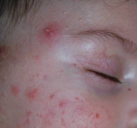
Neonatal acne (acne neonatorum) typically presents as small closed comedones on the forehead, nose, and cheeks (Figure 1).4 Accompanying sebaceous hyperplasia often is noted.5 Less frequently, open comedones, inflammatory papules, and pustules may develop.6 Neonatal acne may be evident at birth or appear during the first 4 weeks of life7 and is more commonly seen in boys.8
Etiology
Several factors may be pivotal in the etiology of neonatal acne, including increased sebum excretion, stimulation of the sebaceous glands by maternal or neonatal androgens,4 and colonization of sebaceous glands by Malassezia species.2 Increased sebum excretion occurs during the neonatal period due to enlarged sebaceous glands,2 which may result from the substantial production of β-hydroxysteroids from the relatively large adrenal glands.9,10 After 6 months of age, the size of the sebaceous glands and the sebum excretion rate decrease.9,10
Both maternal and neonatal androgens have been implicated in the stimulation of sebaceous glands in neonatal acne.2 The neonatal adrenal gland produces high levels of dehydroepiandrosterone,2 which stimulate sebaceous glands until around 1 year of age when dehydroepiandrosterone levels drop off as a consequence of involution of the neonatal adrenal gland.11 Testicular androgens provide additional stimulation to the sebaceous glands, which may explain why neonatal acne is more common in boys.1 Neonatal acne may be an inflammatory response to Malassezia species; however, Malassezia was not isolated in a series of patients,12 suggesting that neonatal acne is an early presentation of comedonal acne and not a response to Malassezia.2,12
Differential Diagnosis
There are a number of acneform eruptions that should be considered in the differential diagnosis,3 including bacterial folliculitis, secondary syphilis,13 herpes simplex virus and varicella zoster virus,14 and skin colonization by fungi of Malassezia species.15 Other neonatal eruptions such as erythema toxicum neonatorum,16 transient neonatal pustular melanosis, and milia and pustular miliaria, as well as a drug eruption associated with hydantoin, lithium, or halogens should be considered.17 The relationship between neonatal acne and neonatal cephalic pustulosis, which is characterized by papules and pustules without comedones, is controversial; some consider them to be 2 different entities,14 while others do not.18
Treatment
Guardians should be reassured that neonatal acne is mild, self-limited, and generally resolves spontaneously without scarring in approximately 1 to 3 months.1,2 In most cases, no treatment is needed.19 If necessary, comedones may be treated with azelaic acid cream 20% or tretinoin cream 0.025% to 0.05%.1,2 For inflammatory lesions, erythromycin solution 2% and benzoyl peroxide gel 2.5% may be used.1,20 Severe or recalcitrant disease warrants a workup for congenital adrenal hyperplasia, a virilizing tumor, or underlying endocrinopathy.19
Infantile Acne Vulgaris
Clinical Presentation
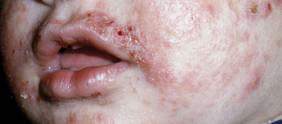
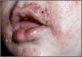
Infantile acne vulgaris shares similarities with neonatal acne21,22 in that they both affect the face, predominantly the cheeks, and have a male predominance (Figure 2).1,10 However, by definition, onset of infantile acne typically occurs later than acne neonatorum, usually at 3 to 6 months of age.1,4 Lesions are more pleomorphic and inflammatory than in neonatal acne. In addition to closed and open comedones, infantile acne may be first evident with papules, pustules, severe nodules, and cysts with scarring potential (Figure 3).1,2,5 Accordingly, treatment may be required. Most cases of infantile acne resolve by 4 or 5 years of age, but some remain active into puberty.1 Patients with a history of infantile acne have an increased incidence of acne vulgaris during adolescence compared to their peers, with greater severity and enhanced risk for scarring.4,23

Etiology
The etiology of infantile acne remains unclear.2 Similar to neonatal acne, infantile acne may be a result of elevated androgens produced by the fetal adrenal glands as well as by the testes in males.11 For example, a child with infantile acne had elevated luteinizing hormone, follicle-stimulating hormone, and testosterone levels.24 Therefore, hyperandrogenism should be considered as an etiology. Other causes also have been suggested. Rarely, an adrenocortical tumor may be associated with persistent infantile acne with signs of virilization and rapid development.25Malassezia was implicated in infantile acne in a 6-month-old infant who was successfully treated with ketoconazole cream 2%.26
Differential Diagnosis
Infantile acne often is misdiagnosed because it is rarely considered in the differential diagnosis. When closed comedones predominate, acne venenata induced by topical creams, lotions, or oils may be etiologic. Chloracne also should be considered.14
Treatment
Guardians should be educated about the likely chronicity of infantile acne, which may require long-term treatment, as well as the possibility that acne may recur in severe form during puberty.1 The treatment strategy for infantile acne is similar to treatment of acne at any age, with topical agents including retinoids (eg, tretinoin, benzoyl peroxide) and topical antibacterials (eg, erythromycin). Twice-daily erythromycin 125 to 250 mg is the treatment of choice when oral antibiotics are indicated. Tetracyclines are contraindicated in treatment of neonatal and infantile acne. Intralesional injections with low-concentration triamcinolone acetonide, cryotherapy, or topical corticosteroids for a short period of time can be used to treat deep nodules and cysts.2 Acne that is refractory to treatment with oral antibiotics alone or combined with topical treatments poses a dilemma, given the potential cosmetic sequelae of scarring and quality-of-life concerns. Because reducing or eliminating dairy intake appears beneficial for adolescents with moderate to severe acne,27 this approach may represent a good option for infantile acne.
Conclusion
Neonatal and infantile acne vulgaris may be overlooked or misdiagnosed. It is important to consider and treat. Early childhood acne may represent a virilization syndrome.
- Jansen T, Burgdorf WH, Plewig G. Pathogenesis and treatment of acne in childhood. Pediatr Dermatol. 1997;14:17-21.
- Antoniou C, Dessinioti C, Stratigos AJ, et al. Clinical and therapeutic approach to childhood acne: an update. Pediatr Dermatol. 2009;26:373-380.
- Kuflik JH, Schwartz RA. Acneiform eruptions. Cutis. 2000;66:97-100.
- Barbareschi M, Benardon S, Guanziroli E, et al. Classification and grading. In: Schwartz RA, Micali G, eds. Acne. Gurgaon, India: Nature Publishing Group; 2013:67-75.
- Mengesha YM, Bennett ML. Pustular skin disorders: diagnosis and treatment. Am J Clin Dermatol. 2002;3:389-400.
- O’Connor NR, McLaughlin MR, Ham P. Newborn skin: part I. common rashes. Am Fam Physician. 2008;77:47-52.
- Nanda S, Reddy BS, Ramji S, et al. Analytical study of pustular eruptions in neonates. Pediatr Dermatol. 2002;19:210-215.
- Yonkosky DM, Pochi PE. Acne vulgaris in childhood. pathogenesis and management. Dermatol Clin. 1986;4:127-136.
- Agache P, Blanc D, Barrand C, et al. Sebum levels during the first year of life. Br J Dermatol. 1980;103:643-649.
- Herane MI, Ando I. Acne in infancy and acne genetics. Dermatology. 2003;206:24-28.
- Lucky AW. A review of infantile and pediatric acne. Dermatology (Basel, Switzerland). 1998;103:643-649.
- Bernier V, Weill FX, Hirigoyen V, et al. Skin colonization by Malassezia species in neonates: a prospective study and relationship with neonatal cephalic pustulosis. Arch Dermatol. 2002;138:215-218.
- Lambert WC, Bagley MP, Khan Y, et al. Pustular acneiform secondary syphilis. Cutis. 1986;37:69-70.
- Antoniou C, Dessinioti C, Stratigos AJ, et al. Clinical and therapeutic approach to childhood acne: an update. Pediatr Dermatol. 2009;26:373-380.
- Borton LK, Schwartz RA. Pityrosporum folliculitis: a common acneiform condition of middle age. Ariz Med. 1981;38:598-601.
- Morgan AJ, Steen CJ, Schwartz RA, et al. Erythema toxicum neonatorum revisited. Cutis. 2009;83:13-16.
- Brodkin RH, Schwartz RA. Cutaneous signs of dioxin exposure. Am Fam Physician. 1984;30:189-194.
- Mancini AJ, Baldwin HE, Eichenfield LF, et al. Acne life cycle: the spectrum of pediatric disease. Semin Cutan Med Surg. 2011;30(suppl 3):S2-S5.
- Katsambas AD, Katoulis AC, Stavropoulos P. Acne neonatorum: a study of 22 cases. Int J Dermatol. 1999;38:128-130.
- Van Praag MC, Van Rooij RW, Folkers E, et al. Diagnosis and treatment of pustular disorders in the neonate. Pediatr Dermatol. 1997;14:131-143.
- Barnes CJ, Eichenfield LF, Lee J, et al. A practical approach for the use of oral isotretinoin for infantile acne. Pediatr Dermatol. 2005;22:166-169.
- Janniger CK. Neonatal and infantile acne vulgaris. Cutis. 1993;52:16.
- Chew EW, Bingham A, Burrows D. Incidence of acne vulgaris in patients with infantile acne. Clin Exp Dermatol. 1990;15:376-377.
- Duke EM. Infantile acne associated with transient increases in plasma concentrations of luteinising hormone, follicle-stimulating hormone, and testosterone. Br Med J (Clinical Res Ed). 1981;282:1275-1276.
- Mann MW, Ellis SS, Mallory SB. Infantile acne as the initial sign of an adrenocortical tumor [published online ahead of print September 14, 2006]. J Am Acad Dermatol. 2007;56(suppl 2):S15-S18.
- Kang SK, Jee MS, Choi JH, et al. A case of infantile acne due to Pityrosporum. Pediatr Dermatol. 2003;20:68-70.
- Di Landro A, Cazzaniga S, Parazzini F, et al. Family history, body mass index, selected dietary factors, menstrual history, and risk of moderate to severe acne in adolescents and young adults [published online ahead of print March 3, 2012]. J Am Acad Dermatol. 2012;67:1129-1135.
Acne vulgaris typically is associated with adolescence and young adulthood; however, it also can affect neonates, infants, and small children.1 Acne neonatorum occurs in up to 20% of newborns. The clinical importance of neonatal acne lies in its differentiation from infectious diseases, the exclusion of virilization as its underlying cause, and the possible implication of severe acne in adolescence.2 Neonatal acne also must be distinguished from acne that is induced by application of topical oils and ointments (acne venenata) and from acneform eruptions induced by acnegenic maternal medications such as hydantoin (fetal hydantoin syndrome) and lithium.3
Neonatal Acne (Acne Neonatorum)
Clinical Presentation
Neonatal acne (acne neonatorum) typically presents as small closed comedones on the forehead, nose, and cheeks (Figure 1).4 Accompanying sebaceous hyperplasia often is noted.5 Less frequently, open comedones, inflammatory papules, and pustules may develop.6 Neonatal acne may be evident at birth or appear during the first 4 weeks of life7 and is more commonly seen in boys.8
Etiology
Several factors may be pivotal in the etiology of neonatal acne, including increased sebum excretion, stimulation of the sebaceous glands by maternal or neonatal androgens,4 and colonization of sebaceous glands by Malassezia species.2 Increased sebum excretion occurs during the neonatal period due to enlarged sebaceous glands,2 which may result from the substantial production of β-hydroxysteroids from the relatively large adrenal glands.9,10 After 6 months of age, the size of the sebaceous glands and the sebum excretion rate decrease.9,10
Both maternal and neonatal androgens have been implicated in the stimulation of sebaceous glands in neonatal acne.2 The neonatal adrenal gland produces high levels of dehydroepiandrosterone,2 which stimulate sebaceous glands until around 1 year of age when dehydroepiandrosterone levels drop off as a consequence of involution of the neonatal adrenal gland.11 Testicular androgens provide additional stimulation to the sebaceous glands, which may explain why neonatal acne is more common in boys.1 Neonatal acne may be an inflammatory response to Malassezia species; however, Malassezia was not isolated in a series of patients,12 suggesting that neonatal acne is an early presentation of comedonal acne and not a response to Malassezia.2,12
Differential Diagnosis
There are a number of acneform eruptions that should be considered in the differential diagnosis,3 including bacterial folliculitis, secondary syphilis,13 herpes simplex virus and varicella zoster virus,14 and skin colonization by fungi of Malassezia species.15 Other neonatal eruptions such as erythema toxicum neonatorum,16 transient neonatal pustular melanosis, and milia and pustular miliaria, as well as a drug eruption associated with hydantoin, lithium, or halogens should be considered.17 The relationship between neonatal acne and neonatal cephalic pustulosis, which is characterized by papules and pustules without comedones, is controversial; some consider them to be 2 different entities,14 while others do not.18
Treatment
Guardians should be reassured that neonatal acne is mild, self-limited, and generally resolves spontaneously without scarring in approximately 1 to 3 months.1,2 In most cases, no treatment is needed.19 If necessary, comedones may be treated with azelaic acid cream 20% or tretinoin cream 0.025% to 0.05%.1,2 For inflammatory lesions, erythromycin solution 2% and benzoyl peroxide gel 2.5% may be used.1,20 Severe or recalcitrant disease warrants a workup for congenital adrenal hyperplasia, a virilizing tumor, or underlying endocrinopathy.19
Infantile Acne Vulgaris
Clinical Presentation
Infantile acne vulgaris shares similarities with neonatal acne21,22 in that they both affect the face, predominantly the cheeks, and have a male predominance (Figure 2).1,10 However, by definition, onset of infantile acne typically occurs later than acne neonatorum, usually at 3 to 6 months of age.1,4 Lesions are more pleomorphic and inflammatory than in neonatal acne. In addition to closed and open comedones, infantile acne may be first evident with papules, pustules, severe nodules, and cysts with scarring potential (Figure 3).1,2,5 Accordingly, treatment may be required. Most cases of infantile acne resolve by 4 or 5 years of age, but some remain active into puberty.1 Patients with a history of infantile acne have an increased incidence of acne vulgaris during adolescence compared to their peers, with greater severity and enhanced risk for scarring.4,23
Etiology
The etiology of infantile acne remains unclear.2 Similar to neonatal acne, infantile acne may be a result of elevated androgens produced by the fetal adrenal glands as well as by the testes in males.11 For example, a child with infantile acne had elevated luteinizing hormone, follicle-stimulating hormone, and testosterone levels.24 Therefore, hyperandrogenism should be considered as an etiology. Other causes also have been suggested. Rarely, an adrenocortical tumor may be associated with persistent infantile acne with signs of virilization and rapid development.25Malassezia was implicated in infantile acne in a 6-month-old infant who was successfully treated with ketoconazole cream 2%.26
Differential Diagnosis
Infantile acne often is misdiagnosed because it is rarely considered in the differential diagnosis. When closed comedones predominate, acne venenata induced by topical creams, lotions, or oils may be etiologic. Chloracne also should be considered.14
Treatment
Guardians should be educated about the likely chronicity of infantile acne, which may require long-term treatment, as well as the possibility that acne may recur in severe form during puberty.1 The treatment strategy for infantile acne is similar to treatment of acne at any age, with topical agents including retinoids (eg, tretinoin, benzoyl peroxide) and topical antibacterials (eg, erythromycin). Twice-daily erythromycin 125 to 250 mg is the treatment of choice when oral antibiotics are indicated. Tetracyclines are contraindicated in treatment of neonatal and infantile acne. Intralesional injections with low-concentration triamcinolone acetonide, cryotherapy, or topical corticosteroids for a short period of time can be used to treat deep nodules and cysts.2 Acne that is refractory to treatment with oral antibiotics alone or combined with topical treatments poses a dilemma, given the potential cosmetic sequelae of scarring and quality-of-life concerns. Because reducing or eliminating dairy intake appears beneficial for adolescents with moderate to severe acne,27 this approach may represent a good option for infantile acne.
Conclusion
Neonatal and infantile acne vulgaris may be overlooked or misdiagnosed. It is important to consider and treat. Early childhood acne may represent a virilization syndrome.
- Jansen T, Burgdorf WH, Plewig G. Pathogenesis and treatment of acne in childhood. Pediatr Dermatol. 1997;14:17-21.
- Antoniou C, Dessinioti C, Stratigos AJ, et al. Clinical and therapeutic approach to childhood acne: an update. Pediatr Dermatol. 2009;26:373-380.
- Kuflik JH, Schwartz RA. Acneiform eruptions. Cutis. 2000;66:97-100.
- Barbareschi M, Benardon S, Guanziroli E, et al. Classification and grading. In: Schwartz RA, Micali G, eds. Acne. Gurgaon, India: Nature Publishing Group; 2013:67-75.
- Mengesha YM, Bennett ML. Pustular skin disorders: diagnosis and treatment. Am J Clin Dermatol. 2002;3:389-400.
- O’Connor NR, McLaughlin MR, Ham P. Newborn skin: part I. common rashes. Am Fam Physician. 2008;77:47-52.
- Nanda S, Reddy BS, Ramji S, et al. Analytical study of pustular eruptions in neonates. Pediatr Dermatol. 2002;19:210-215.
- Yonkosky DM, Pochi PE. Acne vulgaris in childhood. pathogenesis and management. Dermatol Clin. 1986;4:127-136.
- Agache P, Blanc D, Barrand C, et al. Sebum levels during the first year of life. Br J Dermatol. 1980;103:643-649.
- Herane MI, Ando I. Acne in infancy and acne genetics. Dermatology. 2003;206:24-28.
- Lucky AW. A review of infantile and pediatric acne. Dermatology (Basel, Switzerland). 1998;103:643-649.
- Bernier V, Weill FX, Hirigoyen V, et al. Skin colonization by Malassezia species in neonates: a prospective study and relationship with neonatal cephalic pustulosis. Arch Dermatol. 2002;138:215-218.
- Lambert WC, Bagley MP, Khan Y, et al. Pustular acneiform secondary syphilis. Cutis. 1986;37:69-70.
- Antoniou C, Dessinioti C, Stratigos AJ, et al. Clinical and therapeutic approach to childhood acne: an update. Pediatr Dermatol. 2009;26:373-380.
- Borton LK, Schwartz RA. Pityrosporum folliculitis: a common acneiform condition of middle age. Ariz Med. 1981;38:598-601.
- Morgan AJ, Steen CJ, Schwartz RA, et al. Erythema toxicum neonatorum revisited. Cutis. 2009;83:13-16.
- Brodkin RH, Schwartz RA. Cutaneous signs of dioxin exposure. Am Fam Physician. 1984;30:189-194.
- Mancini AJ, Baldwin HE, Eichenfield LF, et al. Acne life cycle: the spectrum of pediatric disease. Semin Cutan Med Surg. 2011;30(suppl 3):S2-S5.
- Katsambas AD, Katoulis AC, Stavropoulos P. Acne neonatorum: a study of 22 cases. Int J Dermatol. 1999;38:128-130.
- Van Praag MC, Van Rooij RW, Folkers E, et al. Diagnosis and treatment of pustular disorders in the neonate. Pediatr Dermatol. 1997;14:131-143.
- Barnes CJ, Eichenfield LF, Lee J, et al. A practical approach for the use of oral isotretinoin for infantile acne. Pediatr Dermatol. 2005;22:166-169.
- Janniger CK. Neonatal and infantile acne vulgaris. Cutis. 1993;52:16.
- Chew EW, Bingham A, Burrows D. Incidence of acne vulgaris in patients with infantile acne. Clin Exp Dermatol. 1990;15:376-377.
- Duke EM. Infantile acne associated with transient increases in plasma concentrations of luteinising hormone, follicle-stimulating hormone, and testosterone. Br Med J (Clinical Res Ed). 1981;282:1275-1276.
- Mann MW, Ellis SS, Mallory SB. Infantile acne as the initial sign of an adrenocortical tumor [published online ahead of print September 14, 2006]. J Am Acad Dermatol. 2007;56(suppl 2):S15-S18.
- Kang SK, Jee MS, Choi JH, et al. A case of infantile acne due to Pityrosporum. Pediatr Dermatol. 2003;20:68-70.
- Di Landro A, Cazzaniga S, Parazzini F, et al. Family history, body mass index, selected dietary factors, menstrual history, and risk of moderate to severe acne in adolescents and young adults [published online ahead of print March 3, 2012]. J Am Acad Dermatol. 2012;67:1129-1135.
Practice Points
- Infantile acne needs to be recognized and treated.
- Acne in early childhood may represent virilization.
Etanercept-Induced Cystic Acne
Case Report
A 35-year-old man with chronic plaque-type psoriasis presented for treatment of a flare-up caused by a combination of hot tub use and 3 missed etanercept injections. The patient had been prescribed subcutaneous etanercept (50 mg once weekly) approximately 2 years prior to presentation for treatment of psoriasis and had used it consistently with 2 brief periods of discontinuation due to upper respiratory infections; treatment was discontinued for 2 weeks until the infections resolved and was restarted at the same dose. Recent hot tub use induced localized folliculitis of the leg and caused koebnerization of his psoriasis. Treatment with etanercept (50 mg twice weekly) was reinitiated, but after 1 month of therapy, the patient developed a nodulocystic eruption on the face. The patient discontinued use of etanercept and subsequently was started on oral minocycline (50 mgonce daily) by his primary care physician. After 6 weeks of minocycline therapy, the patient reported gradual improvement of his cystic acne. At follow-up, physical examination revealed approximately 15 erythematous cystic papules and nodules on the bilateral cheeks. The dose of oral minocycline was increased to 100 mg twice daily and his face subsequently cleared after 12 weeks of treatment.
On rechallenge with etanercept (50 mg weekly) after a separate flare-up of his psoriasis and psoriatic arthritis, the patient again developed nodulocystic acne within 3 weeks of restarting the medication, which confirmed the association between etanercept and the eruption. Etanercept was subsequently discontinued and the patient was started on ustekinumab for treatment of psoriasis and psoriatic arthritis as well as minocycline (100 mg twice daily) for treatment of nodulocystic acne. His acne subsequently cleared after 12 weeks of therapy. The patient’s psoriasis currently is well controlled on a regimen of methotrexate 20 mg weekly, as continued use of ustekinumab was not covered by his health insurance policy.
Comment
Etanercept, along with infliximab and adalimumab, is a tumor necrosis factor α (TNF-α) antagonist. Tumor necrosis factor α is a major cytokine of the immune system that is deregulated in autoimmune disorders, causing inflammation and a variety of systemic effects. Inhibition of TNF-α results in a decrease in inflammatory markers such as IL-1 and IL-6, leading to a reduction in endothelial permeability and leukocyte migration.1 Etanercept is unique in that it is a fully humanized soluble fusion protein consisting of a TNF-α receptor linked to the Fc region of IgG1, whereas infliximab and adalimumab are monoclonal antibodies that target TNF-α. Etanercept works by acting as a decoy receptor for TNF-α, thereby preventing it from binding cell surface receptors and subsequently decreasing inflammation. It is an approved treatment of rheumatoid arthritis, polyarticular juvenile rheumatoid arthritis, psoriatic arthritis, ankylosing spondylitis, and plaque psoriasis.2
Many drugs have been implicated in acneform eruptions mimicking acne vulgaris, such as corticosteroids, cyclosporine, antipsychotics, anticonvulsants, epidermal growth factor receptor inhibitors, antidepressants, danazol, antituberculosis drugs, quinidine, azathioprine, testosterone, and TNF-α antagonists.1 The TNF-α antagonists that have been associated with acne are infliximab and adalimumab, especially when used to treat Crohn disease or rheumatoid arthritis, though there also are reports of acneform eruptions when using these drugs to treat psoriasis.1,3 There have been reports of the use of etanercept in treating severe acne and acne conglobata4,5; our case documents etanercept-induced acne associated with psoriasis treatment. Theoretically, anti–TNF-α agents should suppress acne rather than induce it due to their inhibition of the inflammatory markers TNF-α, IL-1a, and IFN-γ, which are thought to play a role in hypercornification of the infundibulum.6 Thus the mechanism of TNF-α antagonists and associated acneform eruptions remains unknown. This observation should prompt further research to investigate the correlation between the use of TNF-α inhibitors, specifically etanercept, and cystic acne.
- Momin SB, Peterson A, Del Rosso JQ. A status report on drug-associated acne and acneiform eruptions. J Drugs Dermatol. 2010;9:627-636.
- Zhou H. Clinical pharmacokinetics of etanercept: a fully humanized soluble recombinant tumor necrosis factor receptor fusion protein. J Clin Pharmacol. 2005;45:490-497.
- Sun G, Wasko CA, Hsu S. Acneiform eruption following anti-TNF-alpha treatment: a report of three cases. J Drugs Dermatol. 2008;7:69-71.
- Campione E, Mazzotta AM, Bianchi L, et al. Severe acne successfully treated with etanercept. Acta Derm Venereol. 2006;86:256-257.
- Vega J, Sánchez-Velicia L, Pozo T. Efficacy of etanercept in the treatment of acne conglobata [in Spanish]. Actas Dermosifiliogr. 2010;101:553-554.
- Bassi E, Poli F, Charachon A, et al. Infliximab-induced acne: report of two cases. Br J Dermatol. 2007;156:402-403.
Case Report
A 35-year-old man with chronic plaque-type psoriasis presented for treatment of a flare-up caused by a combination of hot tub use and 3 missed etanercept injections. The patient had been prescribed subcutaneous etanercept (50 mg once weekly) approximately 2 years prior to presentation for treatment of psoriasis and had used it consistently with 2 brief periods of discontinuation due to upper respiratory infections; treatment was discontinued for 2 weeks until the infections resolved and was restarted at the same dose. Recent hot tub use induced localized folliculitis of the leg and caused koebnerization of his psoriasis. Treatment with etanercept (50 mg twice weekly) was reinitiated, but after 1 month of therapy, the patient developed a nodulocystic eruption on the face. The patient discontinued use of etanercept and subsequently was started on oral minocycline (50 mgonce daily) by his primary care physician. After 6 weeks of minocycline therapy, the patient reported gradual improvement of his cystic acne. At follow-up, physical examination revealed approximately 15 erythematous cystic papules and nodules on the bilateral cheeks. The dose of oral minocycline was increased to 100 mg twice daily and his face subsequently cleared after 12 weeks of treatment.
On rechallenge with etanercept (50 mg weekly) after a separate flare-up of his psoriasis and psoriatic arthritis, the patient again developed nodulocystic acne within 3 weeks of restarting the medication, which confirmed the association between etanercept and the eruption. Etanercept was subsequently discontinued and the patient was started on ustekinumab for treatment of psoriasis and psoriatic arthritis as well as minocycline (100 mg twice daily) for treatment of nodulocystic acne. His acne subsequently cleared after 12 weeks of therapy. The patient’s psoriasis currently is well controlled on a regimen of methotrexate 20 mg weekly, as continued use of ustekinumab was not covered by his health insurance policy.
Comment
Etanercept, along with infliximab and adalimumab, is a tumor necrosis factor α (TNF-α) antagonist. Tumor necrosis factor α is a major cytokine of the immune system that is deregulated in autoimmune disorders, causing inflammation and a variety of systemic effects. Inhibition of TNF-α results in a decrease in inflammatory markers such as IL-1 and IL-6, leading to a reduction in endothelial permeability and leukocyte migration.1 Etanercept is unique in that it is a fully humanized soluble fusion protein consisting of a TNF-α receptor linked to the Fc region of IgG1, whereas infliximab and adalimumab are monoclonal antibodies that target TNF-α. Etanercept works by acting as a decoy receptor for TNF-α, thereby preventing it from binding cell surface receptors and subsequently decreasing inflammation. It is an approved treatment of rheumatoid arthritis, polyarticular juvenile rheumatoid arthritis, psoriatic arthritis, ankylosing spondylitis, and plaque psoriasis.2
Many drugs have been implicated in acneform eruptions mimicking acne vulgaris, such as corticosteroids, cyclosporine, antipsychotics, anticonvulsants, epidermal growth factor receptor inhibitors, antidepressants, danazol, antituberculosis drugs, quinidine, azathioprine, testosterone, and TNF-α antagonists.1 The TNF-α antagonists that have been associated with acne are infliximab and adalimumab, especially when used to treat Crohn disease or rheumatoid arthritis, though there also are reports of acneform eruptions when using these drugs to treat psoriasis.1,3 There have been reports of the use of etanercept in treating severe acne and acne conglobata4,5; our case documents etanercept-induced acne associated with psoriasis treatment. Theoretically, anti–TNF-α agents should suppress acne rather than induce it due to their inhibition of the inflammatory markers TNF-α, IL-1a, and IFN-γ, which are thought to play a role in hypercornification of the infundibulum.6 Thus the mechanism of TNF-α antagonists and associated acneform eruptions remains unknown. This observation should prompt further research to investigate the correlation between the use of TNF-α inhibitors, specifically etanercept, and cystic acne.
- Momin SB, Peterson A, Del Rosso JQ. A status report on drug-associated acne and acneiform eruptions. J Drugs Dermatol. 2010;9:627-636.
- Zhou H. Clinical pharmacokinetics of etanercept: a fully humanized soluble recombinant tumor necrosis factor receptor fusion protein. J Clin Pharmacol. 2005;45:490-497.
- Sun G, Wasko CA, Hsu S. Acneiform eruption following anti-TNF-alpha treatment: a report of three cases. J Drugs Dermatol. 2008;7:69-71.
- Campione E, Mazzotta AM, Bianchi L, et al. Severe acne successfully treated with etanercept. Acta Derm Venereol. 2006;86:256-257.
- Vega J, Sánchez-Velicia L, Pozo T. Efficacy of etanercept in the treatment of acne conglobata [in Spanish]. Actas Dermosifiliogr. 2010;101:553-554.
- Bassi E, Poli F, Charachon A, et al. Infliximab-induced acne: report of two cases. Br J Dermatol. 2007;156:402-403.
- Tumor necrosis factor α antagonists have been implicated in causing acneform eruptions.
- Discontinuation of the agent along with initiation of oral antibiotics or isotretinoin can treat medication-induced nodulocystic acne.
- Ustekinumab is an alternative non–tumor necrosis factor α antagonist that can be used to treat psoriasis and psoriatic arthritis that has not been associated with acneform eruptions.
OTC topical acne meds can cause severe reactions
The Food and Drug Administration says that some over-the-counter acne products can cause severe hypersensitivity or allergic reactions and is warning consumers to discontinue use immediately and seek emergency medical attention if they experience such symptoms.
The reactions seem to be on the upswing, according to the agency’s review of reports from 1969 to early 2013.
The agency said in safety announcement that it has not determined whether the reactions are being triggered by the active ingredients in the products – benzoyl peroxide or salicylic acid – or by the inactive ingredients or a combination of the inactive and active ingredients.
The products include Proactiv, Neutrogena, MaxClarity, Oxy, Ambi, Aveeno, Clean & Clear, and private label store brands, and are sold as gels, lotions, face washes, solutions, cleansing pads, toners, and face scrubs, among other products, according to the agency.
The FDA has received reports stating that – within minutes to a day or longer of use – consumers have had hypersensitivity reactions that include throat tightness, difficulty breathing, or swelling of the eyes, face, lips, or tongue. Hives and itching are also indications of an allergic reaction.
From 1969 to January 2013, the agency identified 131 cases of hypersensitivity reactions, with the majority reported in 2012-2013. A total of 86% (113) of the cases were in women, and the reactions were reported in people aged 11-78 years.
There were no deaths, but 58 of the consumers were hospitalized. A total of 38 of the 131 cases (29%) were categorized as anaphylactic reactions, and the remainder were categorized as nonanaphylactic hypersensitivity. Almost half of the consumers said they had discontinued use after the reaction, the majority of whom reported some degree of recovery after discontinuing product use, with final outcomes unavailable for the rest. Four of the patients who used the product again reported a recurrence of the reaction.
The agency is encouraging manufacturers to add directions on testing for hypersensitivity to the labels of all OTC topical acne products. Some labels already include those instructions, which direct users to apply a small amount to one or two small affected areas of the skin for 3 days. If there is no reaction – topical or otherwise – then the product can be used according to the directions.
Consumers are also being urged to avoid using a product if they have previously experienced a hypersensitivity reaction with its use.
The FDA says that physicians should be aware that some topical prescription acne drug products also contain warnings about allergic reactions, including anaphylaxis.
Physicians can report adverse events involving OTC topical acne products to the FDA MedWatch program.
On Twitter @aliciaault
Adapalene/benzoyl peroxide gel halves acne lesion counts in 1 month
WAIKOLOA, HAWAII – The fixed-dose combination of adapalene/benzoyl peroxide gel 0.1%/2.5% typically results in reductions of 40%-50% in inflammatory and 30%-40% in noninflammatory acne lesion counts during the first 4 weeks of therapy.
In a pooled analysis of 14 studies totaling 2,358 acne patients aged 9-61 years, the proportion with an Investigator’s Global Assessment (IGA) of moderate or severe acne dropped from 92% at baseline to 51% at 4 weeks, according to Dr. Linda Stein Gold.

The pooled analysis was carried out to provide physicians and patients with information as to what to realistically expect in the first 4 weeks of therapy. The analysis is particularly timely in light of the Food and Drug Administration’s recent expansion of the indication for adapalene/benzoyl peroxide gel 0.1%/2.5% (Epiduo) to include acne patients as young as 9 years of age, noted Dr. Stein Gold, director of clinical research in the department of dermatology at Henry Ford Hospital, Detroit.
Mild skin irritation was common, especially in the first 2 weeks of use.
"It’s a fiction that retinoids are too harsh for younger patients to use," Dr. Stein Gold said. "I tell all my acne patients, especially the younger ones, no matter what topical retinoid they’re using, to use it every other night for the first 2 weeks, make sure their skin is completely dry, and use a gentle cleanser and a good moisturizer," she said at the Hawaii Dermatology Seminar sponsored by Global Academy for Medical Education/Skin Disease Education Foundation. "If they get through those first 2 weeks, they’ll see that the tolerability really improves."
Dr. Stein Gold said she likes the fixed combination of adapalene/benzoyl peroxide gel 0.1%/2.5% because it combines several key elements of cutting-edge acne treatment. Topical retinoids are not mere comedone busters, as formerly thought, but are also effective agents for papules and pustular lesions. While they do not kill Propionibacterium acnes, they down-regulate Toll-like receptor 2, which is produced by the bacterium and induces proinflammatory cytokines. Topical retinoids are a key part of maintenance-of-remission strategies. And adapalene is unique among topical retinoids in that it is inherently stable with benzoyl peroxide (BPO) and is stable in daylight.
BPO is unique in that it has potent antibacterial activity, but never causes P. acnes resistance, even after many years of treatment. BPO is quite effective for inflammatory lesions and moderately effective for comedonal acne lesions, Dr. Stein Gold noted. Also, it is well established that BPO at a concentration of 2.5% is as efficacious as 5% or 10%, and much better tolerated than at the higher concentrations. The molecule size and the use of a vehicle with good penetration into the hair follicle are much more important factors in treatment effectiveness than is the BPO concentration, she added.
In one study of acne patients with antibiotic-resistant P. acnes, including clindamycin-, doxycycline-, erythromycin-, and minocycline-resistant microorganisms, 88% of the antibiotic-resistant P. acnes bacteria were killed after 2 weeks of treatment with adapalene/BPO gel 0.1%/2.5%. After 4 weeks of treatment, 97% of the antibiotic-resistant organisms were dead. That’s testimony to the P. acnes–killing power of BPO, said Dr. Stein Gold.
"There’s a sense among many that with all the newer medications we have for acne, benzoyl peroxide is really your grandfather’s treatment, with no place in today’s modern world. This is totally false. I really feel that benzoyl peroxide should play a central role in all of our acne patients’ treatment regimens, unless of course they’re allergic to it, which occurs in only a small percentage of our patients," she said.
The pooled analysis was funded by Galderma. Dr. Stein Gold is a consultant to Galderma, Stiefel, Medicis, and Warner Chilcott.
SDEF and this news organization are owned by the same parent company.
WAIKOLOA, HAWAII – The fixed-dose combination of adapalene/benzoyl peroxide gel 0.1%/2.5% typically results in reductions of 40%-50% in inflammatory and 30%-40% in noninflammatory acne lesion counts during the first 4 weeks of therapy.
In a pooled analysis of 14 studies totaling 2,358 acne patients aged 9-61 years, the proportion with an Investigator’s Global Assessment (IGA) of moderate or severe acne dropped from 92% at baseline to 51% at 4 weeks, according to Dr. Linda Stein Gold.
The pooled analysis was carried out to provide physicians and patients with information as to what to realistically expect in the first 4 weeks of therapy. The analysis is particularly timely in light of the Food and Drug Administration’s recent expansion of the indication for adapalene/benzoyl peroxide gel 0.1%/2.5% (Epiduo) to include acne patients as young as 9 years of age, noted Dr. Stein Gold, director of clinical research in the department of dermatology at Henry Ford Hospital, Detroit.
Mild skin irritation was common, especially in the first 2 weeks of use.
"It’s a fiction that retinoids are too harsh for younger patients to use," Dr. Stein Gold said. "I tell all my acne patients, especially the younger ones, no matter what topical retinoid they’re using, to use it every other night for the first 2 weeks, make sure their skin is completely dry, and use a gentle cleanser and a good moisturizer," she said at the Hawaii Dermatology Seminar sponsored by Global Academy for Medical Education/Skin Disease Education Foundation. "If they get through those first 2 weeks, they’ll see that the tolerability really improves."
Dr. Stein Gold said she likes the fixed combination of adapalene/benzoyl peroxide gel 0.1%/2.5% because it combines several key elements of cutting-edge acne treatment. Topical retinoids are not mere comedone busters, as formerly thought, but are also effective agents for papules and pustular lesions. While they do not kill Propionibacterium acnes, they down-regulate Toll-like receptor 2, which is produced by the bacterium and induces proinflammatory cytokines. Topical retinoids are a key part of maintenance-of-remission strategies. And adapalene is unique among topical retinoids in that it is inherently stable with benzoyl peroxide (BPO) and is stable in daylight.
BPO is unique in that it has potent antibacterial activity, but never causes P. acnes resistance, even after many years of treatment. BPO is quite effective for inflammatory lesions and moderately effective for comedonal acne lesions, Dr. Stein Gold noted. Also, it is well established that BPO at a concentration of 2.5% is as efficacious as 5% or 10%, and much better tolerated than at the higher concentrations. The molecule size and the use of a vehicle with good penetration into the hair follicle are much more important factors in treatment effectiveness than is the BPO concentration, she added.
In one study of acne patients with antibiotic-resistant P. acnes, including clindamycin-, doxycycline-, erythromycin-, and minocycline-resistant microorganisms, 88% of the antibiotic-resistant P. acnes bacteria were killed after 2 weeks of treatment with adapalene/BPO gel 0.1%/2.5%. After 4 weeks of treatment, 97% of the antibiotic-resistant organisms were dead. That’s testimony to the P. acnes–killing power of BPO, said Dr. Stein Gold.
"There’s a sense among many that with all the newer medications we have for acne, benzoyl peroxide is really your grandfather’s treatment, with no place in today’s modern world. This is totally false. I really feel that benzoyl peroxide should play a central role in all of our acne patients’ treatment regimens, unless of course they’re allergic to it, which occurs in only a small percentage of our patients," she said.
The pooled analysis was funded by Galderma. Dr. Stein Gold is a consultant to Galderma, Stiefel, Medicis, and Warner Chilcott.
SDEF and this news organization are owned by the same parent company.
EXPERT ANALYSIS FROM SDEF HAWAII DERMATOLOGY SEMINAR

Gold-coated microparticles aid acne treatment
PHOENIX – Application of gold-coated microparticles to the skin may enhance laser photothermolysis of sebaceous glands to treat acne, according to three small, preliminary studies.
Inadequate contrast in sebaceous glands has limited the effectiveness of selective photothermolysis to treat acne. Specially engineered 0.150-mcm microparticles of inert gold coating a silica core were designed for surface plasmon resonance at near-infrared wavelengths to optimize contrast and high absorption of light in the sebaceous glands during laser treatment.
In a randomized, controlled crossover study, 48 patients with acne vulgaris received either immediate or delayed treatment for 12 weeks, after which the delayed-treatment group also began receiving the treatment. Immediate treatment included three treatments at 2-week intervals. The treatment consisted of a microparticle suspension gently massaged into facial skin for about 8 minutes, superficial skin cleansing, and pulsed irradiation with two passes of an 800-nm laser. Patients in the delayed-treatment group used an over-the-counter face wash (2% salicylic acid) twice daily.
The mean number of inflammatory lesions decreased by 34% at 12 weeks after treatment started, compared with a 16% reduction in the control group, a statistically significant difference, Dilip Paithankar, Ph.D., reported at the annual meeting of the American Society for Laser Medicine and Surgery.

At 28 weeks after the crossover to treatment for all subjects (40 weeks after the start of the study), the mean number of inflammatory lesions decreased by 61%, reported Dr. Paithankar, chief technology officer at Sebacia, Duluth, Ga., which is developing the gold-coated microparticles treatment.
In another randomized, sham-controlled study, 49 patients with acne vulgaris underwent three laser photothermolysis treatments at weekly intervals using either the gold-coated microparticles or a control vehicle without the particles. Mean inflammatory lesion counts decreased significantly more in the treatment group than in the control group at 8, 12, and 16 weeks of follow-up. In the treatment group, mean inflammatory lesion counts were 44% lower at 8 weeks, 49% lower at 12 weeks, and 53% lower at 16 weeks, compared with baseline. In the control group, mean inflammatory lesion counts were 14% lower, 22% lower, and 31% lower at those time points, respectively, compared with baseline.
Inflammatory lesions were clear or almost clear by the end of the study in 24% in the treatment group and in none of the control patients.

Data from a separate histologic study suggest that using ultrasound to assist delivery of the gold-coated microparticles may improve selective destruction from laser treatment, Dr. Girish Munavalli reported at the meeting. Ultrasound has been used in other settings to enhance delivery of small molecules through the stratum corneum.
The study used an ultrasound horn vibrating in a microparticle suspension placed in an enclosure above preauricular skin in 37 subjects, followed by 800-nm laser treatment. Biopsies taken within 15 minutes showed localized thermal injury to infundibuli and sebaceous glands with no collateral damage to surrounding tissue or to the epidermis, reported Dr. Munavalli, a dermatologist in group practice in Charlotte, N.C. The extent of thermal damage correlated with the duration of the ultrasound.
The level of damage could be expected to improve acne, but that needs confirmation in clinical trials, he said.
In each of the studies, the treatments were well tolerated, with minimal side effects including mild to moderate pain (using no anesthetic) and short-lived erythema and edema.
Dr. Paithankar works for Sebacia. Dr. Munavalli reported having no disclosures; some of his coinvestigators work for Sebacia.
On Twitter @sherryboschert
PHOENIX – Application of gold-coated microparticles to the skin may enhance laser photothermolysis of sebaceous glands to treat acne, according to three small, preliminary studies.
Inadequate contrast in sebaceous glands has limited the effectiveness of selective photothermolysis to treat acne. Specially engineered 0.150-mcm microparticles of inert gold coating a silica core were designed for surface plasmon resonance at near-infrared wavelengths to optimize contrast and high absorption of light in the sebaceous glands during laser treatment.
In a randomized, controlled crossover study, 48 patients with acne vulgaris received either immediate or delayed treatment for 12 weeks, after which the delayed-treatment group also began receiving the treatment. Immediate treatment included three treatments at 2-week intervals. The treatment consisted of a microparticle suspension gently massaged into facial skin for about 8 minutes, superficial skin cleansing, and pulsed irradiation with two passes of an 800-nm laser. Patients in the delayed-treatment group used an over-the-counter face wash (2% salicylic acid) twice daily.
The mean number of inflammatory lesions decreased by 34% at 12 weeks after treatment started, compared with a 16% reduction in the control group, a statistically significant difference, Dilip Paithankar, Ph.D., reported at the annual meeting of the American Society for Laser Medicine and Surgery.
At 28 weeks after the crossover to treatment for all subjects (40 weeks after the start of the study), the mean number of inflammatory lesions decreased by 61%, reported Dr. Paithankar, chief technology officer at Sebacia, Duluth, Ga., which is developing the gold-coated microparticles treatment.
In another randomized, sham-controlled study, 49 patients with acne vulgaris underwent three laser photothermolysis treatments at weekly intervals using either the gold-coated microparticles or a control vehicle without the particles. Mean inflammatory lesion counts decreased significantly more in the treatment group than in the control group at 8, 12, and 16 weeks of follow-up. In the treatment group, mean inflammatory lesion counts were 44% lower at 8 weeks, 49% lower at 12 weeks, and 53% lower at 16 weeks, compared with baseline. In the control group, mean inflammatory lesion counts were 14% lower, 22% lower, and 31% lower at those time points, respectively, compared with baseline.
Inflammatory lesions were clear or almost clear by the end of the study in 24% in the treatment group and in none of the control patients.
Data from a separate histologic study suggest that using ultrasound to assist delivery of the gold-coated microparticles may improve selective destruction from laser treatment, Dr. Girish Munavalli reported at the meeting. Ultrasound has been used in other settings to enhance delivery of small molecules through the stratum corneum.
The study used an ultrasound horn vibrating in a microparticle suspension placed in an enclosure above preauricular skin in 37 subjects, followed by 800-nm laser treatment. Biopsies taken within 15 minutes showed localized thermal injury to infundibuli and sebaceous glands with no collateral damage to surrounding tissue or to the epidermis, reported Dr. Munavalli, a dermatologist in group practice in Charlotte, N.C. The extent of thermal damage correlated with the duration of the ultrasound.
The level of damage could be expected to improve acne, but that needs confirmation in clinical trials, he said.
In each of the studies, the treatments were well tolerated, with minimal side effects including mild to moderate pain (using no anesthetic) and short-lived erythema and edema.
Dr. Paithankar works for Sebacia. Dr. Munavalli reported having no disclosures; some of his coinvestigators work for Sebacia.
On Twitter @sherryboschert
AT LASER 2014
Key clinical point: Inadequate contrast has limited the use of laser targeting of the sebaceous glands; specially designed gold-coated microparticles are designed to improve contrast and promote light absorption.
Major finding: Laser treatment of acne vulgaris preceded by application of gold-coated microparticles decreased mean inflammatory lesion counts by 34% and 49% at 12 weeks in two separate studies, compared with reductions of 16% and 22% in control groups.
Data source: Two randomized, controlled trials of 48 patients and 49 patients, respectively, with acne vulgaris, and a separate histologic study of 37 subjects.
Disclosures: Dr. Paithankar works for Sebacia, which developed the gold-coated microparticles treatment. Dr. Munavalli reported having no disclosures; some of his coinvestigators work for Sebacia.
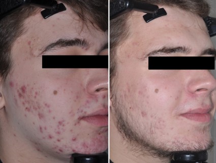
The Cutting Edge: New Research and Developments From the AAD Annual Meeting
As one of the most common skin diseases, acne research, clinical guidelines, and therapeutic innovation are always a hot topic at the Annual Meeting of the American Academy of Dermatology (March 21-25, 2014). A new dimension in the chicken and egg, or rather microcomedone and inflammation, story of acne pathogenesis emerged in a recent Journal of Investigative Dermatology (2014;134:381-388) article, which demonstrated that Propionibacterium acnes triggers a key inflammatory mediator, IL-1β, via the inflammasome (a compilation of inflammatory proteins such as caspases and NOD-like receptors) activation, suggesting a role for inflammasome-mediated inflammation in acne pathogenesis in addition to and independent of toll-like receptor activation. A potential therapeutic target, perhaps?
Drs. Ted Rosen and Joshua Zeichner in 2 independent sessions (Treating Tumors and Inflammatory Skin Diseases With Immunomodulators and Biologics [Rosen] and Acne Treatment Controversies [Zeichner]) discussed the importance of purposeful utilization of oral antibiotics—less is more—to prevent the continued emergence of antimicrobial resistance. Dr. Rosen commented that doxycycline at doses ≥50 mg daily provides serum levels that have an impact on commensal or colonized organisms, while lower doses provide only the anti-inflammatory effects without any bacteriostatic impact. This finding highlights the importance of low-dose controlled-release formulations. Dr. Zeichner also stressed the importance of knowing when to quit; if a patient does not improve in 6 to 8 weeks of therapy, move on. He also commented on the importance of multimechanistic therapy (solo is a no-go), utilizing benzoyl peroxide–containing products and most importantly retinoids from day 1. Dr. Zeichner also stressed the importance of recognizing acne mimics, such as gram-negative folliculitis, and made it clear that hormonally driven acne must not be missed, especially in the adult female population.
Lastly, new directions in acne are emerging, utilizing the science of nanotechnology (nano is equivalent to 1 billionth of a part). Drug delivery with nanomaterials is being fervently pursued across the globe in the field of acne. Nanoparticles can allow for sustained and controlled release of established products, increasing efficacy and stability, compliance due to decreased dosing, and safety by limiting associated irritation and dryness. In a session on nanotechnology, Dr. Rox Anderson presented his work utilizing gold nanoparticles to selectively destroy sebaceous glands via selective photothermolysis. He commented, “Do we really need our sebaceous glands,” citing that babies and infants do just fine without their activity. This work is currently in clinical trials in Europe. Nanotechnology also can be used to deliver previously undeliverable actives, such as the gaseous molecule nitric oxide. It was shown that a nitric oxide–releasing nanoparticle technology effectively penetrated the pilosebaceous unit, killed P acnes in culture, and inhibited inflammatory cytokine production by keratinocytes exposed to P acnes.
Stay tuned for more innovation coming soon!

Acne scar improvements maintained after laser treatment
PHOENIX – Improvements in acne scarring were maintained 3 months after six laser treatments with a diffractive lens array and a picosecond 755-nm alexandrite laser. Changes seen on histology suggest the improvements stem from more than collagen remodeling alone.
Twenty adults with facial acne scars and Fitzpatrick skin types I-V underwent six laser treatments performed every 4-8 weeks at a single center using a 755-nm wavelength, diffractive lens array, pulse duration of 750-850 picoseconds, and repetition rate of 5 Hz, with a fixed spot size of 6 mm and fluence of 0.71 J/cm2.

Three physicians who were blinded to the results evaluated two- and three-dimensional images taken at baseline and at follow-up visits. Analysis of two-dimensional photos produced acne scar improvement scores averaging 1.5 at the 1-month follow-up visit and 1.4 at the 3-month visit on a 4-point scale (with 0 indicating 0-25% improvement and 3 indicating greater than 75% improvement), Dr. Jeremy Brauer and his associates reported.
Scar volume decreased by an average of 24% by 1 month post treatment and 27% by 3 months post treatment, according to analyses of the three-dimensional images, Dr. Brauer said at the annual meeting of the American Society for Laser Medicine and Surgery.
Histology of biopsy specimens taken at baseline and at 3 months after the last treatment showed elongation and increased density of elastic fibers, increased dermal collagen, and increased dermal mucin after treatment. These findings suggest that the improvements in scarring are caused by dermal changes beyond remodeling of collagen alone, said Dr. Brauer of the Laser & Skin Surgery Center in New York.
Dr. Brauer and his associates previously reported that the picosecond laser and diffractive lens array can improve acne scarring with minimal preparation and down time, "in many cases with no anesthesia at all," he said. The investigators offered topical anesthesia and antiviral prophylaxis to patients, but most declined.
The diffractive lens array delivers varying levels of heat at a fixed spot size and fluence. High-energy pulses that are 500 mcm apart deliver 70% of the fluence, with a lower level of heating surrounding these higher-level pulses. In total, approximately 10% of the tissue is exposed to the high-fluence regions.
The 15 women and 5 men in the study ranged in age from 27 to 62 years.
Previous methods of treating acne scarring with lasers, intense pulsed light, or other energy devices typically cause thermal injury that produces collagen synthesis and remodeling. Those procedures usually require anesthesia and can cause collateral damage that may take a long time to heal, he said.
The investigators chose the picosecond 755-nm alexandrite laser because it has been used successfully to remove tattoos or treat pigmented lesions while causing fewer side effects and coupled it with the specialized diffractive lens array.
The study excluded patients with hypersensitivity to light; localized or systemic infection, prior laser treatment in the past 3 months; and a history of skin cancer, keloidal scarring, immunosuppression, or immune deficiency disorder. Also excluded were patients who used isotretinoin in the past year and women who were pregnant, breastfeeding, or contemplating pregnancy.
Dr. Brauer and a coinvestigator reported financial associations with Cynosure, which also loaned equipment for the study.
On Twitter @sherryboschert
PHOENIX – Improvements in acne scarring were maintained 3 months after six laser treatments with a diffractive lens array and a picosecond 755-nm alexandrite laser. Changes seen on histology suggest the improvements stem from more than collagen remodeling alone.
Twenty adults with facial acne scars and Fitzpatrick skin types I-V underwent six laser treatments performed every 4-8 weeks at a single center using a 755-nm wavelength, diffractive lens array, pulse duration of 750-850 picoseconds, and repetition rate of 5 Hz, with a fixed spot size of 6 mm and fluence of 0.71 J/cm2.
Three physicians who were blinded to the results evaluated two- and three-dimensional images taken at baseline and at follow-up visits. Analysis of two-dimensional photos produced acne scar improvement scores averaging 1.5 at the 1-month follow-up visit and 1.4 at the 3-month visit on a 4-point scale (with 0 indicating 0-25% improvement and 3 indicating greater than 75% improvement), Dr. Jeremy Brauer and his associates reported.
Scar volume decreased by an average of 24% by 1 month post treatment and 27% by 3 months post treatment, according to analyses of the three-dimensional images, Dr. Brauer said at the annual meeting of the American Society for Laser Medicine and Surgery.
Histology of biopsy specimens taken at baseline and at 3 months after the last treatment showed elongation and increased density of elastic fibers, increased dermal collagen, and increased dermal mucin after treatment. These findings suggest that the improvements in scarring are caused by dermal changes beyond remodeling of collagen alone, said Dr. Brauer of the Laser & Skin Surgery Center in New York.
Dr. Brauer and his associates previously reported that the picosecond laser and diffractive lens array can improve acne scarring with minimal preparation and down time, "in many cases with no anesthesia at all," he said. The investigators offered topical anesthesia and antiviral prophylaxis to patients, but most declined.
The diffractive lens array delivers varying levels of heat at a fixed spot size and fluence. High-energy pulses that are 500 mcm apart deliver 70% of the fluence, with a lower level of heating surrounding these higher-level pulses. In total, approximately 10% of the tissue is exposed to the high-fluence regions.
The 15 women and 5 men in the study ranged in age from 27 to 62 years.
Previous methods of treating acne scarring with lasers, intense pulsed light, or other energy devices typically cause thermal injury that produces collagen synthesis and remodeling. Those procedures usually require anesthesia and can cause collateral damage that may take a long time to heal, he said.
The investigators chose the picosecond 755-nm alexandrite laser because it has been used successfully to remove tattoos or treat pigmented lesions while causing fewer side effects and coupled it with the specialized diffractive lens array.
The study excluded patients with hypersensitivity to light; localized or systemic infection, prior laser treatment in the past 3 months; and a history of skin cancer, keloidal scarring, immunosuppression, or immune deficiency disorder. Also excluded were patients who used isotretinoin in the past year and women who were pregnant, breastfeeding, or contemplating pregnancy.
Dr. Brauer and a coinvestigator reported financial associations with Cynosure, which also loaned equipment for the study.
On Twitter @sherryboschert
AT LASER 2014
Major finding: Acne scar volume decreased 24% by 1 month after treatment ended and 27% by 3 months post treatment, compared with baseline.
Key clinical point: Histologic data suggest that using a diffractive lens array and a picosecond 755-nm laser to treat acne scars caused dermal changes beyond skin remodeling alone.
Data source: Single-center study of 20 adults and photographic assessments by three physicians who were blinded to the results.
Disclosures: Dr. Brauer and a coinvestigator reported financial associations with Cynosure, which also loaned equipment for the study.

VIDEO: Laser innovations target tattoos, acne, body fat
PHOENIX – Advances in laser technology allow dermatologists to remove tattoos in half the time but at twice the cost. Cold therapy for sebaceous glands theoretically could cure acne. Nonsurgical body sculpting and contouring techniques are making it easier to take away love handles and other unwanted body fat.
These are some of the highlights from the annual meeting of the American Society for Laser Medicine and Surgery. Dr. Jeffrey S. Dover, immediate past president of the ASLMS, explains the details of these developments and how to make the most of them in practice. Dr. Dover is a dermatologist practicing in Chestnut Hill, Mass.
The video associated with this article is no longer available on this site. Please view all of our videos on the MDedge YouTube channel
On Twitter @sherryboschert
AT LASER 2014
Investigational acne treatment targets the sebaceous gland
DENVER – In the not-so-distant future, a treatment option for patients with moderate to severe acne may involve destruction of the sebaceous gland, according to Dr. R. Rox Anderson, director of the Wellman Center for Photomedicine at Massachusetts General Hospital, Boston.
"If you look at the existing devices that are available for treating acne, none of them are really targeting the sebaceous gland itself," said Dr. Anderson. "They’re having secondary effects by eating skin or changing inflammatory reactions."
At the annual meeting of the American Academy of Dermatology, Dr. Anderson presented findings from a randomized phase I study of an investigational technique to target the sebaceous gland that consists of externally delivered plasmonic microparticles followed by pulsed laser irradiation, much like laser hair removal.

From a physiologic standpoint, Dr. Anderson likened the sebaceous gland to a small version of a lung. "It has an outflow tract that puts sebum out, but it doesn’t know how to inhale; it only exhales sebum," he explained. "The question is, can we make [the sebaceous gland] inhale and drive material into the gland itself? The strategy is to come up with a light-absorbing material, force it into the gland, then once the gland is made to absorb light, do selective photothermolysis – the same principles that were developed for hair removal."
Dr. Anderson and his associates randomized 48 adult patients with moderate to severe acne to treatment using a technology being developed by Duluth, Ga.–based company Sebacia, or to a control treatment. The 23 patients in the treatment arm received massage of 3 mL of a topical gold nanoparticle suspension and 800-nm laser irradiation without anesthesia for 12 weeks. In the control group, 25 patients used an over-the-counter face wash that contained salicylic acid, followed by laser treatment for 12 weeks, and then were crossed over to treatment with gold nanoparticles. The laser treatments occurred three times, two weeks apart, and consisted of two passes on a 9-mm square spot delivered at a fluence of 25-35 J/cm3 with a 30-second pulse duration. The study was conducted at two sites in Poland.
At 12 weeks, patients in the nanoparticle group had a 34% reduction in inflammatory lesion counts, compared with 16% in the salicylic acid group, a difference that reached significance (P = .02). At 7 months, patients in the treatment group had a 61% reduction in inflammatory lesion counts, compared with a 50% reduction among patients in the crossover group. "What was interesting to me is the lack of side effects," Dr. Anderson said. The treatment "was very well tolerated, very much like laser hair removal. You don’t have the pustulation and side effects of photodynamic therapy."
The gold nanoparticles used in the study were 150-300 nanometers in diameter and "are basically like a tattoo," Dr. Anderson said. "Once they get into the glands, do they stay there? Or do they come out?" To answer this question, the researchers measured the number of nanograms of material inserted, and observed that the sebaceus gland "constantly spits these particles out," even after laser exposure, he said.
Dr. Anderson characterized selective photothermolysis of the sebaceous gland as a technology "at the beginning, but we’ll see how this goes. It’s not available clinically yet, but maybe we’ll see it in our hands someday."
Dr. Anderson disclosed that he is a consultant for numerous pharmaceutical and device companies, including Sebacia.
DENVER – In the not-so-distant future, a treatment option for patients with moderate to severe acne may involve destruction of the sebaceous gland, according to Dr. R. Rox Anderson, director of the Wellman Center for Photomedicine at Massachusetts General Hospital, Boston.
"If you look at the existing devices that are available for treating acne, none of them are really targeting the sebaceous gland itself," said Dr. Anderson. "They’re having secondary effects by eating skin or changing inflammatory reactions."
At the annual meeting of the American Academy of Dermatology, Dr. Anderson presented findings from a randomized phase I study of an investigational technique to target the sebaceous gland that consists of externally delivered plasmonic microparticles followed by pulsed laser irradiation, much like laser hair removal.
From a physiologic standpoint, Dr. Anderson likened the sebaceous gland to a small version of a lung. "It has an outflow tract that puts sebum out, but it doesn’t know how to inhale; it only exhales sebum," he explained. "The question is, can we make [the sebaceous gland] inhale and drive material into the gland itself? The strategy is to come up with a light-absorbing material, force it into the gland, then once the gland is made to absorb light, do selective photothermolysis – the same principles that were developed for hair removal."
Dr. Anderson and his associates randomized 48 adult patients with moderate to severe acne to treatment using a technology being developed by Duluth, Ga.–based company Sebacia, or to a control treatment. The 23 patients in the treatment arm received massage of 3 mL of a topical gold nanoparticle suspension and 800-nm laser irradiation without anesthesia for 12 weeks. In the control group, 25 patients used an over-the-counter face wash that contained salicylic acid, followed by laser treatment for 12 weeks, and then were crossed over to treatment with gold nanoparticles. The laser treatments occurred three times, two weeks apart, and consisted of two passes on a 9-mm square spot delivered at a fluence of 25-35 J/cm3 with a 30-second pulse duration. The study was conducted at two sites in Poland.
At 12 weeks, patients in the nanoparticle group had a 34% reduction in inflammatory lesion counts, compared with 16% in the salicylic acid group, a difference that reached significance (P = .02). At 7 months, patients in the treatment group had a 61% reduction in inflammatory lesion counts, compared with a 50% reduction among patients in the crossover group. "What was interesting to me is the lack of side effects," Dr. Anderson said. The treatment "was very well tolerated, very much like laser hair removal. You don’t have the pustulation and side effects of photodynamic therapy."
The gold nanoparticles used in the study were 150-300 nanometers in diameter and "are basically like a tattoo," Dr. Anderson said. "Once they get into the glands, do they stay there? Or do they come out?" To answer this question, the researchers measured the number of nanograms of material inserted, and observed that the sebaceus gland "constantly spits these particles out," even after laser exposure, he said.
Dr. Anderson characterized selective photothermolysis of the sebaceous gland as a technology "at the beginning, but we’ll see how this goes. It’s not available clinically yet, but maybe we’ll see it in our hands someday."
Dr. Anderson disclosed that he is a consultant for numerous pharmaceutical and device companies, including Sebacia.
EXPERT ANALYSIS FROM THE AAD ANNUAL MEETING
Major Finding: At 12 weeks, patients in the treatment group had a 34% reduction in inflammatory lesion counts, compared with 16% in the salicylic acid group, a significant difference.
Data Source: A randomized study of 48 patients with moderate to severe acne.
Disclosures: Dr. Anderson disclosed that he is a consultant for numerous pharmaceutical and device companies, including Sebacia.

Childhood rosacea may wear acne mask
WAIKOLOA, HAWAII – Rosacea is generally thought of as a common adult disorder with onset typically at age 30-50 years. But recent evidence indicates it can occur during early childhood, too.
Idiopathic facial aseptic granuloma – an uncommon condition sometimes mistaken for unusually early acne – is actually often an expression of childhood rosacea.

"If you see these atypical skin lesions that look like acne cysts in young children who also have facial flushing, erythema, and perhaps pustules without comedones, be sure to look carefully at the eyes. Many times, they will have ocular rosacea with recurrent chalazions, conjunctival hyperemia, or keratitis. And if you think it’s ocular rosacea, you may want to make a referral to ophthalmology for assistance with ocular rosacea management," Dr. Lawrence F. Eichenfield said at the Hawaii Dermatology Seminar sponsored by Global Academy for Medical Education/Skin Disease Education Foundation.
"I find that many times oral antibiotics are highly useful in this subset," added Dr. Eichenfield, chief of pediatric and adolescent dermatology at Rady Children’s Hospital and professor of clinical pediatrics and medicine at the University of California, San Diego.
The pathogenesis of idiopathic facial aseptic granuloma (IFAG) is poorly understood. French dermatologists were first to identify the link between IFAG and rosacea. In a multicenter study involving 20 girls and 18 boys with IFAG, the investigators determined that 16 of the children, or 42%, met two or more criteria for rosacea (Pediatr. Dermatol. 2013;30:429-32).
Median age at diagnosis of IFAG was 43 months, and the children were subsequently followed for a median of 3.9 years before their evaluation for possible rosacea. Eleven of the 32 (34%) children with a single IFAG lesion met criteria for childhood rosacea, as did 5 of the 6 (83%) children with multiple skin lesions.
"So be aware: Although rosacea is very uncommon in our preteens and teens, it can occur," Dr. Eichenfield observed.
He reported having served as a clinical investigator and/or consultant to Allergan, Galderma, GSK-Stiefel, and Medicis/Valeant.
The SDEF and this news organization are owned by the same parent company.
WAIKOLOA, HAWAII – Rosacea is generally thought of as a common adult disorder with onset typically at age 30-50 years. But recent evidence indicates it can occur during early childhood, too.
Idiopathic facial aseptic granuloma – an uncommon condition sometimes mistaken for unusually early acne – is actually often an expression of childhood rosacea.
"If you see these atypical skin lesions that look like acne cysts in young children who also have facial flushing, erythema, and perhaps pustules without comedones, be sure to look carefully at the eyes. Many times, they will have ocular rosacea with recurrent chalazions, conjunctival hyperemia, or keratitis. And if you think it’s ocular rosacea, you may want to make a referral to ophthalmology for assistance with ocular rosacea management," Dr. Lawrence F. Eichenfield said at the Hawaii Dermatology Seminar sponsored by Global Academy for Medical Education/Skin Disease Education Foundation.
"I find that many times oral antibiotics are highly useful in this subset," added Dr. Eichenfield, chief of pediatric and adolescent dermatology at Rady Children’s Hospital and professor of clinical pediatrics and medicine at the University of California, San Diego.
The pathogenesis of idiopathic facial aseptic granuloma (IFAG) is poorly understood. French dermatologists were first to identify the link between IFAG and rosacea. In a multicenter study involving 20 girls and 18 boys with IFAG, the investigators determined that 16 of the children, or 42%, met two or more criteria for rosacea (Pediatr. Dermatol. 2013;30:429-32).
Median age at diagnosis of IFAG was 43 months, and the children were subsequently followed for a median of 3.9 years before their evaluation for possible rosacea. Eleven of the 32 (34%) children with a single IFAG lesion met criteria for childhood rosacea, as did 5 of the 6 (83%) children with multiple skin lesions.
"So be aware: Although rosacea is very uncommon in our preteens and teens, it can occur," Dr. Eichenfield observed.
He reported having served as a clinical investigator and/or consultant to Allergan, Galderma, GSK-Stiefel, and Medicis/Valeant.
The SDEF and this news organization are owned by the same parent company.
EXPERT ANALYSIS FROM SDEF HAWAII DERMATOLOGY SEMINAR