User login
Phototoxic and Photoallergic Potential of Tazarotene Foam 0.1% in 2 Phase 1 Patch Studies
Update on Liposuction: Clinical Pearls
Treatment of Nodular Basal Cell Carcinoma With Cryotherapy and Reduced Protocol of Imiquimod
A Dermatologist's Dilemma: Treatment Failure or Failure to Treat? [editorial]
Surgery for Hidradenitis Suppurativa: Results Mostly Suboptimal
PRAGUE – More than half of patients who undergo surgical treatment for hidradenitis suppurativa experience one or more indicators of unmet treatment needs – in other words, a suboptimal result – during the subsequent 12 months, according to Dr. Gregor B. Jemec of the University of Copenhagen.
"Very often hidradenitis suppurativa is considered a disease that is treatable only surgically. This study highlights the need for development of effective nonsurgical treatment options for people with hidradenitis suppurativa," he said at the annual congress of the European Academy of Dermatology and Venereology.

Several medical therapies for hidradenitis suppurativa (HS) are backed by reasonably good evidence, but all of them are off label. No approved therapy exists for this chronic, recurrent, scarring, inflammatory skin disease that affects 1%-4% of U.S. adults.
Dr. Jemec presented a retrospective claims-based analysis of the economic burden of failed HS surgery in the United States.
He and his coinvestigators identified 2,668 patients in the Ingenix Employer Solutions database who underwent surgery for HS within 6 months after being diagnosed with the skin disease. The researchers came up with a list of indicators of unmet needs following surgery: development of surgical complications such as scarring, abscess, fistulization, or septicemia; an inpatient stay, office visit, or emergency department visit for HS; or another HS skin surgery. The investigators asked two questions: How many patients experienced one or more of these adverse outcome indicators within 12 months following surgery, and what was the associated economic cost?
Fifty-one percent of patients experienced one or more of the indicators of unmet needs. Fifty-seven percent of this cohort underwent one or more subsequent skin surgeries, mostly excisions. Twenty-three percent of patients in the unmet needs cohort developed abscesses, 15% had septicemia, 5% had an HS hospitalization, and 4% had an emergency department visit for HS.
Patients with unmet needs following HS surgery were significantly older by a mean of 3 years. They also had more comorbid conditions as reflected in their significantly higher mean Charlson Comorbidity Index score. After adjustment for these and other potential confounding variables in a multivariate analysis, the group with indicators of unmet needs had a 63% greater incidence of hospital admission for HS during follow-up than did those without. They also had a 59% greater rate of emergency department visits for HS and a 35% increase in office visits for the skin disease.
Total health care costs during the 12 months following HS surgery averaged $13,235 for patients with indicators of unmet needs, compared with $8,193 for those who were trouble free after surgery. After multivariate adjustment, costs in the unmet needs group were an average of $3,109 higher, with the bulk of this difference being driven by the increased rate of inpatient stays for HS.
This study was funded by Abbott Laboratories, which is developing its tumor necrosis factor inhibitor adalimumab (Humira) as a therapy for HS. Abbott has two pivotal 36-week, randomized, double-blind, placebo-controlled, multinational phase III clinical trials underway. Dr. Jemec is a consultant to the company and has received research grants from and/or serves as a consultant to multiple other pharmaceutical companies.
PRAGUE – More than half of patients who undergo surgical treatment for hidradenitis suppurativa experience one or more indicators of unmet treatment needs – in other words, a suboptimal result – during the subsequent 12 months, according to Dr. Gregor B. Jemec of the University of Copenhagen.
"Very often hidradenitis suppurativa is considered a disease that is treatable only surgically. This study highlights the need for development of effective nonsurgical treatment options for people with hidradenitis suppurativa," he said at the annual congress of the European Academy of Dermatology and Venereology.
Several medical therapies for hidradenitis suppurativa (HS) are backed by reasonably good evidence, but all of them are off label. No approved therapy exists for this chronic, recurrent, scarring, inflammatory skin disease that affects 1%-4% of U.S. adults.
Dr. Jemec presented a retrospective claims-based analysis of the economic burden of failed HS surgery in the United States.
He and his coinvestigators identified 2,668 patients in the Ingenix Employer Solutions database who underwent surgery for HS within 6 months after being diagnosed with the skin disease. The researchers came up with a list of indicators of unmet needs following surgery: development of surgical complications such as scarring, abscess, fistulization, or septicemia; an inpatient stay, office visit, or emergency department visit for HS; or another HS skin surgery. The investigators asked two questions: How many patients experienced one or more of these adverse outcome indicators within 12 months following surgery, and what was the associated economic cost?
Fifty-one percent of patients experienced one or more of the indicators of unmet needs. Fifty-seven percent of this cohort underwent one or more subsequent skin surgeries, mostly excisions. Twenty-three percent of patients in the unmet needs cohort developed abscesses, 15% had septicemia, 5% had an HS hospitalization, and 4% had an emergency department visit for HS.
Patients with unmet needs following HS surgery were significantly older by a mean of 3 years. They also had more comorbid conditions as reflected in their significantly higher mean Charlson Comorbidity Index score. After adjustment for these and other potential confounding variables in a multivariate analysis, the group with indicators of unmet needs had a 63% greater incidence of hospital admission for HS during follow-up than did those without. They also had a 59% greater rate of emergency department visits for HS and a 35% increase in office visits for the skin disease.
Total health care costs during the 12 months following HS surgery averaged $13,235 for patients with indicators of unmet needs, compared with $8,193 for those who were trouble free after surgery. After multivariate adjustment, costs in the unmet needs group were an average of $3,109 higher, with the bulk of this difference being driven by the increased rate of inpatient stays for HS.
This study was funded by Abbott Laboratories, which is developing its tumor necrosis factor inhibitor adalimumab (Humira) as a therapy for HS. Abbott has two pivotal 36-week, randomized, double-blind, placebo-controlled, multinational phase III clinical trials underway. Dr. Jemec is a consultant to the company and has received research grants from and/or serves as a consultant to multiple other pharmaceutical companies.
AT THE ANNUAL CONGRESS OF THE EUROPEAN ACADEMY OF DERMATOLOGY AND VENEREOLOGY
Major Finding: Fifty-one percent of patients who underwent surgery for hidradenitis suppurativa experienced one or more indicators of unmet needs within the following year, underscoring the limitations of the surgical solution to this common inflammatory skin disease.
Data Source: This was a retrospective study of 2,668 patients who underwent surgery for hidradenitis suppurativa, with analysis of their total health care costs for the subsequent 12 months.
Disclosures: This study was funded by Abbott Laboratories, which is developing adalimumab as a potential therapy for hidradenitis suppurativa. The presenter serves as a consultant to the company.

Imaging Unwarranted in Primary Cutaneous Melanoma
ATLANTA – The routine use of imaging for the staging or surveillance of asymptomatic patients with primary cutaneous melanoma appears unwarranted, according to findings from a comprehensive literature review.
Even in high-risk patients, the use of chest x-ray (CXR), computed tomography (CT), magnetic resonance imaging (MRI), and positron emission tomography (PET), or PET-CT, yields unacceptably high rates of false positive results and has minimal impact on treatment plans, Dr. Daniel Eisen reported at the annual meeting of the American Society for Dermatologic Surgery.
The findings, which are based on an extensive systematic search of the Medline database for all relevant imaging studies conducted from 1970 to 2011, did suggest that high-frequency ultrasonography might be of diagnostic value for the early detection of subclinical lymph node metastases when used with routine follow-up in high-risk patients, but additional comparative prospective studies are needed to confirm this, said Dr. Eisen of the University of California, Davis.
Among the notable findings, CXR for staging yielded 14 true positive results in 4,320 patients (0.3%) in the reviewed studies. None of these was mentioned as amenable to surgical intervention. In addition, 326 false positives were reported in 3,447 (9.4%) patients with relevant data.
"So, [with CXR for staging] you have about a 20-fold chance of causing your patient undue psychological or iatrogenic harm," Dr. Eisen said.
CXR for surveillance yielded 198 true positive results in 10,046 patients (2%), and false positive results in 384 of 4,055 patients (9.5%).
Based on findings from studies that reported true positives as well as the number of patients who were eligible for or who underwent surgery as a result, 47 of 4,849 patients (1%) may have benefited from CXR surveillance, Dr. Eisen said.
The literature included fewer studies for CT than for CXR, likely because of the increased expense for CT, he noted.
CT was slightly more sensitive for staging, but false positives still outweighed true positives by at least threefold for staging; among 1,111 patients, there were 112 (10%) false positives vs. 41 (3.6%) true positives. CT results altered treatment in 1 of 338 patients (0.3%).
CT for routine surveillance yielded true positives in 63 of 467 patients (13.5%), and false positives in 13 of 127 patients (10.2%). There were no data on the number of patients eligible for surgical intervention based on CT surveillance, Dr. Eisen said.
Even fewer studies were available for MRI, which was almost exclusively used for cerebral imaging. MRI yielded true positives in 13 of 285 patients (4.5%), and false positives in 1 of 185 patients (0.5%). MRI for routine surveillance was addressed in only one study, which showed a true positive rate in 1 of 43 patients (2.3%).
PET for staging yielded true positives in 40 of 821 patients (4.9%), and false positives in 87 of 821 patients (10.6%). PET failed to detect disease in 159 of the patients, for a false negative rate of 19.3%, and changed management decisions in 21 of 191 patients (11%). Only 2 of these patients (1%) were eligible for surgery, however.
PET for post-treatment surveillance yielded true positives in 10 of 252 patients (4%), false positives in 16 of 218 patients (7.3%), and false negatives in 30 of 204 patients (14.7%).
Based on one study, 3 of 30 patients (10%) were found to be eligible for surgery as a result of PET imaging. PET-CT was more sensitive, but results were similarly disappointing, Dr. Eisen said.
Findings were slightly more encouraging for lymph node ultrasonography. In 11 studies, ultrasound staging yielded true positive rates in 100 of 1,035 patients (10%), and false positive results in 73 of 1,035 patients (7%); 44 patients were spared sentinel lymph node biopsies as a result of ultrasound imaging, and instead, proceeded to complete lymph node dissection, Dr. Eisen said. However, ultrasound for staging yielded a false negative result in 120 of 688 patients (17.4%).
Based on 9 studies, ultrasound for surveillance yielded true positive results in the absence of clinical findings in 79 of 1,266 patients (6.2%), false positive results in 12 of 966 patients for whom data were reported (1.2%), and false negative results in 5 of 806 patients (0.62%).
The data are imperfect, but seem to suggest that use of these imaging technologies can do more harm than good, Dr. Eisen said.
While patients may specifically request imaging to alleviate concerns that their primary cutaneous melanoma has advanced or will advance, it is important to keep in mind that, based on these results, the risk of causing unnecessary stress and iatrogenic harm outweigh the potential benefits, he concluded.
Dr. Eisen reported having no disclosures.
AT THE ANNUAL MEETING OF THE AMERICAN SOCIETY FOR DERMATOLOGIC SURGERY
Major Finding: Among the imaging modalities studied, the use of chest x-ray for staging primary cutaneous melanoma yielded true positives in 0.3% of patients and false positives in 9.4%.
Data Source: The data are derived from a comprehensive review of the Medline database.
Disclosures: Dr. Eisen reported having no disclosures.
Vein Sealed With Superglue-Based Implant
ATLANTA – A cyanoacrylate adhesive–based implant is feasible, safe, and effective for the treatment of great saphenous vein incompetence, according to 1-year follow-up data from the first study of the product in humans.
The treatment requires neither local anesthesia nor use of medical compression stockings, Dr. Thomas Proebstle reported at the annual meeting of the American Society for Dermatologic Surgery.
Of 38 patients with an incompetent great saphenous vein who were treated with a proprietary formulation of cyanoacrylate adhesive (commonly known as superglue), 100% demonstrated complete closure of the vein immediately and at the 48-hour follow-up, as measured using duplex ultrasound and clinical examination; at 1-year follow-up, 92% maintained complete closure of the vein, said Dr. Proebstle of Hirschberg, Germany. One complete recanalization and two partial recanalizations occurred during follow-up – at 1, 3, and 6 months, respectively, he noted.
Most patients (89%) had improvement in leg edema within 48 hours, and all had venous clinical severity score improvement, which changed from a mean of 6.1 at baseline to a mean of 1.1 at 6 months.
The study involved 29 women and 9 men with a median age of 51 years. Treatment was administered by catheter deployment under ultrasound guidance via a repetitive bolus injection algorithm, Dr. Proebstle noted. No tumescent anesthesia or compression stockings were used.
For this study, the mean total volume of endovenous cyanoacrylate adhesive delivered was 1.26 mL. Side effects were mild and self-limited.
The cyanoacrylate adhesive implant, known as the VenaSeal Sapheon closure system, has received European Union regulatory approval. The manufacturer, Sapheon, is currently preparing for U.S. clinical trials and Food and Drug Administration approval, according to the company’s website.
Dr. Proebstle is a consultant for Sapheon. He also owns stock in the company.
AT THE ANNUAL MEETING OF THE AMERICAN SOCIETY FOR DERMATOLOGIC SURGERY
Major Finding: Of 38 patients with an incompetent great saphenous vein who were treated with a proprietary formulation of cyanoacrylate adhesive, 100% demonstrated complete closure of the vein immediately and at the 48-hour follow-up.
Data Source: Data are from a prospective study involving 38 patients.
Disclosures: Dr. Proebstle is a consultant for Sapheon. He also owns stock in the company.
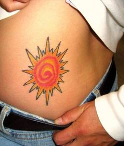
Topical Fluorocarbon Speeds Tattoo Removal Process
ATLANTA – Applying the topical fluorocarbon perfluorodecalin prior to Q-switched laser treatment for tattoo removal allows for immediate retreatment of the tattoo, thereby improving results while decreasing overall treatment time, according to Dr. Roy Geronemus.
The findings have important implications for improving outcomes and patient satisfaction, given that tattoo removal can require 10-20 sessions, depending on factors such as the age and colors of the tattoo, and that tattoos – and thus tattoo removal – continue to increase in popularity, he said. "For a busy practice or impatient patients like we have in New York, this has been a nice advance," Dr. Geronemus said at the annual meeting of the American Society for Dermatologic Surgery.
The approach builds on the "R20 technique" described earlier this year in a study published in the Journal of the American Academy of Dermatology. R20 involves the use of multiple treatment passes that are made at 20-minute intervals.
Typically, after an initial pass, tiny white bubbles form in the superficial papillary dermis, appearing as a whitening of the skin. Retreatment while these bubbles are present elicits a limited reaction.
The R20 technique was developed when investigators found that the bubbles disappear after 20 minutes, and, thus, tested the technique in a study of 12 adults. The patients were randomized to a single treatment pass with a Q-switched alexandrite laser (5.5 J/cm2, 755 nm, 100-nanosecond pulse duration, 3-mm spot size) or to four passes at 20 minute intervals.
The first treatment caused an immediate whitening reaction, but little or no whitening occurred after subsequent passes at 20-minute intervals. However, at 90-day follow-up, significant improvement was seen in the R20 group, compared with the single-treatment group, and light microscopy demonstrated greater dispersion of tattoo ink with the R20 approach. (J. Am. Acad. Dermatol. 2012;66:271-7).
Despite the increased efficacy using this approach, the time required to complete four passes at 20-minute intervals makes it impractical in the clinical setting, said Dr. Geronemus, a dermatologist in private practice in New York.
"So with this idea in mind, we began to look at a concept that would allow us to re-treat tattoos immediately without waiting 20 minutes," he said, explaining that topical perfluorodecalin helps dissolve the gas seen after the application of the Q-switched laser and speeds the resolution of the whitening.
"Rather than waiting 20 minutes, the gas dissolves almost immediately, allowing you to re-treat, and we’re now re-treating three or four times in a matter of minutes rather than waiting the 80 minutes that the R20 technique would take for a four-time treatment session," he said.
In his experience, results with perfluorodecalin are comparable to those seen with the R20 technique – but with greater convenience for the patient.
His observations were confirmed on optical coherence tomography scanning, which demonstrated that cavitation levels are indeed reduced by the use of perfluorodecalin.
Dr. Geronemus is an investigator for Cutera, Cynosure, Palomar, Solta Medical, and Syneron. He is also on the medical advisory board for Cynosure, Lumenis, Photomedex, Syneron, and Zeltiq. He reported that he is a Zeltiq shareholder.
EXPERT ANALYSIS FROM THE ANNUAL MEETING OF THE AMERICAN SOCIETY FOR DERMATOLOGIC SURGERY

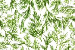
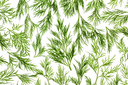
Dill
Dill (Anethum graveolens), an aromatic perennial herb often used as a culinary spice, has been utilized for medical purposes for hundreds of years, at least since medieval times (Wurzbg. Medizinhist. Forsch. 1982;24:411-24). A member of the Umbelliferae (carrot or parsley) family, dill is used in traditional Chinese medicine, and its use in cooking and Uygur medicine is believed to date back to ancient times in China (Evid. Based Complement. Alternat. Med. 2011;2011:659-704).
Based on such past uses, as well as modern research, dill is known for having demonstrated anti-inflammatory, antispasmodic, carminative, aromatic, and galactagogue activity (Medical Herbalism: The Science and Practice of Herbal Medicine. Rochester, Vt.: Healing Arts Press, 2003).
Antimicrobial Activity
In a 2003 study of the essential oil of seeds of dill stored for more than 35 years, Bulgarian researchers tested its antimicrobial activity using various microorganisms. They noted high activity of the essential A. graveolens oil against the mold Aspergillus niger and the yeasts Saccharomyces cerevisiae and Candida albicans (J. Agric. Food. Chem. 2003;51:3854-7). Worth noting from this study is not only the anticandidal properties of dill but its potency even after a long time in storage.
In 2009, Kaur and Arora examined the spices dill, fennel (Foeniculum vulgare), and ajwain (Trachyspermum ammi) for their antibacterial activity. The investigators ascertained antibacterial activity by using agar diffusion assay, minimum inhibitory concentration, and viable cell count studies, and compared effects with those of some standard antibiotics. All three spices in the study exhibited significant activity against a wide range of bacteria, with the exception of Klebsiella pneumoniae and a strain of Pseudomonas aeruginosa. The researchers concluded that the results reveal a scientific basis for the reputed antibacterial effects of these plants and lend credence to their traditional medicinal applications. Further, they suggested that future work may lead to viable antibacterial agents based on these ingredients (BMC Complement. Altern. Med. 2009;9:30).
In 2011, Zeng et al. found that the essential oil produced from dill displays properties effective against vulvovaginal candidiasis in immunosuppressed mice (Evid. Based Complement. Alternat. Med. 2011;659704).
In addition, dill has been found to play an adjuvant role in augmenting the antibacterial activity of nitrofurantoin, which is used to treat urinary tract infections. Researchers used disk-diffusion and agar-dilution methods to determine the effects of essential oils of spearmint (Mentha spicata), dill (A. graveolens), and peppermint (Mentha piperita) and their components on the antibacterial activity of nitrofurantoin against Enterobacter cloacae. They used gas chromatography-mass spectrometry to examine essential oil composition. Dill and spearmint were found to exhibit the most significant effects, with pure carvone and piperitone identified as the most active constituents (Chemotherapy 2007;53:21-5).
Elastogenesis Promotion
In 2006, Cenizo et al. set out to induce elastogenesis in adult dermal fibroblasts by targeting lysyl oxidase (LOX) and lysyl oxidase–like (LOXL) enzymes, which are responsible for elastin cross-linking. LOX and LOXL have been identified as the rate-limiting step in synthesizing mature elastin in adult skin. The expression of LOXL in particular decreases with age. Copious amounts of LOX and LOXL allow the catalysis of immature elastin into desmosine and isodesmosine.
In studying these enzymes, Cenizo and colleagues screened more than 1,000 active ingredients to identify agents that could spur LOXL gene expression in adult dermal fibroblasts. They found that a dill extract was capable of penetrating into the epidermis and dermis in skin engineering and in vitro models, significantly stimulating LOXL gene expression in dermal equivalents (an increase of 64% in mRNA level compared with controls). The researchers also noted increases in elastin detection in dermal equivalents under the dermal-epidermal junction without a corresponding elevation in elastin mRNA. They concluded that LOXL is a suitable target for stimulating elastogenesis, and that dill extract appears to foster such activity (Exp. Dermatol. 2006;15:574-81).
Having shown that dill increases LOX and LOXL expression, along with their respective mRNAs, in 2011, some of the same investigators, this time led by Sohm, assessed the capacity of dill extract to enhance skin elasticity in vitro and in vivo. They reported that skin firmness and elasticity did indeed improve significantly in subjects treated for 56 days with a 1% topical application of dill extract, compared with subjects treated with placebo, based on cutometer measurements, biotribometer measurements, investigator evaluations, subject assessments, and photography. Most volunteers treated with dill extract identified marked enhancements in elasticity, firmness, and jaw line slackness. After 84 days, subjects treated with dill also exhibited significantly reduced mean wrinkle area and length compared with those taking the placebo formulation (Int. J. Cosmet. Sci. 2011;33:157-63). This greater elasticity might be attributed to increased LOX expression stimulated by dill extract application, though this study did not measure LOX or LOXL.
Antioxidant Activity
Recently, investigators compared the radical scavenging and antioxidant activities of the phenolic compounds in six spice plants using spectrophotometric and chromatographic methods. They analyzed onion (Allium cepa), parsley (Petroselinum crispum) roots and leaves, and celery (Apium graveolens) roots and leaves, as well as dill (A. graveolens) leaves, and found that the total amounts of phenolic compounds and radical scavenging activity were greatest in celery leaves and dill extracts (J. Sep. Sci. 2011;34:1261-7).
Combination Therapy
It is worth noting that significant improvement in most measures of photoaged skin was observed after the use of a day and night regimen containing dill extract, blackberry leaf extract, and Zn-Cu(II) bi-mineral complex in patients with mild to moderate photodamage in a small (n = 33), single-center, open-label study led by the author (Baumann LS, Figueras KA, Bell M, Flitter CJ. Unpublished results). This small study supports the notion of dill contributing to cutaneous improvement in combination therapy.
Conclusion
Dill is a culinary spice used worldwide and has a history of traditional use in medicine. Current results suggest reasons for optimism in harnessing the medicinal properties of this plant for various uses in the modern armamentarium, particularly as an antibacterial agent. Much more research is necessary, of course, but recent findings regarding the elastogenesis effects of dill are encouraging and suggest the potential for dermatologic, particularly antiaging, applications.
Differences in the Stratum Corneum Barrier
Despite a growing worldwide population, there is still limited data on the physiology and structural differences in ethnic skin. Although the increased presence of melanin in skin of color confers a photo-protective effect, there are various differences in the stratum corneum that will influence skin physiology in different racial groups.
Barrier function of the skin depends on the structure of the corneocytes, lipid content, and transepidermal water loss.
Compared to white skin, black skin has more corneocyte layers and a more compact stratum corneum with greater intercellular cohesiveness (Semin. Cutan. Med. Surg. 2009;28:115-29). The epidermal barrier in darker skin has been shown to be stronger when exposed to mechanical or chemical challenge. Although the size of the individual corneocytes is the same in black and white skin, the desquamation rate in certain locations is higher in black skin. This is likely because of increased desquamatory enzyme levels, such as cathepsin L2 in the lamellar granules of darker pigmented individuals, leading to an "ashy" manifestation.
Black skin has the highest sebum content of all ethnicities but also the lowest ceramide level, and is, therefore, the most susceptible to transepidermal water loss and xerosis of the skin, when compared with other ethnic groups.
Asian skin has similar corneocyte architecture and transepidermal water loss when compared to white skin, but has decreased corneocyte cohesion, and, thus, a weaker barrier function when exposed to mechanical and chemical stimuli. Compared to white and black patients, Asian patients have the highest amount of ceramides and the least amount of transepidermal water loss.
Although no large, multiethnic group studies have been performed looking at all of the skin barrier physiologic properties and their relation to clinical signs of disease, several small studies do shed light on some of the ethnic variations in skin barrier function. In clinical practice, these small variations should play a role in ethnic-specific treatment regimens for common conditions, such as acne and atopic dermatitis.
In my practice, black patients with acne often have a high sebum content but cannot tolerate drying medications such as benzoyl peroxide. These patients often present with ashy, dry skin in certain areas and oily acne-prone skin in other areas, leading to more complex skin care regimens. Understanding these concepts can help tailor skin treatments for ethnic patients.
- Lily Talakoub, M.D.
Do you have questions about treating patients with darker skin? If so, send them to sknews@elsevier.com.