User login
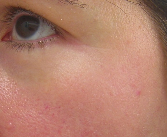
Dysport Trumps Botox for Crow's Feet
A randomized, double-blind study comparing two commercially available botulinum toxin type A injections found abobotulinumtoxinA to be more effective than onabotulinumtoxinA for treating crow's feet.
The study, published online in the Archives of Facial Plastic Surgery (doi:10.1001/archfacial.2011.37), compared the effectiveness of abobotulinumtoxinA, marketed as Dysport, with onabotulinumtoxinA, marketed as Botox, in 90 patients with lateral orbital rhytids.
Investigator and patient assessments of Merz scale scores (a visual tool used to grade wrinkles on a severity scale of 0 to 4) before and at several points after treatment were collected; the primary endpoint was investigator assessment of maximal contraction 30 days post treatment. However, patients also were asked to assess results using the same scale.
AbobotulinumtoxinA was reported by both patients and investigators to be significantly more effective in reducing lines during maximum facial contraction.
Dr. Corey S. Maas of the department of otolaryngology–head and neck surgery at the University of California, San Francisco, who is in private practice, and his colleagues enrolled 90 patients between 31 and 78 years old (mean age 54.5 years) without previous surgery or facial injections for at least 6 months; 13 were men.
All were injected with 10 U of onabotulinumtoxinA on one side of the face and 30 U of abobotulinumtoxinA on the other, according to a 1:3 ratio employed in earlier studies and judged by the principal investigator to be the best comparative dose. The study investigators were blinded as to which side of the face received which treatment, as were patients. Treatments were prepared by a nonblinded nurse.
At baseline, investigators assessed patients as having mean Merz scale scores of 3.68 on the onabotulinumtoxinA-treated sides of their faces at maximal contraction; after treatment, this became 2.33. With abobotulinumtoxinA, investigators assessed patients as having a mean score of 3.64 at baseline and 2.60 after treatment, offering a statistically significant advantage to abobotulinumtoxinA.
Patients, using the same scale, assessed more improvement on the abobotulinumtoxinA side than on the onabotulinumtoxinA side at maximal contraction and favored results from the abobotulinumtoxinA sides 67% of the time.
No statistically significant difference was seen between the two treatments when the lateral orbital rhytids were at rest.
The study was the first to evaluate both products simultaneously in a patient; crow's feet were designated as the candidate wrinkles because of the minimal likelihood of the products diffusing from one side of the face to the other, the investigators wrote.
The investigators offered no explanation for why abobotulinumtoxinA appeared to work better, but wrote in their analysis that because the main pharmacologic difference between the two medications is the hemagglutinin and non-hemagglutinin surrounding each protein, "one may theorize that these differences in efficacy can be ascribed to the hemagglutinin and non-hemagglutinin binding."
Dr. Maas disclosed that he is a consultant for and owns stock in both Medicis Aesthetics (makers of abobotulinumtoxinA) and Allergan (makers of onabotulinumtoxinA). Medicis Aesthetics funded the study.
FROM ARCHIVES OF FACIAL PLASTIC SURGERY
Major Finding: With abobotulinumtoxinA, investigators assessed patients as having a mean score of 3.64 at baseline and 2.60 after treatment, offering a statistically significant advantage to abobotulinumtoxinA.
Data Source: A randomized double-blind trial to compare the effectiveness of abobotulinumtoxinA to onabotulinumtoxinA in 90 patients with lateral orbital rhytids.
Disclosures: Dr. Maas disclosed that he is a consultant for and owns stock in both Medicis Aesthetics (makers of abobotulinumtoxinA) and Allergan (makers of onabotulinumtoxinA). Medicis Aesthetics funded the study.
Microdermabrasion Plus Nd:YAG Laser Improves Melasma
GRAPEVINE, TEX. – The combination of microdermabrasion and low-fluence Q-switched neodymium: YAG laser treatment in conjunction with pigment-reducing skin care produced consistent improvement in two or three treatments for 27 female patients with refractory facial melasma.
Previous attempts to treat melasma using various types of lasers have been associated with significant downtime, punctate hypopigmentation, melasma recurrence, and rebound hyperpigmentation.
Fractional lasers require four to six treatments and are associated with treatment pain, several days of recovery, and a high risk of rebound melasma. Higher-fluence Q-switched Nd:YAG laser therapy, performed with multiple laser passes during weekly treatments, is associated with pain, hair whitening, urticaria, punctuate hypopigmentation, and rebound melasma, said Dr. Arielle N.B. Kauvar at the annual meeting of the American Society for Laser Medicine and Surgery.
In this observational study, the 27 women had phototypes II-V with refractory mixed-type or dermal melasma. Their skin was first cleansed and then treated with diamond-chip microdermabrasion. Immediately after the microdermabrasion, 17 of the women received treatment with the Candela TriVantage (wavelength 1064 nm, nominal pulse width 50 ns, spot size 5 mm, fluence 1.6 J/cm2), while the other 10 women were treated with the Palomar Q-YAG (1064 nm, 5-7 ns, 6 mm, 1.8-2.0 J/cm2).
Patients began using a broad-spectrum sunscreen SPF 40 or higher immediately after treatment with the microdermabrasion and laser. For 2 days after each laser treatment, 4% hydroquinone twice daily plus 0.05% tretinoin at bedtime was applied until the day of the next microdermabrasion/laser treatment.
For patients with sensitive skin, 15% L-ascorbic acid was substituted for the tretinoin and used in the morning. Patients were maintained on skin care long term, said Dr. Kauvar, who is director of New York Laser & Skin Care.
The 27 women had a mean age of 37 years (range, 26-54 years). Four had skin type II, 11 type III, 7 type IV, and 5 type V. The mean number of treatments was 2.6. Mean clearance scores were 3.3 at 3 months, 3.2 at 6 months (25 patients), and 3.3 at 12 months (9); a score of 0 indicates less than 25% clearance, 1 = 25%-50% clearance, 2 = 51%-75%, 3 = 75%-95%, and 4 = greater than 95%.
Of the 27 patients, 22 had greater than 27% clearance of their melasma, while 11 had more than 95% clearance of pigmented patches. Most showed greater than 50% clearance at 1 month after the first treatment session, and only one patient had less than 25% clearance after one treatment, said Dr. Kauvar, who is in the department of dermatology at New York University.
The procedure was not associated with pain. All of the patients experienced very faint erythema that developed after the microdermabrasion, which lasted 30-60 minutes. Seven of the 27 had significant irritation from the skin care regimen, which resolved when the retinoid was discontinued. Another four had mild irritation from the skin care, which was successfully managed with reduction of the hydroquinone and retinoid applications. There was no incidence of hyperpigmentation or hypopigmentation.
Microdermabrasion decreases the scattering of laser light and increases epidermal cell turnover, while the low-fluence Q-switched YAG laser directly damages the melanocytes and melanosomes. The skin care regimen suppresses melanin production and protects against ultraviolet exposure, Dr. Kauvar explained.
"The combination of microdermabrasion and low-fluence Q-switched YAG laser treatment in conjunction with pigment-reducing skin care is a safe and effective treatment for melasma with minimal risks. This treatment offers substantial benefits over more invasive, higher-risk, costly procedures such as nonablative or ablative fractional laser treatment," she said.
Dr. Kauver stated that she has no disclosures.
FROM THE ANNUAL MEETING OF THE AMERICAN SOCIETY FOR LASER MEDICINE AND SURGERY
Major Finding: Of a total of 27 patients, 22 had greater than 27% clearance of their melasma while 11 had more than 95% clearance of pigmented patches.
Data Source: Observational study of 27 women with refractory mixed-type or dermal melasma.
Disclosures: Dr. Kauver stated that she had no disclosures.
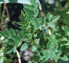
Astragalus
Astragalus membranaceus has a millennia-long tradition of medicinal use in China, but has only recently been considered and investigated by Western medicine. Preliminary indications suggest that this herb offers significant potential, with few side effects, as an alternative or adjuvant therapy for several conditions, including dermatologic.
Native to Mongolia, and northern and eastern China, A. membranaceus, a member of the pea family, is a perennial herb that has been used in Traditional Chinese Medicine (TCM) as a tonic for thousands of years (Zhonghua Shi Yan He Lin Chuang Bing Du Xue Za Zhi 1998;12:269-71). The dried root of the plant has been traditionally used to treat various digestive ailments, stomach ulcers, colds and influenza, fever, allergies, anemia, uterine bleeding and prolapsed uterus, as well as wounds, and, in combination with other herbs, to treat dry or peeling skin, bruises and other minor skin conditions.
Recently, the herb has come to be recognized for its medical potential as an antioxidant and for its immunomodulatory activity (Yao Xue Xue Bao 1992;27:5-9). In fact, the effectiveness of Astragalus to boost the immune system has been well established over the last several years (Integr. Cancer Ther. 2003;2:247-67; J. Clin. Lab. Immunol. 1988;25:119-23; J. Clin. Lab.Immunol. 1988;25:125-9).
Antioxidant Properties of A. membranaceus
A study conducted of 21 species of herbs used for medicinal purposes to evaluate relative antioxidant as well as nicotine degradation activity revealed that A. membranaceus exhibited significant antioxidant activity and nicotine degradation activity. The herb was subsequently included in a medicinal tea (with 10 other species) and studied, displaying success as a smoking cessation treatment in 100 human males (Am. J.Chin. Med. 2005;33:127-38). Antioxidant effects have also been associated with the polysaccharides of the A. membranaceus variant A. mongholicus. The effects of Astragalus polysaccharide, an active component, on Escherichia coli endotoxin–induced liver damage in mice were found to increase longevity, as the most significant of several benefits. The protective effects of Astragalus polysaccharide were ascribed to its antioxidant activity (Yao Xue Xue Bao 1992;27:5-9). A. mongholicus, also used for centuries in TCM, has been shown in animal models and clinical trials to exhibit immunomodulating activity (Mem. Inst Oswaldo Cruz 1991;86 Suppl 2:159-64). In addition, a novel lectin, isolated from the roots of A. mongholicus, was recently found to exert antifungal activity against Botrytis cincerea, Fusarium oxysporum, Colletorichum sp., and Drechslera turcia (Arch. Biochem. Biophys. 2005;442:72-81)
Astragalus is an adaptogen and is usually used in conjunction with other herbs such as ginseng and echinacea.
Anti-aging Properties and Cutaneous Applications
In a recent study, investigators evaluated the anti-aging effects of astragalosides (AST), major active components of Astragalus species, by ascertaining the influence of AST on motor and memory manifestations of D-galactose (D-gal)–induced senescent and middle-aged mice.
Ten-week treatment with AST was found to improve age-related changes in memory and motor response, as well as ameliorate the diminished cellular immunity in the murine test subjects. Investigators concluded from the enhanced brain activity and immunomodulatory results that AST confers an anti-aging effect on D-gal–induced senescent and middle-aged mice (Acta Pharmacol. Sin. 2003;24:230-4). An earlier investigation of the hairy root of A. membranaceus (HRA), in which the extract was administered over 50 days to senescent mice treated with D-galactose, revealed several benefits. The hairy root was found to enhance memory, elevate superoxide dismutase antioxidant activity in brain and liver, and foster natural killer (NK) cell activity in immunocompromised mice as well as reduce malondialdehyde content in rat ischemia-reperfusion kidney and lower the creatinine level in rat blood. Investigators concluded that HRA is similar to natural A. membranaceus in its antioxidant capacity as well as its immunomodulatory and senility-preventing activity (Zhongguo Zhong Yao Za Zhi 1999;24:619-21, 639).
In a study evaluating 15 extracts of herbs used in TCM, investigators found that A. membranaceus was one of three botanicals to inhibit 5-lipoxygenase, one of the enzymes, along with elastase, considered important therapeutic targets in the treatment of psoriasis and other cutaneous conditions (J Pharm Pharmacol. 2003;55:1275-82).
Antiviral Properties of A. membranaceus
Astragalus extract has also been shown to impart an anticarcinogenic effect in mice by spurring cytotoxic activity and cytokine production (Cancer Invest. 1999;17:30-5).
Investigators conducted a study on the anti-herpes simplex virus activity of A. membranaceus (in suppository or ointment form) combined with recombinant human interferon (IFN) alpha 2b in human diploid cell culture. Results showed that combination treatment was significantly more effective than placebo or IFN alone. The authors concluded that this combination treatment including A. membranaceus was suitable in suppository form for the treatment of cervicitis and, in ointment form, for the treatment of herpetic lesions on the skin (Zhonghua Shi Yan He Lin Chuang Bing Du Xue Za Zhi 1998;12:269-71). More recent studies have also demonstrated clear HSV-1 inhibiting activity and low cytotoxicity exhibited by A. membranaceus (Di Yi Jun Yi Da Xue Xue Bao 2004;24:57-8). In a study with 106 patients with herpes simplex keratitis, evidence showed that A. membranaceus significantly improved imbalances in serum cytokines and enhanced immune function (Zhongguo Zhong Xi Yi Jie He Za Zhi 2004;24:121-3).
A. membranaceus was also shown to exert significant anti-viral activity in a study on mice infected with coxsackie B-3 virus (CVB3) as the herb was demonstrated to suppress viral replication in a viral myocarditis model (Chin. Med.Sci. J. 1995;10:146-50).
In an examination of the effects of 14 Chinese medicinal herbs on lipid peroxidation, investigators found that A. membranaceus conferred significant protection of rat heart mitochondria, inhibiting oxygen consumption and malondialdehyde production (Am J Chin Med. 1994;22:63-70).
In a study over 15 years ago, anti-senility effects were demonstrated by the Chinese herbal formulation Shou Xing Bu Zhi (composed of 13 herbs, including A. membranaceus) in mice. After 3 months of oral administration, liver and brain tissue lipofuscin was markedly decreased in young (1 month old) and adult (11 months) animals, lipid peroxidation was similarly diminished in adult mice, and hydroxyproline of skin was reduced in young and adult mice. Investigators concluded that the herbal combination agent was effective in retarding several markers of aging (Zhong Xi Yi Jie He Za Zhi 1989;9:226-7, 198).
A TCM decoction containing Angelica sinensis and A. membranaceus used for stimulating red blood cell production and bolstering cardiovascular function was shown in a rat model to confer myocardial protection against ischemia-reperfusion injury (Phytother.Res. 2000;14:195-9).
Various other herbal formulations containing Astragalus have been found to exert a range of health benefits. Injection of Qi-Xue, a Chinese herb combination that contains A. monogholicus, along with Angelica sinensis and Panax ginseng, is thought to prevent severe hypoxic pulmonary hypertension by enhancing heart function (Zhongguo Yi Xue Ke Xue Yuan Xue Bao 1990;12:51-5). A. membranaceus root is also among a cocktail of herbs contained in Hochuekkito, a Kampo (traditional Japanese herbal) medication recently found in a study of 95 patients to be effective, in combination with dietary changes, in treating recalcitrant atopic dermatitis (Drugs Exp. Clin. Res. 2004;30:197-202). An open-label study of a drug mixture containing five Chinese medicinal herbs including A. membranaceus for treatment of people living with HIV has shown some promise, but requires more investigation. The study revealed that the formulation was safe and effective at decreasing viral load, but an immunologic response in the form of an elevated CD4 count was not established (J. Med .Assoc. Thai. 2004;87:1065-70).
Finally, A. membranaceus has also been demonstrated, in vitro, to significantly improve human sperm motility (Am. J. Chin. Med. 1992;20:289-94).
Astragalus root is available in several forms, including oral, injectable (in the clinical setting), and topical.
Conclusion
Research on A. membranaceus has revealed a remarkable array of medicinal properties, many of which have clear potential for dermatological applications. Much more research is necessary, though, to determine the appropriate medicinal role(s) for this ancient herb.
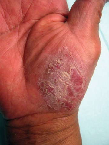
308-nm Excimer Laser Effective for Treating Palmoplantar Psoriasis
GRAPEVINE, TEX. – The 308-nm excimer laser significantly reduced palmoplantar psoriasis severity in a study of 30 patients.
Multiple studies have demonstrated the efficacy of the excimer laser in the treatment of plaque psoriasis, but few have investigated its use for palmoplantar psoriasis. "Palmoplantar psoriasis is difficult to treat and often recalcitrant to traditional therapies such as corticosteroids. Since the excimer laser can selectively treat psoriatic plaque with higher fluences than is tolerated with traditional phototherapy, it could be a therapeutic modality for the thicker skin on the palms and soles," said Dr. David Goldberg, director of dermatologic laser research at Mount Sinai School of Medicine, New York.
The 30 patients were aged 18-75 years, with mild to severe psoriasis on their hands and/or feet. All discontinued other treatments 4 weeks prior to starting the study. By study design, patients could receive up to 16 biweekly laser treatments over the course of 3 months with an excimer laser (PHAROS EX-308, RA Medical Systems). Fluences ranged from 400-600 mJ/cm2 depending on disease severity. Short pulses were delivered with a flexible handpiece, with a maximal output of 15 mJ/pulse (J. Cosmet. Laser Ther. 2011;13:47-9).
Each treatment was tailored to the individual patient’s response from the previous session. If there was no response or minimal erythema, the dose was increased by 30%. If there was moderate erythema, the dose was increased by 20%, and if significant erythema, the dose was increased by 10% until the patient could not tolerate further increases. For severe reactions or blistering, the dose was decreased by 20%. The number of sessions ranged from 7 to 14, with a mean of 11.

All of the subjects had some improvement by week 5, as measured by the Psoriasis Area and Severity Index (PASI). At the end of the treatments, all showed 50%-100% reductions in scaling, erythema, and flattened plaques. No patient had a relapse detected at the 3-month follow-up, but two-thirds of the patients had relapses by 6 months. There was no evidence of persistent pigmentary changes or scarring. Periodic retreatments will be required, Dr. Goldberg said.
"Although no treatment can be expected to ‘cure’ palmoplantar psoriasis, our data do support the use of the excimer laser to treat patients with hand and foot psoriasis. The excimer laser should be strongly considered for patients with palmoplantar psoriasis unresponsive to other treatments," he concluded.
The excimer laser study was provided on loan by the manufacturer during the course of the study. Dr. Goldberg stated that he had no other disclosures.
GRAPEVINE, TEX. – The 308-nm excimer laser significantly reduced palmoplantar psoriasis severity in a study of 30 patients.
Multiple studies have demonstrated the efficacy of the excimer laser in the treatment of plaque psoriasis, but few have investigated its use for palmoplantar psoriasis. "Palmoplantar psoriasis is difficult to treat and often recalcitrant to traditional therapies such as corticosteroids. Since the excimer laser can selectively treat psoriatic plaque with higher fluences than is tolerated with traditional phototherapy, it could be a therapeutic modality for the thicker skin on the palms and soles," said Dr. David Goldberg, director of dermatologic laser research at Mount Sinai School of Medicine, New York.
The 30 patients were aged 18-75 years, with mild to severe psoriasis on their hands and/or feet. All discontinued other treatments 4 weeks prior to starting the study. By study design, patients could receive up to 16 biweekly laser treatments over the course of 3 months with an excimer laser (PHAROS EX-308, RA Medical Systems). Fluences ranged from 400-600 mJ/cm2 depending on disease severity. Short pulses were delivered with a flexible handpiece, with a maximal output of 15 mJ/pulse (J. Cosmet. Laser Ther. 2011;13:47-9).
Each treatment was tailored to the individual patient’s response from the previous session. If there was no response or minimal erythema, the dose was increased by 30%. If there was moderate erythema, the dose was increased by 20%, and if significant erythema, the dose was increased by 10% until the patient could not tolerate further increases. For severe reactions or blistering, the dose was decreased by 20%. The number of sessions ranged from 7 to 14, with a mean of 11.
All of the subjects had some improvement by week 5, as measured by the Psoriasis Area and Severity Index (PASI). At the end of the treatments, all showed 50%-100% reductions in scaling, erythema, and flattened plaques. No patient had a relapse detected at the 3-month follow-up, but two-thirds of the patients had relapses by 6 months. There was no evidence of persistent pigmentary changes or scarring. Periodic retreatments will be required, Dr. Goldberg said.
"Although no treatment can be expected to ‘cure’ palmoplantar psoriasis, our data do support the use of the excimer laser to treat patients with hand and foot psoriasis. The excimer laser should be strongly considered for patients with palmoplantar psoriasis unresponsive to other treatments," he concluded.
The excimer laser study was provided on loan by the manufacturer during the course of the study. Dr. Goldberg stated that he had no other disclosures.
FROM THE ANNUAL MEETING OF THE AMERICAN SOCIETY FOR LASER MEDICINE AND SURGERY
Major Finding: At the end of the treatments, all patients showed 50%-100% reductions in scaling, erythema, and flattened plaques
Data Source: The 30 patients were aged 18-75 years, with mild to severe psoriasis on their hands and/or feet.
Disclosures: The excimer laser study was provided on-loan by the manufacturer during the course of the study. Dr. Goldberg stated that he had no other disclosures.
Blog: The Cosmetic Surgery Serenity Prayer
If there ever were a field in which it's easy to overdo things, it's aesthetic medicine. In fact, some clinicians develop what Dr. Val S. Lambros called "surgeon dysmorphia syndrome," which occurs when "the surgeon thinks something looks so good that he makes everyone look that way."

At the SDEF Summit in Aesthetic Medicine meeting in Dana Point, Calif., Dr. Lambros shared his version of the Serenity Prayer that he modified as a way to remind his colleagues to avoid developing surgeon dysmorphia syndrome. Known as the Facelift Serenity Prayer, it reads:
Grant me the ability to change the things I can,The serenity to let go of the things that I can't,
And the wisdom to avoid overdoing the things I can change to fix the things I can't.
Dr. Lambros, a clinical instructor at the University of California, Irvine, Aesthetic and Plastic Surgery Institute, said that he created the prayer prior to addressing a group of plastic surgeons about 4 years ago.
"It really could be called the Cosmetic Surgery Serenity Prayer," he said.
If there ever were a field in which it's easy to overdo things, it's aesthetic medicine. In fact, some clinicians develop what Dr. Val S. Lambros called "surgeon dysmorphia syndrome," which occurs when "the surgeon thinks something looks so good that he makes everyone look that way."
At the SDEF Summit in Aesthetic Medicine meeting in Dana Point, Calif., Dr. Lambros shared his version of the Serenity Prayer that he modified as a way to remind his colleagues to avoid developing surgeon dysmorphia syndrome. Known as the Facelift Serenity Prayer, it reads:
Grant me the ability to change the things I can,The serenity to let go of the things that I can't,
And the wisdom to avoid overdoing the things I can change to fix the things I can't.
Dr. Lambros, a clinical instructor at the University of California, Irvine, Aesthetic and Plastic Surgery Institute, said that he created the prayer prior to addressing a group of plastic surgeons about 4 years ago.
"It really could be called the Cosmetic Surgery Serenity Prayer," he said.
SDEF: When to Discard and Replace Lasers, Factors Considered
DANA POINT, CALIF. - Between 1985 and 2011, Dr. Gordon Sasaki retired or discarded an estimated 15 devices used for aesthetic plastic surgery, including two erbium lasers, three CO2 lasers, and an external radiofrequency system for tissue lifting.
Factors that played into the dismissal of these devices included obsolescence of technology, the rise of improved, similar technologies, and competing devices in the office.
At the Summit in Aesthetic Medicine sponsored by Skin Disease Education Foundation (SDEF), Dr. Sasaki, clinical professor of plastic surgery at Loma Linda (Calif.) University Medical Center, shared why he chose to retire or let go of certain devices and embrace others.
Sometimes he stopped working with a device after comparing the cost of buying or leasing it with its positive return on investment, he said. Other factors he considered included the cost of disposable equipment, warranty renewals, and unanticipated costs for reparative procedures when a patient had unsatisfactory results or complications.
One device Dr. Sasaki retired was a high frequency eradicator, which is a low-powered, nongrounded electrosurgical device used for desiccation of subdermal lesions and fulguration of superficial lesions. His rationale included its cost to purchase ($1,000-$1,500), cost of the disposable tip ($1.50 each), and nontransportability from room to room.
He replaced it with the Medi-Pak handheld battery-powered cautery device, which achieves acceptable results yet costs only $9.95 per device, has no disposable components, and can be used multiple times until loss of battery power. After battery power loss, a new device is purchased.
Dr. Sasaki, who has a private aesthetic plastic surgery practice in Pasadena, Calif., also retired a nonablative monopolar radiofrequency device he used for tissue lifting and volumetric heating of dermis, septae, and fat. The initial cost of the second-generation device was $65,000, with a warranty that ranged from $2,800 to $3,300 per year. The annual cost of disposable tips added another $450-$949 per year. "Although the device produced satisfactory results in some patients, it also had unpredictable responses, as well as patient and operator fatigue, especially for body contouring procedures," he said. "There was low patient request for this device, and new, improved technology became available."
He put aside the nonablative radiofrequency device in favor of a multilevel focused and imaged ultrasonic tissue lifting device that provides an imaging level of treatment, precise thermal coagulation points, and more predictable patient responses. In his experience, 80%-90% of his patients showed clinical improvement by 3 months, and results tend to last 1-1.5 years. "There is minimal to moderate pain during treatment, and there is a high patient request for this procedure," Dr. Sasaki said.
Costs of the unit, including a 3-year warranty, were $87,500, he said. Additional warranties cost $10,000 per year, and the cost of a disposable transducer amounts to $2,100, which can last for at least four patient treatments, depending on the area of treatment.
Dr. Sasaki said that he currently shares a fractional CO2 resurfacing laser, in part because of its high purchase cost ($152,000, plus $240 for a box of 30 disposable tips), but also because of patient and operator fatigue, complex patient recovery, unpredictable and unsatisfactory patient responses, and low patient requests for the technology.
As a substitute for selected patients, Dr. Sasaki uses croton oil peels for patients who request facial resurfacing. Concentrations ranging from 0.0125% to 0.4% can be prepared, he said, and the procedure does not require application of a topical or local anesthesia. Costs are reasonable. In his practice a 1-ounce container of phenol costs $23.75 and a 1-ounce container of croton oil costs $106. "The preferred treatments are in patients with Fitzpatrick I-IV skin types," he said. "There are predictable and satisfactory patient responses."
Dr. Sasaki disclosed that he has been a paid consultant for many laser and device companies.
SDEF and this news organization are owned by Elsevier.
EXPERT ANALYSIS FROM THE SDEF SUMMIT IN AESTHETIC MEDICINE
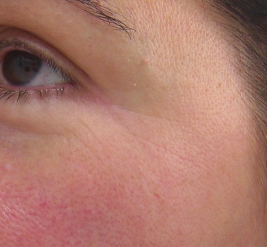
SDEF: Fractionated Radiofrequency Smoothes Wrinkles, New Findings Show
DANA POINT, CALIF. – In the clinical experience of Dr. George J. Hruza, bipolar fractionated radiofrequency provides good wrinkle effacement, especially in the periorbital area.
"The benefits of bipolar fractionated radiofrequency include a limited downtime, no adverse events are seen, and any skin type can be treated," Dr. Hruza said at the Summit in Aesthetic Medicine sponsored by Skin Disease Education Foundation (SDEF).
Dr. Hruza, clinical professor of dermatology at St. Louis University, presented findings from a study of 22 adults with Fitzpatrick skin types I-IV who were treated with bipolar fractionated radiofrequency for visible wrinkles and/or elastosis. He and his associates used the Food and Drug Administration–cleared eMatrix radiofrequency device (Syneron) to treat two of the following facial regions per patient: the periorbital region, the perioral region, the cheeks, and the forehead.
Both 64 and 144 pin tips were used; energy was delivered at a range of 2-14 joules per pulse, or 20 J/double pulse, for a maximum duration of 50 milliseconds per pulse. Each patient underwent three treatments at 3 weeks apart. Topical anesthesia was used, and the patients were followed up a month after the last treatment.
Photographic analysis at the 1-month follow-up revealed that fine lines, smoothness, tightness, and brightness improved in about half of the patients by at least 40%. Periorbital photographic results demonstrated a mean improvement in fine lines of at least 30%, with almost all patients showing clinically significant improvement. A 6-month follow-up study of the patients showed persistence of the improvement.
Dr. Hruza noted that patients may experience micro peeling for 2-4 days after undergoing bipolar fractionated radiofrequency.
An emerging technology for deeper skin lesions is dermal bipolar fractionated radiofrequency, which delivers radiofrequency energy within the dermis via micro-needle electrode pairs. "Ninety-six percent of energy is absorbed in the dermis, and the thermal profile is confined along and between the needles," Dr. Hruza said. "This creates a controlled lesion and a fractionated zone of thermal injury."
Tumescent anesthesia is recommended for bipolar dermal fractionated radiofrequency, he said, because it protects deeper skin structures and reduces bleeding. The procedure typically requires 100-300 insertions in the lower face and upper neck. The needle entry points typically close in 2 hours.
Dr. Hruza said that he had no relevant disclosures.
SDEF and this news organization are owned by Elsevier.
FROM THE SDEF SUMMIT IN AESTHETIC MEDICINE
Major Finding: Photographic analysis at 1 month follow-up revealed that fine lines, smoothness, tightness, and brightness improved in about half of patients by at least 40%.
Data Source: A study of 22 adults with Fitzpatrick skin types I-IV who were treated with bipolar fractionated radiofrequency for visible wrinkles and/or elastosis.
Disclosures: Dr. Hruza said that he had no relevant disclosures. SDEF and this news organization are owned by Elsevier.
SDEF: Remain Skeptical Over Efficacy of Noninvasive Fat Removal Devices
DANA POINT, CALIF. – Practice skepticism when it comes to the efficacy of noninvasive fat removal devices, advised Dr. Matthew A. Avram.
"Fat removal has a long history of 'snake oil' salesmanship," Dr. Avram said at the Summit in Aesthetic Medicine, which was sponsored by Skin Disease Education Foundation (SDEF). "You can count on this to continue, because many of these devices do little, if anything. It is important to critically assess these technologies in this emerging field."
Focused Ultrasound
Dr. Avram, faculty director for procedural dermatology training at Harvard Medical School and director of the dermatology laser and cosmetic center at Massachusetts General Hospital, both in Boston, discussed the evidence surrounding devices for noninvasive fat reduction.
One non–Food and Drug Administration–cleared modality being studied is focused ultrasound, which delivers mechanical, nonthermal energy to the thigh, abdomen, and flanks. A study of 30 patients who were treated once a month for 3 months demonstrated a circumference reduction of 2-4 cm in the treated sites (Lasers Surg. Med. 2007;39:315-23). Liver function tests, a lipid panel, and liver ultrasound showed no adverse systemic effects from the procedure.
However, Dr. Avram noted that the study is limited because there was no untreated control group and that circumference "is an inherently imprecise measure of improvement that can be manipulated." MRI would prove objective improvement, he said, but it was not performed in this trial.
In a subsequent study from Hong Kong, 53 patients underwent treatment once a month for 3 months for body contouring (Lasers Surg. Med. 2009; 41:751-9). No significant changes were observed in circumference and caliper measurements, and patients rated their satisfaction as poor.
High-Intensity Focused Ultrasound
High-intensity focused ultrasound, which is also not FDA cleared, involves the rapid heating of adipocytes that are purported to produce coagulative necrosis and cell death in adipose tissue. A retrospective chart review of the modality that was used in 85 patients who underwent one treatment session showed a mean 4.6 cm decrease in waist circumference after 3 months (Aesth. Plast. Surg. 2010;34:577-82). Adverse events that lasted 4-12 weeks occurred in 12% of subjects and included prolonged tenderness, ecchymosis, nodules, and edema, as well as procedural pain in one patient, which required discontinuation of the procedure.
Dr. Avram called high-intensity focused ultrasound a promising technology, "but it's difficult to assess its efficacy. Further study of this technology is needed."
Monopolar Radiofrequency
Monopolar radiofrequency, conventionally used for tissue tightening of the face, has produced lipoatrophy with aggressive settings as a complication. "Perhaps this can be harnessed to effectively treat fat," he said, noting that clinical studies are currently underway.
Low-Level Light Therapy
Low-level light therapy, an FDA-cleared modality for fat removal, uses a multiple head, low-level diode laser at an energy level of 635 nm, which is "roughly equivalent to a laser pointer," Dr. Avram said. In one randomized trial, 59 patients received three treatments of the technology or sham treatment per week for 2 weeks (Lasers Surg. Med. 2009;41:799-809). At 2 weeks, mean circumference reductions in the treatment group were 0.98 inches at the waist, 1.05 inches at the hip, and 0.85 inches at the right thigh and 0.65 inches at the left thigh. Circumference increased in the 2 weeks following treatment.
Dr. Avram said the study was poorly designed because there was no untreated control group, the duration of treatment was only 2 weeks, and there was no ultrasound or other noninvasive evidence of decreased fat layer. "Treat with extreme skepticism," he advised.
Cryolipolysis
Perhaps the most promising technology is cryolipolysis, he said, which is FDA cleared for noninvasive fat removal. Cryolipolysis involves the noninvasive cooling of fat to selectively cause cell death without damage to surrounding tissue.
The mechanism of action of cryolipolysis is believed to involve selective crystallization of lipids in fat cells at temperatures near freezing. "Apoptotic fat cell death is followed by slow dissolution of the fat cell and a gradual release of lipids," Dr. Avram explained. "The inflammatory process results in fat layer reduction over 2-4 months."
When human studies of the technology were first conducted in 2008, enrollment was restricted to 32 patients whose "love handles" were treated at a cooling intensity factor (CIF) of 33 for 60 minutes, and progressed to higher rates of energy extractions for 45 minutes per application site. One side was treated; the untreated contralateral side served as the control.
Efficacy was evaluated at 4 months post treatment via visual assessment as a primary end point, as well as ultrasound and histology. The ultrasound results demonstrated an average 23% decrease in fat layer thickness.
"In this initial group of 32 love handle patients treated once, discernible changes were seen on the treated side vs. baseline of the treated side, and compared to the untreated contralateral control," Dr. Avram said of the findings presented during a poster session at the 2008 annual meeting of the American Society for Dermatologic Surgery. "This unique study design [using each patient as his or her own control], provided very powerful evidence that this was a true treatment effect rather than a change in the patient's diet or exercise pattern during the 4 months after this single procedure exposure."
Common effects after cryolipolysis include redness, which lasts minutes to a few hours; bruising, which may last for a few weeks; and temporary dulling of sensation in the treated area, which typically resolves in 1-8 weeks. No postprocedural changes in pigmentation or laboratory abnormalities have been observed, he said.
About 1 in 2,000 patients experiences severe pain beginning 3-7 days post treatment, which translates into 26 reported cases out of 60,000 treatments. "We are not sure why these occur, but these cases completely resolve with no sequelae," Dr. Avram said.
He emphasized that cryolipolysis is not a replacement for liposuction. "It is not a weight-loss device," he said. "It's best suited for local fat removal resistant to exercise in relatively fit patients."
Dr. Avram disclosed holding stock options in Zeltiq Aesthetics, which manufactures cryolipolysis equipment. SDEF and this news organization are owned by Elsevier.
EXPERT ANALYISIS FROM THE SDEF SUMMIT IN AESTHETIC MEDICINE
Scrubbing Often During Nail Removal Reduces Infection Rates
LAS VEGAS – Preoperative scrubs before nail avulsion fail to reduce postoperative infection, recent data suggest.
However, studies have identified best practices for reducing bacterial counts – such as irrigating after nail plate avulsion though there is no evidence that the measures reduce postoperative infection rates.

"Every study that has looked at rates of colonization or recolonization after your scrub showed significant bacterial presence. As good as our scrubs are, they're probably not good enough," Dr. Nathaniel J. Jellinek said at the annual meeting of the American College of Mohs Surgery.
A recent study measured bacterial counts after a 7-minute surgical scrub and again after avulsion of the nail plate. The bacterial counts were essentially the same. "It's as if the surgical scrub didn't do any good," said Dr. Jellinek, who was not involved in the study (Dermatol. Surg. 2010;36:1258-65).
The investigators then irrigated the nail bed with saline, which reduced bacterial counts by 95%. That's "an easy thing to do intraoperatively that you might consider for your practice," said Dr. Jellinek of the department of dermatology at Brown University, Providence, R.I.
Studies published mainly in the podiatric and orthopedic literature show that postoperative infection rates are higher after nail surgery than after skin surgery. Rates of bacterial colonization before and after surgical preparation of nails make the term "sterile surgery" inaccurate for these procedures, which might better be considered clean-contaminated or even contaminated surgery, he said in an interview after his presentation.
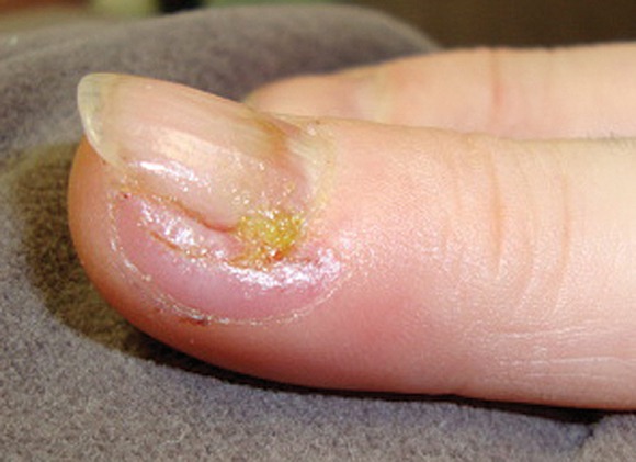
Other studies have shown that applying alcohol and chlorhexidine may be superior to chloroxylenol and povidone-iodine to reduce bacterial counts before nail surgery. Bacterial persistence and recolonization also can be reduced by the use of bristled brushes, soaked gauze pads, scrubbing to the interdigital webs, and repeat scrubs.
"At this point, it's all hypothetical whether it decreases infection. But we know that infection rates are unacceptably high, so it can only help if you decrease the bacterial count," Dr. Jellinek said.
He has added multiple prophylactic measures to his practice, where he performs a lot of nail surgery.
First, Dr. Jellinek informs the patient that the nail is a dirty site, he reviews wound care, and educates the patient to pay attention to risk factors for infection and avoid putting fingers or toes in dirty places.
He recommends strict sterile preoperative and intraoperative techniques. A nurse or medical assistant should scrub the surgical area for several minutes. "We're talking not 10 or 30 seconds, but 2, 3, 5 minutes," he said.
The scrub should use multiple agents. He prefers using a bristle brush to scrub with chlorhexidine, gauze pads soaked in 70% isopropyl alcohol, and maybe even povidone-iodine paint. He lets all that sit, and applies a sterile glove over the digit, hand, or foot after the scrub.
During surgery, once the nail plate is avulsed, he recommends either performing a repeat scrub or at least irrigating the nail bed with sterile saline.
Dr. Jellinek said recent data have lowered his threshold for using prophylactic antibiotics. He has come to appreciate that nail infections may be caused by different organisms than those that cause most skin infections and require different antibiotics for treatment.
As a result, "I actually have very few nail infections, but I can't pinpoint which of those five or six things that I've done give me those results," he said.
Studies of prophylactic measures to avoid postoperative nail infections include:
- Preoperative skin and nail preparation of the foot: Comparison of the efficacy of 4 different methods in reducing bacterial load (J. Am. Acad. Dermatol. 2009;61:986-92).
- Chlorhexidine Provides Superior Skin Decontamination in Foot and Ankle Surgery: A Prospective Randomized Study (Clin. Orthop. Relat. Res. 2005;438:204-8).
- Preoperative Skin Preparation of the Foot and Ankle: Bristles and Alcohol Are Better (J. Bone Joint Surg. 2005;87:986-92).
- Efficacy of Surgical Preparation in Solutions in Foot and Ankle Surgery (J. Bone Joint Surg. 2005;87:980-5).
Dr. Jellinek said he had no relevant financial disclosures.
LAS VEGAS – Preoperative scrubs before nail avulsion fail to reduce postoperative infection, recent data suggest.
However, studies have identified best practices for reducing bacterial counts – such as irrigating after nail plate avulsion though there is no evidence that the measures reduce postoperative infection rates.
"Every study that has looked at rates of colonization or recolonization after your scrub showed significant bacterial presence. As good as our scrubs are, they're probably not good enough," Dr. Nathaniel J. Jellinek said at the annual meeting of the American College of Mohs Surgery.
A recent study measured bacterial counts after a 7-minute surgical scrub and again after avulsion of the nail plate. The bacterial counts were essentially the same. "It's as if the surgical scrub didn't do any good," said Dr. Jellinek, who was not involved in the study (Dermatol. Surg. 2010;36:1258-65).
The investigators then irrigated the nail bed with saline, which reduced bacterial counts by 95%. That's "an easy thing to do intraoperatively that you might consider for your practice," said Dr. Jellinek of the department of dermatology at Brown University, Providence, R.I.
Studies published mainly in the podiatric and orthopedic literature show that postoperative infection rates are higher after nail surgery than after skin surgery. Rates of bacterial colonization before and after surgical preparation of nails make the term "sterile surgery" inaccurate for these procedures, which might better be considered clean-contaminated or even contaminated surgery, he said in an interview after his presentation.
Other studies have shown that applying alcohol and chlorhexidine may be superior to chloroxylenol and povidone-iodine to reduce bacterial counts before nail surgery. Bacterial persistence and recolonization also can be reduced by the use of bristled brushes, soaked gauze pads, scrubbing to the interdigital webs, and repeat scrubs.
"At this point, it's all hypothetical whether it decreases infection. But we know that infection rates are unacceptably high, so it can only help if you decrease the bacterial count," Dr. Jellinek said.
He has added multiple prophylactic measures to his practice, where he performs a lot of nail surgery.
First, Dr. Jellinek informs the patient that the nail is a dirty site, he reviews wound care, and educates the patient to pay attention to risk factors for infection and avoid putting fingers or toes in dirty places.
He recommends strict sterile preoperative and intraoperative techniques. A nurse or medical assistant should scrub the surgical area for several minutes. "We're talking not 10 or 30 seconds, but 2, 3, 5 minutes," he said.
The scrub should use multiple agents. He prefers using a bristle brush to scrub with chlorhexidine, gauze pads soaked in 70% isopropyl alcohol, and maybe even povidone-iodine paint. He lets all that sit, and applies a sterile glove over the digit, hand, or foot after the scrub.
During surgery, once the nail plate is avulsed, he recommends either performing a repeat scrub or at least irrigating the nail bed with sterile saline.
Dr. Jellinek said recent data have lowered his threshold for using prophylactic antibiotics. He has come to appreciate that nail infections may be caused by different organisms than those that cause most skin infections and require different antibiotics for treatment.
As a result, "I actually have very few nail infections, but I can't pinpoint which of those five or six things that I've done give me those results," he said.
Studies of prophylactic measures to avoid postoperative nail infections include:
- Preoperative skin and nail preparation of the foot: Comparison of the efficacy of 4 different methods in reducing bacterial load (J. Am. Acad. Dermatol. 2009;61:986-92).
- Chlorhexidine Provides Superior Skin Decontamination in Foot and Ankle Surgery: A Prospective Randomized Study (Clin. Orthop. Relat. Res. 2005;438:204-8).
- Preoperative Skin Preparation of the Foot and Ankle: Bristles and Alcohol Are Better (J. Bone Joint Surg. 2005;87:986-92).
- Efficacy of Surgical Preparation in Solutions in Foot and Ankle Surgery (J. Bone Joint Surg. 2005;87:980-5).
Dr. Jellinek said he had no relevant financial disclosures.
EXPERT ANALYSIS FROM THE ANNUAL MEETING OF THE AMERICAN COLLEGE OF MOHS SURGERY
Thrombotic Risk More Concern Than Bleeding After Mohs
LAS VEGAS – A perioperative hemorrhage in a patient undergoing Mohs surgery is a bloody mess, but a thrombosis causing a stroke is a catastrophe.
"Bleeding is overemphasized in our specialty, because it's dramatic. But it is very inconsequential in contrast to thrombosis, which is probably underappreciated," Dr. Clark C. Otley said at the annual meeting of the American College of Mohs Surgery.
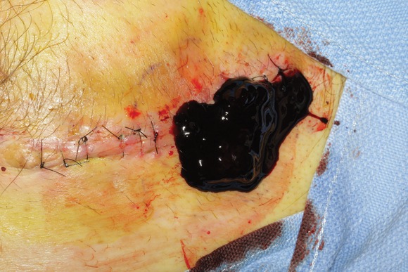
He reviewed the literature on the risks for hemorrhage in patients on anticoagulant therapy who undergo Mohs surgery and the risks for thrombosis in patients who stop their anticoagulant therapy before Mohs.
"The bottom line is that bleeding happens, but nobody dies. That's in contrast to the thrombotic data, where people do die," said Dr. Otley, professor of dermatology at the Mayo Clinic, Rochester, Minn.
The consequences of a thrombotic episode are so much greater than the impact of a hemorrhagic complication that he recommended continuing medically necessary anticoagulants in most cases. "Take extra care with clopidogrel plus aspirin and with warfarin. Taking patients off these, especially if they have a fresh stent, is not the right thing to do," he said.
A survey of 168 American College of Mohs Surgery members in 2003 gathered reports of thrombotic complications within 3 days of Mohs surgery in 46 patients who had stopped anticoagulant therapy, Dr. Otley noted. These included stroke in 24 patients, transient ischemic attack in 8, MI in 5, cerebral emboli or death in 3 patients each, pulmonary embolus in 2, and blindness in 1 patient (J. Am. Acad. Dermatol 2003;48:233-7).
A separate survey of more than 270 dermasurgeons in 2005 found thrombotic complications in 126 patients who stopped anticoagulants for Mohs surgery, including stroke in 39 patients, transient ischemic attack in 25, MI in 19, unstable angina in 17, death in 15, deep venous thrombosis in 7, and pulmonary embolus in 4 patients (Dermatol. Surg. 2007;33:1189-97).

Case reports are emerging of thrombotic episodes in patients on newer anticoagulants who stop therapy for cutaneous surgery. For instance, he noted, a patient who stopped ticlopidine and aspirin developed a deep venous thrombosis. A patient who stopped clopidogrel and ardeparin thrombosed a prosthetic valve. A patient who stopped clopidogrel and aspirin had an MI.
These problems typically present as emergencies, which the Mohs surgeon may not see, Dr. Otley noted. "You may have had more patients experience this than you know about," he said.
Most of the data on the risk of hemorrhagic complications in patients undergoing superficial cutaneous surgery while on anticoagulants focus on traditional agents such as warfarin, aspirin, and NSAIDs, with some data on heparin. Little or no data exist on the risks with newer, more potent anticoagulants.
A total of 10 of 11 studies of patients on warfarin, aspirin, or NSAIDs found no increased risk of perioperative severe hemorrhagic complications, Dr. Otley said. One study of 21 patients on warfarin found an increased risk of complications including persistent bleeding, hematoma, infection, or graft loss. The most severe complication observed in any of the 11 studies was hematoma.
A meta-analysis of the data found a significantly increased risk if moderate and severe hemorrhagic complications were combined as an outcome, but not for severe complications alone, he noted (Dermatol. Surg. 2008;34:160-5).
Clopidogrel (Plavix) is new enough that there is little consensus on how to manage patients on this potent anticoagulant during cutaneous surgery. "This is the one that we’re seeing a ton of patients on," Dr. Otley said. "If your patient has a fresh stent, you would be insane to take that patient off this medication."
Patients on clopidogrel usually are on other anticoagulants, too. Data suggest there is a significant 28-fold higher risk of severe hemorrhagic complications in patients on any clopidogrel-containing regimen, compared with patients not taking anticoagulants, and a significant eightfold higher risk in patients on clopidogrel plus aspirin, compared with patients on aspirin alone. If you compare patients on clopidogrel with patients not on anticoagulant therapy, the difference in risk for severe hemorrhagic complications is not significant, probably due to the small number of patients on clopidogrel alone, he said.
"Nobody has died of hemorrhagic complications, to my knowledge," while having cutaneous surgery on clopidogrel. However, stopping the drug increases the risk of death from thrombotic complications, Dr. Otley said.
There are no data yet on cutaneous surgery complications in patients on dabigatran, "the new kid on the block," and a drug that "you're going to be hearing a lot about," he said. Dabigatran is as effective as warfarin for preventing stroke in high-risk patients.
If patients scheduled for Mohs surgery are on dabigatran, "I'd probably have them continue it unless your hematologist says otherwise," Dr. Otley said. He urged Mohs surgeons to start collecting data on any complications in patients on dabigatran.
To prevent bleeding complications from Mohs surgery, pay attention to hematologic parameters and monitor blood pressure. Hypertension is the leading cause of excessive intraoperative bleeding, he said. Administer sedatives for anxiolysis, use epinephrine as needed, apply pressure dressings, and put in a drain if you think a patient is going to bleed. Use blood products if needed.
In the OR, dislodge temporary clots, eliminate dead space, apply pressure dressings, and use other techniques for secondary prevention of bleeding complications, he advised. "There's no better time to have someone bleed than while you have them open, so rub all those clots off and recoagulate," he said.
Patients with hemorrhagic complications often apologize to Dr. Otley, thinking they did something that caused the bleeding. "I can guarantee you thrombotic patients will not be apologizing to you," he said.
Dr. Otley said he has no relevant conflicts of interest.
LAS VEGAS – A perioperative hemorrhage in a patient undergoing Mohs surgery is a bloody mess, but a thrombosis causing a stroke is a catastrophe.
"Bleeding is overemphasized in our specialty, because it's dramatic. But it is very inconsequential in contrast to thrombosis, which is probably underappreciated," Dr. Clark C. Otley said at the annual meeting of the American College of Mohs Surgery.
He reviewed the literature on the risks for hemorrhage in patients on anticoagulant therapy who undergo Mohs surgery and the risks for thrombosis in patients who stop their anticoagulant therapy before Mohs.
"The bottom line is that bleeding happens, but nobody dies. That's in contrast to the thrombotic data, where people do die," said Dr. Otley, professor of dermatology at the Mayo Clinic, Rochester, Minn.
The consequences of a thrombotic episode are so much greater than the impact of a hemorrhagic complication that he recommended continuing medically necessary anticoagulants in most cases. "Take extra care with clopidogrel plus aspirin and with warfarin. Taking patients off these, especially if they have a fresh stent, is not the right thing to do," he said.
A survey of 168 American College of Mohs Surgery members in 2003 gathered reports of thrombotic complications within 3 days of Mohs surgery in 46 patients who had stopped anticoagulant therapy, Dr. Otley noted. These included stroke in 24 patients, transient ischemic attack in 8, MI in 5, cerebral emboli or death in 3 patients each, pulmonary embolus in 2, and blindness in 1 patient (J. Am. Acad. Dermatol 2003;48:233-7).
A separate survey of more than 270 dermasurgeons in 2005 found thrombotic complications in 126 patients who stopped anticoagulants for Mohs surgery, including stroke in 39 patients, transient ischemic attack in 25, MI in 19, unstable angina in 17, death in 15, deep venous thrombosis in 7, and pulmonary embolus in 4 patients (Dermatol. Surg. 2007;33:1189-97).
Case reports are emerging of thrombotic episodes in patients on newer anticoagulants who stop therapy for cutaneous surgery. For instance, he noted, a patient who stopped ticlopidine and aspirin developed a deep venous thrombosis. A patient who stopped clopidogrel and ardeparin thrombosed a prosthetic valve. A patient who stopped clopidogrel and aspirin had an MI.
These problems typically present as emergencies, which the Mohs surgeon may not see, Dr. Otley noted. "You may have had more patients experience this than you know about," he said.
Most of the data on the risk of hemorrhagic complications in patients undergoing superficial cutaneous surgery while on anticoagulants focus on traditional agents such as warfarin, aspirin, and NSAIDs, with some data on heparin. Little or no data exist on the risks with newer, more potent anticoagulants.
A total of 10 of 11 studies of patients on warfarin, aspirin, or NSAIDs found no increased risk of perioperative severe hemorrhagic complications, Dr. Otley said. One study of 21 patients on warfarin found an increased risk of complications including persistent bleeding, hematoma, infection, or graft loss. The most severe complication observed in any of the 11 studies was hematoma.
A meta-analysis of the data found a significantly increased risk if moderate and severe hemorrhagic complications were combined as an outcome, but not for severe complications alone, he noted (Dermatol. Surg. 2008;34:160-5).
Clopidogrel (Plavix) is new enough that there is little consensus on how to manage patients on this potent anticoagulant during cutaneous surgery. "This is the one that we’re seeing a ton of patients on," Dr. Otley said. "If your patient has a fresh stent, you would be insane to take that patient off this medication."
Patients on clopidogrel usually are on other anticoagulants, too. Data suggest there is a significant 28-fold higher risk of severe hemorrhagic complications in patients on any clopidogrel-containing regimen, compared with patients not taking anticoagulants, and a significant eightfold higher risk in patients on clopidogrel plus aspirin, compared with patients on aspirin alone. If you compare patients on clopidogrel with patients not on anticoagulant therapy, the difference in risk for severe hemorrhagic complications is not significant, probably due to the small number of patients on clopidogrel alone, he said.
"Nobody has died of hemorrhagic complications, to my knowledge," while having cutaneous surgery on clopidogrel. However, stopping the drug increases the risk of death from thrombotic complications, Dr. Otley said.
There are no data yet on cutaneous surgery complications in patients on dabigatran, "the new kid on the block," and a drug that "you're going to be hearing a lot about," he said. Dabigatran is as effective as warfarin for preventing stroke in high-risk patients.
If patients scheduled for Mohs surgery are on dabigatran, "I'd probably have them continue it unless your hematologist says otherwise," Dr. Otley said. He urged Mohs surgeons to start collecting data on any complications in patients on dabigatran.
To prevent bleeding complications from Mohs surgery, pay attention to hematologic parameters and monitor blood pressure. Hypertension is the leading cause of excessive intraoperative bleeding, he said. Administer sedatives for anxiolysis, use epinephrine as needed, apply pressure dressings, and put in a drain if you think a patient is going to bleed. Use blood products if needed.
In the OR, dislodge temporary clots, eliminate dead space, apply pressure dressings, and use other techniques for secondary prevention of bleeding complications, he advised. "There's no better time to have someone bleed than while you have them open, so rub all those clots off and recoagulate," he said.
Patients with hemorrhagic complications often apologize to Dr. Otley, thinking they did something that caused the bleeding. "I can guarantee you thrombotic patients will not be apologizing to you," he said.
Dr. Otley said he has no relevant conflicts of interest.
EXPERT ANALYSIS FROM THE ANNUAL MEETING OF THE AMERICAN COLLEGE OF MOHS SURGERY