User login
Portulaca oleracea (purslane)
Portulaca oleracea, also known as purslane, has long been used in various traditional medicine systems to relieve pain and edema.1Portulaca oleracea is a warm-climate annual plant originally found in the Middle East, North Africa, and the Indian subcontinent and now cultivated in the Arabian peninsula; Japan, where it is an abundant garden plant from spring to fall;2,3 and throughout the world.
The use of P. oleracea, a member of the Portulacaceae family, as a vegetable as well as herbal medicine dates back several centuries.4 In modern times, purslane has been found to be rich in antioxidants, particularly omega-3 fatty acids, vitamins C and E, beta-carotene, melatonin, and glutathione, as well as several minerals.5,6,7 Currently, it is considered one of the top ten most common plants in the world, and one of the most-used medical plants according to the World Health Organization.6 It is considered a weed in the United States, but is eaten in many parts of the world.

Antioxidant activity
Using two different assays, Uddin et al. determined in 2012 that P. oleracea cultivars exhibited significant antioxidant activity through various growth stages. In addition, the researchers suggested that purslane could provide multiple minerals as well as antioxidants in the context of nutraceutical products and functional food.7 Early this year, Silva et al. studied the antioxidant activity of P. oleracea leaves, flowers, and stems from two different locations in Portugal, with assays revealing significantly greater antioxidant activity in the stems of both samples compared to the leaves and flowers. However, the phenolic extracts of all three plant sections from both samples were found to protect DNA against hydroxyl radicals. The investigators concluded that their findings, particularly related to high antioxidant activity, support the potential benefits of purslane consumption to human health.8
A 2014 analysis of 13 collected purslane accessions revealed significant mineral content (particularly potassium, followed by nitrogen, sodium, calcium, magnesium, phosphorus, iron, zinc, and manganese) and showed that antioxidant activity was more strongly associated with ornamental as opposed to common purslane, the latter of which was richer in mineral content.6
Anti-inflammatory activity
In 2000, Chan et al. found that a 10% ethanolic extract of the dried leaves and stem of a P. oleracea cultivar displayed significant anti-inflammatory and analgesic properties after topical and intraperitoneal, but not oral, administration in comparison to diclofenac sodium, a synthetic drug used as active control. They added that these activities corresponded to the reputed effects of the traditional uses of the wild species.9
Wound healing activity
Rashed et al. reported in 2003 that a crude extract of P. oleracea accelerates wound healing. They used Mus musculus JVI-1 to show that fresh homogenized crude aerial parts of the plant topically applied on excision wound surfaces reduced wound surface areas and increased tensile strength. The best documented contraction was associated with a single dose of 50 mg, followed by two doses of 25 mg each.10
Oral lichen planus treatment
In 2010, Agha-Hosseini et al. conducted a randomized double-blind placebo-controlled 3-month study to assess the effectiveness of purslane in the treatment of oral lichen planus. Thirty-seven symptomatic patients (confirmed by biopsy) were divided into a purslane treatment group (n = 20) and a placebo group (n = 17). The investigators reported that partial to complete clinical improvement was observed in 83% of the treatment group, with no response in the remaining 17%, whereas partial improvement was seen in 17% of the placebo group, 73% had no response, and the condition was aggravated in 10% of the placebo group. No adverse side effects were reported in either group, and the researchers concluded that purslane was clinically effective in treating oral lichen planus and warrants consideration as a treatment option for the disorder.5
Other activities
In 2001, Radhakrishnan et al. identified several neuropharmacological actions, particularly anti-nociceptive and muscle-relaxing activity, with a range of effects on the central and peripheral nervous system observed in animal studies.1 The betacyanins found in P. oleracea have subsequently been found to confer a protective effect against neurotoxicity, specifically, ameliorating the D-galactose-induced cognitive deficits in senescent mice.11P. oleracea also has been shown to efficiently eliminate the endocrine-disrupting chemical bisphenol A from a hydroponic solution.12
In 2012, Yan et al. showed that three newly isolated homoisoflavonoids, known as portulacanones, and the compound 2,2’-dihydroxy-4’,6’-dimethoxychalcone selectively exhibited in vitro cytotoxic activities against four human cancer cell lines.2
Conclusions
This antioxidant-rich plant is found throughout the world and has long been associated with traditional health care. Modern research into its potential dermatologic uses is ongoing, but the evidence is relatively scarce. There are indications that the antioxidant, anti-inflammatory, and wound healing activity reportedly exhibited by purslane may be harnessed for various cutaneous applications. However, much more research is necessary to determine how extensive a role purslane may play in skin care.
References
1.J. Ethnopharmacol. 2001;76:171-6
2.Phytochemistry 2012;80:37-41
3.J. Biosci. Bioeng. 2007;103:420-6
4.J. Ethnopharmacol. 2000;73:445-51
5.Phytother. Res. 2010;24:240-4
6.Biomed. Res. Int. 2014;2014:296063
7.Int. J. Mol. Sci. 2012;13:10257-67
8.Nat Prod. Commun. 2014;9:45-50
9. J. Ethnopharmacol. 2000;73:445-51
10. J. Ethnopharmacol. 2003;88:131-6
11. Phytomedicine. 2010 Jun;17:527-32
12. Biosci. Biotechnol. Biochem. 2012;76:1015-7
Dr. Baumann is chief executive officer of the Baumann Cosmetic & Research Institute in the Design District in Miami. She founded the Cosmetic Dermatology Center at the University of Miami in 1997. Dr. Baumann wrote the textbook “Cosmetic Dermatology: Principles and Practice” (New York: McGraw-Hill, 2002), and a book for consumers, “The Skin Type Solution” (New York: Bantam Dell, 2006). She has contributed to the Cosmeceutical Critique column in Dermatology News since January 2001. Her latest book, “Cosmeceuticals and Cosmetic Ingredients,” was published in November 2014. Dr. Baumann has received funding for clinical grants from Allergan, Aveeno, Avon Products, Evolus, Galderma, GlaxoSmithKline, Kythera Biopharmaceuticals, Mary Kay, Medicis Pharmaceuticals, Neutrogena, Philosophy, Topix Pharmaceuticals, and Unilever.
Portulaca oleracea, also known as purslane, has long been used in various traditional medicine systems to relieve pain and edema.1Portulaca oleracea is a warm-climate annual plant originally found in the Middle East, North Africa, and the Indian subcontinent and now cultivated in the Arabian peninsula; Japan, where it is an abundant garden plant from spring to fall;2,3 and throughout the world.
The use of P. oleracea, a member of the Portulacaceae family, as a vegetable as well as herbal medicine dates back several centuries.4 In modern times, purslane has been found to be rich in antioxidants, particularly omega-3 fatty acids, vitamins C and E, beta-carotene, melatonin, and glutathione, as well as several minerals.5,6,7 Currently, it is considered one of the top ten most common plants in the world, and one of the most-used medical plants according to the World Health Organization.6 It is considered a weed in the United States, but is eaten in many parts of the world.
Antioxidant activity
Using two different assays, Uddin et al. determined in 2012 that P. oleracea cultivars exhibited significant antioxidant activity through various growth stages. In addition, the researchers suggested that purslane could provide multiple minerals as well as antioxidants in the context of nutraceutical products and functional food.7 Early this year, Silva et al. studied the antioxidant activity of P. oleracea leaves, flowers, and stems from two different locations in Portugal, with assays revealing significantly greater antioxidant activity in the stems of both samples compared to the leaves and flowers. However, the phenolic extracts of all three plant sections from both samples were found to protect DNA against hydroxyl radicals. The investigators concluded that their findings, particularly related to high antioxidant activity, support the potential benefits of purslane consumption to human health.8
A 2014 analysis of 13 collected purslane accessions revealed significant mineral content (particularly potassium, followed by nitrogen, sodium, calcium, magnesium, phosphorus, iron, zinc, and manganese) and showed that antioxidant activity was more strongly associated with ornamental as opposed to common purslane, the latter of which was richer in mineral content.6
Anti-inflammatory activity
In 2000, Chan et al. found that a 10% ethanolic extract of the dried leaves and stem of a P. oleracea cultivar displayed significant anti-inflammatory and analgesic properties after topical and intraperitoneal, but not oral, administration in comparison to diclofenac sodium, a synthetic drug used as active control. They added that these activities corresponded to the reputed effects of the traditional uses of the wild species.9
Wound healing activity
Rashed et al. reported in 2003 that a crude extract of P. oleracea accelerates wound healing. They used Mus musculus JVI-1 to show that fresh homogenized crude aerial parts of the plant topically applied on excision wound surfaces reduced wound surface areas and increased tensile strength. The best documented contraction was associated with a single dose of 50 mg, followed by two doses of 25 mg each.10
Oral lichen planus treatment
In 2010, Agha-Hosseini et al. conducted a randomized double-blind placebo-controlled 3-month study to assess the effectiveness of purslane in the treatment of oral lichen planus. Thirty-seven symptomatic patients (confirmed by biopsy) were divided into a purslane treatment group (n = 20) and a placebo group (n = 17). The investigators reported that partial to complete clinical improvement was observed in 83% of the treatment group, with no response in the remaining 17%, whereas partial improvement was seen in 17% of the placebo group, 73% had no response, and the condition was aggravated in 10% of the placebo group. No adverse side effects were reported in either group, and the researchers concluded that purslane was clinically effective in treating oral lichen planus and warrants consideration as a treatment option for the disorder.5
Other activities
In 2001, Radhakrishnan et al. identified several neuropharmacological actions, particularly anti-nociceptive and muscle-relaxing activity, with a range of effects on the central and peripheral nervous system observed in animal studies.1 The betacyanins found in P. oleracea have subsequently been found to confer a protective effect against neurotoxicity, specifically, ameliorating the D-galactose-induced cognitive deficits in senescent mice.11P. oleracea also has been shown to efficiently eliminate the endocrine-disrupting chemical bisphenol A from a hydroponic solution.12
In 2012, Yan et al. showed that three newly isolated homoisoflavonoids, known as portulacanones, and the compound 2,2’-dihydroxy-4’,6’-dimethoxychalcone selectively exhibited in vitro cytotoxic activities against four human cancer cell lines.2
Conclusions
This antioxidant-rich plant is found throughout the world and has long been associated with traditional health care. Modern research into its potential dermatologic uses is ongoing, but the evidence is relatively scarce. There are indications that the antioxidant, anti-inflammatory, and wound healing activity reportedly exhibited by purslane may be harnessed for various cutaneous applications. However, much more research is necessary to determine how extensive a role purslane may play in skin care.
References
1.J. Ethnopharmacol. 2001;76:171-6
2.Phytochemistry 2012;80:37-41
3.J. Biosci. Bioeng. 2007;103:420-6
4.J. Ethnopharmacol. 2000;73:445-51
5.Phytother. Res. 2010;24:240-4
6.Biomed. Res. Int. 2014;2014:296063
7.Int. J. Mol. Sci. 2012;13:10257-67
8.Nat Prod. Commun. 2014;9:45-50
9. J. Ethnopharmacol. 2000;73:445-51
10. J. Ethnopharmacol. 2003;88:131-6
11. Phytomedicine. 2010 Jun;17:527-32
12. Biosci. Biotechnol. Biochem. 2012;76:1015-7
Dr. Baumann is chief executive officer of the Baumann Cosmetic & Research Institute in the Design District in Miami. She founded the Cosmetic Dermatology Center at the University of Miami in 1997. Dr. Baumann wrote the textbook “Cosmetic Dermatology: Principles and Practice” (New York: McGraw-Hill, 2002), and a book for consumers, “The Skin Type Solution” (New York: Bantam Dell, 2006). She has contributed to the Cosmeceutical Critique column in Dermatology News since January 2001. Her latest book, “Cosmeceuticals and Cosmetic Ingredients,” was published in November 2014. Dr. Baumann has received funding for clinical grants from Allergan, Aveeno, Avon Products, Evolus, Galderma, GlaxoSmithKline, Kythera Biopharmaceuticals, Mary Kay, Medicis Pharmaceuticals, Neutrogena, Philosophy, Topix Pharmaceuticals, and Unilever.

VIDEO: Get excited about the excimer laser for dermatitis
ASHEVILLE, N.C. – Excimer lasers can be used for patients with allergic contact dermatitis for whom topical therapies are unsuccessful or undesirable, according to Dr. Alison Ehrlich of George Washington University in Washington.
In an interview at the annual meeting of the Noah Worcester Dermatological Society, Dr. Ehrlich discussed her use of the excimer laser for dermatitis patients and shared some tips for successful treatment.
The video associated with this article is no longer available on this site. Please view all of our videos on the MDedge YouTube channel
AT NOAH 57

VIDEO: Dermatologists should embrace tissue adhesive
ASHEVILLE, N.C. – Cutaneous adhesives have many advantages in dermatologic surgery, according to Dr. John West of Seaport Dermatology and Mohs Surgery in Mystic, Conn.
In a video interview at the annual meeting of the Noah Worcester Dermatological Society, Dr. West explained some of the benefits of tissue adhesives for both doctors and patients and shared several pearls for optimal use of these products.
The video associated with this article is no longer available on this site. Please view all of our videos on the MDedge YouTube channel
AT NOAH 57

Aesthetic Dermatology: Sun protection after aesthetic procedures
As summertime approaches, opportunities for outdoor activities increase. For many of our patients, summer inspires a desire to have aesthetic procedures in preparation for outdoor events, such as weddings and vacations. We must, however, be mindful that increased sun exposure after some aesthetic procedures can mean an increased risk of complications.
The main complication we worry about with sun exposure is, of course, hyperpigmentation. The risk is low with injectable procedures such as botulinum toxin and fillers, but sun protection is still encouraged, especially in skin types III-VI. The risk increases greatly with chemical peels and laser and light-based procedures, such as intense pulsed light, vascular lasers, pigment lasers, laser hair removal, and especially nonablative and ablative resurfacing (including nonlaser resurfacing such as dermabrasion).
Sun protection should be encouraged, even with seemingly less invasive procedures, such as electrodessication. I once had a patient with type-IV skin tell me at her first visit that, years before, she had electrodessication on her face for DPN (dermatosis papulosa nigra), a procedure she had done on several occasions without complications and great results. However, she went to a party on a boat the weekend after the procedure and developed hyperpigmentation at the procedure areas, and she still had a few dark macules several years later.
At a follow-up visit, she said the doctor told her she should not have gone out on the boat and should have worn sunscreen. Of course, she was highly upset that she wasn’t advised about sun protection at the time of the procedure. This is one of several stories I’ve heard or seen of complications and postinflammatory hyperpigmentation after an aesthetic procedure, when the patients felt that the treating physician or practitioner did not counsel them about sun exposure during the consultation or treatment visit. It seems intuitive, but I’ve made it a habit to make sun protection part of my counseling routine.
In my practice, we often give patients sunscreen to apply immediately after a procedure. Specifically encouraging the use of zinc- and/or titanium-based, broad-spectrum, noncomedogenic physical blockers that are SPF 30 or higher may help reduce the risk of potential irritation or allergy and subsequent postinflammatory pigmentary alteration from chemical blocking ingredients. We provide a postprocedure handout, and the medical assistant also will counsel about sun protection when applying it to the patient or reviewing postprocedure instructions. So the patient is counseled at least three times: By me during consultation or pre-procedure, by the medical assistant post procedure, and by written instructions.
Vigorous sun protection is encouraged for at least 1 week after any aesthetic procedure (and longer if the downtime is longer or if multiple treatments are required). Some practices also use antioxidant serums to reduce free radicals, encourage healing, and reduce the risk of hyperpigmentation after procedures. Wide-brimmed hats also are encouraged, particularly after resurfacing or photodynamic therapy (PDT). We give patients sun-protective hats when they leave our office after PDT. We counsel them to practice vigorous sun protection for at least 1 week and to avoid sitting by a window for 48 hours after the procedure so as to not reactivate the levulan.
Delaying more high-risk procedures, such as laser treatments, until after the summer months may be appropriate if sun cannot be avoided to mitigate the risk of complications. If a patient comes to the office for a laser procedure and is visibly more tan than at the time of the last treatment, I will counsel about risks, adjust the settings appropriately, or even delay the treatment altogether to a time when the tan has faded. This is particularly important for lasers and light treatments for which melanin is the target chromosphere, such as intense pulsed light and laser hair removal. Although UV exposure is more intense in the summer, in our practice in Southern California we follow these principles year-round for the safety of our patients.
Dr. Wesley and Dr. Talakoub are co-contributors to a monthly Aesthetic Dermatology column in Dermatology News. Dr. Talakoub is in private practice in McLean, Va. Dr. Wesley practices dermatology in Beverly Hills, Calif. This month’s column is by Dr. Wesley.

Cosmetic Corner: Dermatologists Weigh in on OTC Acne Treatments
To improve patient care and outcomes, leading dermatologists offered their recommendations on the top OTC acne treatments. Consideration must be given to:
- ClarityMD Deep Pore Cleanser and Clarifying Gel
- Clear Complexion Concealer
- Effaclar Duo
- PanOxyl 10% Acne Foaming Wash
Cutis invites readers to send us their recommendations. Cleansers for rosacea patients, face washes, and products for babies will be featured in upcoming editions of Cosmetic Corner. Please e-mail your recommendation(s) to cutis@frontlinemedcom.com.
Disclaimer: Opinions expressed herein do not necessarily reflect those of Cutis or Frontline Medical Communications Inc and shall not be used for product endorsement purposes. Any reference made to a specific commercial product does not indicate or imply that Cutis or Frontline Medical Communications Inc endorses, recommends, or favors the product mentioned. No guarantee is given to the effects of recommended products.
ABA: Childhood burn survivors risk more physical, mental disorders
CHICAGO – Adult survivors of childhood burns have significantly higher rates of Axis I mental and physical disorders years after their injury, a population-based study shows.
“We think it is really important to screen for, identify, and treat these illnesses not only in that acute period and shortly after the burn injury, but well into adulthood,” study author James Stone said at the annual meeting of the American Burn Association.
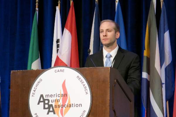
He reported on 745 adult burn survivors identified using administrative data from a regional pediatric burn center registry in Manitoba, Canada, who were matched 1:5 with 3,725 controls from the general Manitoba population based on age, sex, and geographic location. The burn survivors had an average age of 5.9 years at the time of burn injury, burns involved an average 12% of total body surface area, and 65% of burn survivors were male. The average follow-up was nearly 15 years (range 2.8-24.7 years).
In unadjusted univariate analysis, adult survivors had significantly higher rates than matched controls for any lifetime physical disorder (rate ratio, 1.17), arthritis (RR, 1.23), cancer (RR, 1.94), diabetes (RR, 1.69), fractures (RR, 1.45), and total respiratory morbidity (RR, 1.15).
After adjustment for gender, geography, and income, any physical disorder (RR, 1.15; P value < .01), arthritis (RR, 1.24; P < .01), fractures (RR, 1.37; P .001), and total respiratory morbidity (RR, 1.13; P < .05) remained significant, Mr. Stone, from the University of Manitoba, Winnipeg, Canada, reported.
Further, 81% of burn survivors had a lifetime physical illness compared with 69% of controls.
“The fact that 81% of our burn cohort was diagnosed with a physical illness is definitely concerning,” he said. “We hypothesize that the prolonged hyperinflammatory and hypermetabolic state that has been previously reported makes these individuals more susceptible to these illnesses down the road.”
The burn cohort also had significantly higher unadjusted rate ratios for any Axis 1 mental disorder (RR, 1.62), major depressive disorder (RR, 1.64), anxiety (RR, 1.57), substance abuse (RR, 2.86), and suicide attempts (RR, 5.00).
All disorders remained statistically significant after adjustment with rate ratios of 1.54 (P < .001), 1.54 (P < .001), 1.50 (P < .001), 2.35 (P < .001), and 4.33 (P < .01), respectively.
The high rates of substance abuse and suicide attempts are consistent with previous clinical interview studies, but are still cause for great concern, Mr. Stone said.
The risk for any mental or physical disorder was not significantly impacted by burn location or by burns that affected more than 30% of total body surface area. Age older than 5 years at the time of the burn significantly increased the risk of any mental disorder (relative risk, 1.92; P < .001).
Limitations of the study include the potential for bias because the data relied on individuals presenting to physicians, discrepancies between ICD codes for physician billings and hospital claims, and some survivors may have moved out of the province, Mr. Stone said. The study, however, had a sample size three times greater than the next largest study of its kind, and importantly, matched burn patients to the general population.
CHICAGO – Adult survivors of childhood burns have significantly higher rates of Axis I mental and physical disorders years after their injury, a population-based study shows.
“We think it is really important to screen for, identify, and treat these illnesses not only in that acute period and shortly after the burn injury, but well into adulthood,” study author James Stone said at the annual meeting of the American Burn Association.
He reported on 745 adult burn survivors identified using administrative data from a regional pediatric burn center registry in Manitoba, Canada, who were matched 1:5 with 3,725 controls from the general Manitoba population based on age, sex, and geographic location. The burn survivors had an average age of 5.9 years at the time of burn injury, burns involved an average 12% of total body surface area, and 65% of burn survivors were male. The average follow-up was nearly 15 years (range 2.8-24.7 years).
In unadjusted univariate analysis, adult survivors had significantly higher rates than matched controls for any lifetime physical disorder (rate ratio, 1.17), arthritis (RR, 1.23), cancer (RR, 1.94), diabetes (RR, 1.69), fractures (RR, 1.45), and total respiratory morbidity (RR, 1.15).
After adjustment for gender, geography, and income, any physical disorder (RR, 1.15; P value < .01), arthritis (RR, 1.24; P < .01), fractures (RR, 1.37; P .001), and total respiratory morbidity (RR, 1.13; P < .05) remained significant, Mr. Stone, from the University of Manitoba, Winnipeg, Canada, reported.
Further, 81% of burn survivors had a lifetime physical illness compared with 69% of controls.
“The fact that 81% of our burn cohort was diagnosed with a physical illness is definitely concerning,” he said. “We hypothesize that the prolonged hyperinflammatory and hypermetabolic state that has been previously reported makes these individuals more susceptible to these illnesses down the road.”
The burn cohort also had significantly higher unadjusted rate ratios for any Axis 1 mental disorder (RR, 1.62), major depressive disorder (RR, 1.64), anxiety (RR, 1.57), substance abuse (RR, 2.86), and suicide attempts (RR, 5.00).
All disorders remained statistically significant after adjustment with rate ratios of 1.54 (P < .001), 1.54 (P < .001), 1.50 (P < .001), 2.35 (P < .001), and 4.33 (P < .01), respectively.
The high rates of substance abuse and suicide attempts are consistent with previous clinical interview studies, but are still cause for great concern, Mr. Stone said.
The risk for any mental or physical disorder was not significantly impacted by burn location or by burns that affected more than 30% of total body surface area. Age older than 5 years at the time of the burn significantly increased the risk of any mental disorder (relative risk, 1.92; P < .001).
Limitations of the study include the potential for bias because the data relied on individuals presenting to physicians, discrepancies between ICD codes for physician billings and hospital claims, and some survivors may have moved out of the province, Mr. Stone said. The study, however, had a sample size three times greater than the next largest study of its kind, and importantly, matched burn patients to the general population.
AT THE ABA ANNUAL MEETING
Key clinical point: Adult survivors of childhood burn injuries have increased rates of Axis I mental and physical disorders.
Major finding: 81% of burn survivors had a physical disorder vs. 69% of matched controls.
Data source: Population-based study in 745 adult survivors of childhood burns.
Disclosures: The study was funded by grants from the University of Manitoba and the Manitoba Firefighters Burn Fund. The authors declared no conflicts of interest.

ABA: Rehab time linked to outcomes in all burn patients
CHICAGO – Rehabilitation time is directly associated with a reduced rate of contracture and a better range of motion, irrespective of the extent of burn injury, results from the prospective ACT study showed.
The Acuity, Contractures, Time (ACT) study examined outcomes and rehabilitation time in patients with burns involving 10% or less of their total body surface area (n = 177) and in those with burns involving more than 10% of total body surface area (n = 130). Joint range of motion was recorded based on cutaneous functional units at the time of discharge for all 307 patients enrolled from September 2010 through December 2013 at five verified burn centers across the United States. Most patients were men (71%).
Overall, 79% of the patients had burn scar contracture. Based on range of motion for 8,068 joints analyzed, 66% had burn scar contracture.
For patients with burns affecting 10% or less of their bodies, only rehabilitation time per cutaneous functional unit significantly differed between those with and without burn scar contracture (4.6 minutes vs. 2.4 minutes; P<i/>= .002). The same was true for patients with burns affecting more than 10% of their bodies (3.3 minutes vs. 1.4 minutes; P<i/> < .0001).
“One of the impetuses to do this study in this manner was the realization of what most therapists say all the time: They don’t have enough time to treat their patients in the hospital,” Reg Richard, P.T., said at the annual meeting of the American Burn Association.
During a discussion of the study at the meeting, Mr. Richard said he hoped the results would be used to improve rehabilitation services for burn patients and called on the ABA to lead the charge.
ABA President David Ahrenholz of Regions Hospital Burn Center, St. Paul, Minn., said in an interview that coverage of rehabilitation services varies from insurer to insurer, and physicians can’t point to an absolute number to say this is the minimum number of rehabilitation days needed to obtain a good outcome in a specific patient.
“This is the first pass of an unbelievably large data set,” Dr. Ahrenholz said “I think it will be transformative ... to help the insurers to understand that it isn’t just a number of sessions, but that we’re looking for an outcome and that the therapy should continue until we get the desired outcome.”
In the ACT study, a univariate analysis found patients with no contracture were significantly more likely than those with contracture to have shorter hospital stays (12 days vs. 14 days; P value = .02), burns with a lower percentage of body surface area involvement (5% vs. nearly 10%; P<i/> < .0001), fewer skin grafts (2.3% vs. 4%; P<i/> = .001), more rehabilitation time per total body surface area involvement (6.1 minutes vs. 4.5 minutes; P<i/> = .003 ), and more rehabilitation time per cutaneous functional unit (4.4 minutes vs. 2 minutes; P<i/> < .0001), Mr. Richard from the U.S. Army Institute of Surgical Research, JBSA Fort Sam Houston, Texas, reported.
In multivariate regression analysis, rehabilitation time per cutaneous functional unit was the only significant predictor of preventing burn scar contracture and lost range of motion for patients with small burns (Odds ratio, 1.07; 95% confidence interval, 1.02-1.12) and for those with larger burns (OR, 1.36; 95% CI, 1.18-1.74).
Further analyses are warranted, as the results were based on the variables used and present analysis methods, he said.
On Twitter @pwendl
AT THE ABA ANNUAL MEETING
Key clinical point: Rehabilitation time was associated with reduced risk of scar contracture and lost range of motion in burn patients.
Major finding: For patients with burns affecting 10% or less of their bodies, only rehabilitation time per cutaneous functional unit significantly differed between those with and without burn scar contracture (4.6 minutes vs. 2.4 minutes; P = .002). The same was true for patients with burns affecting more than 10% of their bodies (3.3 minutes vs. 1.4 minutes; P < .0001).
Data source: Prospective, observational study in 307 burn patients.
Disclosures: The study was funded by a U.S. Department of Defense grant.

VIDEO: Lasers take on the toughest scars
ASHEVILLE, N.C. – Advances in laser and light sources are making it possible to greatly improve the treatment of atrophic and hypertrophic scars, according to Dr. Michael Gold of the Gold Skin Care Center in Nashville, Tenn. In a video interview at the annual meeting of the Noah Worcester Dermatological Society, Dr. Gold explained how laser technology is being used today to manage contracture and other elements of the most challenging scars, such as those sustained by military personnel.
The video associated with this article is no longer available on this site. Please view all of our videos on the MDedge YouTube channel
AT NOAH 57

Long-term Cosmetic Use of Botulinum Toxin Type A
In the United States, the cosmetic use of botulinum toxin type A (BTX-A) has continued to grow over the last 15 years, according to multispecialty data recently released by the American Society for Aesthetic Plastic Surgery. During these years, many of our patients, if not ourselves, have undergone treatment faithfully every 3 to 6 months to combat the signs of aging. Subsequently, with the monitoring of adverse events (AEs), the US Food and Drug Administration has issued a black box warning that covers serious side effects—respiratory compromise and death—associated with treatment, yet most of what is listed in the black box warning pertains to medical use rather than cosmetic use. However, with the ever-growing indications for BTX-A, we must be cognizant of the fact that our patients may be receiving concomitant treatment with BTX-A for medical conditions (eg, migraines, hyperhidrosis, achalasia, dysphonia, dystonia, strabismus) by other specialists. Thus, there is a need to understand the potential side effects associated with BTX-A treatments and long-term consequences.
In a January 21 article published online in Pharmacology Yiannakopoulou looked at national monitoring programs through the US Food and Drug Administration and the Danish Medicines Agency. Many of the AEs reported were related to medical use of BTX-A, including anaphylaxis, death, generalized weakness, and dysphagia. Serious AEs related to the cosmetic use of BTX-A included thyroid eye disease, sarcoidal granuloma, pseudoaneurysm of the frontal branch of the superior temporal artery, and severe respiratory failure. Additionally, a patient receiving BTX-A for palmar and axillary hyperhidrosis developed botulinumlike generalized weakness. Upon review of epidemiological studies, the incidence and types of AEs were covered. The vast majority of these events were related to the medical use of BTX-A, which could stem from the lack of long-term studies on cosmetic patients and the lack of reporting of many AEs. The author summarized that minimizing potential AEs relies on proper injection technique, proper storage of the medication, proper dosing, and thorough knowledge of the anatomy.
What’s the issue?
Botulinum toxin type A remains one of the most gratifying treatments for both physicians and patients alike. However, with the potential for abuse, as in the case of the recent fake Botox Cosmetic that has shown up on the market in the United States), we must remain vigilant for AEs. Furthermore, we must continue to emphasize to patients that it is a medical treatment and deserves all the attention and respect we give to other medical interventions. However, as the Yiannakopoulou review has shown, proper injection techniques have a low rate of AEs in the cosmetic use of BTX-A. Have you seen an increase in the number of cosmetic BTX-A patients receiving concomitant treatments with BTX-A for medical conditions? If so, how do you manage them?

Spray-dried fibrin sealant for surgical use approved by FDA
A product that contains a spray-dried, blended formulation of fibrinogen and thrombin, derived from human plasma, has been approved for use in helping control surgical bleeding, the Food and Drug Administration announced.
The approved indication for the fibrin sealant is for use with an absorbable gelatin sponge; it is the first spray-dried fibrin sealant approved by the FDA, according to its statement. The product, which will be marketed as Raplixa, can be applied from the product vial or sprayed onto the site of bleeding with a spray device.
“This approval provides surgeons an additional option to help control bleeding during surgery when needed,” Dr. Karen Midthun, director of the FDA’s Center for Biologics Evaluation and Research, said in the FDA’s statement. “The spray-drying process used to manufacture Raplixa produces dried powders that can be combined into a single vial. This eliminates the need to combine the fibrinogen and thrombin before use and allows the product to be stored at room temperature,” she added.
Approval was based on a study of 719 people undergoing different types of surgical procedures, which showed that use of the fibrin sealant with an absorbable gelatin sponge reduced the time required to achieve hemostasis, compared with the use of a sponge alone. The manufacturing process includes viral inactivation and removal to reduce the risk of transmitting of blood-borne viruses, the FDA statement said.
The approved indication is “to provide adjunctive hemostasis for mild to moderate bleeding in adults undergoing surgery when control of bleeding by standard surgical techniques (such as suture, ligature and cautery) is ineffective or impractical,” according to an April 30 statement issued by the Medicines Company. The statement said that the product does not need to be thawed, reconstituted or mixed before use, and it describes the spray device as “a low-pressure spray applicator designed to deliver Raplixa to larger bleeding surfaces in difficult to reach areas.”
The product is manufactured by ProFibrix BV, a subsidiary of the Medicines Company.