User login
Cosmeceutical Critique: Benzoyl peroxide
Benzoyl peroxide (BPO) has been used for more than 45 years for the treatment of acne, and has recently been enjoying renewed popularity, thanks to its performance in recent studies of both prescription and over-the-counter formulations (J. Drugs Dermatol. 2013;12:180-5). In fact, BPO is one of the two most common ingredients in OTC acne products (Semin. Cutan. Med. Surg. 2008;27:170-6). The prescription form is used alone or in combination with tretinoin, adapalene, or clindamycin. BPO, originally sourced from the coal tar component chlorhydroxyquinoline, is now typically prepared by treating hydrogen peroxide with benzoyl chloride (Dermatol. Clin. 2009;27:17-24). Because it can generate reactive oxygen species and commonly leads to skin irritation, its use is somewhat limited.
Antibacterial uses
BPO imparts bactericidal activity by releasing highly reactive oxygen (free radicals) that can oxidize proteins in bacterial cell membranes. It also exhibits antibacterial action against Propionibacterium acnes and Corynebacterium acnes, the bacteria implicated in the pathophysiology of acne (Dermatol. Ther. 2012;25:6-11), as well as Staphylococcus capitis, S. epidermis, S. hominis, P. avidum, P. granulosum, and the yeast Pityrosporum ovale (J. Appl. Bacteriol. 1983;54:379-82).
Acne
Many studies over the years have shown that topically applied BPO effectively treats acne (Expert Opin. Pharmacother. 2009;10:2555-62). These ameliorative results, which include enhancing the benefits of other topical antimicrobials, are thought to arise because BPO, a highly lipophilic molecule, penetrates through the sebum and into the pilosebaceous unit, and exerts bactericidal, keratolytic, and anti-inflammatory activity (Skin Pharmacol. Physiol. 2006;19:283-9). BPO may contribute to the antiacne efficacy of other antimicrobials by preventing bacterial resistance and promoting penetration into the sebum, keratin, and polysaccharides to reach the target bacteria. Specifically, the oxidative activity of BPO helps eliminate the biofilm polysaccharides secreted by P. acnes, thus expediting the delivery of other agents to the bacteria (Int. J. Dermatol. 2006;45:872; Int. J. Dermatol. 2003;42:925-7).
Not surprisingly, several studies have shown that the antiacne efficacy of a combination of BPO with other antimicrobials, such as clindamycin, is greater than that of either agent used alone. Simpson et al. demonstrated that the use of clindamycin and BPO together led to a 61% decline in inflammatory lesions after 3 months, as compared with 39% and 35%, respectively, when the agents were used alone (J. Am. Acad. Dermatol. 1997;37:590-5). BPO is frequently paired with salicylic acid to treat acne (Clin. Exp. Dermatol. 2011;36:840-3).
Acne often improves more rapidly with BPO treatment than with retinoids and other acne therapies, and data suggest that the faster clearing of acne lesions and comedones is most likely because of its keratolytic activity (Dermatol. Clin. 2009;27:17-24; J. Dermatolog. Treat. 2003;14:166-71). However, the dryness and irritation associated with BPO usage may undermine patient compliance. Several studies have suggested that BPO is effective in cleanser formulations, which seem to reduce irritation (Clin. Exp. Dermatol. 2011;36:840-3).
Photocarcinogenicity
Reports that BPO predisposed mice to skin cancer, particularly when they were exposed to ultraviolet radiation, prompted the Food and Drug Administration to form an advisory committee in 1992 to review the safety of BPO. The committee called for additional photocarcinogenicity studies while suggesting that BPO products include animal safety data on the labels. BPO-containing acne products were kept on the market. In the ensuing two decades, newer safety studies have led the FDA to change the classification of BPO to category I, deeming the OTC topical treatment of acne to be generally recognized as safe and effective (GRASE) (Fed. Regist. 2010;75:9767-77).
Photoaging
When BPO breaks down into benzoic acid in the skin, benzoyloxy, a free radical, forms as an intermediate (Prog. Clin. Biol. Res. 1995;391:245). Benzoyloxy can decarboxylate into a phenyl radical. These free radicals produce oxidative stress, which may cause DNA strand breaks in keratinocytes or may harm proteins or lipids. In addition to becoming a free radical, BPO depletes membrane and cytosolic antioxidants (Toxicology 2001;165:225-34). No retrospective trials looking at the effects of long-term use of BP on photoaging have been performed, so the role of BPO in photoaging is not clear. One study in mice found that topical BP has some of the same effects on skin as UVB (J. Invest. Dermatol. 1999;112:933-38).
Other safety issues
Acne is not uncommon among pregnant women. Although safety studies of BPO use by pregnant women have not been performed, various authors suggest that only about 5% of topically applied BPO is absorbed systemically, implying that topical BPO can be safely used during pregnancy (Int. J. Dermatol. 2002;41:197-203; Can. Fam. Physician 2011;57:665-7; Drugs 2013;73:779-87; Dermatol. Ther. 2013;26:302-11).
In approximately 1% of patients, topical BPO causes contact or irritant dermatitis (Contact Dermatitis 1999;41:233; Contact Dermatitis 1996;34:68-9). The use of barrier repair moisturizers may reduce the incidence of irritation, though this has not been proven.
Usage considerations
BPO use for acne is linked to a reduction in antibiotic resistance (J. Drugs Dermatol. 2013;12:s73-6). Because BPO, a potent oxidizer, eliminates bacteria by generating reactive oxygen species in the sebaceous follicle, it is important to consider the chemical compatibility of BPO with other agents (J. Am. Acad. Dermatol. 1981;4:31-7). Martin et al. showed that BPO tends to degrade tretinoin to about 80% of initial content, an effect that is markedly enhanced by indoor light. However, even in the presence of light, adapalene is not degraded by BPO (Br. J. Dermatol. 1998;139 Suppl 52:8-11). But the order in which products are applied is important, given that BPO can inactivate other ingredients.
Studies have demonstrated that the use of BPO in body washes leads to greater efficacy when the product is left on for 5 minutes before rinsing (J. Drugs Dermatol. 2010;9:622-5; J. Clin. Aesthet. Dermatol. 2010;3:26-9). Notably, the efficacy of BPO in cleansing products is comparable to that observed in leave-on products, but BPO provokes less irritation than leave-on formulations (J. Drugs Dermatol. 2009;8:657-61; Skinmed. 2005;4:370).
Conclusion
BPO remains quite effective in acne therapy, and it is one of the few acne medications available both over the counter and by prescription in the United States. BPO helps prevent antibiotic resistance to erythromycin and clindamycin, which makes it an important ingredient in many acne skin care regimens. However, it is pro-oxidant, and clinicians and patients should take into account the risk of BPO contributing to skin aging because of the free radicals it produces.
Dr. Baumann is chief executive officer of the Baumann Cosmetic & Research Institute in Miami Beach. She founded the cosmetic dermatology center at the University of Miami in 1997. Dr. Baumann wrote the textbook "Cosmetic Dermatology: Principles and Practice" (McGraw-Hill, April 2002), and a book for consumers, "The Skin Type Solution" (Bantam, 2006). She has contributed to the Cosmeceutical Critique column in Skin & Allergy News since January 2001 and joined the editorial advisory board in 2004. Dr. Baumann has received funding for clinical grants from Allergan, Aveeno, Avon Products, Galderma, Mary Kay, Medicis Pharmaceuticals, Neutrogena, Philosophy, Stiefel, Topix Pharmaceuticals, and Unilever.
VIDEO: Spruce up your suturing; try these techniques
ORLANDO – Inspired by sewing lessons during her childhood, Dr. Cyndi Yag-Howard has developed several suturing techniques that help promote better wound healing and yield more aesthetically pleasing results.
She has published several of her techniques, including the SICM (subcutaneous inverted cross mattress) stitch (Dermatol. Surg. 2011;37:1503-5) and the Zipper stitch (Dermatol. Surg. 2013;39:1400-2), and she has a few more in the works. In a video interview after her presentation at the annual meeting of the Florida Society of Dermatology and Dermatologic Surgery, Dr. Yag-Howard, who is in private practice in Naples, Fla., shared the how-to’s of several of her suturing techniques.
On Twitter @naseemmiller
AT FSDDS 2014

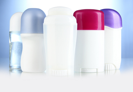
Cosmetic Corner: Dermatologists Weigh in on Antiperspirants
To improve patient care and outcomes, leading dermatologists offered their recommendations on the top antiperspirants. Consideration must be given to:
- Certain Dri
- Degree Dry Protection for Men
- Dove Clinical Protection
- Dove Go Sleeveless
- Green Tea Cream Deodorant
- Secret Clinical Strength
- Tom’s of Maine Unscented Naturally Dry Antiperspirant
Cutis invites readers to send us their recommendations. Stretch mark reducers, exfoliators, and OTC hair removal will be featured in upcoming editions of Cosmetic Corner. Please e-mail your recommendation(s) to the Editorial Office. ((link to cutis@frontlinemedcom.com))
Disclaimer: Opinions expressed herein do not necessarily reflect those of Cutis or Frontline Medical Communications Inc. and shall not be used for product endorsement purposes. Any reference made to a specific commercial product does not indicate or imply that Cutis or Frontline Medical Communications Inc. endorses, recommends, or favors the product mentioned. No guarantee is given to the effects of recommended products.
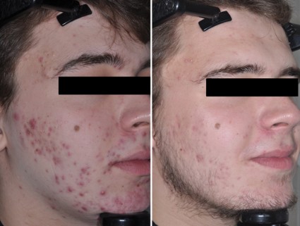
Gold-coated microparticles aid acne treatment
PHOENIX – Application of gold-coated microparticles to the skin may enhance laser photothermolysis of sebaceous glands to treat acne, according to three small, preliminary studies.
Inadequate contrast in sebaceous glands has limited the effectiveness of selective photothermolysis to treat acne. Specially engineered 0.150-mcm microparticles of inert gold coating a silica core were designed for surface plasmon resonance at near-infrared wavelengths to optimize contrast and high absorption of light in the sebaceous glands during laser treatment.
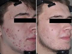
In a randomized, controlled crossover study, 48 patients with acne vulgaris received either immediate or delayed treatment for 12 weeks, after which the delayed-treatment group also began receiving the treatment. Immediate treatment included three treatments at 2-week intervals. The treatment consisted of a microparticle suspension gently massaged into facial skin for about 8 minutes, superficial skin cleansing, and pulsed irradiation with two passes of an 800-nm laser. Patients in the delayed-treatment group used an over-the-counter face wash (2% salicylic acid) twice daily.
The mean number of inflammatory lesions decreased by 34% at 12 weeks after treatment started, compared with a 16% reduction in the control group, a statistically significant difference, Dilip Paithankar, Ph.D., reported at the annual meeting of the American Society for Laser Medicine and Surgery.

At 28 weeks after the crossover to treatment for all subjects (40 weeks after the start of the study), the mean number of inflammatory lesions decreased by 61%, reported Dr. Paithankar, chief technology officer at Sebacia, Duluth, Ga., which is developing the gold-coated microparticles treatment.
In another randomized, sham-controlled study, 49 patients with acne vulgaris underwent three laser photothermolysis treatments at weekly intervals using either the gold-coated microparticles or a control vehicle without the particles. Mean inflammatory lesion counts decreased significantly more in the treatment group than in the control group at 8, 12, and 16 weeks of follow-up. In the treatment group, mean inflammatory lesion counts were 44% lower at 8 weeks, 49% lower at 12 weeks, and 53% lower at 16 weeks, compared with baseline. In the control group, mean inflammatory lesion counts were 14% lower, 22% lower, and 31% lower at those time points, respectively, compared with baseline.
Inflammatory lesions were clear or almost clear by the end of the study in 24% in the treatment group and in none of the control patients.

Data from a separate histologic study suggest that using ultrasound to assist delivery of the gold-coated microparticles may improve selective destruction from laser treatment, Dr. Girish Munavalli reported at the meeting. Ultrasound has been used in other settings to enhance delivery of small molecules through the stratum corneum.
The study used an ultrasound horn vibrating in a microparticle suspension placed in an enclosure above preauricular skin in 37 subjects, followed by 800-nm laser treatment. Biopsies taken within 15 minutes showed localized thermal injury to infundibuli and sebaceous glands with no collateral damage to surrounding tissue or to the epidermis, reported Dr. Munavalli, a dermatologist in group practice in Charlotte, N.C. The extent of thermal damage correlated with the duration of the ultrasound.
The level of damage could be expected to improve acne, but that needs confirmation in clinical trials, he said.
In each of the studies, the treatments were well tolerated, with minimal side effects including mild to moderate pain (using no anesthetic) and short-lived erythema and edema.
Dr. Paithankar works for Sebacia. Dr. Munavalli reported having no disclosures; some of his coinvestigators work for Sebacia.
On Twitter @sherryboschert
PHOENIX – Application of gold-coated microparticles to the skin may enhance laser photothermolysis of sebaceous glands to treat acne, according to three small, preliminary studies.
Inadequate contrast in sebaceous glands has limited the effectiveness of selective photothermolysis to treat acne. Specially engineered 0.150-mcm microparticles of inert gold coating a silica core were designed for surface plasmon resonance at near-infrared wavelengths to optimize contrast and high absorption of light in the sebaceous glands during laser treatment.
In a randomized, controlled crossover study, 48 patients with acne vulgaris received either immediate or delayed treatment for 12 weeks, after which the delayed-treatment group also began receiving the treatment. Immediate treatment included three treatments at 2-week intervals. The treatment consisted of a microparticle suspension gently massaged into facial skin for about 8 minutes, superficial skin cleansing, and pulsed irradiation with two passes of an 800-nm laser. Patients in the delayed-treatment group used an over-the-counter face wash (2% salicylic acid) twice daily.
The mean number of inflammatory lesions decreased by 34% at 12 weeks after treatment started, compared with a 16% reduction in the control group, a statistically significant difference, Dilip Paithankar, Ph.D., reported at the annual meeting of the American Society for Laser Medicine and Surgery.
At 28 weeks after the crossover to treatment for all subjects (40 weeks after the start of the study), the mean number of inflammatory lesions decreased by 61%, reported Dr. Paithankar, chief technology officer at Sebacia, Duluth, Ga., which is developing the gold-coated microparticles treatment.
In another randomized, sham-controlled study, 49 patients with acne vulgaris underwent three laser photothermolysis treatments at weekly intervals using either the gold-coated microparticles or a control vehicle without the particles. Mean inflammatory lesion counts decreased significantly more in the treatment group than in the control group at 8, 12, and 16 weeks of follow-up. In the treatment group, mean inflammatory lesion counts were 44% lower at 8 weeks, 49% lower at 12 weeks, and 53% lower at 16 weeks, compared with baseline. In the control group, mean inflammatory lesion counts were 14% lower, 22% lower, and 31% lower at those time points, respectively, compared with baseline.
Inflammatory lesions were clear or almost clear by the end of the study in 24% in the treatment group and in none of the control patients.
Data from a separate histologic study suggest that using ultrasound to assist delivery of the gold-coated microparticles may improve selective destruction from laser treatment, Dr. Girish Munavalli reported at the meeting. Ultrasound has been used in other settings to enhance delivery of small molecules through the stratum corneum.
The study used an ultrasound horn vibrating in a microparticle suspension placed in an enclosure above preauricular skin in 37 subjects, followed by 800-nm laser treatment. Biopsies taken within 15 minutes showed localized thermal injury to infundibuli and sebaceous glands with no collateral damage to surrounding tissue or to the epidermis, reported Dr. Munavalli, a dermatologist in group practice in Charlotte, N.C. The extent of thermal damage correlated with the duration of the ultrasound.
The level of damage could be expected to improve acne, but that needs confirmation in clinical trials, he said.
In each of the studies, the treatments were well tolerated, with minimal side effects including mild to moderate pain (using no anesthetic) and short-lived erythema and edema.
Dr. Paithankar works for Sebacia. Dr. Munavalli reported having no disclosures; some of his coinvestigators work for Sebacia.
On Twitter @sherryboschert
AT LASER 2014
Key clinical point: Inadequate contrast has limited the use of laser targeting of the sebaceous glands; specially designed gold-coated microparticles are designed to improve contrast and promote light absorption.
Major finding: Laser treatment of acne vulgaris preceded by application of gold-coated microparticles decreased mean inflammatory lesion counts by 34% and 49% at 12 weeks in two separate studies, compared with reductions of 16% and 22% in control groups.
Data source: Two randomized, controlled trials of 48 patients and 49 patients, respectively, with acne vulgaris, and a separate histologic study of 37 subjects.
Disclosures: Dr. Paithankar works for Sebacia, which developed the gold-coated microparticles treatment. Dr. Munavalli reported having no disclosures; some of his coinvestigators work for Sebacia.
Pyoderma Gangrenosum Following Gastric Bypass Surgery
Laser settings tested against melasma
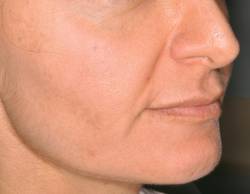
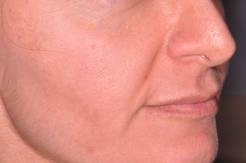
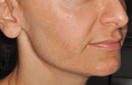
PHOENIX – Treating moderate to severe melasma with a 50-ns, 1,064-nm Q-switched neodynium:YAG laser produced equivalent temporary results with less pain than did a 5-ns, 1,064-nm Q-switched Nd:YAG laser in a split-face study of 10 women.
In the 6-month study, scores on a melasma area and severity index decreased by 16% using the 50-ns pulse width, and by 27% using the 5-ns pulse width at month 4, which was 1 month after the last of three monthly treatments that combined microdermabrasion, laser therapy, and a topical therapy regimen. Changes from both lasers were statistically significant compared with baseline, but not significantly different between laser groups, Dr. Vineet Mishra reported at the annual meeting of the American Society for Laser Medicine and Surgery.

The investigators sought to optimize laser settings for treating melasma to see whether lasers could be used in combination with other melasma therapies, Dr. Mishra said in an interview.
"During the study, everybody improved," Dr. Mishra said.
Melanin (as measured by a reflectance spectrophotometer at each visit) decreased by 20% with the 50-ns pulse width and by 17% with the 5-ns pulse width during the study.
However, pigment relapsed in both treatment areas after the study, to a level not significantly different from that at baseline. This was not unexpected when treating melasma, and was "likely due to sun exposure in sunny San Diego," said Dr. Mishra, director of Mohs surgery and procedural dermatology at the University of Texas Health Science Center, San Antonio.
Patients who completed the study rated pain from the procedure as a mean of 1.2 on the side of the face treated with the 50-ns laser and 2.9 with the 5-ns laser, using a 10-point scale with 0 representing no pain and 10 the worst pain, Dr. Mishra said.
Each treatment session consisted of microdermabrasion of the entire face followed by treatment with the 50-ns laser on one side and treatment with the 5-ns laser on the other side. Two laser passes were applied using a fluence of 1.6 J/cm2 and a 5-mm spot size. For 2 weeks prior to each treatment session and starting 2 days after each laser treatment, patients were asked to follow a daily skin care regimen that consisted of topical 4% hydroquinone, 0.05% tretinoin cream, and sunscreen with a sun protection factor of 50.
"Lasers have exploded on the market" but can be tricky to use against melasma, Dr. Mishra said. Too-high levels of laser energy can cause postinflammatory hyperpigmentation, which can worsen melasma, he noted.
Melasma is a difficult condition to treat, but "combination therapy is optimal," Dr. Mishra added in an interview. Next steps for research should include longer-term studies to determine how patients would fare if they continued with the combination treatment regimen. In addition, using a picosecond laser for melasma "would be a new frontier" to explore in long-term studies, he said.
The study won an award at the meeting.
The study received funding from Syneron and Candela, which market the 50-ns laser, and one of Dr. Mishra’s coinvestigators reported financial associations with Syneron and Candela and with Cynosure, which markets the 5-ns laser.
On Twitter @sherryboschert
PHOENIX – Treating moderate to severe melasma with a 50-ns, 1,064-nm Q-switched neodynium:YAG laser produced equivalent temporary results with less pain than did a 5-ns, 1,064-nm Q-switched Nd:YAG laser in a split-face study of 10 women.
In the 6-month study, scores on a melasma area and severity index decreased by 16% using the 50-ns pulse width, and by 27% using the 5-ns pulse width at month 4, which was 1 month after the last of three monthly treatments that combined microdermabrasion, laser therapy, and a topical therapy regimen. Changes from both lasers were statistically significant compared with baseline, but not significantly different between laser groups, Dr. Vineet Mishra reported at the annual meeting of the American Society for Laser Medicine and Surgery.
The investigators sought to optimize laser settings for treating melasma to see whether lasers could be used in combination with other melasma therapies, Dr. Mishra said in an interview.
"During the study, everybody improved," Dr. Mishra said.
Melanin (as measured by a reflectance spectrophotometer at each visit) decreased by 20% with the 50-ns pulse width and by 17% with the 5-ns pulse width during the study.
However, pigment relapsed in both treatment areas after the study, to a level not significantly different from that at baseline. This was not unexpected when treating melasma, and was "likely due to sun exposure in sunny San Diego," said Dr. Mishra, director of Mohs surgery and procedural dermatology at the University of Texas Health Science Center, San Antonio.
Patients who completed the study rated pain from the procedure as a mean of 1.2 on the side of the face treated with the 50-ns laser and 2.9 with the 5-ns laser, using a 10-point scale with 0 representing no pain and 10 the worst pain, Dr. Mishra said.
Each treatment session consisted of microdermabrasion of the entire face followed by treatment with the 50-ns laser on one side and treatment with the 5-ns laser on the other side. Two laser passes were applied using a fluence of 1.6 J/cm2 and a 5-mm spot size. For 2 weeks prior to each treatment session and starting 2 days after each laser treatment, patients were asked to follow a daily skin care regimen that consisted of topical 4% hydroquinone, 0.05% tretinoin cream, and sunscreen with a sun protection factor of 50.
"Lasers have exploded on the market" but can be tricky to use against melasma, Dr. Mishra said. Too-high levels of laser energy can cause postinflammatory hyperpigmentation, which can worsen melasma, he noted.
Melasma is a difficult condition to treat, but "combination therapy is optimal," Dr. Mishra added in an interview. Next steps for research should include longer-term studies to determine how patients would fare if they continued with the combination treatment regimen. In addition, using a picosecond laser for melasma "would be a new frontier" to explore in long-term studies, he said.
The study won an award at the meeting.
The study received funding from Syneron and Candela, which market the 50-ns laser, and one of Dr. Mishra’s coinvestigators reported financial associations with Syneron and Candela and with Cynosure, which markets the 5-ns laser.
On Twitter @sherryboschert
AT LASER 2014
Key clinical point: Finding the optimal settings for lasers in patients with melasma could help improve the condition as part of a combination treatment regimen.
Major finding: Both the 50-ns and 5-ns pulse widths significantly decreased melasma area and severity index scores temporarily, but mean pain scores were significantly lower with the 50-ns laser (1.2) than with the 5-ns laser (2.9).
Data source: A prospective, randomized split-face comparison of 50-ns and 5-ns 1,064-nm Q-switched Nd:YAG laser therapy, plus dermabrasion and topical therapy for melasma.
Disclosures: The study received funding from Syneron and Candela, which market the 50-ns laser, and one of Dr. Mishra’s coinvestigators reported financial associations with Syneron and Candela and with Cynosure, which markets the 5-ns laser.
Early look: Localized cryolysis reduced sebum by 40%
PHOENIX – One cycle of selective transepidermal cooling on the backs of 11 men with healthy, normal skin produced a transient 40% reduction in sebum production by sebaceous glands in a controlled pilot study.
Sebum production fell significantly by 1 and 2 weeks after treatment, compared with production prior to treatment, but by week 4, was not significantly different than at baseline, Dr. H. Ray Jalian and his associates reported at the annual meeting of the American Society for Laser Medicine and Surgery. Sebum production did not change significantly on untreated patches of skin that served as controls.

The results give hope that selective cryolysis of sebaceous glands might be a noninvasive treatment for acne vulgaris, which should be explored in additional studies, said Dr. Jalian of the Wellman Center for Photomedicine, Massachusetts General Hospital, Boston.
The study won a top award at the meeting and piqued the interest of attendees. Dr. Jeffrey Dover, immediate past president of the ASLMS, explained the excitement over this small pilot study.
"If you extend that to the future, after they do more studies; more cold for longer [time periods], or maybe more applications may produce prolonged sebaceous gland inactivity," he noted. "And maybe [achieve] permanent sebaceous gland inactivity, which theoretically could be a cure for acne – there could be Accutane-like results with an applicator that’s cold, with no side effects," said Dr. Dover, a dermatologist in Chestnut Hill, Mass. "Now, that would be an advance for dermatology," he said in an interview.
Sebumeter measurements at 2 weeks (the primary outcome measure) showed a 40% reduction in sebum production for two temperature settings, compared with baseline.
The study involved the off-label use of a handheld cooling applicator that has been approved for body sculpting to destroy fat in some body areas. Lipid-rich tissue is susceptible to cold injury, and the researchers hoped to target the lipid content of the sebaceous glands.
Participants were randomized to treatment on select areas of their backs at temperatures of –10° C or –15° C, and they received either a single 20-minute cooling cycle or two 10-minute cooling cycles with rewarming in between. Some skin areas on the scapulas served as control sites, and each subject had two treatment sites and one control site. Sebum measurements and standardized clinical photos were taken just before treatment and on follow-up visits at 3 days (72 hours) and at 1, 2, and 4 weeks.
Among secondary outcomes, a smaller but significant sebum reduction occurred at 1 week, but sebum production did not differ significantly at 72 hours or at 4 weeks, compared with baseline. Transient pain reported during treatment (average on a Visual Analog Scale score of approximately 3 on a 10-point scale) disappeared quickly post treatment and at follow-up visits. "It actually was a very-well-tolerated procedure," Dr. Jalian said.
Transient post-treatment erythema, dysesthesia, and edema also cleared quickly, though one subject had erythema lasting approximately 72 hours, according to blinded evaluators.
"Perhaps the most interesting part of our study was the histology," Dr. Jalian noted. Biopsies from a subset of participants showed histologic evidence of sebaceous gland damage after treatment that persisted at weeks 1 and 2.
There was no clinical or histologic evidence of damage to tissue outside of the treated areas.
"Selective cryolysis of sebaceous glands is achievable through noninvasive cooling," said Dr. Jalian. Future studies may examine whether repeated treatments could maintain the reduction in sebum output beyond 2 weeks, he added.
Previous studies have shown that a 30%-50% reduction in sebum is needed to produce a 50% improvement in acne (Br. J. Dermatol. 2010;163:683-8).
The human pilot study followed preclinical experiments on the ears of mice, sheep, and pigs that showed histologic damage in sebaceous glands after treatment, Dr. Jalian said.
The study was funded by Zeltiq Aesthetics, which makes the handheld cooling device. Dr. Jalian reported no other relevant disclosures.
On Twitter @sherryboschert
PHOENIX – One cycle of selective transepidermal cooling on the backs of 11 men with healthy, normal skin produced a transient 40% reduction in sebum production by sebaceous glands in a controlled pilot study.
Sebum production fell significantly by 1 and 2 weeks after treatment, compared with production prior to treatment, but by week 4, was not significantly different than at baseline, Dr. H. Ray Jalian and his associates reported at the annual meeting of the American Society for Laser Medicine and Surgery. Sebum production did not change significantly on untreated patches of skin that served as controls.
The results give hope that selective cryolysis of sebaceous glands might be a noninvasive treatment for acne vulgaris, which should be explored in additional studies, said Dr. Jalian of the Wellman Center for Photomedicine, Massachusetts General Hospital, Boston.
The study won a top award at the meeting and piqued the interest of attendees. Dr. Jeffrey Dover, immediate past president of the ASLMS, explained the excitement over this small pilot study.
"If you extend that to the future, after they do more studies; more cold for longer [time periods], or maybe more applications may produce prolonged sebaceous gland inactivity," he noted. "And maybe [achieve] permanent sebaceous gland inactivity, which theoretically could be a cure for acne – there could be Accutane-like results with an applicator that’s cold, with no side effects," said Dr. Dover, a dermatologist in Chestnut Hill, Mass. "Now, that would be an advance for dermatology," he said in an interview.
Sebumeter measurements at 2 weeks (the primary outcome measure) showed a 40% reduction in sebum production for two temperature settings, compared with baseline.
The study involved the off-label use of a handheld cooling applicator that has been approved for body sculpting to destroy fat in some body areas. Lipid-rich tissue is susceptible to cold injury, and the researchers hoped to target the lipid content of the sebaceous glands.
Participants were randomized to treatment on select areas of their backs at temperatures of –10° C or –15° C, and they received either a single 20-minute cooling cycle or two 10-minute cooling cycles with rewarming in between. Some skin areas on the scapulas served as control sites, and each subject had two treatment sites and one control site. Sebum measurements and standardized clinical photos were taken just before treatment and on follow-up visits at 3 days (72 hours) and at 1, 2, and 4 weeks.
Among secondary outcomes, a smaller but significant sebum reduction occurred at 1 week, but sebum production did not differ significantly at 72 hours or at 4 weeks, compared with baseline. Transient pain reported during treatment (average on a Visual Analog Scale score of approximately 3 on a 10-point scale) disappeared quickly post treatment and at follow-up visits. "It actually was a very-well-tolerated procedure," Dr. Jalian said.
Transient post-treatment erythema, dysesthesia, and edema also cleared quickly, though one subject had erythema lasting approximately 72 hours, according to blinded evaluators.
"Perhaps the most interesting part of our study was the histology," Dr. Jalian noted. Biopsies from a subset of participants showed histologic evidence of sebaceous gland damage after treatment that persisted at weeks 1 and 2.
There was no clinical or histologic evidence of damage to tissue outside of the treated areas.
"Selective cryolysis of sebaceous glands is achievable through noninvasive cooling," said Dr. Jalian. Future studies may examine whether repeated treatments could maintain the reduction in sebum output beyond 2 weeks, he added.
Previous studies have shown that a 30%-50% reduction in sebum is needed to produce a 50% improvement in acne (Br. J. Dermatol. 2010;163:683-8).
The human pilot study followed preclinical experiments on the ears of mice, sheep, and pigs that showed histologic damage in sebaceous glands after treatment, Dr. Jalian said.
The study was funded by Zeltiq Aesthetics, which makes the handheld cooling device. Dr. Jalian reported no other relevant disclosures.
On Twitter @sherryboschert
AT LASER 2014
Key clinical point: Cryolysis of the sebaceous glands might provide a noninvasive option for acne treatment.
Major finding: Sebum excretion was 40% lower 2 weeks after transepidermal cryolysis treatment of sebaceous glands, compared with baseline.
Data source: A prospective, controlled pilot study of 11 healthy men who were treated on selected areas on their backs.
Disclosures: The study was funded by Zeltiq Aesthetics, which makes the handheld cooling device used in the study. Dr. Jalian reported no other relevant disclosures.

Three laser/light devices lead injury reports
PHOENIX – Radiofrequency devices, diode lasers, and intense pulsed light devices had the most reports of suspected injuries from 1991 through 2013, based on data from a Food and Drug Administration database.
During that period, 1,212 medical device reports were submitted for suspected injuries from laser or light device use or malfunction to the FDA’s MAUDE (Manufacturer and User Facility Device Experience) database, as well as another 45 that were excluded from the study because of insufficient data. The FDA requires device users and manufacturers to submit the reports and collects voluntary reports from patients. Each report may contain more than one adverse event.
Blisters or burns were the most common problems reported to the MAUDE database for each of the top three devices, Dr. Anne Marie Tremaine and her associates reported at the annual meeting of the American Society for Laser Medicine and Surgery.
Radiofrequency devices were the subject of 270 reports that included 294 adverse events. These reports included 119 burns or blisters; 85 incidents of fat loss, depression, or divot; 34 scars; 25 incidents of dyschromia (both hyper- and hypopigmentation); and 12 cases of sagging skin. In addition, a few reports noted burns in the area of the return pad, paresthesia or nerve palsy, dry eye or "floaters," and cracked dental crowns.
Insufficient coupling fluid and tearing or breakdown in the device tip membrane topped the reasons given for problems related to radiofrequency treatment, reported Dr. Tremaine of Massachusetts General Hospital’s Wellman Center for Photomedicine, Boston. Complications also were ascribed to tumescent anesthesia, intradermal lidocaine injections, and general anesthesia.
The most common treatment modality associated with adverse events was laser hair removal using diode lasers or intense pulsed light. The 252 reports on diode lasers included 297 burns or blisters, 42 incidents of dyschromia, 11 scars, and 8 cases of swelling. The 158 reports on adverse events from intense pulsed light included 126 burns or blisters; 24 incidents of dyschromia; 20 scars; 13 patients with bruising; 12 incidents of fat loss, depression, or divot; and smaller numbers of eye injury or delayed wound healing.
Many of the complications in the reports were attributed to improper settings or improperly maintained device tips.
"Physicians should be aware of the adverse events and malfunctions of the devices used in dermatology," Dr. Tremaine said. The MAUDE database "will help uncover otherwise unreported events seen in routine practice among all types of practitioners" to supplement data from initial studies and reports in the medical literature. This information should help physicians better inform patients about the potential risks of treatment, she said.
Dr. Tremaine and her associates searched the MAUDE database using terms including names of device manufacturers from a comprehensive list, specific product names, and the wavelengths and technology of the devices used in dermatology.
However, because adverse events are underreported to MAUDE, which provides only passive surveillance, the incidence or prevalence of problems with laser or light devices could not be discerned from this database, the investigators noted.
Dr. Tremaine reported having no disclosures.
On Twitter @sherryboschert
AT LASER 2014
Major finding: Radiofrequency devices had the highest number of adverse-event reports (270), followed by diode lasers (252) and intense pulsed light devices (158).
Data source: A study of 1,212 medical device reports for laser or light devices in the FDA’s MAUDE database from 1991 through 2013.
Disclosures: Dr. Tremaine reported having no disclosures.
Laser therapy improved severe hyperhidrosis
PHOENIX – Laser treatment of severe hyperhidrosis significantly reduced underarm sweating after 6 months in two separate but similar studies of 20 and 13 patients.
Patients with scores of 3 or 4 on the 4-point Hyperhidrosis Disease Severity Scale (HDSS) underwent a single treatment to acutely damage eccrine glands using a 1,440-nm Nd:YAG laser with a new 800-mcm side-firing fiber designed to deliver targeted energy. Patients received tumescent anesthesia in each axilla.

Sixteen of 20 patients in one study (78%) reported at least a 2-point improvement in HDSS scores at a 6-month follow-up visit, and 17 (83%) reported at least a 1-point improvement at 1 year, compared with baseline, Dr. Bruce Katz and his associates reported at the annual meeting of the American Society for Laser Medicine and Surgery.
Average HDSS scores were 3.6 before treatment, 2.3 at 3 months, 1.7 at 6 months, and 2.2 at 1 year, said Dr. Katz, a clinical professor of dermatology at Mount Sinai Hospital, New York.
The side-firing fiber inserted through a 150-mm handpiece and set at 10-15 W delivered 1,200-1,500 J to each of four 5 cm2 × 5 cm2 areas in each armpit. Both eccrine and apocrine glands were reduced based on post-treatment histological studies. Side effects included edema, bruising, and numbness, but these conditions resolved in 1-2 weeks.
Before the study, 13 patients (65%) had an HDSS score of 4. Five patients had tried treatment with botulinum toxin type A before the study and found it effective, with results lasting an average of 6 months, but they had discontinued that therapy because of the high cost, Dr. Katz said.
In a second study of 13 patients, HDSS scores improved by a mean of 1.3 at the 6-month follow-up visit, Dr. Alina Fratila and her associates reported in a separate presentation at the meeting.
Gravimetric measurements (in milligrams per minute) showed a mean 78% reduction in sweating at 6 months, said Dr. Fratila of the Fountain of Youth Clinic in Bonn, Germany.
Physicians rated the improvement in sweat as 71%, and patients rated it as 55%. The laser in this study was set at 7.5 W, delivering an average of 6,000 J per axilla (or 1,500 J per 5 cm2 × 5 cm2 quadrant).
Ten patients had tried a prescription antiperspirant prior to participating in the study, but they stopped because they found it either irritating or ineffective. Four patients had tried botulinum toxin therapy but stopped because of the cost, Dr. Fratila said.
The laser treatment has a low risk of side effects and causes essentially no downtime, Dr. Fratila said. She added that she plans to follow the patients to assess results after 1 and 2 years.
An estimated 3% of the U.S. population has primary focal hyperhidrosis, comprising nearly 8 million people, Dr. Katz said.
Other treatment options for primary focal hyperhidrosis include topical over-the-counter antiperspirants, aluminum chloride 10%-35% topical antiperspirant, intradermal injections of botulinum toxin type A, and surgical resection of local sweat glands.
Dr. Katz reported financial associations with Cynosure, which makes the laser systems used in his study, and with Allergan, Alma, and other companies. Dr. Fratila reported having no disclosures.
On Twitter @sherryboschert
PHOENIX – Laser treatment of severe hyperhidrosis significantly reduced underarm sweating after 6 months in two separate but similar studies of 20 and 13 patients.
Patients with scores of 3 or 4 on the 4-point Hyperhidrosis Disease Severity Scale (HDSS) underwent a single treatment to acutely damage eccrine glands using a 1,440-nm Nd:YAG laser with a new 800-mcm side-firing fiber designed to deliver targeted energy. Patients received tumescent anesthesia in each axilla.
Sixteen of 20 patients in one study (78%) reported at least a 2-point improvement in HDSS scores at a 6-month follow-up visit, and 17 (83%) reported at least a 1-point improvement at 1 year, compared with baseline, Dr. Bruce Katz and his associates reported at the annual meeting of the American Society for Laser Medicine and Surgery.
Average HDSS scores were 3.6 before treatment, 2.3 at 3 months, 1.7 at 6 months, and 2.2 at 1 year, said Dr. Katz, a clinical professor of dermatology at Mount Sinai Hospital, New York.
The side-firing fiber inserted through a 150-mm handpiece and set at 10-15 W delivered 1,200-1,500 J to each of four 5 cm2 × 5 cm2 areas in each armpit. Both eccrine and apocrine glands were reduced based on post-treatment histological studies. Side effects included edema, bruising, and numbness, but these conditions resolved in 1-2 weeks.
Before the study, 13 patients (65%) had an HDSS score of 4. Five patients had tried treatment with botulinum toxin type A before the study and found it effective, with results lasting an average of 6 months, but they had discontinued that therapy because of the high cost, Dr. Katz said.
In a second study of 13 patients, HDSS scores improved by a mean of 1.3 at the 6-month follow-up visit, Dr. Alina Fratila and her associates reported in a separate presentation at the meeting.
Gravimetric measurements (in milligrams per minute) showed a mean 78% reduction in sweating at 6 months, said Dr. Fratila of the Fountain of Youth Clinic in Bonn, Germany.
Physicians rated the improvement in sweat as 71%, and patients rated it as 55%. The laser in this study was set at 7.5 W, delivering an average of 6,000 J per axilla (or 1,500 J per 5 cm2 × 5 cm2 quadrant).
Ten patients had tried a prescription antiperspirant prior to participating in the study, but they stopped because they found it either irritating or ineffective. Four patients had tried botulinum toxin therapy but stopped because of the cost, Dr. Fratila said.
The laser treatment has a low risk of side effects and causes essentially no downtime, Dr. Fratila said. She added that she plans to follow the patients to assess results after 1 and 2 years.
An estimated 3% of the U.S. population has primary focal hyperhidrosis, comprising nearly 8 million people, Dr. Katz said.
Other treatment options for primary focal hyperhidrosis include topical over-the-counter antiperspirants, aluminum chloride 10%-35% topical antiperspirant, intradermal injections of botulinum toxin type A, and surgical resection of local sweat glands.
Dr. Katz reported financial associations with Cynosure, which makes the laser systems used in his study, and with Allergan, Alma, and other companies. Dr. Fratila reported having no disclosures.
On Twitter @sherryboschert
AT LASER 2014
Key clinical point: Patients with severe hyperhidrosis who underwent laser treatment had minimal, short-term side effects and essentially no downtime.
Major finding: HDSS scores of underarm sweating 6 months after laser treatment improved by at least 2 points in one study and by a mean of 1.3 points in another study.
Data source: Separate single-center, uncontrolled trials of 20 and 13 patients, respectively, with severe primary focal hyperhidrosis.
Disclosures: Dr. Katz reported financial associations with Cynosure, which makes the laser systems used in the study, and with Allergan, Alma, and other companies. Dr. Fratila reported having no disclosures.

Bruises Be Gone! Treatment of Bruising

An article in Dermatologic Surgery, “Comparative Study on Bruise Reduction Treatments After Bruise Induction Using the Pulsed Dye Laser,” (2013;39:1459-1464) compared the of effectiveness different modalities in reducing time for bruise resolution. The investigators compared cold compresses; hydrogen peroxide; over-the-counter bruise serum containing primrose oil, vitamin E oil, and glycerin; and pulsed dye laser (PDL). Seventeen patients (Fitzpatrick skin types I–IV) were enrolled and had bruise induction with a PDL to produce five 2×2-cm zones of bruising on the lower abdomen. Excluding the control, bruises were randomly treated using a cold compress, bruise serum, 3% hydrogen peroxide–soaked gauze, or PDL. Subjects and 2 blinded physician evaluators graded the bruise severity using a visual scale on days 0, 3, and 7. The investigators found that treatment did not result in statistically significantly shorter bruise resolution time than in controls. They reported that PDL-treated bruises took a longer time to resolve than controls.
What’s the issue?
Although this study found that the PDL-treated bruises took a longer time to resolve, there have been other studies that have shown PDL to hasten time to bruise resolution, which could be due to the fact that the initial bruises in this particular study were actually induced by PDL. In another study, DeFatta et al (Arch Facial Plast Surg. 2009;11:99-103) found maximal efficacy when PDL was performed on or after postoperative day 5. It was reasoned that treatment before then was less effective because of the depth of red blood cell extravasation and overlying inflammation and edema. Karen and Hale (Dermatol Surg. 2010;36:1328-1331) reported that the optimal time for PDL treatment was between 48 and 72 hours, suggesting that hemoglobin predominates during this period instead of the bilirubin predominance seen in the later stages of ecchymosis. Their parameters were 7.5 J/cm2, 10-mm spot, 6-millisecond pulse duration, and cryogen 30 milliseconds with a 20-millisecond delay for a single pass. What do you use for posttreatment bruising?
