User login
Tests can identify leukemia risk in newborns with Down syndrome
SAN DIEGO – Research into hundreds of babies with Down syndrome is providing valuable insight into the genetic roots of leukemia and offering a route to identify newborns at high risk.
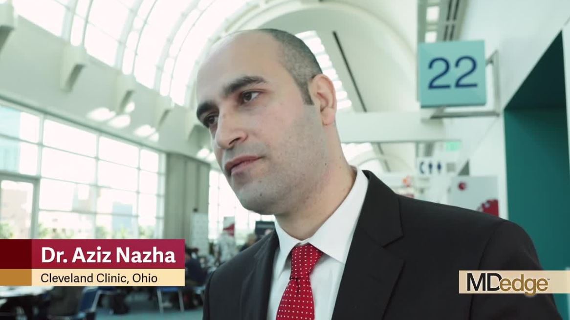
“We can now identify children at high risk of developing myeloid leukemia within 4 years” through blood or genetic tests, Irene Roberts, MD, a pediatric hematologist at the University of Oxford’s (England) MRC Weatherall Institute of Molecular Medicine, said at the annual meeting of the American Society of Hematology.
About 2%-3% of children with Down syndrome will develop acute lymphocytic leukemia (ALL) or acute myeloid leukemia (AML), according to the National Cancer Institute, rates that are much higher than in the general population.
Research suggests that among children aged 0-4 years with Down syndrome, the standardized incidence ratio (SIR) of AML is 114, compared with other children, Dr. Roberts said. The SIR of ALL is 27 in children aged 1-4 years, she said.
For people with Down syndrome aged 0-60 years, the SIRs are 12 and 13 in AML and ALL, respectively, she said.
In her presentation, Dr. Roberts focused on AML that appears before age 4 years and is preceded by a neonatal preleukemia – transient abnormal myelopoiesis (TAM) – that only occurs in Down syndrome. In most cases, TAM, which occurs with GATA1 mutations, resolves on its own after birth, she said. But in others, the GATA1 mutations continue and cause AML to develop.
Dr. Roberts highlighted her institution’s Oxford Down Syndrome Cohort Study and offered an update to a 2013 report (Blood. 2013 Dec 5;122[24]:3908–17). The study recruited 471 neonates with Down syndrome and followed them for up to 4 years: 341 with no GATA1 mutation and 130 (28%) with the mutation. Dr. Roberts called the latter number a “very high frequency.”
Of those with the mutation, 7 patients (5%) developed AML at a median age of 16 months. None of those without the mutation developed AML.
Also, among the 130 neonates with the mutation, 42% were considered to have “clinical” TAM (more than 10% blasts) and 58% were considered to have “silent” TAM (fewer than 10% blasts).
“We predicted that these babies with clinical TAM would have more severe clinical disease ... and that in fact turned out to be the case,” Dr. Roberts said.
Why is the GATA1 mutation so significant? Research suggests that platelet production is abnormal in neonates with Down syndrome, compared with neonates without it, regardless of whether they have the mutation, Dr. Roberts said.
The mutation doesn’t reduce further platelet count, but does disrupt megakaryopoiesis – the process of the production of platelets. As a result, giant platelets and megakaryocyte fragments are more common, she explained.
Moving forward, research data can be used to identify which children are most at risk, Dr. Roberts said. Newborns with Down syndrome are more likely to survive without leukemia if they have silent TAM, compared with those who have clinical TAM, and if they have an estimated variant allele frequency above 15%, according to findings from the Oxford study.
Children at high risk of AML before age 4 years can be identified by analyzing the percentage of blasts on a smear and/or by analyzing mutation of GATA1, according to Dr. Roberts. However, this cannot be accomplished by the use of a complete blood count (CBC) test, she said, which is used to check for leukemia.
Dr. Roberts called for the development of more guidelines for screening newborns with Down syndrome for leukemia risk. The British Society for Haematology issued testing guidelines, coauthored by Dr. Roberts, in 2018 (Br J Haematol. 2018 Jul;182[2]:200-11).
Dr. Roberts reported having no financial disclosures.
SAN DIEGO – Research into hundreds of babies with Down syndrome is providing valuable insight into the genetic roots of leukemia and offering a route to identify newborns at high risk.
“We can now identify children at high risk of developing myeloid leukemia within 4 years” through blood or genetic tests, Irene Roberts, MD, a pediatric hematologist at the University of Oxford’s (England) MRC Weatherall Institute of Molecular Medicine, said at the annual meeting of the American Society of Hematology.
About 2%-3% of children with Down syndrome will develop acute lymphocytic leukemia (ALL) or acute myeloid leukemia (AML), according to the National Cancer Institute, rates that are much higher than in the general population.
Research suggests that among children aged 0-4 years with Down syndrome, the standardized incidence ratio (SIR) of AML is 114, compared with other children, Dr. Roberts said. The SIR of ALL is 27 in children aged 1-4 years, she said.
For people with Down syndrome aged 0-60 years, the SIRs are 12 and 13 in AML and ALL, respectively, she said.
In her presentation, Dr. Roberts focused on AML that appears before age 4 years and is preceded by a neonatal preleukemia – transient abnormal myelopoiesis (TAM) – that only occurs in Down syndrome. In most cases, TAM, which occurs with GATA1 mutations, resolves on its own after birth, she said. But in others, the GATA1 mutations continue and cause AML to develop.
Dr. Roberts highlighted her institution’s Oxford Down Syndrome Cohort Study and offered an update to a 2013 report (Blood. 2013 Dec 5;122[24]:3908–17). The study recruited 471 neonates with Down syndrome and followed them for up to 4 years: 341 with no GATA1 mutation and 130 (28%) with the mutation. Dr. Roberts called the latter number a “very high frequency.”
Of those with the mutation, 7 patients (5%) developed AML at a median age of 16 months. None of those without the mutation developed AML.
Also, among the 130 neonates with the mutation, 42% were considered to have “clinical” TAM (more than 10% blasts) and 58% were considered to have “silent” TAM (fewer than 10% blasts).
“We predicted that these babies with clinical TAM would have more severe clinical disease ... and that in fact turned out to be the case,” Dr. Roberts said.
Why is the GATA1 mutation so significant? Research suggests that platelet production is abnormal in neonates with Down syndrome, compared with neonates without it, regardless of whether they have the mutation, Dr. Roberts said.
The mutation doesn’t reduce further platelet count, but does disrupt megakaryopoiesis – the process of the production of platelets. As a result, giant platelets and megakaryocyte fragments are more common, she explained.
Moving forward, research data can be used to identify which children are most at risk, Dr. Roberts said. Newborns with Down syndrome are more likely to survive without leukemia if they have silent TAM, compared with those who have clinical TAM, and if they have an estimated variant allele frequency above 15%, according to findings from the Oxford study.
Children at high risk of AML before age 4 years can be identified by analyzing the percentage of blasts on a smear and/or by analyzing mutation of GATA1, according to Dr. Roberts. However, this cannot be accomplished by the use of a complete blood count (CBC) test, she said, which is used to check for leukemia.
Dr. Roberts called for the development of more guidelines for screening newborns with Down syndrome for leukemia risk. The British Society for Haematology issued testing guidelines, coauthored by Dr. Roberts, in 2018 (Br J Haematol. 2018 Jul;182[2]:200-11).
Dr. Roberts reported having no financial disclosures.
EXPERT ANALYSIS FROM ASH 2018
Chemo for solid tumors and risk of tMDS/AML

Chemotherapy for solid tumors is associated with an increased risk of therapy-related myelodysplastic syndromes or acute myeloid leukemia (tMDS/AML), according to a retrospective analysis.
Long-term, population-based cohort data showed the risk of tMDS/AML was significantly elevated after chemotherapy for 22 solid tumor types.
The relative risk of tMDS/AML was 1.5- to 39.0-fold greater among patients treated for these tumors than among the general population.
Lindsay M. Morton, PhD, of the National Institutes of Health in Rockville, Maryland, and her colleagues reported these findings in JAMA Oncology.
“We undertook an investigation to quantify tMDS/AML risks after chemotherapy for solid tumors in the modern treatment era, 2000-2014, using United States cancer registry data from the National Cancer Institute’s Surveillance, Epidemiology, and End Results Program,” the investigators wrote.
They retrospectively analyzed data from 1619 patients with tMDS/AML who were diagnosed with an initial primary solid tumor from 2000 to 2013.
Patients were given initial chemotherapy and lived for at least 1 year after treatment. Subsequently, Dr. Morton and her colleagues linked patient database records with Medicare insurance claim information to confirm the accuracy of chemotherapy data.
“Because registry data do not include treatment details, we used an alternative database to provide descriptive information on population-based patterns of chemotherapeutic drug use,” the investigators noted.
The team found the risk of developing tMDS/AML was significantly increased following chemotherapy administration for 22 of 23 solid tumor types, excluding colon cancer.
The standardized incidence ratio (SIR) for tMDS/AML ranged from 1.5 to 39.0, and the excess absolute risk (EAR) ranged from 1.4 to 23.6 cases per 10,000 person-years.
SIRs were greatest in patients who received chemotherapy for malignancy of the bone (SIR=39.0, EAR=23.6), testis (SIR, 12.3, EAR=4.4), soft tissue (SIR=10.4, EAR=12.6), fallopian tube (SIR=8.7, EAR=16.0), small cell lung (SIR=8.1, EAR=19.9), peritoneum (SIR=7.5, EAR=15.8), brain or central nervous system (SIR=7.2, EAR=6.0), and ovary (SIR=5.8, EAR=8.2).
The investigators also found that patients who were given chemotherapy at a young age had the highest risk of developing tMDS/AML.
“For patients treated with chemotherapy at the present time, approximately three-quarters of tMDS/AML cases expected to occur within the next 5 years will be attributable to chemotherapy,” the investigators said.
They acknowledged a key limitation of this study was the limited data on patient-specific chemotherapy and dosing information. Given these limitations, Dr. Morton and her colleagues said, “the exact magnitude of our risk estimates, including the proportions of excess cases, should therefore be interpreted cautiously.”
This study was supported by the Intramural Research Program of the National Institutes of Health, National Cancer Institute, and the California Department of Public Health. The authors reported no conflicts of interest.
Higher AML, MDS risk linked to solid tumor chemotherapy
There is an increased risk for therapy-related myelodysplastic syndrome or acute myeloid leukemia (tMDS/AML) following chemotherapy for the majority of solid tumor types, according to an analysis of cancer registry data.
These findings suggest a substantial expansion in the patients at risk for tMDS/AML because, in the past, excess risks were established only after chemotherapy for cancers of the lung, ovary, breast, soft tissue, testis, and brain or central nervous system,” Lindsay M. Morton, PhD, of the National Institutes of Health, and her colleagues wrote in JAMA Oncology.
The researchers retrospectively analyzed data from 1,619 patients with tMDS/AML who were diagnosed with an initial primary solid tumor from 2000 to 2013. Data came from the National Cancer Institute’s Surveillance, Epidemiology, and End Results (SEER) Program and Medicare claims.
Study participants were given initial chemotherapy and lived for at least 1 year after treatment. Subsequently, Dr. Morton and her colleagues linked patient database records with Medicare insurance claim information to confirm the accuracy of chemotherapy data.
“Because registry data [does] not include treatment details, we used an alternative database to provide descriptive information on population-based patterns of chemotherapeutic drug use,” the researchers wrote in JAMA Oncology.
After statistical analysis, the researchers found that the risk of developing tMDS/AML was significantly elevated following chemotherapy administration for 22 of 23 solid tumor types, excluding colon cancer. They reported a 1.5-fold to more than 10-fold increased relative risk for tMDS/AML in those patients who received chemotherapy for those 22 solid cancer types, compared with the general population.
The relative risks were highest after chemotherapy for bone, soft-tissue, and testis cancers.
The researchers found that the absolute risk of developing tMDS/AML was low. Excess absolute risks ranged from 1.4 to greater than 15 cases per 10,000 person-years, compared with the general population, in those 22 solid cancer types. The greatest absolute risks were for peritoneum, small-cell lung, bone, soft-tissue, and fallopian tube cancers.
“For patients treated with chemotherapy at the present time, approximately three-quarters of tMDS/AML cases expected to occur within the next 5 years will be attributable to chemotherapy,” they added.
The researchers acknowledged a key limitation of the study was the limited data on dosing and patient-specific chemotherapy. As a result, Dr. Morton and her colleagues called for a cautious interpretation of the magnitude of the risk.
The study was supported by the Intramural Research Program of the National Institutes of Health, National Cancer Institute, and the California Department of Public Health. The authors reported having no conflicts of interest.
SOURCE: Morton LM et al. JAMA Oncol. 2018 Dec 20. doi: 10.1001/jamaoncol.2018.5625.
Possibly the most clinical relevant finding of the study by Lindsay M. Morton, PhD, and her colleagues is that patients who received chemotherapy for solid tumor treatment at a younger age were at the highest relative risk for tMDS/AML.
The incidence of tMDS/AML was highest among patients treated with chemotherapy for bone, soft-tissue, and testicular cancers, where the median age of onset is often by 30 years, and the mean onset occurs before age 50.
The researchers also noted an increased risk for tMDS/AML associated with prolonged survival from primary tumors.
Going forward, research should consider those patients at highest risk for tMDS/AML and risk-assessment models for these therapy-related myeloid neoplasms should take into account the clonal evolution of subclinical mutations into overt disease.
The study findings point to the unanswered question of how best to perform risk assessment of chemotherapy in solid tumors. That risk stratification could include the probability of the specific chemotherapy agent initiating disease, the benefit of tumor regression from chemotherapy, and the potential consequences of tumor progression if chemotherapy is not administered.
Shyam A. Patel, MD, PhD, is with the department of medicine at Stanford (Calif.) University. Dr. Patel reported having no financial disclosures. These comments are adapted from his accompanying editorial (JAMA Oncol. 2018 Dec 20. doi: 10.1001/jamaoncol.2018.5617 ).
There is an increased risk for therapy-related myelodysplastic syndrome or acute myeloid leukemia (tMDS/AML) following chemotherapy for the majority of solid tumor types, according to an analysis of cancer registry data.
These findings suggest a substantial expansion in the patients at risk for tMDS/AML because, in the past, excess risks were established only after chemotherapy for cancers of the lung, ovary, breast, soft tissue, testis, and brain or central nervous system,” Lindsay M. Morton, PhD, of the National Institutes of Health, and her colleagues wrote in JAMA Oncology.
The researchers retrospectively analyzed data from 1,619 patients with tMDS/AML who were diagnosed with an initial primary solid tumor from 2000 to 2013. Data came from the National Cancer Institute’s Surveillance, Epidemiology, and End Results (SEER) Program and Medicare claims.
Study participants were given initial chemotherapy and lived for at least 1 year after treatment. Subsequently, Dr. Morton and her colleagues linked patient database records with Medicare insurance claim information to confirm the accuracy of chemotherapy data.
“Because registry data [does] not include treatment details, we used an alternative database to provide descriptive information on population-based patterns of chemotherapeutic drug use,” the researchers wrote in JAMA Oncology.
After statistical analysis, the researchers found that the risk of developing tMDS/AML was significantly elevated following chemotherapy administration for 22 of 23 solid tumor types, excluding colon cancer. They reported a 1.5-fold to more than 10-fold increased relative risk for tMDS/AML in those patients who received chemotherapy for those 22 solid cancer types, compared with the general population.
The relative risks were highest after chemotherapy for bone, soft-tissue, and testis cancers.
The researchers found that the absolute risk of developing tMDS/AML was low. Excess absolute risks ranged from 1.4 to greater than 15 cases per 10,000 person-years, compared with the general population, in those 22 solid cancer types. The greatest absolute risks were for peritoneum, small-cell lung, bone, soft-tissue, and fallopian tube cancers.
“For patients treated with chemotherapy at the present time, approximately three-quarters of tMDS/AML cases expected to occur within the next 5 years will be attributable to chemotherapy,” they added.
The researchers acknowledged a key limitation of the study was the limited data on dosing and patient-specific chemotherapy. As a result, Dr. Morton and her colleagues called for a cautious interpretation of the magnitude of the risk.
The study was supported by the Intramural Research Program of the National Institutes of Health, National Cancer Institute, and the California Department of Public Health. The authors reported having no conflicts of interest.
SOURCE: Morton LM et al. JAMA Oncol. 2018 Dec 20. doi: 10.1001/jamaoncol.2018.5625.
FROM JAMA ONCOLOGY
Key clinical point:
Major finding: Treatment with chemotherapy was linked with a 1.5-fold to more than 10-fold increased risk for tMDS/AML.
Study details: A retrospective analysis of 1,619 patients with tMDS/AML who were diagnosed with an initial primary solid tumor from 2000 to 2013.
Disclosures: The study was supported by the Intramural Research Program of the National Institutes of Health, National Cancer Institute, and the California Department of Public Health. The authors reported having no conflicts of interest.
Source: Morton LM et al. JAMA Oncol. 2018 Dec 20. doi: 10.1001/jamaoncol.2018.5625.
Quizartinib improves survival of FLT3-mutated AML
Single-agent therapy with quizartinib slightly but significantly prolonged survival – compared with salvage chemotherapy – for patients with relapsed/refractory acute myeloid leukemia (AML) bearing the FLT3-ITD mutation, results of the phase 3 randomized QuANTUM-R trial showed.

Median overall survival (OS), the trial’s primary endpoint, was 6.2 months for 245 patients randomized to quizartinib, compared with 4.7 months for 122 patients assigned to salvage chemotherapy, a difference that translated into a hazard ratio (HR) for death of 0.76 (P = .0177), reported Jorge E. Cortes, MD, of the University of Texas MD Anderson Cancer Center in Houston.
“This study is the first study that demonstrates in a randomized fashion an overall survival benefit in the salvage setting for patients with FLT-3 mutated refractory or relapsed AML,” he said at the annual meeting of the American Society of Hematology. “I will also add that these results you saw here are very consistent with all the trials previously with quizartinib with more than 1,000 patients treated.”
Quizartinib, a tyrosine kinase inhibitor (TKI), has previously been shown to be associated with higher response rates among patients with AML bearing the FLT3-ITD mutation than in patients with AML without the deleterious mutation.
Investigators in the QuANTUM-R trial enrolled 367 adults with FTL3-ITD mutated AML that was refractory to the most recent line of therapy or had relapsed within 6 months of first remission, with or without hematopoietic stem cell transplant (HSCT).
The patients had all received at least one cycle of standard-dose induction therapy containing an anthracycline or mitoxantrone, and had a 3% or greater FLT3-ITD allelic ratio in their AML cells.
The patients were randomly assigned on a 2:1 basis to receive either quizartinib or salvage chemotherapy. Quizartinib was dosed 30 mg per day for 15 days, which could be titrated upward to 60 mg daily if the corrected QT interval by Fredericia (QTcF) was 450 ms or less on day 16.
Chemotherapy was the investigator’s choice of one of three specified regimens: either low-dose cytarabine (LoDAC); mitoxantrone, etoposide, and intermediate-dose cytarabine (MEC); or fludarabine, cytarabine, and granulocyte-colony stimulating factor (G-CSF) with idarubicin (FLAG-IDA). Up to two cycles of MEC or FLAG-IDA were permitted; quizartinib and LoDAC were given until lack of benefit, unacceptable toxicity, or until the patient went on to HSCT.
The analysis was by intention-to-treat. In the quizartinib arm, 241 of the 245 randomized patients (98.4%) received treatment. In the chemotherapy arm, 94 of 122 randomized patients (77%) received chemotherapy. Of this group, 22 received LoDAC, 25 received MEC, and 47 received FLAG-IDA.
The median treatment duration was 97 days in the quizartinib arm versus 28 days (one cycle) in the chemotherapy arm.
The 1-year overall survival rate was 27% for patients assigned to quizartinib, compared with 20% for patients assigned to chemotherapy.
An analysis of OS by subgroup indicated a trend or significant benefit for quizartinib in all categories, including age over or under 65 years, sex, low or high-intensity chemotherapy, response to prior therapy, FLT3 variant allele frequency, prior allogenic HSCT, and AML risk score.
For the secondary endpoint of event-free survival in the ITT population, there was no significant difference between the study arms. In a per-protocol analysis, however, median event-free survival was better with quizartinib, at 1.4 months versus 0.0 months (P = .006).
In all, 32% of patients assigned to quizartinib went on to HSCT, compared with 12% of patients randomized to chemotherapy.
Rates of treatment-emergent adverse events (TEAEs) were similar between the study arms, despite higher total drug exposure in patients randomized to quizartinib. The most frequent grade 3 or greater TEAEs in each arm were infections and cytopenia-related events.
Two patients discontinued quizartinib due to QTcF prolongation. Grade 3 QTcF (greater than 500 ms) occurred in 3% of patients treated with quizartinib, but no grade 4 cases were seen.
The adverse event profile for patients who resumed quizartinib following HSCT was similar to that of patients who received the drug pretransplant.
The combination of standard chemotherapy, with or without quizartinib, is currently being explored in the phase 3 QuANTUM-First trial, Dr. Cortes said.
Daiichi Sankyo sponsored the trial. Dr. Cortes reported financial relationships with Daiichi Sankyo, Pfizer, Arog, Astellas Pharma, and Novartis.
SOURCE: Cortes JE et al. ASH 2018, Abstract 563.
Single-agent therapy with quizartinib slightly but significantly prolonged survival – compared with salvage chemotherapy – for patients with relapsed/refractory acute myeloid leukemia (AML) bearing the FLT3-ITD mutation, results of the phase 3 randomized QuANTUM-R trial showed.
Median overall survival (OS), the trial’s primary endpoint, was 6.2 months for 245 patients randomized to quizartinib, compared with 4.7 months for 122 patients assigned to salvage chemotherapy, a difference that translated into a hazard ratio (HR) for death of 0.76 (P = .0177), reported Jorge E. Cortes, MD, of the University of Texas MD Anderson Cancer Center in Houston.
“This study is the first study that demonstrates in a randomized fashion an overall survival benefit in the salvage setting for patients with FLT-3 mutated refractory or relapsed AML,” he said at the annual meeting of the American Society of Hematology. “I will also add that these results you saw here are very consistent with all the trials previously with quizartinib with more than 1,000 patients treated.”
Quizartinib, a tyrosine kinase inhibitor (TKI), has previously been shown to be associated with higher response rates among patients with AML bearing the FLT3-ITD mutation than in patients with AML without the deleterious mutation.
Investigators in the QuANTUM-R trial enrolled 367 adults with FTL3-ITD mutated AML that was refractory to the most recent line of therapy or had relapsed within 6 months of first remission, with or without hematopoietic stem cell transplant (HSCT).
The patients had all received at least one cycle of standard-dose induction therapy containing an anthracycline or mitoxantrone, and had a 3% or greater FLT3-ITD allelic ratio in their AML cells.
The patients were randomly assigned on a 2:1 basis to receive either quizartinib or salvage chemotherapy. Quizartinib was dosed 30 mg per day for 15 days, which could be titrated upward to 60 mg daily if the corrected QT interval by Fredericia (QTcF) was 450 ms or less on day 16.
Chemotherapy was the investigator’s choice of one of three specified regimens: either low-dose cytarabine (LoDAC); mitoxantrone, etoposide, and intermediate-dose cytarabine (MEC); or fludarabine, cytarabine, and granulocyte-colony stimulating factor (G-CSF) with idarubicin (FLAG-IDA). Up to two cycles of MEC or FLAG-IDA were permitted; quizartinib and LoDAC were given until lack of benefit, unacceptable toxicity, or until the patient went on to HSCT.
The analysis was by intention-to-treat. In the quizartinib arm, 241 of the 245 randomized patients (98.4%) received treatment. In the chemotherapy arm, 94 of 122 randomized patients (77%) received chemotherapy. Of this group, 22 received LoDAC, 25 received MEC, and 47 received FLAG-IDA.
The median treatment duration was 97 days in the quizartinib arm versus 28 days (one cycle) in the chemotherapy arm.
The 1-year overall survival rate was 27% for patients assigned to quizartinib, compared with 20% for patients assigned to chemotherapy.
An analysis of OS by subgroup indicated a trend or significant benefit for quizartinib in all categories, including age over or under 65 years, sex, low or high-intensity chemotherapy, response to prior therapy, FLT3 variant allele frequency, prior allogenic HSCT, and AML risk score.
For the secondary endpoint of event-free survival in the ITT population, there was no significant difference between the study arms. In a per-protocol analysis, however, median event-free survival was better with quizartinib, at 1.4 months versus 0.0 months (P = .006).
In all, 32% of patients assigned to quizartinib went on to HSCT, compared with 12% of patients randomized to chemotherapy.
Rates of treatment-emergent adverse events (TEAEs) were similar between the study arms, despite higher total drug exposure in patients randomized to quizartinib. The most frequent grade 3 or greater TEAEs in each arm were infections and cytopenia-related events.
Two patients discontinued quizartinib due to QTcF prolongation. Grade 3 QTcF (greater than 500 ms) occurred in 3% of patients treated with quizartinib, but no grade 4 cases were seen.
The adverse event profile for patients who resumed quizartinib following HSCT was similar to that of patients who received the drug pretransplant.
The combination of standard chemotherapy, with or without quizartinib, is currently being explored in the phase 3 QuANTUM-First trial, Dr. Cortes said.
Daiichi Sankyo sponsored the trial. Dr. Cortes reported financial relationships with Daiichi Sankyo, Pfizer, Arog, Astellas Pharma, and Novartis.
SOURCE: Cortes JE et al. ASH 2018, Abstract 563.
REPORTING FROM ASH 2018
Key clinical point:
Major finding: The hazard ratio for death with quizartinib was 0.76 (P = .0177).
Study details: A randomized phase 3 trial comparing quizartinib to salvage chemotherapy on a 2:1 basis in 367 adults with FLT3-ITD mutated AML.
Disclosures: Daiichi Sankyo sponsored the trial. Dr. Cortes reported financial relationships with Daiichi Sankyo, Pfizer, Arog, Astellas Pharma, and Novartis.
Source: Cortes JE et al. ASH 2018, Abstract 563.
FDA approves first treatment for BPDCN
The U.S. Food and Drug Administration (FDA) has approved tagraxofusp-erzs (Elzonris) to treat patients age 2 and older who have blastic plasmacytoid dendritic cell neoplasm (BPDCN).
Tagraxofusp-erzs (formerly SL-401) is a CD123-directed cytotoxin that is the first FDA-approved treatment for BPDCN.
Tagraxofusp-erzs will be commercially available in early 2019, according to Stemline Therapeutics, makers of the drug.
The prescribing information for tagraxofusp-erzs contains a boxed warning noting that the drug is associated with an increased risk of capillary leak syndrome (CLS), which may be life-threatening or fatal.
The FDA previously granted tagraxofusp-erzs breakthrough therapy and orphan drug designations and assessed the drug under priority review.
The FDA’s approval of tagraxofusp-erzs was based on a phase 1 trial (STML-401-0114; NCT02113982).
The trial enrolled 47 patients with BPDCN, including 32 who were treatment-naïve and 15 who were previously treated.
Patients received tagraxofusp-erzs intravenously on days 1-5 of a 21-day cycle for multiple consecutive cycles. The trial had a dose-escalation stage (stage 1), an expansion stage (stage 2), a confirmatory stage (stage 3), and a stage that enabled uninterrupted access to tagraxofusp-erzs (stage 4).
In the confirmatory stage, 13 patients with treatment-naïve BPDCN received tagraxofusp-erzs at the recommended dose and schedule—12 mcg/kg daily for 5 days of a 21-day cycle.
Efficacy was based on the rate of complete response (CR) or clinical complete response (CRc). CRc was defined as CR with residual skin abnormality not indicative of active disease.
The CR/CRc rate was 53.8% (7/13), and the median duration of CR/CRc was not reached (range, 3.9 to 12.2 months).
The safety of tagraxofusp-erzs was assessed in 94 adults with treatment-naïve or previously treated myeloid malignancies, including 58 patients with BPDCN, who were treated at the recommended dose and schedule.
There were two fatal adverse events—both CLS. Eleven percent of patients discontinued treatment with tagraxofusp-erzs due to an adverse event. The most common of these were hepatic toxicities and CLS.
The most common adverse events overall were CLS (55%), nausea (49%), fatigue (45%), peripheral edema (43%), pyrexia (43%), and weight increase (31%).
The most common laboratory abnormalities were decreases in albumin (77%), platelets (67%), hemoglobin (60%), calcium (57%), and sodium (50%), as well as increases in glucose (87%), alanine aminotransferase (82%), and aspartate aminotransferase (79%).
NCI director: Data failures cost lives
SAN DIEGO – A couple years ago, hematologist-oncologist Norman E. “Ned” Sharpless, MD, was gobsmacked by a groundbreaking study into treatments for acute myeloid leukemia. While the findings offered valuable new insight into the best drug options, they left Dr. Sharpless quaking, and not with delight. “I can recall that my knees buckled,” he said. 
Why? Because the findings, he told colleagues at the annual meeting of the American Society of Hematology, came too late for many patients to benefit. “One could say that’s great news, this is medical progress,” he said. “But I saw this as a clear failure of data aggregation.”
Now, Dr. Sharpless is in a position to do more than fume and speak out. He became the director of the National Cancer Institute in 2017 and he’s made “big data” one of his four priorities for the NCI under his leadership.
“While data security is crucial, there are also costs to not aggregating data and sharing it,” he said. “It means giving patients the wrong drug, it means patients having to die.”
While Dr. Sharpless said he’s disappointed by the progress on data in medicine, he had praise to offer, too. In conversations over his first year-plus on the job, he said, he’s learned that “it’s a great time to be a cancer scientist and a cancer doctor in the United States. ... It’s undeniably a great time to be a blood doctor or blood scientist. We’re making progress at a rate that is faster and greater than at any point in my career as an oncologist. Just look at all the new stuff we’ve got!”
In hematology, great strides are being made in areas such as the treatment of leukemia and lymphoma, he said. Progress is also boosting treatment in areas such as melanoma and lung, breast, ovarian, and head and neck cancer.
“Some of you will correctly point out that this progress is not enough. In some cases, treatments are moderately effective and not curative. These are singles or even doubles, but we still need home runs. We still have too many patients dying of cancer, including blood cancer,” he said. “From my perspective, it’s important to be very clear-eyed. While we have a long way to go to end suffering in all patients, we have to be willing to admit that progress has been impressive.”
Dr. Sharpless touted the Cancer Moonshot, which will allocate $1.8 billion in federal funds for cancer research over 7 years. And he mentioned his four priority areas at NCI: Workforce development, basic science, big data, and clinical trials. Initiatives in these areas include prioritization of research by early-career investigators and increased funding for trials, he said.
As for data, he said, “I’ve been trying to explain to congressional leaders why getting control of our data is important.”
Dr. Sharpless likes to point to his own encounter in his kitchen in 2016 – the one that buckled his knees – with an issue of the New England Journal of Medicine. There he found a study that examined molecular determinants of response to decitabine in acute myeloid leukemia and myelodysplastic syndromes (N Engl J Med. 2016 Nov 24;375[21]:2023-36).
“I can still close my eyes now and literally see the faces of patients whom I gave ... a very toxic regimen, some of whom had very bad outcomes,” he said. “I know in retrospect, based on certain statistics, I probably used the wrong drug in some of these patients. If we’d been aggregating data in a deliberate way, from the get-go of AML, a result like this would have fallen out immediately. I’m concerned we’re still making these types of mistakes for other cancer subtypes today.”
Moving forward, he said, the goal is “to create large, multimodal data sets ... And put them in the cloud and make them available to the research community in the most useful format possible, in a way that’s safe and secure. We have to do these things because the costs of not harnessing data are too great.”
Dr. Sharpless reported several past financial relationships with G1 Therapeutics, Healthspan Diagnostics, and Unity Biotechnology.
SAN DIEGO – A couple years ago, hematologist-oncologist Norman E. “Ned” Sharpless, MD, was gobsmacked by a groundbreaking study into treatments for acute myeloid leukemia. While the findings offered valuable new insight into the best drug options, they left Dr. Sharpless quaking, and not with delight. “I can recall that my knees buckled,” he said.
Why? Because the findings, he told colleagues at the annual meeting of the American Society of Hematology, came too late for many patients to benefit. “One could say that’s great news, this is medical progress,” he said. “But I saw this as a clear failure of data aggregation.”
Now, Dr. Sharpless is in a position to do more than fume and speak out. He became the director of the National Cancer Institute in 2017 and he’s made “big data” one of his four priorities for the NCI under his leadership.
“While data security is crucial, there are also costs to not aggregating data and sharing it,” he said. “It means giving patients the wrong drug, it means patients having to die.”
While Dr. Sharpless said he’s disappointed by the progress on data in medicine, he had praise to offer, too. In conversations over his first year-plus on the job, he said, he’s learned that “it’s a great time to be a cancer scientist and a cancer doctor in the United States. ... It’s undeniably a great time to be a blood doctor or blood scientist. We’re making progress at a rate that is faster and greater than at any point in my career as an oncologist. Just look at all the new stuff we’ve got!”
In hematology, great strides are being made in areas such as the treatment of leukemia and lymphoma, he said. Progress is also boosting treatment in areas such as melanoma and lung, breast, ovarian, and head and neck cancer.
“Some of you will correctly point out that this progress is not enough. In some cases, treatments are moderately effective and not curative. These are singles or even doubles, but we still need home runs. We still have too many patients dying of cancer, including blood cancer,” he said. “From my perspective, it’s important to be very clear-eyed. While we have a long way to go to end suffering in all patients, we have to be willing to admit that progress has been impressive.”
Dr. Sharpless touted the Cancer Moonshot, which will allocate $1.8 billion in federal funds for cancer research over 7 years. And he mentioned his four priority areas at NCI: Workforce development, basic science, big data, and clinical trials. Initiatives in these areas include prioritization of research by early-career investigators and increased funding for trials, he said.
As for data, he said, “I’ve been trying to explain to congressional leaders why getting control of our data is important.”
Dr. Sharpless likes to point to his own encounter in his kitchen in 2016 – the one that buckled his knees – with an issue of the New England Journal of Medicine. There he found a study that examined molecular determinants of response to decitabine in acute myeloid leukemia and myelodysplastic syndromes (N Engl J Med. 2016 Nov 24;375[21]:2023-36).
“I can still close my eyes now and literally see the faces of patients whom I gave ... a very toxic regimen, some of whom had very bad outcomes,” he said. “I know in retrospect, based on certain statistics, I probably used the wrong drug in some of these patients. If we’d been aggregating data in a deliberate way, from the get-go of AML, a result like this would have fallen out immediately. I’m concerned we’re still making these types of mistakes for other cancer subtypes today.”
Moving forward, he said, the goal is “to create large, multimodal data sets ... And put them in the cloud and make them available to the research community in the most useful format possible, in a way that’s safe and secure. We have to do these things because the costs of not harnessing data are too great.”
Dr. Sharpless reported several past financial relationships with G1 Therapeutics, Healthspan Diagnostics, and Unity Biotechnology.
REPORTING FROM ASH 2018
Algorithm uncovers DS in AML patients on IDH inhibitors

SAN DIEGO—An algorithm has proven effective for identifying differentiation syndrome (DS) in patients taking ivosidenib or enasidenib, according to a speaker at the 2018 ASH Annual Meeting.
The U.S. Food and Drug Administration (FDA) recently announced that DS is going unnoticed in some patients with acute myeloid leukemia (AML) who are taking the IDH2 inhibitor enasidenib (Idhifa) or the IDH1 inhibitor ivosidenib (Tibsovo).
Though both drug labels include boxed warnings detailing the risk of DS, the FDA found evidence to suggest that DS is underdiagnosed, which can result in fatalities.
The FDA performed a systematic analysis of DS in AML patients taking either drug to determine if an algorithm could uncover a higher incidence of DS than was previously reported.
Kelly J. Norsworthy, MD, of the FDA, described the results of this analysis at ASH as abstract 288.
The analysis included patients with relapsed/refractory AML treated on a phase 1 study of ivosidenib (NCT02074839, AG120-C-001) and a phase 1/2 study of enasidenib (NCT01915498, AG221-C-001).
There were 179 patients treated with the approved dose of ivosidenib and 214 treated with the approved dose of enasidenib.
The researchers searched for DS events in the first 90 days of therapy. Patients were categorized as having DS if they had at least one investigator-reported DS event (IDH DS or retinoic acid syndrome) or if they had at least two signs or symptoms of DS, according to revised Montesinos criteria, within 7 days.
The signs/symptoms included:
- Dyspnea
- Unexplained fever
- Weight gain
- Unexplained hypotension
- Acute renal failure
- Pulmonary infiltrates or pleuropericardial effusion
- Multiple organ dysfunction.
“We added an event for multiple organ dysfunction since this adverse event could satisfy multiple Montesinos criteria,” Dr. Norsworthy said.
“Although leukocytosis is not a diagnostic criterion for DS, it is frequently seen in association with DS, so we performed an additional query for concomitant leukocytosis,” she added.
The researchers looked for adverse events of leukocytosis, hyperleukocytosis, white blood cell count increase, and leukocyte count greater than 10 Gi/L within 7 days of clinical signs/symptoms.
DS incidence
The algorithm suggested 40% of patients in each treatment group had potential DS—72 of 179 patients treated with ivosidenib and 86 of 214 patients treated with enasidenib.
“We reviewed case narratives and laboratory data from the algorithmically defined cases of DS to adjudicate whether cases were DS or unlikely DS due to an alternative explanation, most commonly due to a clinical course inconsistent with DS or confirmed infection,” Dr. Norsworthy said.
The reviewer-adjudicated incidence of DS was 19% in both groups—34 patients on ivosidenib and 41 patients on enasidenib.
“This contrasts with the DS incidence of 11% to 14% reported by investigators,” Dr. Norsworthy said. “Thus, there was a subset of patients where the syndrome was not recognized by investigators.”
Characteristics of DS
The median time to DS onset in this analysis was 20 days (range, 1 to 78) in the ivosidenib group and 19 days in the enasidenib group (range, 1 to 86).
In both treatment groups, most patients had moderate DS—71% (n=24) in the ivosidenib group and 80% (n=33) in the enasidenib group. Moderate DS was defined as meeting two to three of the aforementioned criteria for DS.
Fewer patients had severe DS (four or more criteria)—24% (n=8) in the ivosidenib group and 12% (n=5) in the enasidenib group.
For the remaining patients, DS severity could not be determined—6% (n=2) in the ivosidenib group and 10% (n=4) in the enasidenib group. These were investigator-reported cases of DS.
Most DS cases in the ivosidenib and enasidenib groups—68% (n=23) and 66% (n=27), respectively— included grade 3 or higher adverse reactions.
Two patients in each group died of DS—6% and 5%, respectively. Only one of these cases was recognized as DS and treated with steroids, Dr. Norsworthy noted.
She also pointed out that most patients with DS had leukocytosis—79% (n=27) in the ivosidenib group and 61% (n=25) in the enasidenib group.
In addition, rates of complete response (CR) and CR with incomplete hematologic recovery (CRh) were numerically lower among patients with DS, although the confidence intervals (CI) overlap.
Among patients on ivosidenib, the CR/CRh rate was 18% (95% CI, 7-35) in those with DS and 36% (95% CI, 28-45) in those without DS.
Among patients on enasidenib, the CR/CRh rate was 18% (95% CI, 7-33) in those with DS and 25% (95% CI, 18-32) in those without DS.
“[F]irm conclusions regarding the impact on response cannot be inferred based on this post-hoc subgroup analysis,” Dr. Norsworthy stressed.
Predicting DS
Dr. Norsworthy noted that baseline patient and disease characteristics were similar between patients with and without DS.
The researchers did see a trend toward higher blasts in the marrow and peripheral blood as well as higher white blood cell counts at baseline among patients with DS.
“However, there did not appear to be a distinct baseline white blood cell count or absolute blast cell count cutoff above which DS was more common,” Dr. Norsworthy said.
She added that the patient numbers are small, so it’s not possible to make firm conclusions about prognostic factors for DS.
In closing, Dr. Norsworthy said the algorithmic approach used here “led to the recognition of additional cases of DS not identified by investigators or review committee determination for patients treated with the IDH inhibitors ivo and ena.”
“Increased recognition of the signs and symptoms of DS through the framework of the Montesinos criteria may lead to early diagnosis and treatment, which may decrease severe complications and mortality. Furthermore, integration of the algorithm into clinical trials of differentiating therapies, in a prospective fashion, may help to systematically monitor the incidence and severity of DS.”
Dr. Norsworthy declared no conflicts of interest.
In IDH-mutated AML, first-line IDH inhibitors plus chemo may boost remission
SAN DIEGO – In patients with newly diagnosed acute myeloid leukemia (AML) bearing IDH1 or IDH2 mutations, combinations of either ivosidenib (Tibsovo, for IDH1) or enasidenib (Idhifa, for IDH2) with standard induction and consolidation regimens are safe and well tolerated and are associated with encouraging remission rates, results of a phase 1 trial indicate.
In the open-label, phase 1 trial, ivosidenib plus chemotherapy was associated with elimination of minimal residual disease (MRD) by flow cytometry in 88% of treated patients and with IDH1-mutation clearance in 41% of patients.
Enasidenib plus chemotherapy was associated with elimination of MRD in 45% of patients and with IDH2-mutation clearance in 25% of patients, said Eytan M. Stein, MD, from Memorial Sloan Kettering Cancer Center in New York.
“The overall survival rates are robust, with greater than 75% 1-year survival in both ivosidenib- and enasidenib-treated patients,” he said at the annual meeting of the American Society of Hematology.
Both ivosidenib and enasidenib are approved in the United States for treatment of patients with relapsed or refractory AML bearing either IDH1 or IDH2 mutations, respectively. In this trial, the investigators explored the therapeutic potential of the IDH inhibitors in patients with previously untreated disease.
Dr. Stein and his colleagues investigated combining each of the agents with standard induction therapy with either daunorubicin 60 mg/m2 per day or idarubicin 12 mg/m2 per day for 3 days, plus cytarabine 200 mg/m2 per day for 7 days. Patients with IDH1 mutations received ivosidenib 500 mg once daily, and those with IDH2 mutations received enasidenib 100 mg once daily.
After induction, patients with a complete remission (CR), CR with incomplete recovery of hematologic counts (CRi), or CR with incomplete recovery of platelets (CRp) could receive up to four cycles of consolidation therapy while continuing the IDH inhibitor. Patients who completed consolidation or were ineligible for consolidation could continue on maintenance therapy with their assigned drug until the end of the study.
The drugs were discontinued in patients who went on to hematopoietic stem cell transplant.
The most frequent co-occurring baseline mutations were DNMT3A, NPM1, and NRAS for patients with IDH1 mutations, and DNMT3A, SRSF2, and ASXL1 for patients with IDH2 mutations.
A total of 60 patients were assigned to ivosidenib and chemotherapy and 93 were assigned to enasidenib/chemotherapy. The median patient age was about 63 years in each arm.
All patients in each arm received a least one induction dose and about 48% in each arm received at least some consolidation dosing. In all, 18% of patients in the ivosidenib arm and 19% in the enasidenib arm went on to maintenance.
Treatment discontinuations occurred in 55% of patients in the ivosidenib group and 84% in the enasidenib group. The primary reason for discontinuation included HSCT, adverse events, progressive disease, and death (one and four patients in the respective arms).
Adverse events of interest, regardless of attribution, included the IDH differentiation syndrome in two patients on ivosidenib and one on enasidenib, leukocytosis, QT interval prolongation, and increased blood bilirubin.
The 30-day and 60-day mortality rates were 5% and 8% in the ivosidenib arm and 5% and 9% in the enasidenib arm, respectively.
Best overall response rates (CR+CRi+CRp) among all patients were 80% in the ivosidenib arm and 72% in the enasidenib arm. In each trial arm, the response rates were higher among patients with de novo AML, compared with secondary AML.
Of 12 ivosidenib-treated patients who had IDH1-mutation clearance, 10 had clearance at the end of induction therapy, and 2 achieved clearance during or after consolidation. Of 15 ivosidenib-treated patients who became MRD negative, 12 had achieved it by the end of induction, and 3 became MRD negative during consolidation.
In the enasidenib arm, 15 patients had IDH2 mutation clearance (11 after induction, 4 during consolidation) and 9 became MRD negative (7 after induction and 2 during or after consolidation).
The probability of surviving to 1 year after the start of induction among ivosidenib-treated patients was 79%; the median overall survival had not been reached and was not estimable at the time of data cutoff. The probability of surviving to 1 year among patients in the enasidenib arm was 75%. In this group, too, median overall survival had not been reached.
The clinical benefit of adding either IDH inhibitor to induction, consolidation, and maintenance therapy for patients with newly diagnosed AML with IDH mutations will be further evaluated in a randomized, phase 3 trial, Dr. Stein said.
The study was funded by Agios Pharmaceuticals and Celgene. Dr. Stein reported consulting with those companies and others.
SOURCE: Stein EM et al. ASH 2018, Abstract 560.
REPORTING FROM ASH 2018
Key clinical point: The IDH1 inhibitor ivosidenib and IDH2 inhibitor enasidenib combined with induction and consolidation therapy showed promising efficacy against newly diagnosed acute myeloid leukemia.
Major finding: The 1-year survival rates were greater than 75% for patients with previously untreated acute myeloid leukemia who received either of the IDH-mutated inhibitors.
Study details: An open-label, prospective trial in 153 patients with newly diagnosed acute myeloid leukemia.
Disclosures: The study was funded by Agios Pharmaceuticals and Celgene. Dr. Stein reported consulting with those companies and others.
Source: Stein EM et al. ASH 2018, Abstract 560.
FLT3 inhibitor moves forward in newly diagnosed AML
SAN DIEGO – In an ongoing phase 1 study, the oral FMS-like tyrosine kinase 3
The dose escalation/expansion study coupled the oral agent with induction and consolidation chemotherapy and was aimed at establishing the dosing and safety of gilteritinib.
The findings – reported at the annual meeting of the American Society of Hematology – mean that the FLT3 inhibitor will next be compared with the current standard of care, which has a 60%-65% remission rate, according to Keith W. Pratz, MD, of Johns Hopkins University in Baltimore.
“The later-phase clinical studies that we will be doing with gilteritinib will look to compare midostaurin-based chemotherapy with gilteritinib and looking for outcomes, with hopes of improving upon this 60%-65% remission rate,” Dr. Pratz said in a video interview.
Gilteritinib was recently approved by the Food and Drug Administration for relapsed/refractory AML patients with FLT3 mutations.
Dr. Pratz said the approval will provide a needed new treatment option in that patient population, which has had a low response rate to conventional therapy, in the range of 10%-15%.
“There really wasn’t a standard approved therapy prior to this and this will be what is given to most patients who have a FLT3 mutation and relapse,” he said.
Dr. Pratz reported consultancy and research funding from Astellas, which markets gilteritinib, as well as other companies.
REPORTING FROM ASH 2018
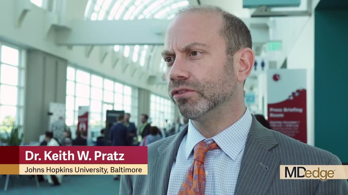
Model bests IPSS-R for predicting survival, risk for AML in myelodysplastic syndrome
SAN DIEGO – A newly developed personalized model that “harnesses the power of artificial intelligence” to predict overall survival and transformation to acute myeloid leukemia (AML) in patients with myelodysplastic syndromes outperforms both the original and revised International Prognostic Scoring Systems (IPSS, IPSS-R), according to Aziz Nazha, MD.
The machine learning model, which was built using clinical and genomic data derived from myelodysplastic syndrome (MDS) patients diagnosed according to 2008 World Health Organization criteria, incorporates information beyond that included in the IPSS and IPSS-R, and provides patient-specific survival probabilities at different time points, Dr. Nazha of Cleveland Clinic reported during a press briefing at the annual meeting of the American Society of Hematology.
The model was developed in a combined training cohort of 1,471 patients from the Cleveland Clinic and Munich Leukemia Laboratory and was validated in a separate cohort of 831 patients from the Moffitt Cancer Center in Tampa, Fla.
The concordance index – a measure for comparing the accuracy of the various models – was 0.80 for overall survival (OS), and 0.78 for AML transformation vs. 0.66 and 0.73, respectively, for IPSS, and 0.67 and 0.73, respectively, for IPSS-R, Dr. Nazha said. The new “geno-clinical” model also outperformed mutations-only analysis, mutations plus cytogenetics analysis, and mutations plus cytogenetics plus age analyses for both OS and AML transformation.
Adding mutational variant allelic frequency did not significantly improve prediction accuracy, he noted.
Dr. Nazha and his colleagues are developing a web application tool that can be used to run the trained model to calculate patient-specific, time-specific OS and AML transformation probabilities. He discussed the new model and its implications for personalized prognosis and treatment in this video interview.
Improved risk assessment helps patients understand their disease and “establish expectations about their journey with their disease,” and it is also extremely important for treating physicians, he said.
“All of our consensus guidelines and treatment recommendations are based on risk,” he explained, noting that the approach varies greatly for higher- and lower-risk patients.
This model represents a potential new focus on “personalized prediction” in addition to the increasing focus on personalized treatment and takes into account the heterogeneous outcomes seen in patients with MDS, he said.
Dr. Nazha reported consultancy for Karyopharma and Tolero, and data-monitoring committee membership for MEI.
SOURCE: Nazha A et al. ASH 2018, Abstract 793.
REPORTING FROM ASH 2018