User login
Elderly-onset atopic dermatitis is on the rise
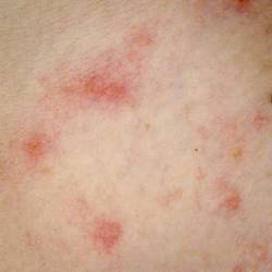
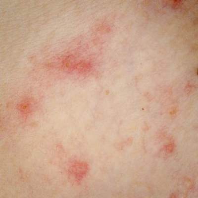
VANCOUVER – Atopic dermatitis arising de novo in patients in their 60s or older with no history of the disease poses a diagnostic challenge, and a low threshold for biopsy is warranted, Dr. Thomas Bieber said at the World Congress of Dermatology.
“The diagnosis is not very easy, and if you are not sure what you are facing, I urge you to take biopsies in order to exclude cutaneous T-cell lymphoma before treating the patient with any kind of active compound,” cautioned Dr. Bieber, professor and chair of the department of dermatology and allergy at the University of Bonn (Germany).

Very-late-onset atopic dermatitis and cutaneous T-cell lymphoma (CTCL) may look quite similar clinically. There is but a single exception: Primary CTCL usually doesn’t itch, while pruritus is a prominent feature of atopic dermatitis arising in seniors, he added.
New-onset atopic dermatitis at an advanced age is increasing in prevalence, as is true of atopic dermatitis across the rest of the age spectrum. Dr. Bieber said that statistic is certainly borne out in his own clinical practice, where with the graying of the population he is seeing more cases.
He credited Dr. Ryoji Tanei of Tokyo Metropolitan Geriatric Hospital with doing pioneering work in bringing this particular variant of atopic dermatitis to wider attention (J. Clin. Med. 2015;4:979-97). Roughly 30% of patients with atopic dermatitis in their 60s or older report they never had the disease before. Another 20% had atopic dermatitis in childhood, while it arose in early adulthood in the rest.
Atopic dermatitis arising de novo in seniors is a special form of the disease that characteristically involves the face, neck, and trunk while sparing the flexural areas which are so prominently involved in younger patients. The eczema is often erythrodermic. Older men are affected threefold more often than women.
The disorder is characterized by extraordinarily high serum IgE levels: a mean of 8,000 IU in one series reported by Dr. Tanei.
This very-late-onset form of atopic dermatitis tends not to fade away over time. Dr. Tanei has reported that many affected patients die with the inflammatory skin disease, never outgrowing it.
Very-late-onset atopic dermatitis is often resistant to topical therapies; repeated courses of oral corticosteroids may be required.
The differential diagnosis is quite different than in children, where genetic immunodeficiency syndromes are a real concern. While CTCL is the biggie in the differential diagnosis of very-late-onset atopic dermatitis, other conditions that need to be considered include psoriasis, contact dermatitis, pityriasis rubra pilaris, and pityriasis rosea.
Dr. Bieber is a consultant to and recipient of research grants from numerous pharmaceutical companies having an interest in dermatology.
VANCOUVER – Atopic dermatitis arising de novo in patients in their 60s or older with no history of the disease poses a diagnostic challenge, and a low threshold for biopsy is warranted, Dr. Thomas Bieber said at the World Congress of Dermatology.
“The diagnosis is not very easy, and if you are not sure what you are facing, I urge you to take biopsies in order to exclude cutaneous T-cell lymphoma before treating the patient with any kind of active compound,” cautioned Dr. Bieber, professor and chair of the department of dermatology and allergy at the University of Bonn (Germany).
Very-late-onset atopic dermatitis and cutaneous T-cell lymphoma (CTCL) may look quite similar clinically. There is but a single exception: Primary CTCL usually doesn’t itch, while pruritus is a prominent feature of atopic dermatitis arising in seniors, he added.
New-onset atopic dermatitis at an advanced age is increasing in prevalence, as is true of atopic dermatitis across the rest of the age spectrum. Dr. Bieber said that statistic is certainly borne out in his own clinical practice, where with the graying of the population he is seeing more cases.
He credited Dr. Ryoji Tanei of Tokyo Metropolitan Geriatric Hospital with doing pioneering work in bringing this particular variant of atopic dermatitis to wider attention (J. Clin. Med. 2015;4:979-97). Roughly 30% of patients with atopic dermatitis in their 60s or older report they never had the disease before. Another 20% had atopic dermatitis in childhood, while it arose in early adulthood in the rest.
Atopic dermatitis arising de novo in seniors is a special form of the disease that characteristically involves the face, neck, and trunk while sparing the flexural areas which are so prominently involved in younger patients. The eczema is often erythrodermic. Older men are affected threefold more often than women.
The disorder is characterized by extraordinarily high serum IgE levels: a mean of 8,000 IU in one series reported by Dr. Tanei.
This very-late-onset form of atopic dermatitis tends not to fade away over time. Dr. Tanei has reported that many affected patients die with the inflammatory skin disease, never outgrowing it.
Very-late-onset atopic dermatitis is often resistant to topical therapies; repeated courses of oral corticosteroids may be required.
The differential diagnosis is quite different than in children, where genetic immunodeficiency syndromes are a real concern. While CTCL is the biggie in the differential diagnosis of very-late-onset atopic dermatitis, other conditions that need to be considered include psoriasis, contact dermatitis, pityriasis rubra pilaris, and pityriasis rosea.
Dr. Bieber is a consultant to and recipient of research grants from numerous pharmaceutical companies having an interest in dermatology.
EXPERT ANALYSIS FROM WCD 2015

WCD: Methotrexate found safer but less effective than cyclosporine in atopic dermatitis
VANCOUVER, B.C. – In the first head-to-head trial in atopic dermatitis, cyclosporine was about five times more effective in terms of SCORAD 50 but was associated with more adverse events, Dr. Catherine Goujon-Henry reported at the World Congress of Dermatology.
Based on the findings, dermatologists should consider treating AD patients with a relatively high dose of methotrexate, such as 15 mg weekly, or first stabilize flaring patients with cyclosporine and then switch them to methotrexate for long-term disease control, said Dr. Goujon-Henry, who is with the Clinical Research Unit in Immunology at Lyon-Sud Hospital in Lyon, France.
Methotrexate is recommended in atopic dermatitis but has never been directly compared with cyclosporine, Dr. Goujon-Henry noted. For the study, she and her colleagues randomized 97 adults with moderate to severe AD to 8 weeks of treatment with either methotrexate at 15 mg per week or cyclosporine at 2.5 mg per kg per day. Patients only were allowed to use topical glucocorticoids or tacrolimus during the first month of the trial.
Only 8% of the methotrexate group achieved SCORAD 50 by week 8, compared with 42% of cyclosporine patients, Dr. Goujon-Henry reported. Furthermore, only 35% of the methotrexate group achieved EASI 50 compared with 68% of the cyclosporine group, she said. In addition, Dermatology Life Quality Index (DLQI) scores were 5 or less for only 37% of methotrexate patients, compared with 56% of cyclosporine patients, she reported.
The protocol called for increasing the methotrexate dose to 25 mg per week and the cyclosporine dose to 5 mg per kg per day for patients who did not achieve SCORAD 50 by week 8. Dose increases were performed for 74% of the methotrexate group and for 58% of the cyclosporine group, Dr. Goujon-Henry said. At week 12, 15 of the 50 methotrexate patients were considered non-responders and went off study, as did three cyclosporine patients. At this point, 43% of the methotrexate patients who remained on study had achieved SCORAD 50, as had 62% of cyclosporine patients.
Also at week 16, two-thirds of methotrexate patients and 79% of cyclosporine patients had achieved EASI 50, and 61% of methotrexate patients and 77% of cyclosporine patients scored 5 or less on the DLQI, Dr. Goujon-Henry reported. However, about 53% of cyclosporine patients experienced drug-related adverse events, compared with 40% of the methotrexate group, she said. Serious adverse events that led to treatment discontinuation included arterial hypertension and hypertrichosis for the cyclosporine group, and lymphopenia and hepatitis for the methotrexate group, she reported.
Taken together, the findings support using methotrexate as first-line therapy for moderate to severe atopic dermatitis, but patients need to expect a slower treatment response in exchange for the better safety profile, Dr. Goujon-Henry said.
She disclosed no relevant conflicts of interest.
AT WDC 2015
Key clinical point: Methotrexate has a better safety profile but is less effective than cyclosporine in clearing moderate to severe atopic dermatitis.
Major finding: Only 8% of the methotrexate group achieved SCORAD 50 at week 8, compared with 42% of those in the cyclosporine group.
Data source: Randomized, controlled, single-blind, non-inferiority trial of methotrexate and cyclosporine in adults with atopic dermatitis.
Disclosures: Dr. Goujon-Henry disclosed no relevant conflicts of interest.
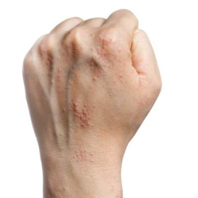
WCD: Smoking tied to worse occupational hand eczema
VANCOUVER, B.C. – Occupational hand eczema is worse and more persistent in smokers than nonsmokers, a large prospective cohort study found.
“Tobacco smoking is associated with work absenteeism and with not staying in the workforce due to occupational hand eczema. Smoking confers a worse prognosis and interferes with the outcome of prevention programs,” Dr. Richard Brans said at the World Congress of Dermatology.
Hand eczema is the most common occupational skin disease. Smoking might worsen signs and symptoms by inducing proinflammatory effects in the skin, said Dr. Brans, a dermatologist at the University of Osnabrück, Germany.
To better assess the link between smoking and hand eczema, he and his associates carried out a prospective 3-year study of 1,095 patients from throughout Germany. The patients initially had attended a 6-week residential treatment program for hand eczema that was followed by a 3-week outpatient program. Smokers comprised about half of the patients and resembled nonsmokers in terms of gender, general atopy, and degree of professional or occupational exposures, such as wetting or soiling the hands at work, Dr. Brans said. However, smokers were significantly younger than nonsmokers and were more likely to have allergic contact dermatitis, he noted.
The inpatient phase of the program markedly benefited both smokers and nonsmokers, but notably, smokers had significantly worse symptoms and signs of hand eczema at all time points assessed, Dr. Brans said. Furthermore, smokers missed an average of 37 days of work because of occupational hand eczema in the year before the program, compared with only 25 days for nonsmokers (P = .001), and smokers continued to miss more days of work because of hand eczema in the year after completing the program (P = .023), he reported. Significantly more smokers also left their professions because of their hand eczema, even after completing the prevention program (P = .021), he added.
The study found no link between number of cigarettes smoked per day and severity of hand eczema, Dr. Brans said. Smoking history was self-reported, and the study design excluded patients who changed their smoking behavior during follow-up, he noted. In addition, the researchers did not assess whether other factors associated with smoking might have confounded the association between smoking and severity of hand eczema, he said.
Dr. Brans reported no relevant disclosures.
AT WDC 2015
Key clinical point: Smoking might worsen the signs and symptoms of occupational hand eczema.
Major finding: Smokers had significantly worse symptoms and signs of hand eczema at all time points assessed.
Data source: Three-year prospective study of 1,095 smokers and nonsmokers with occupational hand eczema.
Disclosures: Dr. Brans reported no relevant conflicts of interest.
WCD: Restoring microbiome might ease atopic dermatitis
VANCOUVER, B.C. – Manipulating the skin microbiome might one day help control atopic dermatitis, Dr. Thomas Bieber said at the World Congress of Dermatology.
Scientists used to focus mainly on pathogens when considering how skin microbes affect atopic dermatitis (AD), said Dr. Bieber, professor of dermatology at the University of Bonn (Germany). But while Staphylococcus aureus is “the constant companion” of patients with AD, data from recent studies have shown that commensal microorganisms found on and within healthy skin undergo a “dialogue” with epithelial cells to help regulate antimicrobial peptides, which in turn inhibit pathogen growth, he said.
Flares in atopic dermatitis are marked by dramatic rises in S. aureus and corresponding decreases in commensal microbes, he noted. Whether an AD flare causes this microbial shift or results from it remains unclear, “but based on this knowledge, we are thinking about strategies to modulate and restore the microbiome. We have discovered a new kind of link to how the microbiome might affect atopic dermatitis,” Dr. Bieber explained.
In the future, such strategies might include formulating emollients to rebalance microbial diversity, he said, pointing to the landmark trial of fecal transplantation in patients with recurrent Clostridium difficile infections (N. Engl. J. Med. 2013;368:407-15). “That study was very impressive – the protocol was stopped due to the overwhelming benefits,” he said. “In terms of skin, could we imagine something similar?”
Restoring skin microbial diversity also might help prevent flares and worsening of AD, said Dr. Bieber. “We can control atopic dermatitis by controlling inflammation, dryness, improving the microbiota with appropriate emollients, and trying to hit early and efficiently,” he said. “We should not only think in terms of treatment, but in terms of prevention. Proactive management of atopic dermatitis does not restore or improve the diversity of the microbiome. It is probably not sufficient, and it’s just one part of the approach.”
Other future treatments for AD might target the dendritic cells that normally “bridge” the innate and adaptive immune systems, or the toll-like receptors of innate immune cells, which help regulate production of antimicrobial peptides by commensal microorganisms, Dr. Bieber said.
Recent research has shown that Th2 T cells inhibit antimicrobial peptide production in patients with atopic dermatitis, and that their dendritic cells have a “defective sensing mechanism toward staphylococcal-derived products,” he noted. “They are kind of neutralized or paralyzed, are not able to induce th17 immune response, and are not able to turn on antimicrobial production in the skin.” Restoring this sensing mechanism might one day help reestablish the normal, healthy diversity of the epidermal microbiome and prevent or alleviate AD, he said.
Dr. Bieber reported serving as a consultant, advisory board member, or speaker bureau member for L’Oreal, Galderma, Sanofi, Regeneron, Pfizer, and Novartis.
EXPERT ANALYSIS FROM WCD 2015
Majority of eczema appears after childhood
Though atopic dermatitis is often regarded as infrequent in adulthood, only 40% of study subjects with current eczema reported onset of the disease in childhood, with most beginning after adulthood, according to data published in the Journal of the European Academy of Dermatology and Venereology.
In a national multicenter, population-based study, Dr. Giancarlo Pesce of the University of Verona (Italy) and his associates administered the GEIRD (Gene-Environment Interactions in Respiratory Diseases) screening questionnaire to 10,464 randomly selected adults aged 20-44 years and gathered data on the frequency and prevalence of doctor-diagnosed eczema, asthma, and hay fever, as well as sociodemographic characteristics and environmental determinants such as smoking habits, traffic, and local pollution levels.
The prevalence of current eczema was 8.1%; the prevalence of eczema with asthma and/or hay fever (EAH), which was adopted as a proxy of atopic dermatitis, was 3.4%. About 60% of the subjects with current eczema reported onset of the disease in adulthood. After adjusting for confounders, the risk of having eczema and EAH was 57% higher in women than in men, and the risk for EAH was 86% higher in women than in men.
Read the full article here: J. Eur. Acad. Dermatol. Venereol. 2015;29:1180-7 (doi: 10.1111/jdv.12784).
Consider combining therapies for atopic dermatitis
Cyclosporine and oral antihistamine therapy showed inconsistent effects on clinical and laboratory markers of atopic dermatitis in older children and adults, based on data from a study of 48 patients.
A total of 25 patients aged 16-42 years received oral cyclosporine and 23 patients aged 15-32 years received oral antihistamine therapy. The patients’ laboratory findings were reviewed, including high-sensitivity C-reactive protein (CRP) and thymus and activation-regulated chemokine (TARC).

Overall, TARC levels were significantly lower after oral cyclosporine therapy, compared with before therapy. Basophil counts in peripheral blood, however, were significantly higher after the therapy than before. After antihistamine therapy, serum high-sensitivity CRP levels and basophil counts in peripheral blood were significantly decreased, compared with before therapy.
“A combination of these two therapies may be more effective for the treatment of AD in adults,” noted study author Dr. Tsutomu Ohtsuka of International University of Health and Welfare Hospital in Nasushiobara, Japan.
The study was published in the June issue of the International Journal of Dermatology (Int. J. Dermatol. 2015;54:648-55 [doi:10.1111/ijd.12374]).
Find the full article online here.
Cyclosporine and oral antihistamine therapy showed inconsistent effects on clinical and laboratory markers of atopic dermatitis in older children and adults, based on data from a study of 48 patients.
A total of 25 patients aged 16-42 years received oral cyclosporine and 23 patients aged 15-32 years received oral antihistamine therapy. The patients’ laboratory findings were reviewed, including high-sensitivity C-reactive protein (CRP) and thymus and activation-regulated chemokine (TARC).
Overall, TARC levels were significantly lower after oral cyclosporine therapy, compared with before therapy. Basophil counts in peripheral blood, however, were significantly higher after the therapy than before. After antihistamine therapy, serum high-sensitivity CRP levels and basophil counts in peripheral blood were significantly decreased, compared with before therapy.
“A combination of these two therapies may be more effective for the treatment of AD in adults,” noted study author Dr. Tsutomu Ohtsuka of International University of Health and Welfare Hospital in Nasushiobara, Japan.
The study was published in the June issue of the International Journal of Dermatology (Int. J. Dermatol. 2015;54:648-55 [doi:10.1111/ijd.12374]).
Find the full article online here.

Hand eczema linked to anxiety, not depression
Adults with chronic hand eczema showed significantly higher levels of anxiety but no difference in depression, compared with healthy controls, based on data from a review of 71 patients. The patients were assessed for anxiety and depression with the Hospital Anxiety and Depression Scale (HADS), and also for compulsive behavior with the Leyton Trait Scale.

Overall quality of life was evaluated according to the Dermatology Life Quality Index (DLQI), and the average score in the patient population was 11.11.
Patients with hand dermatitis had significantly higher scores on the HADS-Anxiety subscale compared to healthy controls, but there was no significant difference in HADS-Depression subscale scores between the groups, noted lead author Dr. Anargyros Kouris of Andreas Sygros Hospital, Athens, and colleagues.
“Hand eczema treatment should address the severity of skin lesions as well as the psychological impact of hand eczema,” the researchers concluded (Contact Dermatitis 2015 June [doi: 10.1111/cod.12366]).
Find the full article online here.
Adults with chronic hand eczema showed significantly higher levels of anxiety but no difference in depression, compared with healthy controls, based on data from a review of 71 patients. The patients were assessed for anxiety and depression with the Hospital Anxiety and Depression Scale (HADS), and also for compulsive behavior with the Leyton Trait Scale.
Overall quality of life was evaluated according to the Dermatology Life Quality Index (DLQI), and the average score in the patient population was 11.11.
Patients with hand dermatitis had significantly higher scores on the HADS-Anxiety subscale compared to healthy controls, but there was no significant difference in HADS-Depression subscale scores between the groups, noted lead author Dr. Anargyros Kouris of Andreas Sygros Hospital, Athens, and colleagues.
“Hand eczema treatment should address the severity of skin lesions as well as the psychological impact of hand eczema,” the researchers concluded (Contact Dermatitis 2015 June [doi: 10.1111/cod.12366]).
Find the full article online here.
FROM CONTACT DERMATITIS
VIDEO: Get excited about the excimer laser for dermatitis
ASHEVILLE, N.C. – Excimer lasers can be used for patients with allergic contact dermatitis for whom topical therapies are unsuccessful or undesirable, according to Dr. Alison Ehrlich of George Washington University in Washington.
In an interview at the annual meeting of the Noah Worcester Dermatological Society, Dr. Ehrlich discussed her use of the excimer laser for dermatitis patients and shared some tips for successful treatment.
The video associated with this article is no longer available on this site. Please view all of our videos on the MDedge YouTube channel
AT NOAH 57

Safely patch test children for contact dermatitis
ASHEVILLE, N.C. – “Children really do develop contact dermatitis, and they are frequently mislabeled as cases of eczema,” Dr. Bruce Brod said at the annual meeting of the Noah Worcester Dermatological Society.
When taking a history in a child with potential contact dermatitis, keep the most likely allergens in mind, especially nickel, said Dr. Brod of the University of Pennsylvania, Philadelphia. Nickel remains the most common allergen in adults and young children, and more than a quarter of patients are positive on patch testing.
Consider all possible sources of nickel. In addition to the old standbys of jewelry and buckles, ask patients and families about the use of flip-style cell phones, as well as first generation iPads (the cases contained nickel). “Old cell phones do not make great toys,” Dr. Brod emphasized.
Patch testing children with potential contact dermatitis makes sense in several situations, including cases of new-onset dermatitis; progressing or deteriorating dermatitis; involvement of specific body sites, such as the face, eyelids, or neck folds; an increase in the total body surface area affected; clinical presentation of dyshidrosis; and dermatitis that resists standard therapies and only improves with oral or extremely potent topical steroids, he said.
Children with atopic dermatitis are more prone to irritation from patch testing, so shorten the exposure time and use a lower concentration of allergens such as nickel, formaldehyde, and rubber additives, advised Dr. Brod. Shorten the exposure time for children younger than 5 years of age, whether or not they have atopy, he added. Videos or video games can work well as a distraction.
When taking a history, consider the most the common allergens in children, defined in a recent study of patch testing results (Dermatitis 2014;25:345-55), said Dr. Brod. He offered the mnemonic MAFLPP (More Allergies for Lovable Pediatric Patients) to characterize the top categories: metals (nickel and cobalt), antibiotics (neomycin and bacitracin), fragrance (fragrances and balsam of Peru), lanolin, phenylenediamine, and preservatives (including quatemium-15 and methylisothiazolinone).
After patch testing, describe the allergen to patients and their families, and explain where it is found, Dr. Brod said. The site mypatchlink.com has helpful information. Also remind patients to read product labels, and to check pharmacy websites such as drugstore.com or cvs.com.
Members of the American Contact Dermatitis Society can access the Contact Allergen Management Program (CAMP) database to help patients identify allergen-free products based on their patch test results, he said.
Dr. Brod had no relevant financial conflicts to disclose.
EXPERT ANALYSIS FROM NOAH 57
Aspirin desensitization making headway in U.S.
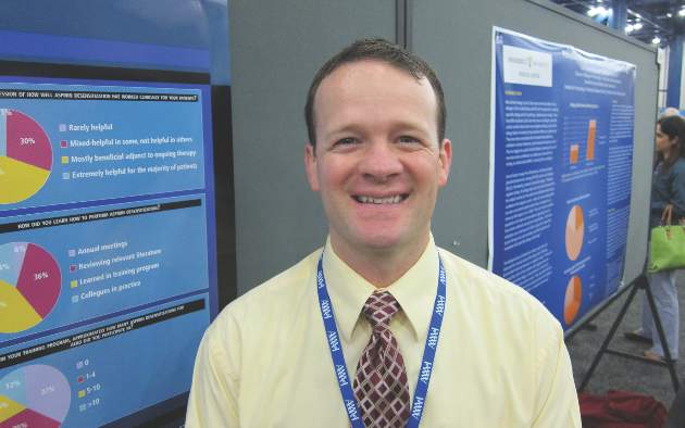
HOUSTON – About 63% of allergists and fellows in training perform aspirin desensitization for aspirin-exacerbated respiratory disease, according to a national survey.
That figure is lower than it should be, given the wealth of published evidence that aspirin desensitization is a safe and effective component of the treatment of aspirin-exacerbated respiratory disease (AERD), Dr. Jeremy D. Waldram asserted in presenting the survey findings at the annual meeting of the American Academy of Allergy, Asthma, and Immunology.
Moreover, the figure likely overcalls the true rate, since participation in the survey was voluntary and fans of aspirin desensitization were probably more inclined to complete the 16-item questionnaire, added Dr. Waldram, a fellow in allergy and immunology at the Scripps Clinic in San Diego.

Was he surprised to find that aspirin desensitization isn’t more widely utilized?
“I think the number that surprised me more was that among the 37.5% of allergists who don’t do aspirin desensitization, almost 30% of them don’t even refer their patients to others who do the procedure. We don’t know why they don’t refer out; it wasn’t a question included in the survey. Perhaps they see patients who are of a less severe phenotype,” he said in an interview.
The 684 survey responses represented a 15% response rate. While 37.5% of respondents indicated they don’t perform aspirin desensitization, 73% of those who reported doing the procedure said they do an average of 1-5 cases annually.
Among allergists who don’t perform aspirin desensitization, safety concerns were the leading reason cited. Indeed, 70% of those who don’t do aspirin desensitization indicated safety risks were the main reason. More than one reason could be given, however, and 30% of allergists cited poor compensation for the procedure as a deterrent, nearly 60% said the logistics of monitoring care were too onerous, and one-third said they didn’t perform aspirin desensitization because they hadn’t been trained to do it.
Of allergists who reported doing aspirin desensitization, 52% perform the procedure in an outpatient setting unattached to a hospital. Another 21% do so in an outpatient clinic that’s physically attached to a hospital.
Within the past 5 years, 9% of respondents said that they’ve had a patient react severely to aspirin desensitization, requiring an unanticipated transfer to a higher level of care. That’s contrary to the experience among allergists at the Scripps Clinic, which is widely credited with pioneering the outpatient approach.
“We essentially do all our aspirin desensitizations for AERD in the outpatient setting. In 1,500 treated patients we’ve never had one that we had to transfer to a higher level of care. We don’t have any special setup. It’s a typical outpatient clinic. We usually don’t start IVs or do anything above and beyond,” Dr. Waldram said.
While 26% of respondents reported they generally recommend aspirin desensitization immediately upon identifying a patient history that supports the diagnosis of AERD, another 54% said they usually recommend the procedure to patients only after they’ve failed to improve on typical medical therapy.
Twenty percent of physicians rated aspirin desensitization as “extremely helpful for the majority of patients,” another 49% said they find it most beneficial as an adjuvant to ongoing medical therapy.
Forty-four percent of allergists who perform aspirin desensitization reported that they learned to do the procedure during fellowship training. Fourteen percent said they learned to the procedure at an annual meeting, and 36% picked it up by reviewing the relevant literature.
Several allergists commented that had Dr. Waldram’s survey been conducted even a couple of years ago the rate of utilization of aspirin desensitization would have been far lower. They interpreted his reported 62.5% rate as a sign of progress. Dr. Waldram said he believes the key to further boosting utilization of aspirin desensitization lies in increasing exposure to the procedure during fellowship training. He noted that internal medicine-trained fellows who responded to the survey had a significantly higher aspirin desensitization utilization rate than those who came to their allergy fellowship with a background in pediatrics.
The hallmarks of AERD are difficult-to-treat nasal polyps, chronic eosinophilic sinusitis, and asthma in a patient with sensitivity to aspirin and other COX-1 inhibitors.
Dr. Waldram reported having no financial conflicts with regard to his study, which was conducted free of commercial support.
HOUSTON – About 63% of allergists and fellows in training perform aspirin desensitization for aspirin-exacerbated respiratory disease, according to a national survey.
That figure is lower than it should be, given the wealth of published evidence that aspirin desensitization is a safe and effective component of the treatment of aspirin-exacerbated respiratory disease (AERD), Dr. Jeremy D. Waldram asserted in presenting the survey findings at the annual meeting of the American Academy of Allergy, Asthma, and Immunology.
Moreover, the figure likely overcalls the true rate, since participation in the survey was voluntary and fans of aspirin desensitization were probably more inclined to complete the 16-item questionnaire, added Dr. Waldram, a fellow in allergy and immunology at the Scripps Clinic in San Diego.
Was he surprised to find that aspirin desensitization isn’t more widely utilized?
“I think the number that surprised me more was that among the 37.5% of allergists who don’t do aspirin desensitization, almost 30% of them don’t even refer their patients to others who do the procedure. We don’t know why they don’t refer out; it wasn’t a question included in the survey. Perhaps they see patients who are of a less severe phenotype,” he said in an interview.
The 684 survey responses represented a 15% response rate. While 37.5% of respondents indicated they don’t perform aspirin desensitization, 73% of those who reported doing the procedure said they do an average of 1-5 cases annually.
Among allergists who don’t perform aspirin desensitization, safety concerns were the leading reason cited. Indeed, 70% of those who don’t do aspirin desensitization indicated safety risks were the main reason. More than one reason could be given, however, and 30% of allergists cited poor compensation for the procedure as a deterrent, nearly 60% said the logistics of monitoring care were too onerous, and one-third said they didn’t perform aspirin desensitization because they hadn’t been trained to do it.
Of allergists who reported doing aspirin desensitization, 52% perform the procedure in an outpatient setting unattached to a hospital. Another 21% do so in an outpatient clinic that’s physically attached to a hospital.
Within the past 5 years, 9% of respondents said that they’ve had a patient react severely to aspirin desensitization, requiring an unanticipated transfer to a higher level of care. That’s contrary to the experience among allergists at the Scripps Clinic, which is widely credited with pioneering the outpatient approach.
“We essentially do all our aspirin desensitizations for AERD in the outpatient setting. In 1,500 treated patients we’ve never had one that we had to transfer to a higher level of care. We don’t have any special setup. It’s a typical outpatient clinic. We usually don’t start IVs or do anything above and beyond,” Dr. Waldram said.
While 26% of respondents reported they generally recommend aspirin desensitization immediately upon identifying a patient history that supports the diagnosis of AERD, another 54% said they usually recommend the procedure to patients only after they’ve failed to improve on typical medical therapy.
Twenty percent of physicians rated aspirin desensitization as “extremely helpful for the majority of patients,” another 49% said they find it most beneficial as an adjuvant to ongoing medical therapy.
Forty-four percent of allergists who perform aspirin desensitization reported that they learned to do the procedure during fellowship training. Fourteen percent said they learned to the procedure at an annual meeting, and 36% picked it up by reviewing the relevant literature.
Several allergists commented that had Dr. Waldram’s survey been conducted even a couple of years ago the rate of utilization of aspirin desensitization would have been far lower. They interpreted his reported 62.5% rate as a sign of progress. Dr. Waldram said he believes the key to further boosting utilization of aspirin desensitization lies in increasing exposure to the procedure during fellowship training. He noted that internal medicine-trained fellows who responded to the survey had a significantly higher aspirin desensitization utilization rate than those who came to their allergy fellowship with a background in pediatrics.
The hallmarks of AERD are difficult-to-treat nasal polyps, chronic eosinophilic sinusitis, and asthma in a patient with sensitivity to aspirin and other COX-1 inhibitors.
Dr. Waldram reported having no financial conflicts with regard to his study, which was conducted free of commercial support.
AT 2015 AAAAI ANNUAL MEETING
Key clinical point: Aspirin desensitization for patients with aspirin-exacerbated respiratory disease is catching on among U.S. allergists.
Major finding: Roughly 63% of allergists and allergy fellows who responded to a national survey indicated they perform aspirin desensitization for aspirin-exacerbated respiratory disease.
Data source: This was a 16-question survey of aspirin desensitization practices among U.S. allergists and allergy fellows. The national survey drew 684 responses.
Disclosures: The presenter reported having no financial conflicts with regard to his study, which was funded without commercial support.