User login
ACOG spells out risk assessment for hereditary cancer syndromes
To identify patients at increased risk for certain types of cancer, ob.gyns. should perform and regularly update hereditary cancer risk assessments, according to a new recommendation from the American College of Obstetricians and Gynecologists.
Patients at increased risk for hereditary cancer syndromes should be referred to a cancer genetics specialist for further assessment and counseling, according to the opinion from ACOG’s Committee on Genetics (Obstet. Gynecol. 2015;125:1538-43).

Personal and family histories provide key information to determine the appropriateness of genetic counseling, which is distinct from genetic testing. Discussed in the counseling visit, the decision for genetic testing is based on family history, pedigree analysis, and, in some cases, pathology reports and confirmation of cancer deaths.
Patient intake forms are used in many office settings to gather relevant information. Online versions of intake forms, such as the Surgeon General’s “My Family Health Portrait,” can facilitate documentation of comprehensive histories. The American Society of Clinical Oncology also has identified some key elements of an adequate family history for assessing cancer risk, including first- and second-degree relatives, maternal and paternal sides, European Jewish ancestry, age of onset and type of cancer, and results of predisposition tests in any relative.
There are several indicators for the presence of hereditary cancer syndromes, including cancer diagnosed at an unusually young age, the presence of several different types of cancer, several close relatives with the same type of cancer, unusual presentation (such as breast cancer in a man), certain birth defects (such as skin growths or skeletal abnormalities associated with inherited cancer syndromes), and Ashkenazi Jewish ancestry.
The ACOG committee opinion identifies common hereditary cancer syndromes involved in gynecologic cancers, including hereditary breast and ovarian cancer syndrome and Lynch, Li-Fraumeni, Cowden, and Peutz-Jeghers syndromes.
Mutations in DNA repair genes BRCA1 and BRCA2 cause hereditary breast and ovarian cancer syndrome and account for 5%-15% of all breast and ovarian cancers in the United States. The carrier frequency in the general population is 1 in 500, but it is much higher (1 in 40) in the Ashkenazi Jewish population. Women with hereditary breast and ovarian cancer syndrome have a 65%-74% lifetime risk of breast cancer and 39%-46% (BRCA1) or 12%-20% (BRCA2) risk of ovarian cancer.
Also caused by DNA repair defects, Lynch syndrome, or hereditary nonpolyposis colorectal cancer, has a prevalence of 1 in 600 to 1 in 3,000. Individuals with Lynch syndrome have increased risk of colon (52%-82%), endometrial (25%-60%), and ovarian (4%-24%) cancers. Malignancies associated with Lynch syndrome include uterine, gastric, small bowel, hepatobiliary, breast, brain, and sebaceous skin cancers.
The presence of Li-Fraumeni syndrome increases the risk of osteosarcoma, breast cancer, colon cancer, adrenocortical carcinoma, leukemia and lymphoma, and brain cancer. Caused by mutations in the tumor suppressor gene TP53, individuals with the syndrome have a 90% risk of cancer by age 60 years. Patients diagnosed with a Li-Fraumeni syndrome–associated malignancy should be referred to a cancer genetics specialist for counseling, according to the committee opinion.
Individuals with the relatively rare (1 in 200,000) Cowden syndrome usually have pathognomonic skin lesions, including papillomatous papules on the face and mucous membranes, by age 30 years. The presence of Cowden syndrome increases the lifetime risk of breast cancer to 25%-50% and endometrial cancer to 5%-10%. Clinicians should consult frequently updated guidelines to determine which patients should be referred for genetic evaluation.
Mutations in serine/threonine kinase 11 cause Peutz-Jehgers syndrome. Two of the following three criteria characterize the syndrome: two or more hamartomatous polyps in the GI tract; mucocutaneous hyperpigmentation of the mouth, lips, nose, eyes, genitalia, or fingers; and a family history of Peutz-Jeghers syndrome. Women with Peutz-Jeghers syndrome have a 50% increased lifetime risk of breast cancer and increased risk of ovarian, uterine, and cervical cancer.
To identify patients at increased risk for certain types of cancer, ob.gyns. should perform and regularly update hereditary cancer risk assessments, according to a new recommendation from the American College of Obstetricians and Gynecologists.
Patients at increased risk for hereditary cancer syndromes should be referred to a cancer genetics specialist for further assessment and counseling, according to the opinion from ACOG’s Committee on Genetics (Obstet. Gynecol. 2015;125:1538-43).
Personal and family histories provide key information to determine the appropriateness of genetic counseling, which is distinct from genetic testing. Discussed in the counseling visit, the decision for genetic testing is based on family history, pedigree analysis, and, in some cases, pathology reports and confirmation of cancer deaths.
Patient intake forms are used in many office settings to gather relevant information. Online versions of intake forms, such as the Surgeon General’s “My Family Health Portrait,” can facilitate documentation of comprehensive histories. The American Society of Clinical Oncology also has identified some key elements of an adequate family history for assessing cancer risk, including first- and second-degree relatives, maternal and paternal sides, European Jewish ancestry, age of onset and type of cancer, and results of predisposition tests in any relative.
There are several indicators for the presence of hereditary cancer syndromes, including cancer diagnosed at an unusually young age, the presence of several different types of cancer, several close relatives with the same type of cancer, unusual presentation (such as breast cancer in a man), certain birth defects (such as skin growths or skeletal abnormalities associated with inherited cancer syndromes), and Ashkenazi Jewish ancestry.
The ACOG committee opinion identifies common hereditary cancer syndromes involved in gynecologic cancers, including hereditary breast and ovarian cancer syndrome and Lynch, Li-Fraumeni, Cowden, and Peutz-Jeghers syndromes.
Mutations in DNA repair genes BRCA1 and BRCA2 cause hereditary breast and ovarian cancer syndrome and account for 5%-15% of all breast and ovarian cancers in the United States. The carrier frequency in the general population is 1 in 500, but it is much higher (1 in 40) in the Ashkenazi Jewish population. Women with hereditary breast and ovarian cancer syndrome have a 65%-74% lifetime risk of breast cancer and 39%-46% (BRCA1) or 12%-20% (BRCA2) risk of ovarian cancer.
Also caused by DNA repair defects, Lynch syndrome, or hereditary nonpolyposis colorectal cancer, has a prevalence of 1 in 600 to 1 in 3,000. Individuals with Lynch syndrome have increased risk of colon (52%-82%), endometrial (25%-60%), and ovarian (4%-24%) cancers. Malignancies associated with Lynch syndrome include uterine, gastric, small bowel, hepatobiliary, breast, brain, and sebaceous skin cancers.
The presence of Li-Fraumeni syndrome increases the risk of osteosarcoma, breast cancer, colon cancer, adrenocortical carcinoma, leukemia and lymphoma, and brain cancer. Caused by mutations in the tumor suppressor gene TP53, individuals with the syndrome have a 90% risk of cancer by age 60 years. Patients diagnosed with a Li-Fraumeni syndrome–associated malignancy should be referred to a cancer genetics specialist for counseling, according to the committee opinion.
Individuals with the relatively rare (1 in 200,000) Cowden syndrome usually have pathognomonic skin lesions, including papillomatous papules on the face and mucous membranes, by age 30 years. The presence of Cowden syndrome increases the lifetime risk of breast cancer to 25%-50% and endometrial cancer to 5%-10%. Clinicians should consult frequently updated guidelines to determine which patients should be referred for genetic evaluation.
Mutations in serine/threonine kinase 11 cause Peutz-Jehgers syndrome. Two of the following three criteria characterize the syndrome: two or more hamartomatous polyps in the GI tract; mucocutaneous hyperpigmentation of the mouth, lips, nose, eyes, genitalia, or fingers; and a family history of Peutz-Jeghers syndrome. Women with Peutz-Jeghers syndrome have a 50% increased lifetime risk of breast cancer and increased risk of ovarian, uterine, and cervical cancer.

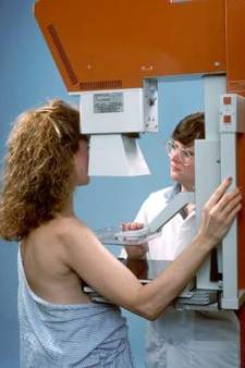
A quarter of women with dense breasts at high interval cancer risk
About a quarter of women with dense breasts are at high risk for interval cancer, so mandating supplemental or alternative screening methods for all such women is not justified, according to a report published May 18 in Annals of Internal Medicine.
Twenty-one states currently have some type of breast density notification law, with most requiring that women be informed if their breasts are heterogeneously or extremely dense on screening digital mammography and suggesting that they discuss the need for supplemental imaging with their health care providers. To assess the real-world risk of interval cancers in women with dense breasts, the researchers analyzed 831,455 digital screening mammographies performed from January 2002 through October 2011 on 365,426 women across the country who were aged 40-74 years and whose medical records included complete demographic and breast health information.
Overall, 47% of the study participants had extremely dense or heterogeneously dense breasts. A total of 2,696 of these women were diagnosed as having invasive breast cancer within 1 year of undergoing screening mammography, according to Dr. Karla Kerlikowske of the University of California, San Francisco, and the San Francisco Veterans Affairs Medical Center, and her associates.
The researchers observed interval cancer rates greater than 1 case per 1,000 mammography exams among 24% of women aged 40-74 years with dense breasts. The 5-year risk was categorized as low to average for 51.0% of those with heterogeneously dense breasts and 52.5% of those with extremely dense breasts, with interval cancer rates of 0.58 to 0.63 and 0.72 to 0.89 case per 1,000 mammography examinations, respectively (Ann. Intern. Med. 2015 May 18 [doi:10.7326/M14-1465]).
Breast density alone was not a good predictor of interval cancer risk. Other factors, such as patient age, race and ethnicity, and family history, should be combined with breast density to determine whether supplemental or alternative imaging is warranted, the investigators wrote.
“Digital mammography has sufficiently high breast cancer detection and reasonably low rates of false-positive results for routine use, even among women with dense breasts,” Dr. Kerlikowske and her associates wrote.
The study was funded primarily by the National Cancer Institute, as well as by many state public health departments and cancer registries. One of the authors reported being a consultant for GE and Bayer and receiving grants or having pending grants from GE.
The study by Kerlikowske et al. provides compelling evidence that breast density shouldn’t be the sole criterion to guide decisions about supplemental breast cancer screening.
The findings suggest that legislation regarding this issue is premature. Fully enacting this legislation would require considerable additional screening resources for approximately half the screening population, with high costs and unclear long-term benefit.
Instead, combining breast density information with a risk assessment, such as the BCSC 5-year risk model, into the mammography report would assist primary care providers to counsel patients on the appropriateness of supplemental screening. At present, many of these physicians are ill-prepared to discuss breast density with their patients, even in states that have already enacted these laws.
Nancy C. Dolan, M.D., and Mita Sanghavi Goel, M.D., are at Northwestern University, Chicago. Dr. Goel reported receiving grants from the Avon Foundation. These comments are adapted from an editorial in Annals of Internal Medicine (Ann. Intern. Med. 2015 May 18 [doi:10.7326/M15-0821]).
About a quarter of women with dense breasts are at high risk for interval cancer, so mandating supplemental or alternative screening methods for all such women is not justified, according to a report published May 18 in Annals of Internal Medicine.
Twenty-one states currently have some type of breast density notification law, with most requiring that women be informed if their breasts are heterogeneously or extremely dense on screening digital mammography and suggesting that they discuss the need for supplemental imaging with their health care providers. To assess the real-world risk of interval cancers in women with dense breasts, the researchers analyzed 831,455 digital screening mammographies performed from January 2002 through October 2011 on 365,426 women across the country who were aged 40-74 years and whose medical records included complete demographic and breast health information.
Overall, 47% of the study participants had extremely dense or heterogeneously dense breasts. A total of 2,696 of these women were diagnosed as having invasive breast cancer within 1 year of undergoing screening mammography, according to Dr. Karla Kerlikowske of the University of California, San Francisco, and the San Francisco Veterans Affairs Medical Center, and her associates.
The researchers observed interval cancer rates greater than 1 case per 1,000 mammography exams among 24% of women aged 40-74 years with dense breasts. The 5-year risk was categorized as low to average for 51.0% of those with heterogeneously dense breasts and 52.5% of those with extremely dense breasts, with interval cancer rates of 0.58 to 0.63 and 0.72 to 0.89 case per 1,000 mammography examinations, respectively (Ann. Intern. Med. 2015 May 18 [doi:10.7326/M14-1465]).
Breast density alone was not a good predictor of interval cancer risk. Other factors, such as patient age, race and ethnicity, and family history, should be combined with breast density to determine whether supplemental or alternative imaging is warranted, the investigators wrote.
“Digital mammography has sufficiently high breast cancer detection and reasonably low rates of false-positive results for routine use, even among women with dense breasts,” Dr. Kerlikowske and her associates wrote.
The study was funded primarily by the National Cancer Institute, as well as by many state public health departments and cancer registries. One of the authors reported being a consultant for GE and Bayer and receiving grants or having pending grants from GE.
FROM ANNALS OF INTERNAL MEDICINE
Key clinical point: Among women with dense breasts, 24% are at risk for interval cancer.
Major finding: Risk of interval cancer was categorized as low to average for 51.0% of women with heterogeneously dense breasts and 52.5% of those with extremely dense breasts.
Data source: A prospective cohort analysis of 831,455 digital screening mammographies in Breast Cancer Surveillance Consortium registries across the country.
Disclosures: This study was funded primarily by the National Cancer Institute, as well as by many state public health departments and cancer registries. One of the authors reported being a consultant for GE and Bayer and grants or pending grants from GE.

Treatment differences between urban and rural women with hormone receptor-positive early-stage breast cancer based on 21-gene assay recurrence score result
Treatment of metastatic breast cancer with nab-paclitaxel in the community practice setting: a US oncology survey
Neoadjuvant chemotherapy for triple negative breast cancer improves conservation
SAN DIEGO – More women with triple negative breast cancer are able to have breast-conserving therapy (BCT) when they receive neoadjuvant systemic chemotherapy.
In a recent trial, 42% of women who were considered ineligible for BCT became eligible for the tissue-sparing surgery after their course of neoadjuvant chemotherapy (NACT) was administered. However, surgeons and patients must work together to decide on the best course of action, according to Dr. David Ollila of the University of North Carolina, Chapel Hill.

The relationship between NACT and the option to have breast-sparing surgery had not been well understood previously, especially for women with aggressive triple negative breast cancer, Dr. Ollila said at the annual meeting of the American Surgical Association.
What has been known is that women with breast cancer who receive NACT show overall improved pathological complete response (Lancet 2014;384:164-72), which is associated with better event-free and overall survival.
The current study, conducted within the larger CALGB 40603 randomized phase II clinical trial, captured the number of patients who converted from BCT-ineligible to BCT-eligible after NACT and before surgery. Investigators also tracked pathological complete response, defined as tumor-free margins on pathologic examination, for those receiving each type of surgery. Dr. Ollila presented his findings on behalf of his coinvestigators in the Alliance for Clinical Trials in Oncology.
The study used a 2x2 factorial design to compare paclitaxel with and without carboplatin followed by doxorubicin plus cyclophosphamide with and without bevacizumab. Before NACT, the surgeon determined whether or not the patient was a candidate for BCT and if not, why not. The process was repeated after NACT, with the surgeon again documenting his or her reasoning for the choice.
Complete data were available for 404 women, distributed evenly across chemotherapy treatment arms. Before NACT, 219 (54%) were judged by their surgeons to be BCT candidates, and 197 (90%) were still deemed eligible for BCT after NACT. Of the 185 (46%) judged ineligible for BCT before chemotherapy, 72 (42%) became eligible for tissue sparing surgery after NACT. “We achieved a very high conversion rate ... from BCT ineligible to eligible,” Dr. Ollila noted.
Overall, just over two-thirds of the 404 women (n = 275, 68%) became candidates for BCT before surgery, and of those, about two-thirds (n = 191, 69%) went on to have an attempted BCT surgery. Surgery was successful for 178 of these women. “Neoadjuvant chemotherapy led to BCT in 93% of selected triple negative patients,”said Dr. Ollila.
Pathological complete response did not differ significantly among the women who received BCT or mastectomy, whether the decision was made before or after chemotherapy.
Discussant Dr. Lisa Newman of the University of Michigan observed that NACT allows some breathing space for the patient and her surgeon to weigh choices, and when indicated, to receive genetic testing.
“We know from numerous population-based and institutional studies that many are opting for bilateral mastectomy, even if a lumpectomy would be optimal,”said Dr. Newman.
She asked whether patient decision making was tracked for this study. Dr. Ollila replied, “We did not include patient factors. We do not have prospective data on what the patient was thinking because we focused on the surgeon.” However, he said, plans are underway to quantify the patient perspective during the decision-making process.
The takeaway message, said Dr. Ollila, is that “We are letting people think that mastectomy is the best option; I think that sequential surgery is all right. I think we just need to try breast conserving therapy more often than we are.”
The study was partially sponsored by the Breast Cancer Research Foundation. The authors reported no conflicts of interest.
The complete manuscript of this study and its presentation at the American Surgical Association’s 135th Annual Meeting, April 2015, in San Diego, California, are anticipated to be published in the Annals of Surgery pending editorial review.
SAN DIEGO – More women with triple negative breast cancer are able to have breast-conserving therapy (BCT) when they receive neoadjuvant systemic chemotherapy.
In a recent trial, 42% of women who were considered ineligible for BCT became eligible for the tissue-sparing surgery after their course of neoadjuvant chemotherapy (NACT) was administered. However, surgeons and patients must work together to decide on the best course of action, according to Dr. David Ollila of the University of North Carolina, Chapel Hill.
The relationship between NACT and the option to have breast-sparing surgery had not been well understood previously, especially for women with aggressive triple negative breast cancer, Dr. Ollila said at the annual meeting of the American Surgical Association.
What has been known is that women with breast cancer who receive NACT show overall improved pathological complete response (Lancet 2014;384:164-72), which is associated with better event-free and overall survival.
The current study, conducted within the larger CALGB 40603 randomized phase II clinical trial, captured the number of patients who converted from BCT-ineligible to BCT-eligible after NACT and before surgery. Investigators also tracked pathological complete response, defined as tumor-free margins on pathologic examination, for those receiving each type of surgery. Dr. Ollila presented his findings on behalf of his coinvestigators in the Alliance for Clinical Trials in Oncology.
The study used a 2x2 factorial design to compare paclitaxel with and without carboplatin followed by doxorubicin plus cyclophosphamide with and without bevacizumab. Before NACT, the surgeon determined whether or not the patient was a candidate for BCT and if not, why not. The process was repeated after NACT, with the surgeon again documenting his or her reasoning for the choice.
Complete data were available for 404 women, distributed evenly across chemotherapy treatment arms. Before NACT, 219 (54%) were judged by their surgeons to be BCT candidates, and 197 (90%) were still deemed eligible for BCT after NACT. Of the 185 (46%) judged ineligible for BCT before chemotherapy, 72 (42%) became eligible for tissue sparing surgery after NACT. “We achieved a very high conversion rate ... from BCT ineligible to eligible,” Dr. Ollila noted.
Overall, just over two-thirds of the 404 women (n = 275, 68%) became candidates for BCT before surgery, and of those, about two-thirds (n = 191, 69%) went on to have an attempted BCT surgery. Surgery was successful for 178 of these women. “Neoadjuvant chemotherapy led to BCT in 93% of selected triple negative patients,”said Dr. Ollila.
Pathological complete response did not differ significantly among the women who received BCT or mastectomy, whether the decision was made before or after chemotherapy.
Discussant Dr. Lisa Newman of the University of Michigan observed that NACT allows some breathing space for the patient and her surgeon to weigh choices, and when indicated, to receive genetic testing.
“We know from numerous population-based and institutional studies that many are opting for bilateral mastectomy, even if a lumpectomy would be optimal,”said Dr. Newman.
She asked whether patient decision making was tracked for this study. Dr. Ollila replied, “We did not include patient factors. We do not have prospective data on what the patient was thinking because we focused on the surgeon.” However, he said, plans are underway to quantify the patient perspective during the decision-making process.
The takeaway message, said Dr. Ollila, is that “We are letting people think that mastectomy is the best option; I think that sequential surgery is all right. I think we just need to try breast conserving therapy more often than we are.”
The study was partially sponsored by the Breast Cancer Research Foundation. The authors reported no conflicts of interest.
The complete manuscript of this study and its presentation at the American Surgical Association’s 135th Annual Meeting, April 2015, in San Diego, California, are anticipated to be published in the Annals of Surgery pending editorial review.
AT THE ASA ANNUAL MEETING
Key clinical point: Neoadjuvant chemotherapy (NACT) for triple negative breast cancer resulted in increased eligibility for breast-conserving therapy (BCT).
Major finding: NACT allowed 42% of women with triple negative breast cancer previously ineligible for BCT to become BCT candidates.
Data source: CALGB 40603, a prospective, randomized controlled trial for women with stage II-III triple negative breast cancer.
Disclosures: The study was partially sponsored by the Breast Cancer Research Foundation. The authors reported no conflicts of interest.

Similar 5-year outcomes from accelerated partial-breast irradiation, whole-breast irradiation
Treatment with accelerated partial-breast irradiation (APBI) versus conventional whole-breast irradiation for women with early-stage breast cancer resulted in similar tumor recurrence rates and overall survival, but APBI was associated with less toxicity, according to a report published in the European Journal of Cancer.
“There was no evidence of significant differences regarding the true incidence of recurrence nor new-onset ipsilateral tumors. [Overall survival] did not differ between the two treatment groups, with the same number of deaths related to” breast cancer, wrote Dr. Lorenzo Livi, a radiation oncologist at the University of Florence, Italy, and his colleagues (Eur. J. Cancer 2015;51:451-63).
Six of the 520 patients, three in each study arm, had ipsilateral breast tumor recurrence. No significant differences were observed in contralateral breast cancer occurrence, distant metastases, or overall survival. The low rate of events at the median 5-year follow up “underlines the importance of longer follow up in addition to an appropriate selection of patient candidates for APBI,” they wrote, urging caution in interpreting the results because longer follow-up is required.
Most of the patient cohort had tumor grade G1-2 (89%), positive estrogen-receptor status (95%), negative nodal status (86%), and were human epidermal growth factor receptor 2 negative (96%).
The APBI group had significantly fewer adverse events than the whole-breast irradiation (WBI) group (P < .0001). Erythema was the most frequently observed event in both arms of the study, at 20% for APBI and 66.5% for WBI. The most represented late skin adverse event was grade 1-2 fibrosis (11% in WBI, 4.5% in APBI). No grade-3 toxicity was recorded in either study arm.
Potential advantages of APBI include shorter treatment time, lower costs, and improved cosmesis, compared with convention treatment. Cosmetic results for both groups in the study were rated excellent/good in greater than 90% of patients, with APBI having slightly better outcomes (P = .045).
The randomized, phase III trial was conducted at the radiation-oncology department of the University of Florence (Italy) between 2005 and 2013 and compared APBI using intensity-modulated radiotherapy (IMRT) with conventional, tangential-field WBI. For the WBI arm, a total dose of 50 Gy was given in 25 fractions, followed by a radiation boost of 10 Gy in five fractions. For the APBI group, a dose of 30 Gy in five fractions at 6 Gy/fraction was given (treatment time, 2 weeks), which is equivalent to 54 Gy in a standard 2-Gy fractionation.
Compared with conformal techniques, IMRT produces optimal dosimeter results while allowing easier, less time-consuming treatment delivery. Additionally, APBI appears to be more cost effective than conventional WBI radiation therapy. The trial “demonstrated excellent results in terms of safety, with a very low rate of local recurrences. APBI using the IMRT technique with the administration of 30 Gy in five non-consecutive fractions should be part of the multidisciplinary discussion to offer a tailored treatment for the patient,” Dr. Livi and his colleagues wrote.
The investigators did not declare any outside funding or conflicts of interest.
FROM THE EUROPEAN JOURNAL OF CANCER
Key clinical point: Patients with early-stage breast cancer had similar outcomes after 5 years whether they received APBI or WBI .
Major finding: 6 of 520 patients, three in each study arm, had ipsilateral breast tumor recurrence.
Data source: From 2005 to 2013, the single-center phase III study randomized 520 patients to receive APBI or WBI; clinicians, investigators, and patients were aware of arm assignments.
Disclosures: The investigators did not declare any outside funding or conflicts of interest.
Low-risk luminal A breast cancers may not require radiation Tx
Some women with the luminal A subtype of breast cancer appear to be at low risk of relapse and may not need breast radiotherapy, the results of a retrospective analysis suggested.
Among 501 patients with tissue blocks that were retrospectively analyzed for breast cancer risk markers, patients with luminal A tumors had an overall risk for 10-year ipsilateral breast recurrence (IBR) of 5.2%, compared with 10.5% for the luminal B subtype and 21.3% for the high-risk subtypes. In addition, a subanalysis of patients with clinical low-risk luminal A disease showed that the 10-year IBR was not significantly better with tamoxifen plus radiotherapy (RT) than with tamoxifen alone (5.0% vs. 1.3%, P = .42), Dr. Fei-Fei Liu of the Princess Margaret Cancer Centre in Toronto and associates reported (Journ. Clin. Onc. 2015 May 11 [doi:10.1200/JCO.2014.57.7999]).
“These results suggest that when luminal A subtype is combined with clinical and pathologic factors, a subgroup of patients with a low risk of IBR may be defined for whom the benefits of RT are small. However, omitting RT and using intrinsic subtyping and clinical factors is a substantial change in care. The breast cancer community would likely require additional prospective evidence before this becomes standard of practice,” the investigators wrote.
They looked at tissue samples from patients enrolled in the Toronto–British Columbia trial, in which older patients with node-negative breast cancer were randomly assigned to tamoxifen or tamoxifen plus radiotherapy. The samples were analyzed with a panel of six immunohistochemical (IHC) markers to identify whether intrinsic subtypes could predict radiotherapy benefit. The markers were estrogen receptor, progesterone receptor, human epidermal growth factor receptor 2 (HER2), cytokeratin 5/6, epidermal growth factor receptor, and Ki-67.
Of the 501 samples analyzed, 265 were determined to be the luminal A subtype, 165 were luminal B, and the remaining 71 were high-risk subtypes, including luminal HER2, HER2 enriched, basal-like and triple negative.
In an exploratory analysis, the authors sought to identify women with a 10-year IBR risk below 5% who might be spared radiotherapy, and found that when they added clinicopathologic features to intrinsic subtyping, there was a subgroup of 151 low-risk luminal A patients with a 10-year 1BR rate of just 3.1%.
Canadian investigators have initiated a prospective, single-arm clinical trial open to women aged 55 or older with pT1N0 grade 1/2 luminal A breast cancer. The patients will undergo breast-conserving surgery and endocrine therapy without radiotherapy. The trial will be stopped if the projected risk of IBR exceeds 5%, Dr. Liu and colleagues said.
The study was supported by the Canadian Institutes of Health Research, the Guglietti Foundation, the Princess Margaret Cancer Foundation, the Campbell Family Institute for Cancer Research, and the Ministry of Health and Long-term Planning, Province of Ontario. Dr. Liu reported no conflicts of interest. Four of her coauthors reported having financial relationships with industry.
Given the overall lower rates of local recurrence seen in modern studies after breast-conserving surgery and radiation therapy, coupled with the possibility of being able to more precisely stratify patients on the basis of risk of recurrence with RT, the question of omitting RT in the treatment of patients with early-stage disease once again comes to the forefront. The reanalysis of the prospective Toronto–British Columbia trial by Liu et al. that accompanies this editorial is a positive step forward in this regard.
This study, coupled with the earlier subtype-based analyses, paves the way for other prospective initiatives using the tumor’s biologic identity to select patients with such a low risk of local recurrence that RT can be avoided without adversely affecting survival.
Dr. Jennifer R. Bellon is at the Dana-Farber Cancer Institute, and is with the department of radiation oncology at Harvard Medical School, Boston. These remarks are taken from an editorial accompanying the study by Dr. Liu and associates (Journ. Clin. Onc. 2015 May 11 [doi:10.1200/JCO.2015.61.2069]).
Some women with the luminal A subtype of breast cancer appear to be at low risk of relapse and may not need breast radiotherapy, the results of a retrospective analysis suggested.
Among 501 patients with tissue blocks that were retrospectively analyzed for breast cancer risk markers, patients with luminal A tumors had an overall risk for 10-year ipsilateral breast recurrence (IBR) of 5.2%, compared with 10.5% for the luminal B subtype and 21.3% for the high-risk subtypes. In addition, a subanalysis of patients with clinical low-risk luminal A disease showed that the 10-year IBR was not significantly better with tamoxifen plus radiotherapy (RT) than with tamoxifen alone (5.0% vs. 1.3%, P = .42), Dr. Fei-Fei Liu of the Princess Margaret Cancer Centre in Toronto and associates reported (Journ. Clin. Onc. 2015 May 11 [doi:10.1200/JCO.2014.57.7999]).
“These results suggest that when luminal A subtype is combined with clinical and pathologic factors, a subgroup of patients with a low risk of IBR may be defined for whom the benefits of RT are small. However, omitting RT and using intrinsic subtyping and clinical factors is a substantial change in care. The breast cancer community would likely require additional prospective evidence before this becomes standard of practice,” the investigators wrote.
They looked at tissue samples from patients enrolled in the Toronto–British Columbia trial, in which older patients with node-negative breast cancer were randomly assigned to tamoxifen or tamoxifen plus radiotherapy. The samples were analyzed with a panel of six immunohistochemical (IHC) markers to identify whether intrinsic subtypes could predict radiotherapy benefit. The markers were estrogen receptor, progesterone receptor, human epidermal growth factor receptor 2 (HER2), cytokeratin 5/6, epidermal growth factor receptor, and Ki-67.
Of the 501 samples analyzed, 265 were determined to be the luminal A subtype, 165 were luminal B, and the remaining 71 were high-risk subtypes, including luminal HER2, HER2 enriched, basal-like and triple negative.
In an exploratory analysis, the authors sought to identify women with a 10-year IBR risk below 5% who might be spared radiotherapy, and found that when they added clinicopathologic features to intrinsic subtyping, there was a subgroup of 151 low-risk luminal A patients with a 10-year 1BR rate of just 3.1%.
Canadian investigators have initiated a prospective, single-arm clinical trial open to women aged 55 or older with pT1N0 grade 1/2 luminal A breast cancer. The patients will undergo breast-conserving surgery and endocrine therapy without radiotherapy. The trial will be stopped if the projected risk of IBR exceeds 5%, Dr. Liu and colleagues said.
The study was supported by the Canadian Institutes of Health Research, the Guglietti Foundation, the Princess Margaret Cancer Foundation, the Campbell Family Institute for Cancer Research, and the Ministry of Health and Long-term Planning, Province of Ontario. Dr. Liu reported no conflicts of interest. Four of her coauthors reported having financial relationships with industry.
Key clinical point: Patients with low-risk luminal A breast cancers may not need radiation therapy.
Major finding: Older women with low-risk luminal A breast tumors had a 1.3% 10-year same-breast recurrence rate when treated with tamoxifen alone.
Data source: A retrospective intrinsic subtype analysis of 501 breast tissue samples.
Disclosures: The study was supported by the Canadian Institutes of Health Research, the Guglietti Foundation, the Princess Margaret Cancer Foundation, the Campbell Family Institute for Cancer Research, and the Ministry of Health and Long-Term Planning, Province of Ontario. Dr. Liu reported no conflicts of interest. Four of her coauthors reported having financial relationships with industry.
Cardiac monitoring falls short in elderly breast cancer patients given trastuzumab
Nearly two-thirds of older women receiving trastuzumab for breast cancer do not get adequate cardiac monitoring, despite the known risks for cardiotoxicity, investigators reported.
Only 36% of 2,203 patients with a median age of 72 years who received trastuzumab (Herceptin)-based adjuvant therapy had adequate cardiac monitoring, based on a review (J. Clin. Oncol 2015 May 11 [doi:10.1200/JCO.2014.58.9465]).
“Our study shows that cardiac monitoring is an area that requires improvement. Actions to increase the rates of cardiac monitoring in this vulnerable population are needed, and adequate cardiac monitoring among trastuzumab-treated patients should be considered a marker of quality of care. Efforts to further disseminate current guidelines should be a priority for our hospitals, training programs, and medical societies,” wrote Dr. Mariana Chavez-MacGregor and colleagues from the University of Texas MD Anderson Cancer Center in Houston.
The authors reviewed Medicare data on patients aged 66 years and older with stage I-III breast cancer treated with trastuzumab-based therapy. They found that 79% of the patients had a baseline cardiac evaluation and that 68% had cardiac tests within 4 months of starting on therapy. Only 43% of patients, however, received subsequent monitoring once every 4 months as recommended in treatment guidelines, and only 36% received optimal monitoring with baseline exam and scheduled follow-up evaluations, the researchers reported.
In multivariable models controlling for demographic/socioeconomic factors (age, race, marital status, education, income, urban/rural residence), clinical stage, comorbidities, and anthracycline and taxane use, factors associated with the likelihood of getting optimal monitoring included more recent year of diagnosis, anthracycline exposure, female prescribing physician, and physician age. Physicians who graduated from medical school after 1990 were significantly more likely to recommend monitoring.
“We estimated the relative contributions of physician and patient-level effects to the variance of the observation; 15.3% of the variance in the adequacy of cardiac monitoring was attributable to physician factors, and only 5.2% of the variance was attributable to measured patient factors,” the authors wrote.
Dr. Chavez-MacGregor disclosed financial relationships with Genomic Health, Roche, and Novartis.
FROM THE JOURNAL OF CLINICAL ONCOLOGY
Key clinical point: Older patients on trastuzumab-based adjuvant therapy require baseline and regular cardiac monitoring.
Major finding: Only 36% of women aged 66 years and older received optimal monitoring during trastuzumab-based therapy.
Data source: Review of SEER-Medicare and Texas Cancer Registry Medicare data on 2,203 women with a mean age of 72 years.
Disclosures: Dr. Chavez-MacGregor disclosed financial relationships with Genomic Health, Roche, and Novartis.
ASA: Radiation lowers local recurrence risk for DCIS patients with close or positive margins
SAN DIEGO – Radiation may benefit women with ductal carcinoma in situ (DCIS) who have breast-conserving surgery if their tumor margins are close or positive; however, wide tumor margins alone also may convey the same protection from local recurrence, Dr. Kimberly Van Zee reported at the annual meeting of the American Surgical Association.
Dr. Van Zee and her colleagues on the breast surgery service at New York’s Sloan Kettering Cancer Center examined the data from a large institutional database of DCIS patients to assess the relative benefit of radiation for various margin widths. They discovered that after adjusting for the other variables, patients with the widest tumor margins saw very little reduction in risk of 10-year recurrence when radiation was added – only 6%. However, this was still a significant difference and represented a hazard ratio of 0.54. Radiation gave patients with positive margins an absolute 18% risk reduction, for a hazard ratio of 0.10.
“We know that radiation reduces risk in all subsets of women with DCIS undergoing breast-conserving surgery,” she said. “But we really wanted to evaluate the relationship between margin width and recurrence and find the best margin width for DCIS with breast-conserving surgery.”

Over 20% of breast cancers are DCIS, and though overall mortality is low, as many as one in three patients will have local recurrence of their cancer. Radiation reduces the risk of local recurrence by about 50%, but it does not reduce the already low mortality associated with DCIS, she said.
Since radiation for DCIS may be associated with an increased risk for cardiovascular disease and certain rare malignancies, Dr. Van Zee said she and her colleagues were interested in identifying those women who were already at low risk for recurrence and would see little increased benefit from radiation.
Dr. Van Zee and her associates conducted a retrospective review of a prospectively collected database of women with DCIS who received treatment at Sloan Kettering Cancer Center between 1978 and 2010. The database contained multiple patient- and procedure-specific variables that were also factored into multivariable analysis in order to evaluate the relationships between margin width and recurrence, and to account for the contribution of radiation to reducing the risk of recurrence in women who received breast-conserving surgery for DCIS.
Overall, the database contained data for nearly 3,000 patients. Of the 2,996 studied, 72% were over the age of 50 and about 67% were postmenopausal. In 87% of cases, the diagnosis was made radiologically rather than clinically, and 60% of the patients had low or intermediate nuclear grade disease.
Dr. Van Zee and her colleagues assessed the 10-year recurrence rate for the 2,788 women whose excision margin width was known. Only 3% of these women had positive margins, and 75% had margin widths greater than 2 mm.
On multivariable analysis, wider margin width was associated with a significantly decreased 10-year risk of recurrence, but only for individuals who had not received radiation (P less than .0001). The hazard ratios for recurrence became progressively lower as margins widened, dropping to 0.31 for a margin of 10 mm or more.
Dr. Van Zee noted that the study was limited by its retrospective nature and the relatively small number of cases with positive margins. Also, cases with positive or close margins usually had more limited or focal disease at the margins, so recurrence rate estimates for this group may have underestimated risk of recurrence for those who had more significant disease.
During the discussion following her presentation, Dr. Van Zee noted that multiple factors are related to the risk of local recurrence, and that nomograms exist to help calculate risk and guide the decision to recommend radiation in women with close margins.
Dr. Patrick Borgen, chairman of the department of surgery at Maimonides Medical Center, New York, remarked that “biology beats technique. A growing wealth of information exists that there is a reservoir of DCIS that will progress so slowly as not to be significant. Is the next step in refining our approach better class prediction using genomic profiling?”
Dr. Van Zee agreed that genomic profiling will play a role, but noted that a study comparing DCIS score and multiple clinical variables would be expensive and archival pathology specimens would be difficult to obtain. Studies will mostly have to be prospective, she said.
The complete manuscript of this study and its presentation at the American Surgical Association’s 135th Annual Meeting, April 2015, in San Diego, California, are anticipated to be published in the Annals of Surgery pending editorial review.
This article was updated May 11, 2015.
SAN DIEGO – Radiation may benefit women with ductal carcinoma in situ (DCIS) who have breast-conserving surgery if their tumor margins are close or positive; however, wide tumor margins alone also may convey the same protection from local recurrence, Dr. Kimberly Van Zee reported at the annual meeting of the American Surgical Association.
Dr. Van Zee and her colleagues on the breast surgery service at New York’s Sloan Kettering Cancer Center examined the data from a large institutional database of DCIS patients to assess the relative benefit of radiation for various margin widths. They discovered that after adjusting for the other variables, patients with the widest tumor margins saw very little reduction in risk of 10-year recurrence when radiation was added – only 6%. However, this was still a significant difference and represented a hazard ratio of 0.54. Radiation gave patients with positive margins an absolute 18% risk reduction, for a hazard ratio of 0.10.
“We know that radiation reduces risk in all subsets of women with DCIS undergoing breast-conserving surgery,” she said. “But we really wanted to evaluate the relationship between margin width and recurrence and find the best margin width for DCIS with breast-conserving surgery.”
Over 20% of breast cancers are DCIS, and though overall mortality is low, as many as one in three patients will have local recurrence of their cancer. Radiation reduces the risk of local recurrence by about 50%, but it does not reduce the already low mortality associated with DCIS, she said.
Since radiation for DCIS may be associated with an increased risk for cardiovascular disease and certain rare malignancies, Dr. Van Zee said she and her colleagues were interested in identifying those women who were already at low risk for recurrence and would see little increased benefit from radiation.
Dr. Van Zee and her associates conducted a retrospective review of a prospectively collected database of women with DCIS who received treatment at Sloan Kettering Cancer Center between 1978 and 2010. The database contained multiple patient- and procedure-specific variables that were also factored into multivariable analysis in order to evaluate the relationships between margin width and recurrence, and to account for the contribution of radiation to reducing the risk of recurrence in women who received breast-conserving surgery for DCIS.
Overall, the database contained data for nearly 3,000 patients. Of the 2,996 studied, 72% were over the age of 50 and about 67% were postmenopausal. In 87% of cases, the diagnosis was made radiologically rather than clinically, and 60% of the patients had low or intermediate nuclear grade disease.
Dr. Van Zee and her colleagues assessed the 10-year recurrence rate for the 2,788 women whose excision margin width was known. Only 3% of these women had positive margins, and 75% had margin widths greater than 2 mm.
On multivariable analysis, wider margin width was associated with a significantly decreased 10-year risk of recurrence, but only for individuals who had not received radiation (P less than .0001). The hazard ratios for recurrence became progressively lower as margins widened, dropping to 0.31 for a margin of 10 mm or more.
Dr. Van Zee noted that the study was limited by its retrospective nature and the relatively small number of cases with positive margins. Also, cases with positive or close margins usually had more limited or focal disease at the margins, so recurrence rate estimates for this group may have underestimated risk of recurrence for those who had more significant disease.
During the discussion following her presentation, Dr. Van Zee noted that multiple factors are related to the risk of local recurrence, and that nomograms exist to help calculate risk and guide the decision to recommend radiation in women with close margins.
Dr. Patrick Borgen, chairman of the department of surgery at Maimonides Medical Center, New York, remarked that “biology beats technique. A growing wealth of information exists that there is a reservoir of DCIS that will progress so slowly as not to be significant. Is the next step in refining our approach better class prediction using genomic profiling?”
Dr. Van Zee agreed that genomic profiling will play a role, but noted that a study comparing DCIS score and multiple clinical variables would be expensive and archival pathology specimens would be difficult to obtain. Studies will mostly have to be prospective, she said.
The complete manuscript of this study and its presentation at the American Surgical Association’s 135th Annual Meeting, April 2015, in San Diego, California, are anticipated to be published in the Annals of Surgery pending editorial review.
This article was updated May 11, 2015.
AT THE ASA ANNUAL MEETING
Key clinical point: Ductal carcinoma in situ patients with close or positive tumor margins may benefit more from radiation.
Major finding: Radiation gave patients with positive margins an absolute 18% risk reduction, for a hazard ratio of 0.10.
Data source: Retrospective review of a prospective database of DCIS patients undergoing breast-conserving surgery from 1978 to 2010.
Disclosures: The authors reported no disclosures.

Mammographic breast density is a strong risk factor for breast cancer
Breast density is a strong, prevalent, and potentially modifiable risk factor for breast cancer, which makes it of special interest to clinicians whose jobs involve breast cancer risk prediction. That was the theme of a talk by Karla Kerlikowske, MD, of the UCSF Helen Diller Family Comprehensive Cancer Center in San Francisco. Dr. Kerlikowske delivered the John I. Brewer Memorial Lecture May 3 at the 2015 Annual Clinical Meeting of the American College of Obstetricians and Gynecologists in San Francisco.
Mammographic breast density is a radiologic term, Dr. Kerlikowske explained. “The only way to really know someone’s breast density is if they have a mammogram.” The whiter the mammogram, the denser the breast. The darker the mammogram, the fattier the breast.
According to the American College of Radiology, the following 4 categories of breast composition are defined by the “visually estimated” content of fibroglandular-density tissue within the breasts:
A. The breasts are almost entirely fatty.
B. There are scattered areas of fibroglandular density.
C. The breasts are heterogeneously dense, which may obscure small masses.
D. The breasts are extremely dense, which lowers the specificity of mammography.
Categories C and D signify dense breasts, which contain a high degree of collagen, epithelial cells, and stroma. In the United States, more than 25 million women are thought to have dense breasts.
Women who have a family history of breast cancer are more likely to have dense breasts. And women who have dense breasts have an elevated risk of breast cancer. They also have a higher risk of advanced disease, as well as a higher risk of large, high-grade, and lymph node-positive tumors, said Dr. Kerlikowske.
Breast-density legislation is increasing
Twenty-two states now have laws mandating that women found to have heterogeneously dense or extremely dense breasts be notified of their status, said Dr. Kerlikowske. That prompts the question: How should these patients be managed?
Breast density declines with age. Breast density also is influenced by body mass index (BMI). As BMI increases, density declines.
Breast density also can be affected by medications, such as hormone therapy and tamoxifen, Dr. Kerlikowske said.
For example, breast density declines about 1% to 2% per year in postmenopause. In postmenopausal women who take estrogen alone, breast density increases slightly. “But the real increase is for people who take estrogen plus progestin,” said Dr. Kerlikowske. “It’s thought that the progestin component is what increases breast density.” Estrogen-progestin therapy confers the same risk of breast cancer as that faced by a premenopausal woman with dense breasts.
As for tamoxifen, it reduces breast density by 2% to 3% per year in postmenopausal women, Dr. Kerlikowske said. “People who have a decrease of more than 10% in breast density are those who have a reduction in breast cancer.” If a woman doesn’t have that reduction with tamoxifen—about half of women don’t—there is no reduction in breast cancer mortality.
“There’s some thought that you should look at mammograms during the first year of tamoxifen use and, if you don’t see a change, consider switching to another medication,” said Dr. Kerlikowske.
More frequent mammograms and supplemental imaging are options for detecting cancers early. Among the modalities that have been studied in this regard are ultrasonography, tomosynthesis, and breast magnetic resonance imaging (MRI).
“If you do more tests, such as ultrasound, you will definitely find additional lesions,” said Dr. Kerlikowske. “There’s no question. But what are the harms?”
The biopsy rate almost doubles after ultrasonography, compared with mammography. And the number needed to screen to detect cancer is fairly high. For mammography, that number is about 250. For ultrasonography, tomosynthesis, and breast MRI, it is higher.
Tomosynthesis is more cost-effective than supplemental ultrasonography because it decreases the number of false positives, Dr. Kerlikowske said.
What’s the bottom line?
Not every woman with dense breasts is at high risk for breast cancer, said Dr. Kerlikowske. And although breast density is prevalent, it is potentially modifiable.
Nevertheless, breast density confers an elevated risk of breast cancer and can also mask tumors. Women with dense breasts likely should avoid the use of postmenopausal hormone therapy. They also may be candidates for more frequent mammography and/or supplemental imaging.
The Breast Cancer Surveillance Consortium (BCSC) Risk Calculator is the only tool that incorporates breast density. In the works is a new model that also incorporates benign breast disease.
Risk-prediction tool considers density and other factors
A risk-prediction tool from the Breast Cancer Surveillance Consortium (BCSC) is the only model to incorporate breast density. The BCSC Risk Calculator is available free of charge for the iPhone and iPad (an Android version is in the works). The tool takes 5 factors into consideration in estimating a woman’s 5-year risk of developing invasive breast cancer:
• age
• race/ethnicity
• breast density
• family history of breast cancer (first-degree relative)
• personal history of breast biopsy.
The tool is designed for use by health professionals. It is not appropriate for determining risk in women younger than 35 years or older than 79 years; women with a previous diagnosis of breast cancer, lobular carcinoma in situ, ductal carcinoma in situ, or atypical ductal hyperplasia; or women who have undergone breast augmentation. Other risk-prediction models are more appropriate for women with a BRCA mutation.
Share your thoughts! Send your Letter to the Editor to rbarbieri@frontlinemedcom.com. Please include your name and the city and state in which you practice.